Embed Size (px)
Citation preview
The Sulfur Oxygenase Reductase from the Mesophilic BacteriumHalothiobacillus neapolitanus Is a Highly Active Thermozyme
Andreas Veith,a Hugo M. Botelho,b Florian Kindinger,a Cláudio M. Gomes,b and Arnulf Kletzina
Institute of Microbiology and Genetics, Technische Universität Darmstadt, Darmstadt, Germany,a and Instituto de Tecnologia Química e Biológica, Universidade Nova deLisboa, Oeiras, Portugalb
A biochemical, biophysical, and phylogenetic study of the sulfur oxygenase reductase (SOR) from the mesophilic gammaproteo-bacterium Halothiobacillus neapolitanus (HnSOR) was performed in order to determine the structural and biochemical proper-ties of the enzyme. SOR proteins from 14 predominantly chemolithoautotrophic bacterial and archaeal species are currentlyavailable in public databases. Sequence alignment and phylogenetic analysis showed that they form a coherent protein family.The HnSOR purified from Escherichia coli after heterologous gene expression had a temperature range of activity of 10 to 99°Cwith an optimum at 80°C (42 U/mg protein). Sulfite, thiosulfate, and hydrogen sulfide were formed at various stoichiometries ina range between pH 5.4 and 11 (optimum pH 8.4). Circular dichroism (CD) spectroscopy and dynamic light scattering showedthat the HnSOR adopts secondary and quaternary structures similar to those of the 24-subunit enzyme from the hyperthermo-phile Acidianus ambivalens (AaSOR). The melting point of the HnSOR was �20°C lower than that of the AaSOR, when analyzedwith CD-monitored thermal unfolding. Homology modeling showed that the secondary structure elements of single subunitsare conserved. Subtle changes in the pores of the outer shell and increased flexibility might contribute to activity at low tempera-ture. We concluded that the thermostability was the result of a rigid protein core together with the stabilizing effect of the 24-subunit hollow sphere.
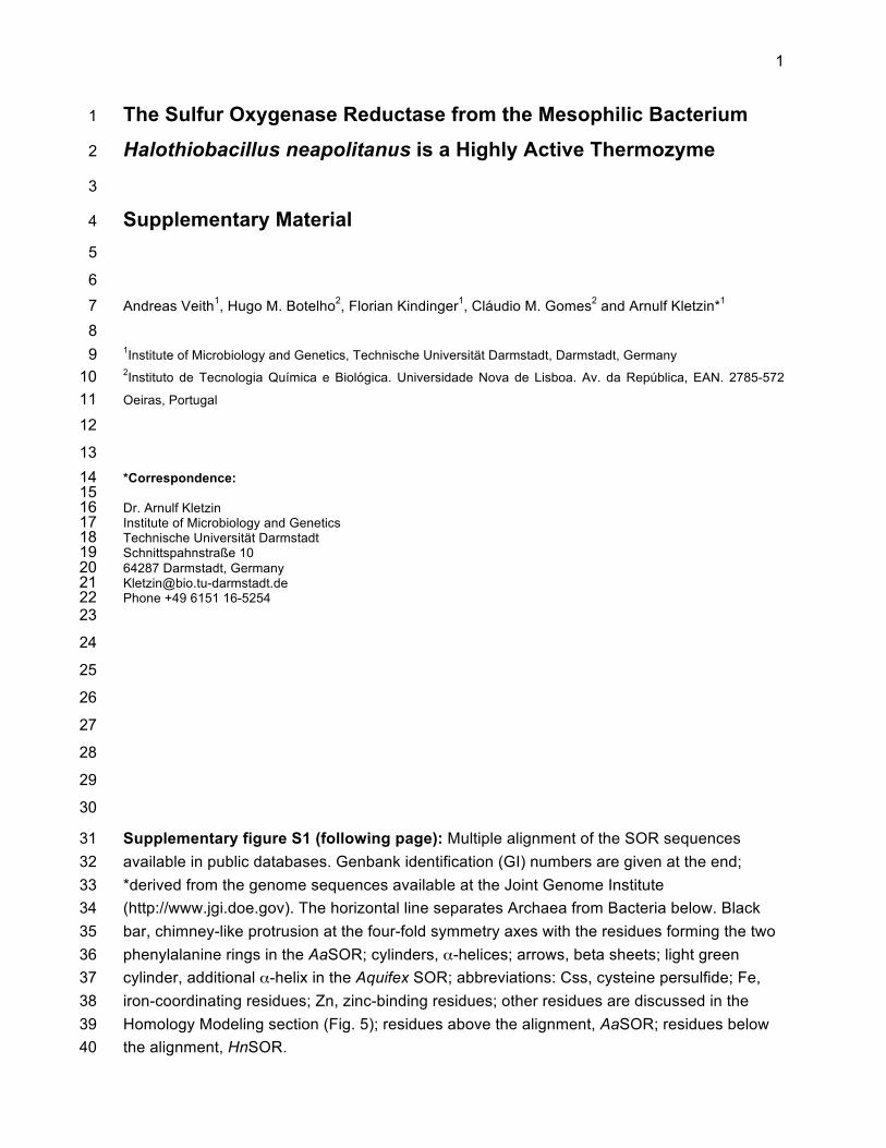
Sulfur oxygenase reductases (SORs) catalyze a dioxygen-dependent disproportionation reaction of elemental sulfur
with sulfite, thiosulfate, and sulfide as products. External cofactorsor electron donors are not required, and the two enzyme activitiescould not be separated (equations 1 to 3) (19, 20, 30, 41).
Oxygenase S0 � O2 � H2O → HSO3� � H� (1)
Disproportionation 3S0 � 3H2O → 2H2S � HSO3� � H�
(2)
Sum 4S0 � O2 � 4H2O → 2H2S � 2HSO3� � 2H� (3)
Thiosulfate is formed rapidly and in a pH-dependent mannerfrom sulfite and excess sulfur at elevated temperatures (equation4) (20, 33), but it is not yet known whether it is a primary reactionproduct as well.
Thiosulfate formation S0 � HSO3�
pH � 4
pH � 6
SSO32�
(4)
SOR is the initial sulfur-oxidizing enzyme in the chemolithoau-totrophic and thermoacidophilic archaea Acidianus ambivalens(AaSOR) and Acidianus tengchongensis (AtSOR), which grow op-timally at 70 to 80°C and pH 1 to 4. The three-dimensional (3D)structures of both highly similar SORs (88% identity) showed thatthe enzymes form large spherical, hollow oligomers with molecu-lar masses of 845 kDa each composed of 24 identical subunits (25,46). Each subunit contains a low-potential mononuclear non-heme iron site as the putative redox-active cofactor and an indis-pensable cysteine, which is persulfurated in the Ac. ambivalensSOR (C31) (25, 45, 46). The iron sites are located in smaller pock-ets within each subunit, which are accessible only from the largeinner cavity. We had shown using site-directed mutagenesis thatthe three Fe-coordinating residues (H86, H90, and E114) and thepersulfurated cysteine C31 are essential for catalysis. Most likely,the cysteine persulfide is involved in sulfur binding. Mutation of
the other two cysteine residues in the protein did not abolish ac-tivity, not even in a double mutant (47). Our current hypothesisabout the reaction mechanism of SOR predicts that the catalyticcycle is initiated by covalent sulfur binding to the active site C31 asa polysulfide chain (R-Sn-SH), followed by hydrolytic cleavage ofthe cysteine polysulfide to sulfide and a polysulfenyl moiety (R-Sn-SOH). Either the sulfenyl group or Fe2� would subsequentlyactivate dioxygen for polysulfenyl oxidation to the final prod-uct(s) sulfite and/or thiosulfate (19).
SORs or sor genes are not widespread in nature. So far, theyhave been reported only from some thermoacidophilic membersof the domain Archaea and (hyper)thermophilic members of thedomain Bacteria. Native SOR enzymes were initially purified fromtwo Acidianus species (8, 20). Later, heterologous gene expressionallowed study of the AaSOR and AtSOR in more detail (5, 41, 45).In addition, the SOR from the hyperthermophilic bacteriumAquifex aeolicus was biochemically characterized, resulting in sim-ilar properties compared to the AaSOR and AtSOR (30). Surpris-ingly, sor genes were recently found in the genomes of a number ofmesophilic and moderately thermophilic bacteria, showing thatSORs are not restricted to hyperthermophiles. The issue is rele-vant for enzymes metabolizing elemental sulfur because the watersolubility of the barely soluble solid substrate decreases signifi-cantly with temperature (478 nM �-S8 at 80°C to 6.1 nM at 4°C
Received 16 November 2011 Accepted 21 November 2011
Published ahead of print 2 December 2011
Address correspondence to Arnulf Kletzin, [email protected].
Supplemental material for this article may be found at http://jb.asm.org/.
Copyright © 2012, American Society for Microbiology. All Rights Reserved.
doi:10.1128/JB.06531-11
0021-9193/12/$12.00 Journal of Bacteriology p. 677–685 jb.asm.org 677
on January 13, 2012 by Biblioteca do IT
QB
http://jb.asm.org/
Dow
nloaded from
[15]), which raises the question whether substrate availability maylimit enzyme activity at mesophilic conditions. There is one reportof a SOR-like enzyme activity in soluble extracts of the mesophilicbacterium Acidithiobacillus thiooxidans, but molecular detailswere not given (43).
In order to investigate the structural and biochemical proper-ties of SORs from a mesophile, we initiated a biochemical, bio-physical, and enzymological study of the enzyme from the gam-maproteobacterium Halothiobacillus neapolitanus (HnSOR) toresolve this issue. The chemolithoautotrophic microbe grows op-timally at 28 to 32°C at more or less neutral pH with thiosulfate orsulfur as electron donors (17). The type strain (NCIMB 8539) wasisolated from corroded concrete sewers in Australia (29) andnamed “pertaining to the seawater at Naples from which this spe-cies was probably first isolated by Nathansohn in 1902” (18). Itseems to be common in sulfide-rich spas and microbial leachingcommunities (3, 49). Apart from the sor gene, the H. neapolitanusgenome encodes a complete SOX complex, a multisubunit sulfuroxidation complex restricted to Bacteria (10). The same applies toAcidithiobacillus caldus. It had been speculated that two sulfur-oxidizing enzyme systems might be used at different temperatures(26). H. neapolitanus does not grow at temperatures exceeding42°C (18), and data regarding the temperature regulation effects,which are hypothetical so far, are lacking for both species.
We show here that recombinant HnSOR is a highly active en-zyme, with a very broad temperature spectrum of activity rangingfrom 10°C to more than 95°C. Also, the results of spectroscopyand in silico modeling suggest that the enzyme adopts secondaryand quaternary structure similar to those from (hyper)thermo-philes. The activity optimization of the HnSOR is discussed as anapparent adaptive response to substrate limitation at mesophilicconditions, which allows efficient sulfur-based energy metabo-lism.
MATERIALS AND METHODSStrain and culture conditions. Halothiobacillus neapolitanus DSM 15147 �NCIMB 8539 (1, 18) was obtained from the Deutsche Sammlung fürMikroorganismen und Zellkulturen (DSMZ; Braunschweig, Germany;http://www.dsmz.de). Cells were grown in liquid culture at 28 to 30°Ceither with 1% (wt/vol) tyndallized elemental sulfur or with 1% (wt/vol)thiosulfate as an electron donor at pH 6.8 in medium 68 recommended onthe DSMZ web server (http://www.dsmz.de/catalogues/catalogue-microorganisms/culture-technology/list-of-media.html). Cells weregrown until the color of bromocresol purple shifted to yellow, indicatinga drop in pH, and subsequently harvested by centrifugation.
DNA procedures and heterologous gene expression. Isolation ofchromosomal H. neapolitanus (DSM15147) DNA was carried out accord-ing to a modified protocol of Sulfolobus solfataricus DNA extraction byS. V. Albers (http://www.rug.nl/gbb/research/researchGroups/molecularMicrobiology/research/extremophiles/DNAsulf.pdf; as of November2011). Six hundred micrograms of sedimented H. neapolitanus cells wasresuspended in 550 �l of TEN buffer (40 mM Tris, 1 mM EDTA, 15 mMNaCl, pH 8). Fifty microliters of a 10% sodium dodecyl sulfate (SDS)(wt/vol) solution was added to the suspension, which was subsequentlyincubated for 45 min at room temperature. After repeated phenol-chloroform-isoamyl alcohol (25:24:1) and chloroform-isoamyl alcohol(24:1) extractions, the genomic DNA was precipitated by the additionof 0.1 volume of a 3 M sodium acetate solution (pH 5.2) and 0.8volume of 2-propanol. After incubation at �20°C for 2 h, the tubeswere centrifuged for 30 min at 16,000 � g in a microcentrifuge. Thepelleted DNA was washed once with 70% ethanol (by volume), centri-fuged for 5 min at 16,000 � g, dried at room temperature, and even-
tually resuspended in 50 �l of TE buffer (10 mM Tris, pH 8, 1 mMEDTA).
The sor gene (GenBank locus tag Hneap_1222) was PCR amplifiedwith the primers HnSOR_fwd (fwd stands for forward) (ACTAGT TAACGA GGGCAA AAAATG TCGAAT GAAAAT CCAATT ATA) andHnSOR_rev (rev stands for reverse) (GTGACG CCAAGC GCTTTG CTTAAG ATGCTT ACGCC) (Biomers, Ulm, Germany) which had beendesigned from the H. neapolitanus genome sequence (strain ATCC23641). The PCR product was purified with the GenElute PCR clean-upkit (Sigma, Taufkirchen, Germany) according to the manufacturer’s rec-ommendations. It was subsequently cleaved with the SpeI and Eco47IIIrestriction enzymes and ligated into the XbaI/Eco47III-digested pASK75vector (38). Positive transformants in Escherichia coli Top 10F cells (In-vitrogen, Darmstadt, Germany) were sequenced and finally introducedinto E. coli BL21(DE3) CodonPlus RIL cells (Agilent, Böblingen, Ger-many).
For heterologous gene expression, 500-ml cultures were grown aero-bically at 37°C in 2� LB medium in notched Erlenmeyer flasks. The ex-pression was induced by the addition of 200 �g/liter anhydrotetracyclinefrom a 2% (wt/vol) stock solution in dimethylformamide at an opticaldensity at 600 nm (OD600) between 0.6 and 0.8. Ferric citrate (100 �M)was added at the time of induction to ensure sufficient iron incorporation.The cultures were incubated for 20 h after induction with vigorous aera-tion and stirring. The Acidianus ambivalens sor gene (EMBL accessionnumber X56616) was expressed heterologously after ligation into thepASK75 vector with a C-terminal Strep tag fusion (38) as described else-where (pASK-SOR.05 plasmid) (45).
Protein purification. The cell pellet obtained by centrifugation waswashed once in approximately 10 volumes (vol/wt) of 100 mM Tris-HClbuffer (pH 8) with 150 mM NaCl (buffer W) and afterwards resuspendedin 5 volumes of the same buffer. Cells were disrupted with a high-pressurehomogenizer (0.18-mm nozzle and 1.35-MPa pressure; Constant Sys-tems, Low March, Daventry, United Kingdom). After the first centrifuga-tion step (10,000 � g for 30 min), the soluble protein-containing super-natant was centrifuged in an ultracentrifuge (100,000 � g for 45 min). Thesoluble total extract from 10 to 20 g of cells (wet weight) was applied eitherto a 1-ml Strep-Tactin gravity flow column or to an 8- to 10-ml Strep-Tactin superflow column (both columns from IBA, Göttingen, Germany)connected to an AKTApurifier 10 (GE Healthcare Bio-Sciences AB, Upp-sala, Sweden). This step was performed repeatedly, when the bindingcapacity of the column was exceeded. The column was equilibrated with 5column volumes (CV) of buffer W prior to loading. The column wassubsequently washed with 6 CV of the same buffer. The protein was elutedby applying 3 CV of buffer E (buffer W with 2.5 mM desthiobiotin; IBA).The column was regenerated with 15 CV of buffer W with 1 mMhydroxyl-azophenyl benzoic acid (HABA) (Sigma). Alternatively, the col-umn was regenerated with 3 CV each of double-distilled water (ddH2O),0.5 M NaOH, and ddH2O instead of the regular HABA solution.
SOR activity and inhibition assays. The SOR activity assay was per-formed as previously described (20, 45). Specific activities were routinelydetermined after incubation of 0.5 to 5 �g of purified enzyme at 80°C in a1-ml solution of 0.1 M sodium citrate– 0.2 M Na2HPO4 (pH 7.2) (27)containing 2% sulfur (wt/vol) and 0.1% Tween 20 (vol/vol). Samples weretaken at appropriate time points (usually after 0, 2, 4, 6, 8, and 10 min),chilled on ice, and centrifuged briefly to sediment elemental sulfur. Theconcentrations of the reaction products hydrogen sulfide, sulfite, andthiosulfate were determined colorimetrically (20) and quantified with cal-ibration curves. The specific activities were calculated from the linearincrease of the reaction products. One unit of enzyme activity was definedas 1 �mol of sulfite plus thiosulfate (oxygenase) or hydrogen sulfide (re-ductase) formed per minute.
The optimal pH and temperature of HnSOR activity were determinedwith the same activity assay and different buffer systems. The citrate-phosphate buffer (27) was used to determine the protein activity in a rangefrom pH 5 to 8. For higher pH values, 0.2 M Na2HPO4 was titrated with a
Veith et al.
678 jb.asm.org Journal of Bacteriology
on January 13, 2012 by Biblioteca do IT
QB
http://jb.asm.org/
Dow
nloaded from

0.1 M Na3PO4 solution to the desired pH. The pH profile was recorded at50°C due to extensive nonenzymatic sulfur disproportionation observedat high temperatures combined with high pH values (20, 40). Usually, 0.75to 2.65 �g of purified HnSOR was added to 1 ml of ice-cold assay buffer.The reaction was started by incubation of the tubes in a heating block at50°C. The concentrations of reaction products and specific activities weredetermined as described above.
Different amounts of a freshly prepared ZnCl2 solution resulting in afinal concentration of 10 to 100 �M Zn2� were added to the reactionmixtures in the SOR activity assay. The reaction mixture was kept on icewater for 30 min after 2 to 5 �g of enzyme had been added. The reactionwas started by heating the samples to 80°C, and specific SOR activitieswere measured as described above. An enzymatic reaction mixture with-out ZnCl2 and a nonenzymatic reaction mixture with inhibitor but with-out SOR were used as controls. For a second control, 2 mM EDTA wasadded to the zinc-containing reaction vials to reverse inhibition. All mea-sured specific activities were plotted against the Zn2� concentration. Theslope of the trend line was used for calculation of the appropriate Ki
values.Biochemical procedures. The protein concentration was determined
by the Coomassie blue method (2). Iron quantification was performedwith pure protein preparations with the 2,4,6-tripyridyl-1,3,5-triazine(TPTZ) method (9). Denaturing SDS-polyacrylamide gel electrophoresis(PAGE) was performed with 10% or 12% polyacrylamide–Tris–tricinegels (35).
CD spectroscopy. Circular dichroism (CD) spectra were measuredwith a JASCO J-815 spectropolarimeter with Peltier temperature control.Typically, 10 accumulations were recorded in the far-UV region (180 to260 nm) at 20°C with a data pitch of 0.2 nm and a 0.1-cm-path-lengthpolarized quartz cuvette. A bandwidth of 2 nm was used with a detectorresponse of 1 s and scanning speed of 200 nm/min. The spectra wereobtained with 0.1 mg/ml of protein in 20 mM Tris-HCl buffer (pH 7.5)and afterwards corrected by subtracting the spectrum of the buffer solu-tion. Thermal unfolding was assessed by measuring the CD signal at 220nm while raising the temperature at a 1°C/min rate.
DLS. Dynamic light scattering (DLS) analyses of recombinant Ac. am-bivalens and H. neapolitanus SORs were performed on a Zetasizer Nanoapparatus (Malvern Instruments, Worcestershire, United Kingdom) at25°C. Samples (0.3 mg/ml in 20 mM Tris-HCl buffer [pH 7.5]) werefiltered through a 0.22-�m-pore-size filter and assayed with a quartz cu-vette with 45-�l volume and 3-mm path length (Hellma, Müllheim, Ger-many). Three measurement cycles were performed for each protein sam-ple. The data were averaged from 14 light scattering periods of 10 s foreach cycle. Average protein diameter values were calculated using thecorresponding Malvern Instruments DTS software.
Phylogenetic analysis and modeling. SOR homologues from othermicroorganisms were detected by BLASTP searches with the Ac. ambiva-lens SOR amino acid sequence as a probe (NCBI accession numberCAA39952.1) against the public database at NCBI (www.ncbi.nlm.nih.gov). The deduced Sulfobacillus and Acidithiobacillus ferrivorans SOR se-quences were identified via tBLASTN analysis in the respective genomesavailable at the DOE Joint Genome Institute (http://www.jgi.doe.gov). AllSOR sequences were aligned by the KALIGN algorithm (24) with manualcorrection. The dendrogram was calculated feeding the alignment into thephylogeny server at the MAFFT site (http://mafft.cbrc.jp/alignment/server/phylogeny.html) with the default parameters and 100 bootstraprepetitions.
3D models of the H. neapolitanus SOR were predicted at the Phyre(http://www.sbg.bio.ic.ac.uk/phyre/html/index.html) (16) and I-Tasserservers (http://zhanglab.ccmb.med.umich.edu/I-TASSER/) (51). Energyminimization of the PHYRE model was performed with UCSF Chimera(http://www.cgl.ucsf.edu/chimera) (31). Clustering was done with CLUS-PRO 2.0 (http://cluspro.bu.edu/login.php) (6, 22) and a single subunit inthe dimer mode. Structure alignments, root mean square deviation
(RMSD) readouts, and the preparation of figures were done with Py-mol (7).
RESULTSPhylogenetic analysis of the SOR protein family. SORs form aconserved protein family with high mutual similarity (Fig. 1)(Pf07682; http://pfam.sanger.ac.uk) but with no recognizablesimilarity to outside proteins. The Halothiobacillus neapolitanusSOR (HnSOR) shared pairwise amino acid identities with otherSORs ranging between 76% (Acidithiobacillus caldus) and 40%(Aquifex aeolicus). The archaeal SORs were about 40 to 42% iden-tical relative to the HnSOR, while Sulfobacillus spp. (47%) were inbetween.
The aligned bacterial SOR amino acid sequences showed sev-eral regions with differences distinguishing them from the ar-chaeal SORs (e.g., positions 91 to 96 and 112 to 117) (see Fig. S1 inthe supplemental material). The dendrogram calculated from thealignment reflects a grouping of the enzymes according to phylo-
FIG 1 Color-coded phylogenetic dendrogram of the available SOR sequencesin GenBank (GI numbers) (for alignment, see Fig. S1 in the supplementalmaterial). The SOR sequences from uncultured (Uncult.) bacteria and otherbacterial species, such as Ferroplasma (Ferropl.), Sulfobacillus (Sulfob.), Desul-fomicrobium (Desulfom.), Acidithiobacillus (Acidithiob.), and Halothiobacillus(Halothiob.) species, are shown. Boldface type indicates that the SOR enzymesfrom this species were biochemically characterized. Underlining indicates thatSOR activity was demonstrated in total cell extracts. Double underlining indi-cates that the X-ray structure is known. The numbers at the nodes of thedendrogram are bootstrap values. The optimal growth temperature is indi-cated by color as follows: red, optimal growth temperature of �70°C; orange,51 to 70°C; green, 40 to 50°C; blue, �30°C; black, optimal growth temperaturenot known. Superscript letters a to d and g indicate the references as follows: a,this work; b, references 21 and 45; c, reference 41, d, reference 14; g, reference30. The superscript e indicates that the sequence was retrieved from the webserver of the Joint Genome Institute (http://www.jgi.doe.gov). The superscriptf indicates that the two sor genes are located on different contigs of the Sulfo-bacillus thermosulfidooxidans genome sequence; they are identical to the twoSOR SA and SB sequences obtained from a bioleaching reactor described inreference 5 and to the SOR sequence of the bacterium SM-1 described by thesame authors.
Sulfur Oxygenase Reductase from H. neapolitanus
February 2012 Volume 194 Number 3 jb.asm.org 679
on January 13, 2012 by Biblioteca do IT
QB
http://jb.asm.org/
Dow
nloaded from

genetic relationship (Fig. 1). The Aquifex SOR was always the leastsimilar enzyme (33 to 40% identity) compared to all other SORs.It contained two insertions and a slightly shortened C terminuscompared to the otherwise constant lengths of the SOR sequences.An outgroup was not included, as no paralogous sequence familywith even minimal similarity was identified so far.
Cloning, expression, and purification of recombinant H.neapolitanus SOR. The H. neapolitanus sor gene (open readingframe [ORF] Hneap_1222) was cloned into the pASK75 expres-sion vector with a C-terminal Strep tag (38). When sequenced forvalidation, the sor genes of strain DSM 15147 used here and ofstrain ATCC 23641 (genome sequence) were identical. When theresulting plasmid pASK_HnSOR was used to produce recombi-nant HnSOR from E. coli BL21 cells, 20 to 30% of the protein wasrecovered in the soluble form, while the remainder precipitated ininclusion bodies. The purification yield was 11 to 18 mg of solubleprotein per liter of 2� LB medium. SDS-PAGE revealed a major36-kDa band characteristic of full-length SOR (Fig. 2). The aver-age iron content of the preparations was 1.6 � 0.3 Fe per proteinmonomer.
Enzymatic properties of recombinant H. neapolitanus SOR.The pH and temperature profiles of HnSOR activity were deter-mined with an enzyme buffer that was modified from those usedpreviously for the Acidianus and Aquifex SORs (20, 30, 45). Opti-mal activity was observed when a 100 mM citrate-phosphate buf-fer was used (instead of Tris-acetate), resulting in 42.4 U/mg ox-ygenase and 4.1 U/mg reductase activities at pH 8 and 80°C. Bothoxygenase and reductase activities were measured at temperaturesbetween 10 and 95°C with an optimal activity at 80°C (Fig. 3B andC). At moderate temperatures (i.e., 10 to 40°C), both enzymeactivities were at similar levels. At temperatures higher than 40°C,oxygenase activity was up to 10-fold higher than reductase activ-ity. The same effect was previously observed for the Aq. aeolicusSOR (30). An equal ratio between both oxygenase products (sul-fite and thiosulfate) was observed at moderate temperatures (10 to40°C). The ratio drastically changed when temperatures exceeded50°C, resulting in an up to sixfold-higher level of thiosulfate at75°C.
The activity assays used to determine the optimal pH for pro-tein activity (Fig. 3A) were carried out at the suboptimal temper-ature of 50°C in order to avoid the interference of nonenzymaticsulfur disproportionation observed at higher temperatures com-bined with a pH of �8 (20, 40). Oxygenase and reductase activitieswere observed between pH 5.4 and 11 with an optimum at pH 8.4(Fig. 3A). At pH 12, it was no longer possible to distinguish be-
FIG 2 Coomassie blue-stained 12% polyacrylamide–Tris–Tricine–SDS gel ofHnSOR purified in a 1-ml Strep-Tactin gravity flow column. The protein waspurified with 0.5-ml volume per wash/elution fraction according to the man-ufacturer’s recommendation (IBA), and 10 �l of eluent was used per lane.Lanes; M, molecular mass markers; FT, flowthrough of E. coli soluble extract;W1 to W3, column wash fractions; E1 to E3, elution fractions. The positions ofmolecular mass markers (in kilodaltons) are indicated to the left of the gel.
FIG 3 (A) pH dependence of HnSOR activity recorded at 50°C. Specific en-zyme activity is shown on the left-hand y axis (�, sulfite plus thiosulfateproduction; �, H2S production) Nonenzymatic sulfur disproportionation isshown on the right-hand y axis (‘, sulfite plus thiosulfate; Œ, H2S). (B) Tem-perature dependence of the specific activities of the Halothiobacillus neapoli-tanus SOR activity at pH 8. Sulfite plus thiosulfate production (�) is shown onthe left-hand y axis, and H2S production (�) is shown on the right-hand y axis.(C) Relative specific activities as a percentage of the value at 80°C. Inset, en-largement of the 0 to 30°C range.
Veith et al.
680 jb.asm.org Journal of Bacteriology
on January 13, 2012 by Biblioteca do IT
QB
http://jb.asm.org/
Dow
nloaded from

tween nonenzymatic sulfur disproportionation and the enzymaticreaction. Under slightly acidic conditions (pH 5.4 to 6.4), reduc-tase and oxygenase activities were at comparable levels. The oxy-genase and reductase activity ratios changed with increasing pH:at pH 7, a 2-fold excess of oxygenase over reductase activity wasobserved, which increased to almost 5-fold at the optimal pH of8.4 (15.9 U/mg oxygenase and 3.27 U/mg reductase activities, re-spectively, at 50°C). Both products of the oxygenase reaction, sul-fite and thiosulfate, were detected at all pH values tested. Sulfitewas the major product under slightly acidic conditions (pH 5.4 to6.4) (not shown). Thiosulfate formation was predominant underneutral and alkaline conditions: at pH 8, thiosulfate productionwas more than 3-fold higher than sulfite formation. The results arein agreement with the higher stability of thiosulfate at neutral andalkaline pH (equation 4).
The enzymatic activity of HnSOR was lost when the protein orprotein extracts were stored at �20°C. Consequently, prepara-tions were kept either at 4°C or at �20°C after the addition of 50%glycerol, which prevented loss of activity. Previous reports aboutthe AaSOR and AtSOR had shown that Zn2� is a potent inhibitorof these enzymes (4, 20, 45, 48). Ki values of HnSOR were 46 �Mfor the oxygenase and 36 �M for the reductase activity, which arevalues comparable with the AaSOR (Table 1) (48).
Structural properties of recombinant H. neapolitanus SOR.In order to compare the folding of mesophilic and (hyper)ther-mophilic SORs, we performed a circular dichroism (CD) study ofthe proteins from H. neapolitanus and Acidianus ambivalens (Fig.4). The CD spectra of both proteins were rather similar, with min-
ima around 220 nm and a shoulder at 209 nm (Fig. 4A). The lowwavelength maximum of HnSOR is not identifiable in the acces-sible spectral window. The signal intensity difference is likely dueto quantification error. The conservation of spectral features sug-gests highly similar secondary structure content in both proteins.In addition, the hydrodynamic diameters as determined by dy-namic light scattering were almost identical for both proteins(17.3 � 1.0 nm for HnSOR and 18.0 � 0.8 nm for AaSOR), show-ing that both proteins adopt comparable oligomeric states.HnSOR was �20°C less thermostable than the enzyme from thehyperthermophile (Fig. 4B).
3D modeling of the H. neapolitanus SOR. We performed ho-mology modeling of the HnSOR with the two Acidianus SORstructures as templates (Protein Data Bank [PDB] identifiers 2cb2and 3bxv) (25, 46) in order to identify features that could giveclues to the activity at low temperature and to temperature stabil-ity. The results obtained with the H. neapolitanus SOR were repro-ducible giving similar models, regardless of the modeling serverand the template. When the modeled HnSOR subunit was super-imposed on each of the templates, the central beta barrel, the nine�-helices (longer than 5 amino acids [aa]) and the active sitepocket were almost identical (Fig. 5A). Minor deviations wereseen in loop or coil regions. The RMSD of the C� chain was 0.16 to0.38 Å.
The two Acidianus SORs are built from 24 subunits, each form-ing a large (15-nm-diameter) hollow sphere with a 432-pointgroup symmetry and an almost impervious surface. The minimalbuilding block is a subunit dimer, so that the holoenzymes repre-
TABLE 1 Properties of microorganisms and their sulfur oxygenase reductasesa
Propertyb H. neapolitanusc Aq. aeolicusd Ac. ambivalense Ac. brierleyif Ac. tengchongensisg
Topt (°C)Microorganism 30 85 80 65–70 70Enzyme 80 80 85 65 70
Sp act (U/mg) at Topt
Oxygenase 42.1 78.8 10.6 0.9 186.7Reductase 4.1 3.05 2.6 NR 45.2
T range (°C) of enzyme 10–99 20–90 50–108 55–�80 50–90
pHopt
Microorganism 6.5–6.9 6.8 2.5 1.5–2.0 2.5Enzyme 8.4 NR 6.5–7.4 7.0 5
pH range of enzyme 5.4–11 5.5–8 5–8.4 NR 3.5–9
Mr
Subunit 35,300 37,674 35,318 35,000h 35,172Holoenzyme (method) 848,000 (DLS) 602,000 (native gel) 844,000 (X-ray) 560,000 (native gel) 845,000 (X-ray)
Zn2� inhibition of oxygenase/reductase Ki � 46/36 �M NR Ki � 45/39 �M NR 27%i
a Properties of five microorganisms (Halothiobacillus neapolitanus, Aquifex aeolicus, Acidianus ambivalens, Acidianus brierleyi, and Acidianus tengchongensis) and their sulfuroxygenase reductases are shown.b Topt, optimum temperature; T range, temperature range; pHopt, optimum pH; NR, not reported; DLS, dynamic light scattering.c Data from this work.d Data from reference 30.e Data from references 20, 45, and 46.f Data from reference 8.g Data from references 11, 25, and 41.h Apparent molecular mass on SDS gel; the gene is not known.i Residual activity with 1 mM Zn2�.
Sulfur Oxygenase Reductase from H. neapolitanus
February 2012 Volume 194 Number 3 jb.asm.org 681
on January 13, 2012 by Biblioteca do IT
QB
http://jb.asm.org/
Dow
nloaded from

sent dodecamers of dimers (45, 46). The dynamic light scatteringresults suggest that the HnSOR adopts a comparable multimeriza-tion state. When two of the modeled HnSOR subunits weredocked to each other, a dimer was formed, which could be super-imposed on the AaSOR structure with an RMSD of 1.7 Å (notshown).
Narrow pores at the 4-fold and 3-fold symmetry axes of theAaSOR provide entrance to the inner cavity of the sphere (Fig. 5Band D). The question arose whether multimerization of the singlesubunit might lead to changes in the pore structures of the en-zyme. When replacing one of the pore-forming subunits at the4-fold symmetry axis of the AaSOR with a copy of the modeledHnSOR, changes in the pore structure became apparent. The phe-nylalanine residue F141 (in AaSOR numbering) was conserved,which forms the outer ring of the two Phe rings. The inner Phering was not present due to replacement of F133 with a valineresidue (Fig. 5B; see Fig. S1 in the supplemental material). In con-sequence, only one of the two access-restricting residues is re-tained in the HnSOR.
The amino acids R99 and S226 define the pore at the 3-foldsymmetry axis of the AaSOR (Fig. 5D). They are connected byhydrogen bonds within and across the subunits. A salt bridge be-tween R99 and E228 of the same subunit also might play a role instabilization (48). Neither of these residues is conserved in theHnSOR; R99 is replaced by a glutamate (E101), S226 is replaced byA226 (see Fig. S1 in the supplemental material), and E228 is replacedby P228. A226 defines the pore diameter when the modeled HnSORsubunit was aligned to the pore-forming regions of the AaSOR(Fig. 5C). E101 is predicted to form a hydrogen bond to the N�2atom of N227, whereas the O�2 atom of the same asparagine couldform an H-bond to the N atom of a lysine (K97) of the neighbor-ing subunit.
DISCUSSIONSources of SORs or sor genes. Once thought to be restricted tosome hyperthermophiles and thermophiles, the sor genes identi-
fied in genomes of mesophilic bacteria gave credit to a report byTano and Imai from 1968 (43) on a SOR-like, glutathione-independent, and sulfur-dismutating enzyme activity in cell ex-tracts of the bacterium Acidithiobacillus thiooxidans (formerlyThiobacillus thiooxidans). The sulfur-dismutating enzyme reac-tion was measured at 30°C in a Warburg apparatus. The productswere H2S and thiosulfate. These results were neither repeated norconfirmed, nor was the enzyme ever purified. In contrast, theGSH-dependent sulfur dioxygenases known from other acidithio-bacilli represent a different and unrelated type of sulfur-oxidizingenzymes, which remained elusive so far (32, 42). Therefore, themolecular basis of sulfur oxidation in these microorganisms is notknown despite their importance in bioleaching and formation ofacid mine drainage (3). The preliminary shotgun genome se-quence of Acidithiobacillus thiooxidans ATCC 19377 available atGenBank does not contain a sor homologue (GenBank accessionnumber AFOH00000000.1). We chose Halothiobacillus neapolita-nus (formerly Thiobacillus neapolitanus) as a representative meso-philic microbe carrying a SOR gene to demonstrate enzyme activ-ity and to study the properties of the protein.
Like the two Acidithiobacillus species (Fig. 1), many but not allof the microorganisms with sor genes are (facultatively) chemo-lithoautotrophic sulfur oxidizers originating from sulfide orsulfur-rich springs, solfataras, or bioleaching environments. Oth-ers would not have been expected to carry a sor gene: for example,Picrophilus torridus is a thermoacidophile described as a strict het-erotroph and Desulfomicrobium baculatum (formerly Desulfovib-rio baculatus) is, as far as we know, a strictly anaerobic sulfate andsulfur reducer (34, 36). The physiological role of the SOR in thesemicroorganisms is unknown.
HnSOR activity within a broad pH and temperature range.The HnSOR is active over a broad pH range (pH 5.4 to 11) with anoptimum at pH 8.4, when measured at 50°C. Measurements at theoptimal temperature at alkaline pH were not possible because ofthe much higher rates of nonenzymatic sulfur disproportionation.
FIG 4 (A) Far-UV circular dichroism spectra of the SOR proteins from Halothiobacillus neapolitanus and Acidianus ambivalens at 0.1 mg/ml protein concen-tration and 25°C. (B) Thermal unfolding curves for the H. neapolitanus and A. ambivalens SOR, as measured from the CD variation at 220 nm. The lines showsigmoidal fits to the CD data. Values have been offset to facilitate comparison.
Veith et al.
682 jb.asm.org Journal of Bacteriology
on January 13, 2012 by Biblioteca do IT
QB
http://jb.asm.org/
Dow
nloaded from

The pH range of the HnSOR extends further into the alkalineregion than for any of the SORs characterized so far (Table 1) (4,20, 30, 41). The pH preference correlates with the slightly alkalineinternal pH of H. neapolitanus (pH of �7.8) (44). In contrast, thepH optimum of the AaSOR is 7 to 7.4, correlating with an internalpH of around 6.5 (28).
The enzymatic activity of the HnSOR covered a temperaturespan of almost 90 K, ranging from 10°C to 99°C (Fig. 3), whichmakes it a thermozyme. Two questions arise from these observa-tions, namely, how the protein maintains its thermostability andhow flexibility is retained sufficiently at low temperatures and lowsubstrate concentrations to enable catalysis.
Even though the HnSOR originates from a mesophilic micro-
organism with an optimal growth temperature around 30°C(maximum 42°C) (17), both oxygenase and reductase activitiespeaked at 80°C. As a comparison, the SOR from the hyperthermo-phile Ac. ambivalens (maximal growth temperature of 88°C) wasactive from 50°C to 108°C (Table 1) (20, 52). Despite the struc-tural similarity between the enzymes from the hyperthermophileand mesophile observed by spectroscopy, the distinct environ-mental conditions of the organisms result in specific adaptations,including the level of stability and their catalytic properties. Highenvironmental temperatures impose the need for thermostabili-zation of the enzymes, resulting in a higher rigidity, less flexibilityand low or no activity at ambient temperatures (50). However,when the comparison is done at their respective optimal catalytic
FIG 5 Three-dimensional modeling results of the HnSOR amino acid sequence with the Acidianus ambivalens SOR (AaSOR; PDB accession number 2cb2) as thetemplate. (A) Cross-eyed stereo view of secondary structure representations of the modeled HnSOR subunit structure (blue) superimposed on AaSOR (gray).The iron atom (Fe) and cysteine persulfide (Css) are shown. The arrows indicate deviations in the coil regions. (B) Side view of the channel at the 4-fold symmetryaxis showing two out of four AaSOR subunits (purple and ochre), while the third subunit is replaced by the modeled HnSOR subunit (cyan), including the outer(F141) and inner (F133) phenylalanine rings (AaSOR only), the conserved methionine ring at its base (M130), and valine and leucine residues in the HnSOR. (C)Three copies of the modeled HnSOR subunit aligned to the respective AaSOR subunits (shown in panel D) at the 3-fold symmetry axis showing the channel-forming residues with the distances in angstroms (yellow broken lines and small white numbers) and the putative hydrogen bonds between K97, N227, and E101.(D) Channel at the 3-fold symmetry axis of the AaSOR formed by R99 and S226. Distances are given between the N atoms of the arginines (yellow dashes), forthe salt bridges to E228 (green dashes), and for the hydrogen bond to the O� of the S226 of the neighboring subunit (white dashes). Panel D reprinted fromreference 48 with permission of the publisher.
Sulfur Oxygenase Reductase from H. neapolitanus
February 2012 Volume 194 Number 3 jb.asm.org 683
on January 13, 2012 by Biblioteca do IT
QB
http://jb.asm.org/
Dow
nloaded from

temperature, the enzymes tend to have identical flexibilities undertheir particular optimal working conditions so that a “corre-sponding state” is maintained regarding conformational flexibil-ity (50). The HnSOR is a remarkable exception to this well-established trend. In fact, the specific HnSOR activity exceeded the(hyperthermophilic) AaSOR activity about 10-fold under optimalconditions (20, 45, 48).
The second question to be answered was about the factors thatmake the HnSOR active at low temperatures. We had observedthat the enzyme should be partially melted at the optimal reactiontemperature. The apparent thermal stability was about 20 K lowerthan the AaSOR (Fig. 4B). The results suggest that the HnSOR hasa higher flexibility compared to the more rigid AaSOR, which isnot active below 50°C (20). The results also suggest that the ther-mal denaturation is reversible; however, these conclusions havenot yet been verified independently by other methods.
In addition, we had shown in a previous study that the widthsof the pores at the 4-fold and 3-fold symmetry axes are crucial forspecific AaSOR activities: the wider the pores are, the higher theactivity becomes (48). In this context, the changes seen in thepores of the outer shell of the HnSOR (Fig. 5B to D) suggest thatthe barriers are less restrictive: at the 4-fold axis, the inner ring ofthe two Phe rings is missing. At the 3-fold axis, the salt bridge-forming arginine and glutamate residues are not present, while theH-bond-forming serine is replaced in favor of an alanine. Thisshould be flexible enough to allow easy passage of substrate and/orproducts. In the HnSOR, a novel and wider hydrogen-bondingnetwork centered at N227 replaced the salt bridges (and the hydro-gen bonds provided by S226 [Fig. 5C and D]) that are considered tostabilize subunit interactions and confer rigidity to the 3-fold axisof the AaSOR.
SORs are structurally conserved. The difference of 40 to 50°Cbetween the maximal or optimal growth temperature and the en-zyme activity optimum is much higher than what would have beenexpected from the overall protein stability. The question of howthis came about may be tentatively explained by one (or more) ofthe following hypotheses. (i) The high optimal temperature ofHnSOR is a trait that persisted (e.g., AaSOR) due to lack of evo-lutionary pressure. (ii) The activity-temperature profile is a con-sequence of the coupling between enzyme activity and proteinstructure: any SOR protein will have such a high optimal temper-ature and intrinsic thermostability. (iii) The existence of HnSORactivity over broad pH and temperature ranges is an adaptivemechanism, which allows metabolic homeostasis upon environ-mental changes. Comparison of the biochemical properties andthe sequences (Table 1; see Fig. S1 in the supplemental material)suggests that the SORs have similar properties independent ofadaptations to the host organism.
We used spectroscopy and a homology modeling approach tomake some predictions regarding the reasons for the thermosta-bility and temperature range of the enzyme. The different catalyticproperties of HnSOR and AaSOR do not reside in major differ-ences in the secondary structure content. The similar far-UV CDspectra indicated that the overall fold is preserved among enzymesfrom mesophilic and thermophilic microorganisms (Fig. 4A).This interpretation is supported by the modeling results, whichshowed little changes in the main secondary structure features ofthe subunit (Fig. 5A). In addition, the oligomeric assembly alsoseems to be conserved, as noted from the similar hydrodynamicproperties of both enzymes. The slightly larger hydrodynamic di-
ameter compared to the diameter obtained by electron micros-copy and X-ray crystallography (�15 nm [20, 25, 45, 46]) reflectsthe conformational dynamics in solution, the effect of the solva-tion shell, and the averaging inherent in an ensemble measure-ment.
SORs feature conserved amino acid sequences regardlesswhether they originate from mesophilic or hyperthermophilic mi-croorganisms (see Fig. S1 in the supplemental material). Conse-quently, the basic building blocks are conserved in the available3D structures and in the models, which includes the central betabarrel with the surrounding alpha helices (PDB identifiers 2cb2and 3bxv). This suggests that they are core features that make theproteins resistant against thermal melting of the subunits, even ifthe individual denaturation temperatures might vary (Fig. 4B).
Subunit interactions are another important parameter forthermostability. It has long been known that hollow spheres areintrinsically stable regardless of whether they are macroscopic ob-jects (37), small molecules like fullerenes (23), or large proteincomplexes like ferritins. Ferritins are intrinsically thermostableand adopt a comparable quaternary structure of 24 subunits in432-point group symmetry (12, 13, 39). The 24 subunits of SORsare assembled from dimers (45, 46), again a feature similar toferritins (39). Docking experiments with the modeled subunitsshowed that dimer formation is feasible. The fit was not optimalbut was in good correlation with the template. Taken together, theresults suggest that these features—thermostable subunits,dimer formation, and oligomerization to yield the 24-mer—are common and intrinsic properties of the SORs in general(hypothesis ii).
Conclusion. SORs were once thought to be restricted to somehyperthermophiles and thermophiles, because the enzyme activ-ity had not been recognized in mesophiles with one exception(43). The data presented here showed that the HnSOR possessesfolding and quaternary structure properties similar to the SORsfrom (hyper)thermophiles. However, the activity at low temper-atures seems to result from a combination of a less thermostableand more flexible oligomer with less restrictive access. In the con-text of the limited bioavailability of sulfur at mesophilic tempera-tures, the basal activity and flexibility of the HnSOR effectivelycontribute to adequate metabolic flows of sulfur metabolites forthe microorganism.
ACKNOWLEDGMENTS
We thank Felicitas Pfeifer (Darmstadt, Germany) for her generosity andencouragement.
A. Veith and A. Kletzin were supported by a grant from the DeutscheForschungsgemeinschaft (Az Kl885-6/1).
REFERENCES1. Boden R, et al. 21 August 2011. Phylogenetic assessment of culture col-
lection strains of Thiobacillus thioparus, and definitive 16S rRNA genesequences for T. thioparus, T. denitrificans, and Halothiobacillus neapoli-tanus. Arch. Microbiol. doi:10.1007/s00203-011-0747-0. [Epub ahead ofprint.]
2. Bradford MM. 1976. A rapid and sensitive method for the quantitation ofmicrogram quantities of protein utilizing the principle of protein-dyebinding. Anal. Biochem. 72:248 –254.
3. Brandl H. 2001. Microbial leaching of metals, p 191–224. In Rehm HJ(ed), Biotechnology, vol 10. Special processes. Wiley-VCH, Weinheim,Germany.
4. Chen ZW, Jiang CY, She Q, Liu SJ, Zhou PJ. 2005. Key role of cysteineresidues in catalysis and subcellular localization of sulfur oxygenase-
Veith et al.
684 jb.asm.org Journal of Bacteriology
on January 13, 2012 by Biblioteca do IT
QB
http://jb.asm.org/
Dow
nloaded from

reductase of Acidianus tengchongensis. Appl. Environ. Microbiol. 71:621– 628.
5. Chen ZW, et al. 2007. Novel bacterial sulfur oxygenase reductases frombioreactors treating gold-bearing concentrates. Appl. Microbiol. Biotech-nol. 74:688 – 698.
6. Comeau SR, Gatchell DW, Vajda S, Camacho CJ. 2004. ClusPro: a fullyautomated algorithm for protein-protein docking. Nucleic Acids Res. 32:W96 –W99.
7. DeLano WL. 2002. The PyMOL Molecular Graphics System, 0.97 ed.DeLano Scientific, San Carlos, CA.
8. Emmel T, Sand W, König WA, Bock E. 1986. Evidence for the existenceof a sulfur oxygenase in Sulfolobus brierleyi. J. Gen. Microbiol. 132:3415–3420.
9. Fischer DS, Price DC. 1964. Simple serum iron method using new sen-sitive chromogen tripyridyl-s-triazine. Clin. Chem. 10:21–25.
10. Friedrich CG, Bardischewsky F, Rother D, Quentmeier A, Fischer J.2005. Prokaryotic sulfur oxidation. Curr. Opin. Microbiol. 8:253–259.
11. He Z, Li Y, Zhou P, Liu S. 2000. Cloning and heterologous expression ofa sulfur oxygenase/reductase gene from the thermoacidophilic archaeonAcidianus sp. S5 in Escherichia coli. FEMS Microbiol. Lett. 193:217–221.
12. Janner A. 2008. Comparative architecture of octahedral protein cages. I.Indexed enclosing forms. Acta Crystallogr. Sect. A 64:494 –502.
13. Janner A. 2008. Comparative architecture of octahedral protein cages. II.Interplay between structural elements. Acta Crystallogr. Sect. A 64:503–512.
14. Janosch C, et al. 2009. Sulfur oxygenase reductase in different Acidithio-bacillus caldus-like strains. Adv. Materials Res. 71–73:239 –243.
15. Kamyshny A. 2009. Solubility of cyclooctasulfur in pure water and seawater at different temperatures. Geochim. Cosmochim. Acta 73:6022– 6028.
16. Kelley LA, Sternberg MJ. 2009. Protein structure prediction on the Web:a case study using the Phyre server. Nat. Protoc. 4:363–371.
17. Kelly DP, Wood AP. 2005. Halothiobacillus, p 58 –59. In Brenner DJ,Krieg NR, Staley JT, Garrity GM (ed), Bergey’s manual of systematic bac-teriology, 2nd ed, vol 2. Springer, New York, NY.
18. Kelly DP, Wood AP. 2000. Reclassification of some species of Thiobacillusto the newly designated genera Acidithiobacillus gen. nov., Halothiobacillusgen. nov. and Thermithiobacillus gen. nov. Int. J. Syst. Evol. Microbiol.50(Part 2):511–516.
19. Kletzin A. 2008. Oxidation of sulfur and inorganic sulfur compounds inAcidianus ambivalens, p 184 –201. In Dahl C, Friedrich CG (ed), Microbialsulfur metabolism. Springer, Berlin, Germany.
20. Kletzin A. 1989. Coupled enzymatic production of sulfite, thiosulfate, andhydrogen sulfide from sulfur: purification and properties of a sulfur oxy-genase reductase from the facultatively anaerobic archaebacterium Desul-furolobus ambivalens. J. Bacteriol. 171:1638 –1643.
21. Kletzin A. 1992. Molecular characterization of the sor gene, which en-codes the sulfur oxygenase reductase of the thermoacidophilic archaeonDesulfurolobus ambivalens. J. Bacteriol. 174:5854 –5859.
22. Kozakov D, et al. 2010. Achieving reliability and high accuracy in auto-mated protein docking: ClusPro, PIPER, SDU, and stability analysis inCAPRI rounds 13–19. Proteins 78:3124 –3130.
23. Kroto HW. 1987. The stability of the fullerenes C-24, C-28, C-32, C-36,C-50, C-60 and C-70. Nature 329:529 –531.
24. Lassmann T, Sonnhammer EL. 2005. Kalign–an accurate and fast mul-tiple sequence alignment algorithm. BMC Bioinformatics 6:298.
25. Li M, et al. 2008. Crystal structure studies on sulfur oxygenase reductasefrom Acidianus tengchongensis. Biochem. Biophys. Res. Commun. 369:919 –923.
26. Mangold S, Valdés J, Holmes D, Dopson M. 2011. Sulfur metabolism inthe extreme acidophile Acidithiobacillus caldus. Front. Microbiol. 2:17.
27. McIlvaine TC. 1921. A buffer solution for colorimetric comparison. J.Biol. Chem. 49:183–186.
28. Moll R, Schäfer G. 1988. Chemiosmotic H� cycling across the plasmamembrane of the thermoacidophilic archaebacterium Sulfolobus acidocal-darius. FEBS Lett. 232:359 –363.
29. Parker CD. 1947. Species of sulphur bacteria associated with the corro-sion of concrete. Nature 159:439.
30. Pelletier N, Leroy G, Guiral M, Giudici-Orticoni MT, Aubert C. 2008.First characterisation of the active oligomer form of sulfur oxygenase re-ductase from the bacterium Aquifex aeolicus. Extremophiles 12:205–215.
31. Pettersen EF, et al. 2004. UCSF Chimera–a visualization system for ex-ploratory research and analysis. J. Comput. Chem. 25:1605–1612.
32. Rohwerder T, Sand W. 2003. The sulfane sulfur of persulfides is the actualsubstrate of the sulfur-oxidizing enzymes from Acidithiobacillus and Aci-diphilium spp. Microbiology 149:1699 –1710.
33. Roy AB, Trudinger PA. 1970. The chemistry of some sulphur com-pounds, p 7–29. In Roy AB, Trudinger PA (ed), The biochemistry of in-organic compounds of sulphur. Cambridge University Press, Cambridge,United Kingdom.
34. Rozanova EP, Nazina TN. 1976. Mesophilic rod-like nonsporeformingbacterium reducing sulfates. Mikrobiologiia 45:825– 830. (In Russian.)
35. Schägger H, von Jagow G. 1987. Tricine-sodium dodecyl sulfate-polyacrylamide gel electrophoresis for the separation of proteins in therange from 1 to 100 kDa. Anal. Biochem. 166:368 –379.
36. Schleper C, et al. 1995. Picrophilus gen. nov., fam. nov.: a novel aerobic,heterotrophic, thermoacidophilic genus and family comprising archaeacapable of growth around pH 0. J. Bacteriol. 177:7050 –7059.
37. Schwerin E. 1922. Zur Stabilität der dünnwandigen Hohlkugel untergleichmäßigem Außendruck. Z. Angew. Math. Mechanik 2:81–91.
38. Skerra A. 1994. Use of the tetracycline promoter for the tightly regulatedproduction of a murine antibody fragment in Escherichia coli. Gene 151:131–135.
39. Stefanini S, et al. 1996. Thermal stability of horse spleen apoferritin andhuman recombinant H apoferritin. Arch. Biochem. Biophys. 325:58 – 64.
40. Steudel R. 2003. Inorganic polysulfanes H2Sn with n � 1. Top. Curr.Chem. 231:99 –125.
41. Sun CW, Chen ZW, He ZG, Zhou PJ, Liu SJ. 2003. Purification andproperties of the sulfur oxygenase/reductase from the acidothermophilicarchaeon, Acidianus strain S5. Extremophiles 7:131–134.
42. Suzuki I. 1965. Oxidation of elemental sulfur by an enzyme system ofThiobacillus thiooxidans. Biochim. Biophys. Acta 104:359 –371.
43. Tano T, Imai K. 1968. Physiological studies on thiobacilli. Part II. Themetabolism of colloidal sulfur by the cell-free enzyme system of Thioba-cillus thiooxidans. Agric. Biol. Chem. 32:51–54.
44. Tsai Y, et al. 2007. Structural analysis of CsoS1A and the protein shell ofthe Halothiobacillus neapolitanus carboxysome. PLoS Biol. 5:e144.
45. Urich T, et al. 2004. The sulphur oxygenase reductase from Acidianusambivalens is a multimeric protein containing a low-potential mononu-clear non-haem iron centre. Biochem. J. 381:137–146.
46. Urich T, Gomes CM, Kletzin A, Frazao C. 2006. X-ray structure of aself-compartmentalizing sulfur cycle metalloenzyme. Science 311:996 –1000.
47. Urich T, et al. 2005. Identification of core active site residues of the sulfuroxygenase reductase from Acidianus ambivalens by site-directed mutagen-esis. FEMS Microbiol. Lett. 248:171–176.
48. Veith A, et al. 2011. Substrate pathways and mechanisms of inhibition inthe sulfur oxygenase reductase of Acidianus ambivalens. Front. Microbiol.2:37.
49. Wood AP, Woodall CA, Kelly DP. 2005. Halothiobacillus neapolitanusstrain OSWA isolated from “The Old Sulphur Well” at Harrowgate (York-shire, England). Syst. Appl. Microbiol. 28:746 –748.
50. Zavodszky P, Kardos J, Svingor Petsko GA. 1998. Adjustment of con-formational flexibility is a key event in the thermal adaptation of proteins.Proc. Natl. Acad. Sci. U. S. A. 95:7406 –7411.
51. Zhang Y. 2008. I-TASSER server for protein 3D structure prediction.BMC Bioinformatics. 9:40.
52. Zillig W, et al. 1986. Desulfurolobus ambivalens gen. nov., sp. nov., anautotrophic archaebacterium facultatively oxidizing and reducing sulfur.Syst. Appl. Microbiol. 8:197–203.
Sulfur Oxygenase Reductase from H. neapolitanus
February 2012 Volume 194 Number 3 jb.asm.org 685
on January 13, 2012 by Biblioteca do IT
QB
http://jb.asm.org/
Dow
nloaded from
1
The Sulfur Oxygenase Reductase from the Mesophilic Bacterium 1
Halothiobacillus neapolitanus is a Highly Active Thermozyme 2
3
Supplementary Material 4
5
6 Andreas Veith1, Hugo M. Botelho2, Florian Kindinger1, Cláudio M. Gomes2 and Arnulf Kletzin*1 7 8 1Institute of Microbiology and Genetics, Technische Universität Darmstadt, Darmstadt, Germany 9 2Instituto de Tecnologia Química e Biológica. Universidade Nova de Lisboa. Av. da República, EAN. 2785-572 10 Oeiras, Portugal 11 12
13 *Correspondence: 14 15 Dr. Arnulf Kletzin 16 Institute of Microbiology and Genetics 17 Technische Universität Darmstadt 18 Schnittspahnstraße 10 19 64287 Darmstadt, Germany 20 [email protected] 21 Phone +49 6151 16-5254 22 23
24
25
26
27
28
29
30
Supplementary figure S1 (following page): Multiple alignment of the SOR sequences 31 available in public databases. Genbank identification (GI) numbers are given at the end; 32 *derived from the genome sequences available at the Joint Genome Institute 33 (http://www.jgi.doe.gov). The horizontal line separates Archaea from Bacteria below. Black 34 bar, chimney-like protrusion at the four-fold symmetry axes with the residues forming the two 35 phenylalanine rings in the AaSOR; cylinders, α-helices; arrows, beta sheets; light green 36 cylinder, additional α-helix in the Aquifex SOR; abbreviations: Css, cysteine persulfide; Fe, 37 iron-coordinating residues; Zn, zinc-binding residues; other residues are discussed in the 38 Homology Modeling section (Fig. 5); residues above the alignment, AaSOR; residues below 39 the alignment, HnSOR. 40

2
41 42 43