Embed Size (px)
Citation preview
Ekowati et al., ALCHEMY Jurnal Penelitian Kimia, Vol. 15(2) 2019, 272-286
272 Copyright © 2019, ALCHEMY Jurnal Penelitian Kimia, ISSN 1412-4092, e ISSN 2443-4183
The Temperature Effect on Ultrasonic-assisted of Synthesis Methyl
Ferulate and Its Antiplatelet Assay
Juni Ekowati*, Rian Putra Pratama, Kholis A. Nofianti, Nuzul W. Diyah
Department of Pharmaceutical Chemistry, Faculty of Pharmacy, Airlangga University, Surabaya, Indonesia
60286
*Corresponding author
E-mail: [email protected]
DOI: 10.20961/alchemy.15.2.29914.272-286
Received 8 Mei 2019, Accepted 5 September 2019, Published 30 September 2019
ABSTRACT
Ferulic acid (FA) has been reported to have antiplatelet activity through indirectly inhibiting P2Y12
receptor. One technique to improve the activity of ferulic acid is to increase its lipophility. In this study the
carboxylic group of ferulic acid was modified to methyl ferulate through the Fisher esterification reaction.
Ultrasonic waves were utilized as source of energy emitted through water as the medium at two various of
temperature, i.e., 55 °C and 65 °C. The purposes of this study are to produce methyl ferulate and to determine
the reaction constant rate (k) and its energy activation (Ea), at temperature of 55 °C and 65 °C. Moreover, the
biological activity as antiplatelet was investigated at dose 20 mg/kg BW. The antiplatelet assay was conducted
by clotting time and bleeding time methods. The results were analyzed by one way ANOVA program (P<0.05).
The yield of methyl ferulate are 50.3% and 67.1% at 55 °C and 65 °C, respectively. The k value at 55°C is
4x10⁻⁵ cons-1 min-1, while that of at 65 °C is 9x10⁻⁵ cons-1min-1. The clotting time and bleeding time of methyl
ferulate obtained were 265 sec and 175 sec, respectively. The antiplatelet activity of methyl ferulate is better
than ferulic acid.
Keywords: antiplatelet, bleeding time, clotting time, ferulic acid, Fisher esterification, methyl ferulate.
INTRODUCTION
Excessive aggregation of platelets in blood vessels causes thromboembolism.
Antithrombotic drugs, which include antiplatelet and anticoagulant therapy, prevent and
treat many cases of heart abnormalities. However, some antithrombotic drugs have several
pharmacological limitations which produce a variety of antithrombotic effects among
patients due to metabolism, and the interactions to the others e.g., other drug interactions,
environment, and genetics, or targets of the drug. The rapid knowledge of pharmacological
antithrombotic drugs and understanding of the mechanism of thrombosis have triggered the
Ekowati et al., ALCHEMY Jurnal Penelitian Kimia, Vol. 15(2) 2019, 272-286
273 Copyright © 2019, ALCHEMY Jurnal Penelitian Kimia, ISSN 1412-4092, e ISSN 2443-4183
development of new antithrombotic drugs which has rapid onset of action, smaller
interactions, and fewer variations in antithrombotic effects (Mega and Simon, 2015).
Ferulic acid has been reported to have antiplatelet activity by indirectly inhibiting
P2Y12 receptor activity (Yang et al., 2013; Zhang et al., 2015). However, a major
inconvenience of FA is its disfavored solubility in both oil and aqueous media constraining
its application in formulations proposed for pharmaceutical products (Antonopoulou et al.,
2017). In an effort to increase the activity of ferulic acid, structural modification is carried
out through the esterification reaction of the carboxylic group to produce an ester compound,
namely methyl ferulate. The purpose of this esterification is to increase the lipophilicity of
the compound, making it easier to enter the cell (Fauchier et al., 2015).
Yang et al. (2013) reported the use of methyl ferulate, in combination as prodrug
with dabigatran. The ester bond of dabigatran-methyl ferulate was expected to be hydrolyzed
in vivo by esterase to release dabigatran and ferulic acid, which has a synergy antithrombotic
effect both in vein and arterial thrombosis via two routes, namely antiplatelet and
antithrombosis.
Methyl ferulate, as ester compound, can be obtained from carboxylic acid through
Fischer esterification reaction (Solomons and Fryhle, 2011), methylation with dimethyl
carbonate (Shieh and Repič, 2002), methylation with a mixture of BF3 and methanol
(Demirbas, 2008), and methylation with dimethyl sulfate (Lamoureux and Agüero, 2009).
Fischer esterification is the formation of ester compounds from a carboxylic acid by reaction
with alcohol and using acid as catalyst (Solomons and Fryhle, 2011). This reaction is
reversible, relatively inexpensive and easy to implement due to the use of acid catalysts and
the ease in obtaining reagents. In addition, compared to the esterification reaction with a base
catalyst, Fischer esterification produces fewer by-products (Pacheco et al., 2014). This
reaction also runs very slowly, but can immediately reach equilibrium in a few hours when
carboxylic acid and alcohol are refluxed together with sulfuric acid or hydrochloric acid in
small amounts.
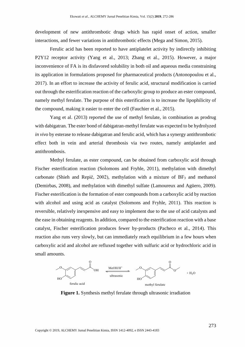
Figure 1. Synthesis methyl ferulate through ultrasonic irradiation
Ekowati et al., ALCHEMY Jurnal Penelitian Kimia, Vol. 15(2) 2019, 272-286
274 Copyright © 2019, ALCHEMY Jurnal Penelitian Kimia, ISSN 1412-4092, e ISSN 2443-4183
Cravotto et al., (2005) showed that Fischer esterification of cholic acid and methanol
assisted by p-toluene sulfuric acid as a catalyst was conducted successfully in three methods,
namely conventional methods (heating with reflux), microwave irradiation, and ultrasonic
waves (Cravotto et al., 2005). The conventional method gave a high yield, which is 93%, in
five hours reaction time, while the esterification reaction using microwave and ultrasonic
waves provided higher reaction yield (96% with ultrasonic waves and 97% with microwave
irradiation) in shorter reaction times (30 minutes for ultrasonic waves and 3 minutes for
microwave irradiation). This shows that reactions assisted by microwave and ultrasonic
irradiation are more efficient, can obtain higher reaction results, and require shorter reaction
times (Cravotto et al., 2005).
This study will evaluate the synthesis of methyl ferulate (Figure 1), and its kinetic
and order reaction using ultrasonic waves. Ultrasonic irradiation has been respected as a
clean and suitable method in organic synthesis throughout the last three decades, contrasted
with conventional methods; the procedure is more accessible. Through the ultrasonic
irradiation method, many organic reactions can be prepared in a higher yield, shorter reaction
time and milder conditions (Arani and Safari, 2011).
According to Pacheco et al. (2014), the effects of ultrasonic waves play a role in
increasing chemical processes, especially in cases where methods require drastic conditions
or long reaction times. Ultrasonic waves are emitted through the wave medium through by
inducing molecular vibrational movements. Therefore, the distance between molecules can
vary to be closer or further due to the movement of the oscillation of the molecule. If the
ultrasonic waves in the medium are getting more intense, one point will be reached where
the intra-molecular force of the fluid cannot maintain its molecular structure intact. As a
result, the molecular structure of the liquid will break and form a cavity. This cavity is a
denomination of a bubble cavity, and this process is called cavitation (Pacheco et al., 2014).
In the present research, synthesis of methyl ferulate was conducted by reacting ferulic
acid, methanol, and sulfuric acid as catalyst (H2SO4) using ultrasonic wave method at 55 °C
and 65 °C. This is because the temperature can influence the possibility of collisions between
molecules in the reaction. The higher the temperature in the reaction, the higher the kinetic
energy produced by compound molecules, thus more collisions occur between molecules.
This phenomenon will increase the reaction rate. At the same time, a reaction at a higher
temperature will also increase the yield percentage.
To conclude that the reaction at a temperature of 65 °C is greater than the reaction at
55 °C, the determination of the Fischer esterification rate synthesis of methyl ferulate was
Ekowati et al., ALCHEMY Jurnal Penelitian Kimia, Vol. 15(2) 2019, 272-286
275 Copyright © 2019, ALCHEMY Jurnal Penelitian Kimia, ISSN 1412-4092, e ISSN 2443-4183
evaluated. Theoretically, every 10 °C temperature increase will increase the reaction rate
two to three times (Singh, 2006).
METHODS
Ferulic acid (1) was bought from Aldrich; dimethyl sulfate, KOH, acetone,
chloroform, sodium hydrogen carbonate, H2SO4, ether, and methanol were purchased from
Merck. The synthesis reaction was conducted by ultrasonic Hwashin Technology
Powersonic 400 series and was observed with TLC using UV lamp on = 254 nm to spot
detection. Melting points were evaluated by Fischer-John melting point apparatus without
correction. UV spectra were achieved by Shimadzu HP 8452 UV-vis spectrophotometer.
The concentration of reactant was monitored through Densitometer Shimadzu. IR spectra
were presented using a Jasco FT-IR 5300 spectrophotometer. The 1HNMR spectra were
obtained from the JEOL JNM-ECS 400 instrument (1H NMR: 400 MHz) using appropriate
DMSO-d6 as a solvent.
Synthesis
A mixture of 400 mg (2.0 mmol) of ferulic acid, 10.0 ml (246.1 mmol) methanol,
and 2 drops (1.86 mmol) of concentrated H2SO4 in a round bottom flask was shaken
homogeneously and reacted using ultrasonic waves at the highest power. This reaction was
conducted at temperatures of 55 ˚C and 65 ˚C. The completion of the reaction was evaluated
by TLC at 0, 30, 60, 90, 180, and 360 minutes reaction time, by using ferulic acid as the
reference compound and chloroform: dichloromethane: methanol = 4: 3: 0.2 as the mobile
phase. After that, the rest of methanol was evaporated, and the residue was then neutralized
by NaHCO3 5% solution. The desired compound was extracted by chloroform and dried by
anhydrous MgSO4. The structure of the obtained compound was confirmed by UV, IR and
1H-NMR spectroscopy.
Determination of Linearity of Densitometry Method
The standard solution was arranged by taking 100 µl, 200 µl, 300 µl, and 500 µl of
the stock solution of 40,000 ppm and diluted by methanol (p.a.) until 1000 µl. Each
concentration was spotted on the TLC plate and eluted by chloroform: dichloromethane:
methanol (4:3:0.2). The area of the resulted spot was determined by a densitometer. The
concentration in ppm (as ordinate) and spot area (as abscissa) were then plotted to form a
line equation and calculated the linearity by linear equation as equation (1).
y = Ax + B (1)

Ekowati et al., ALCHEMY Jurnal Penelitian Kimia, Vol. 15(2) 2019, 272-286
276 Copyright © 2019, ALCHEMY Jurnal Penelitian Kimia, ISSN 1412-4092, e ISSN 2443-4183
The samples of this test were five; the Rtable of those samples was 0.8054. The curve
is linear if Rcalculated is greater than Rtable.
Determination of Reaction Order, Rate Constant (k) and Activation Energy (Ea)
Determination of the reaction rate was carried out by the graph method based on the
integral equation of the reaction rate between the concentration of ferulic acid (Ct) vs. time
(t). The rate constants (k) from each temperature, namely k55 and k65 were calculated after
the rate order was known The reaction rate constant (k) of the two temperatures substituted
in the Arrhenius equation as shown equation (2) to determine the energy of activation as
shown in equation (3).
logk65
k55=
Ea
2,303 R(
T65-T55
T65T55) (2)
Ea= (2,303 R T65 T55
T65-T55) log
k65
k55 (3)
Notes:
k65 = rate constant at 65 oC (cons-1 min-1)
k55 = rate constant at 55 oC (cons-1 min-1)
Ea = activation energy (kcal/mol)
R = gas constant (1.987 calories/mol.K)
T65 = absolute temperature 338 K (65 oC+273)
T55 = absolute temperature 328 K (55 oC+273)
In vivo Antiplatelet Activity Evaluation
Bleeding time assay: Twelve healthy adult male white mice aged 8−12 weeks
weighing 20−22 g, adapted and fed for one week. The mice were divided randomly into four
groups, while each group consisting of three mice. Each mouse was given food in the form
of pellets and drinks in the form of water every day in ad libitum. Test compounds were
given orally at a dose of 20 mg/kg, given for 7 days. On the 8th day, the bleeding time test
was carried out in the test animals. Bleeding time measurement was done by lying down the
mice on the test table. The tip of the tail was cleaned with alcohol 70% then the tail of the
mice was slashed along 2 mm. The dripping blood was placed on a filter paper every 15
seconds to observe whether there is still blood coming out. The duration, started from the
first drop of the blood until the blood stops dripping, was determined as the bleeding time.
The positive control was given oral aspirin dose of 20 mg/kg body weight (BW) in CMC-
Na 0.5%. The negative control group was given oral CMC-Na 0.5%. Other two groups were
given oral FA and MF in the same way and dose as that of aspirin. At the same time, the
Ekowati et al., ALCHEMY Jurnal Penelitian Kimia, Vol. 15(2) 2019, 272-286
277 Copyright © 2019, ALCHEMY Jurnal Penelitian Kimia, ISSN 1412-4092, e ISSN 2443-4183
clotting time was determined from the duration of the first blood drop until the clot formed.
This antiplatelet assay method was approved by the Ethical Commission of Airlangga
University.
In silico test
The in silico test was carried out by docking the molecules against COX-1 (PDB ID
4O1Z) as the target enzyme. 4O1Z is COX-1 crystal structure binding MXM_807 [A] as a
native ligand downloaded in a resolution of 2.90 Å from the RCSB Protein Data Bank
(www.rcsb.org). The 3D structure of the molecules was imported into workspace and
positioned into the cavity aligning to MXM_807 [A]. The molecular docking was performed
against the cavity-x using the MolDock SE algorithm with maximum 1500 iteration. The
enzyme-ligand affinity was determined from the docking score that expressed as the
Moldock Score (MDS). The best docking results must meet the criteria of the lowest energy
pose, and the docked molecule was in the same place as the native ligand.
RESULTS AND DISCUSSION
Synthesis
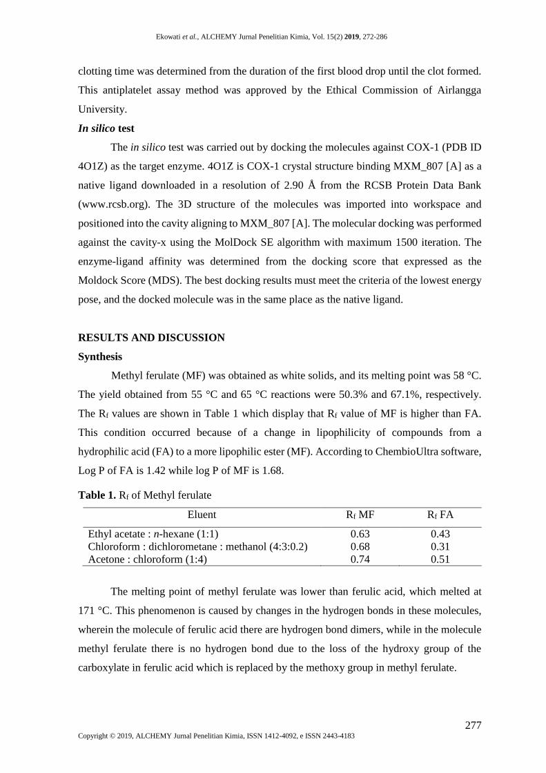
Methyl ferulate (MF) was obtained as white solids, and its melting point was 58 °C.
The yield obtained from 55 °C and 65 °C reactions were 50.3% and 67.1%, respectively.
The Rf values are shown in Table 1 which display that Rf value of MF is higher than FA.
This condition occurred because of a change in lipophilicity of compounds from a
hydrophilic acid (FA) to a more lipophilic ester (MF). According to ChembioUltra software,
Log P of FA is 1.42 while log P of MF is 1.68.
Table 1. Rf of Methyl ferulate
Eluent Rf MF Rf FA
Ethyl acetate : n-hexane (1:1) 0.63 0.43
Chloroform : dichlorometane : methanol (4:3:0.2) 0.68 0.31
Acetone : chloroform (1:4) 0.74 0.51
The melting point of methyl ferulate was lower than ferulic acid, which melted at
171 °C. This phenomenon is caused by changes in the hydrogen bonds in these molecules,
wherein the molecule of ferulic acid there are hydrogen bond dimers, while in the molecule
methyl ferulate there is no hydrogen bond due to the loss of the hydroxy group of the
carboxylate in ferulic acid which is replaced by the methoxy group in methyl ferulate.
Ekowati et al., ALCHEMY Jurnal Penelitian Kimia, Vol. 15(2) 2019, 272-286
278 Copyright © 2019, ALCHEMY Jurnal Penelitian Kimia, ISSN 1412-4092, e ISSN 2443-4183
The spectroscopic data of methyl ferulate were as follow: λmax (nm): 326 nm; IR
(ν;cm-1): 1718 (C=O ester), 3406 (O-H), 1642 (-C=C- alkene), 1590 and 1436 (-C=C-
aromatic), 1264 (-C-O). 1H-NMR (δ: ppm) (400 MHZ, CDCl3): 3.83 (s, 3H), 3.88 (s, 3H),
6.25 (1H, d, J = 16.0 Hz), 6.40 (1H,s), 6.88 (1H, d, J = 8.4 Hz), 7.00 (2H, d, J = 2.0 Hz),
7.60 (1H, d, J = 16.0 Hz). 13C-NMR (δ: ppm) (100 MHZ, CDCl3): 51.74 (1C), 55.91 (1C),
109.75 (1C), 114.72 (1C), 150.74 (1C), 123.05 (2C), 126.79 (1C), 147.13 (1C), 148.33 (1C),
168.13 (1C).
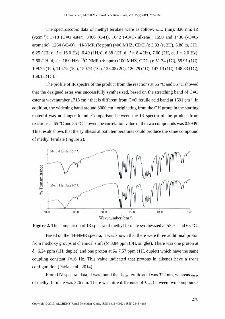
The profile of IR spectra of the product from the reactions at 65 °C and 55 °C showed
that the designed ester was successfully synthesized, based on the stretching band of C=O
ester at wavenumber 1718 cm-1 that is different from C=O ferulic acid band at 1691 cm-1. In
addition, the widening band around 3000 cm-1 originating from the OH group in the starting
material was no longer found. Comparison between the IR spectra of the product from
reactions at 65 °C and 55 °C showed the correlation value of the two compounds was 0.9949.
This result shows that the synthesis at both temperatures could produce the same compound
of methyl ferulate (Figure 2).
Figure 2. The comparison of IR spectra of methyl ferulate synthesized at 55 °C and 65 °C.
Based on the 1H-NMR spectra, it was known that there were three additional proton
from methoxy groups at chemical shift (δ) 3.84 ppm (3H, singlet). There was one proton at
δH 6.24 ppm (1H, duplet) and one proton at δH 7.57 ppm (1H, duplet) which have the same
coupling constant J=16 Hz. This value indicated that protons in alkenes have a trans
configuration (Pavia et al., 2014).
From UV spectral data, it was found that λmax ferulic acid was 322 nm, whereas λmax
of methyl ferulate was 326 nm. There was little difference of λmax between two compounds

Ekowati et al., ALCHEMY Jurnal Penelitian Kimia, Vol. 15(2) 2019, 272-286
279 Copyright © 2019, ALCHEMY Jurnal Penelitian Kimia, ISSN 1412-4092, e ISSN 2443-4183
because the methoxy group in methyl ferulate was not a chromophore group. Moreover, the
electronic transition from σ → σ* in the methyl group required a large amount of energy. As
a result, the shift of λ in the methyl ferulate was not significant (Pavia et al., 2014).
All of the spectral data showed that the same compounds from both reactions at 55
°C and 65 °C were successfully synthesized. The higher the temperature, the higher heat
energy that is received by the molecule, hence the greater the kinetic energy that can be used
by molecules to collide each other in order to initiate a reaction. This theory makes it is easier
for chemical reactions and higher probabilities to form new compounds.
The mechanism reaction for the synthesis of methyl ferulate using Fischer
esterification method is nucleophilic addition (Figure 3). The reaction began with the
protonation of the carbonyl group of ferulic acid by strong acid as catalyst. Protonated
carbonyl groups would be attacked by nucleophilic groups from alcohol. The nucleophilic
groups of carbonyl would take protons and formed water (H2O) that would be eliminated as
a by-product. The protons in the carbonyl group were then released again to reform a strong
acid catalyst (Solomons and Fryhle, 2011).
Figure 3. Reaction mechanism of methyl ferulate synthesis
The synthesis in the present study used the ultrasonic wave method, in which the
ultrasonic apparatus firstly transmitted the wave through the water as the medium, then the
wave propagated to all parts of the solution in the reaction flask. The mechanical activation
process, called cavitation, removed the attraction between molecules in the liquid phase.
Once small air bubbles were formed inside the cavity, these bubbles would absorb energy
from ultrasonic waves and produce the larger cavity.
As the socket grew, the air bubbles inside could no longer absorb ultrasonic energy.
Finally, the fluid around the cavity would go inside and broke the cavity. This rupture of the
air cavity created favorable conditions for chemical reactions. As a result of the vibrational
propagation, kinetic movements of molecules would occur followed by collisions that
triggered chemical reactions. In order to obtain the best condition of reactions, the oriented
Ekowati et al., ALCHEMY Jurnal Penelitian Kimia, Vol. 15(2) 2019, 272-286
280 Copyright © 2019, ALCHEMY Jurnal Penelitian Kimia, ISSN 1412-4092, e ISSN 2443-4183
procedures were conducted at several reaction temperatures. In the condition including
temperature at 35 °C and the moderate intensity of ultrasonic wave, the reaction ran slowly
and the new compound formed in the 120th minutes, based on the new spot observed on
TLC. The next reaction that was carried out at condition of 50 °C in temperature and
moderate intensity of ultrasonic wave, the new compound was found after 90 min. This
results indicated that the reaction proceeds faster by increasing the temperature.
In a further experiment, the temperature was increased to 65 °C, and the ultrasonic
intensity was raised from moderate to high intensity then the new compound was formed
after 60 min reaction. Based on these results, temperature of 65 °C and high ultrasonic
intensity were selected as the condition of methyl ferulate’s synthesis. As a comparison for
the study of reaction rates, 55 °C was chosen as reaction temperature with the same
ultrasonic intensity. Theoretically, the reaction rate was also influenced by the initial
concentration of the reacting compound (Singh, 2006).
To find out whether the TLC-densitometry can be used to monitor the changes of
ferulic acid concentration (reactant), a linearity test was carried out between the ferulic acid
level (ppm) and the percentage area (%). Data processing result the line equation y = 0.001x
+ 3.4833 with the correlation coefficient (R)= 0.9943. The Rtable suitable for this experiment
using 5 samples is 0.8054. The value of Rcalculated, which is greater than Rtable indicated that
the two variables had significant linear relationship.
Determination of Reaction Order
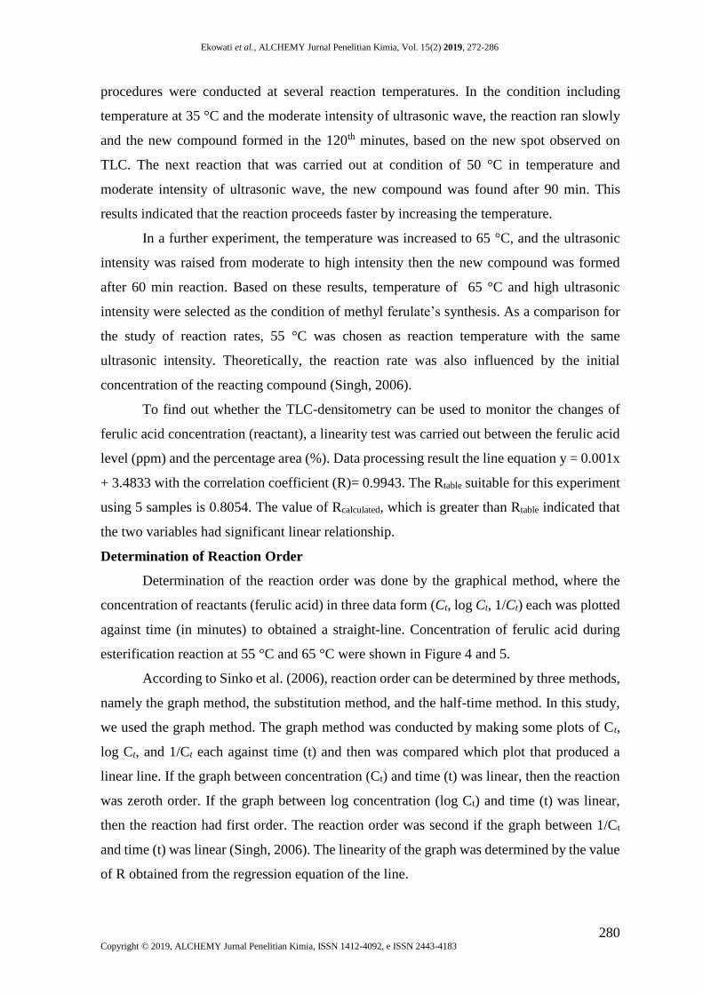
Determination of the reaction order was done by the graphical method, where the
concentration of reactants (ferulic acid) in three data form (Ct, log Ct, 1/Ct) each was plotted
against time (in minutes) to obtained a straight-line. Concentration of ferulic acid during
esterification reaction at 55 °C and 65 °C were shown in Figure 4 and 5.
According to Sinko et al. (2006), reaction order can be determined by three methods,
namely the graph method, the substitution method, and the half-time method. In this study,
we used the graph method. The graph method was conducted by making some plots of Ct,
log Ct, and 1/Ct each against time (t) and then was compared which plot that produced a
linear line. If the graph between concentration (Ct) and time (t) was linear, then the reaction
was zeroth order. If the graph between log concentration (log Ct) and time (t) was linear,
then the reaction had first order. The reaction order was second if the graph between 1/Ct
and time (t) was linear (Singh, 2006). The linearity of the graph was determined by the value
of R obtained from the regression equation of the line.
Ekowati et al., ALCHEMY Jurnal Penelitian Kimia, Vol. 15(2) 2019, 272-286
281 Copyright © 2019, ALCHEMY Jurnal Penelitian Kimia, ISSN 1412-4092, e ISSN 2443-4183
Figure 4. (a) Plot of ferulic acid concentration (ppm) (Ct) vs time in minutes (t) at 55 °C
(b) plot of log Ct vs time in minutes (t) at 55 °C (c) Plot of 1/Ct vs time in minutes (t) at 55
°C.
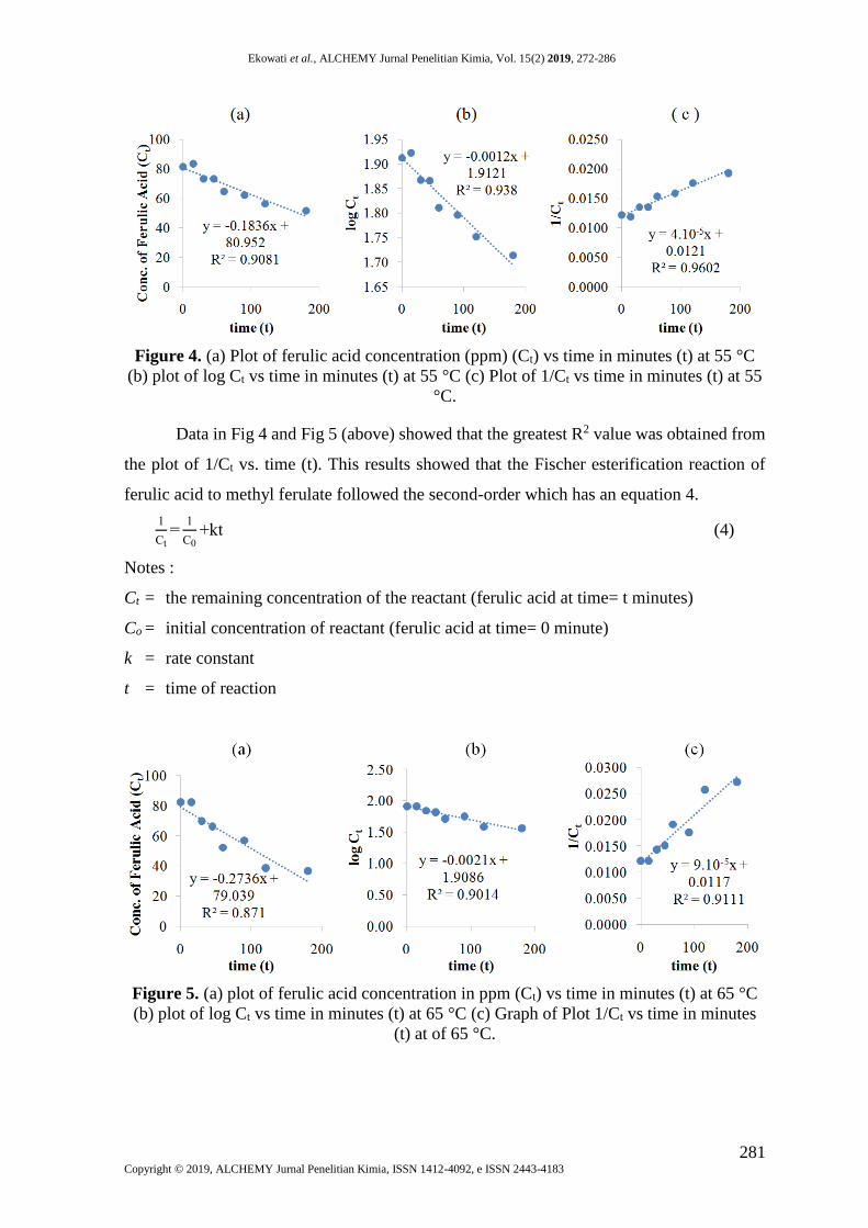
Data in Fig 4 and Fig 5 (above) showed that the greatest R2 value was obtained from
the plot of 1/Ct vs. time (t). This results showed that the Fischer esterification reaction of
ferulic acid to methyl ferulate followed the second-order which has an equation 4.
1
Ct=
1
C0+kt (4)
Notes :
Ct = the remaining concentration of the reactant (ferulic acid at time= t minutes)
Co = initial concentration of reactant (ferulic acid at time= 0 minute)
k = rate constant
t = time of reaction
Figure 5. (a) plot of ferulic acid concentration in ppm (Ct) vs time in minutes (t) at 65 °C
(b) plot of log Ct vs time in minutes (t) at 65 °C (c) Graph of Plot 1/Ct vs time in minutes
(t) at of 65 °C.
Ekowati et al., ALCHEMY Jurnal Penelitian Kimia, Vol. 15(2) 2019, 272-286
282 Copyright © 2019, ALCHEMY Jurnal Penelitian Kimia, ISSN 1412-4092, e ISSN 2443-4183
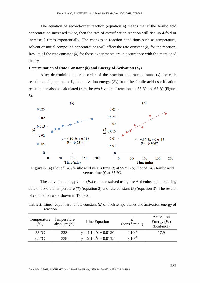
The equation of second-order reaction (equation 4) means that if the ferulic acid
concentration increased twice, then the rate of esterification reaction will rise up 4-fold or
increase 2 times exponentially. The changes in reaction conditions such as temperature,
solvent or initial compound concentrations will affect the rate constant (k) for the reaction.
Results of the rate constant (k) for these experiments are in accordance with the mentioned
theory.
Determination of Rate Constant (k) and Energy of Activation (Ea)
After determining the rate order of the reaction and rate constant (k) for each
reactions using equation 4., the activation energy (Ea) from the ferulic acid esterification
reaction can also be calculated from the two k value of reactions at 55 oC and 65 oC (Figure
6).
Figure 6. (a) Plot of 1/Ct ferulic acid versus time (t) at 55 °C (b) Plot of 1/Ct ferulic acid
versus time (t) at 65 °C.
The activation energy value (Ea) can be resolved using the Arrhenius equation using
data of absolute temperature (T) (equation 2) and rate constant (k) (equation 3). The results
of calculation were shown in Table 2.
Table 2. Linear equation and rate constant (k) of both temperatures and activation energy of
reaction
Temperature
(oC)
Temperature
absolute (K) Line Equation
k
(cons-1 min-1)
Activation
Energy (Ea)
(kcal/mol)
55 °C 328 y = 4.10-5x + 0.0120 4.10-5 17.9
65 °C 338 y = 9.10-5x + 0.0115 9.10-5

Ekowati et al., ALCHEMY Jurnal Penelitian Kimia, Vol. 15(2) 2019, 272-286
283 Copyright © 2019, ALCHEMY Jurnal Penelitian Kimia, ISSN 1412-4092, e ISSN 2443-4183
The results of data processing denoted that the Ea of the Fischer esterification of
ferulic acid was 17.9 kcal/mol. This means that each mole of ferulic acid requires 17.9 kcal
energy to carry out an esterification reaction with the help of a catalyst, then the energy
needed to react 2.10-3 moles of the ferulic acid compound is 0.0358 kcal or equal to 35.8
calories.
Antiplatelet Assay
The result of in vivo antiplatelet evaluation of methyl ferulate compared to the
negative control, positive control (aspirin) and ferulic acid are shown in Figure 7. Based on
the statistical analysis one way ANOVA, it was found that the blood clotting time and
bleeding time of the ferulic acid and methyl ferulate respectively were significantly different
from negative control (P= 0.000 and 0.002) which means that ferulic acid and methyl ferulate
had antiplatelet effects.
According to data in Figure 7, it appears that the change from carboxylic acid to ester
group can increases activity 1.6 times more potent based on the blood clotting time, and 1.35
times higher based on bleeding time data. However, the antiplatelet potency of methyl
ferulate was same with aspirin according to the capability to prolong clotting time and
bleeding time.
Figure 7. Antiplatelet activity histogram of methyl ferulate (MF), ferulic acid (FA), aspirin
(ASP), and control negative (CN). CT = clotting time, BT = bleeding time, (n=5).
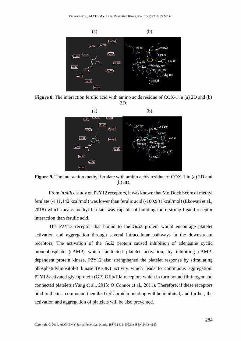
The results of docking in silico against COX-1 enzyme (PDB ID 4O1Z) indicate that
methyl ferulate has greater potency for COX-1 inhibition than ferulic acid. MolDock score
of methyl ferulate is 112.223 kcal/mol, while ferulic acid has 106.779 kcal/mol. As observed
in the Figure 8 and 9, methyl ferulate and ferulic acid showed the same mode of interaction
which involved their same functional groups and the same amino acids, i.e. Ala527 and
Met522, in cavity-4 (vol. 76.8; surface 239.36) of COX-1 (X=240.12; Y=99.88; Z=5.09).
The possible difference comes from the additional interaction of the hydrophobic and steric
interaction of methyl ester moiety with Val349, which are invisible in Figure 9.
Tim
e (s
eco
nd
)
CT
BT
Ekowati et al., ALCHEMY Jurnal Penelitian Kimia, Vol. 15(2) 2019, 272-286
284 Copyright © 2019, ALCHEMY Jurnal Penelitian Kimia, ISSN 1412-4092, e ISSN 2443-4183
(a) (b)
Figure 8. The interaction ferulic acid with amino acids residue of COX-1 in (a) 2D and (b)
3D.
(a) (b)
Figure 9. The interaction methyl ferulate with amino acids residue of COX-1 in (a) 2D and
(b) 3D.
From in silico study on P2Y12 receptors, it was known that MolDock Score of methyl
ferulate (-111,142 kcal/mol) was lower than ferulic acid (-100,981 kcal/mol) (Ekowati et al.,
2018) which means methyl ferulate was capable of building more strong ligand-receptor
interaction than ferulic acid.
The P2Y12 receptor that bound to the Gαi2 protein would encourage platelet
activation and aggregation through several intracellular pathways in the downstream
receptors. The activation of the Gαi2 protein caused inhibition of adenosine cyclic
monophosphate (cAMP) which facilitated platelet activation, by inhibiting cAMP-
dependent protein kinase. P2Y12 also strengthened the platelet response by stimulating
phosphatidylinositol-3 kinase (PI-3K) activity which leads to continuous aggregation.
P2Y12 activated glycoprotein (GP) GIIb/IIIa receptors which in turn bound fibrinogen and
connected platelets (Yang et al., 2013; O’Connor et al., 2011). Therefore, if these receptors
bind to the test compound then the Gαi2-protein bonding will be inhibited, and further, the
activation and aggregation of platelets will be also prevented.
Ekowati et al., ALCHEMY Jurnal Penelitian Kimia, Vol. 15(2) 2019, 272-286
285 Copyright © 2019, ALCHEMY Jurnal Penelitian Kimia, ISSN 1412-4092, e ISSN 2443-4183
CONCLUSIONS
The temperature effects on ultra sonic assisted synthesis of methyl ferulate are to
affect. the reaction rate constant and yield product. The value of reaction rate constant and
yield product at temperature of 65 ⁰C are higher than 55 ⁰C. The presence of a methyl group
of ester moiety increased the antiplatelet activity. The potency of methyl ferulate as
antiplatelet agent was same as aspirin.
ACKNOWLEDGMENTS
This research was funded by PTUPT Grant of Airlangga University in the year 2018.
REFFERENCES
Antonopoulou, I., Leonov, L., Jütten, P., Cerullo, G., Faraco, V., Papadopoulou, A., Kletsas,
D., Ralli, M., Rova, U. and Christakopoulos, P., 2017. Optimized Synthesis of Novel
Prenyl Ferulate Performed by Feruloyl Esterases from Myceliophthora Thermophila
in Microemulsions. Applied Microbiology and Biotechnology, 101(8), 3213-3226.
doi: 10.1007/s00253-017-8089-8.
Arani, N.M. and Safari, J., 2011. A Rapid and Efficient Ultrasound-Assisted Synthesis of 5,
5-diphenylhydantoins and 5,5-diphenyl-2-thiohydantoins. Ultrasonics
Sonochemistry, 18(2), 640-643. doi: 10.1016/j.ultsonch.2010.09.001.
Cravotto, G., Boffa, L., Turello, M., Parenti, M. and Barge, A., 2005. Chemical
Modifications of Bile Acids Under High-Intensity Ultrasound or Microwave
Irradiation. Steroids, 70(2), 77-83. doi: 10.1016/j.steroids.2004.09.007.
Demirbas, A., 2008. Comparison of Transesterification Methods for Production of Biodiesel
from Vegetable Oils and Fats. Energy Conversion and Management, 49(1), 125-130.
doi: 10.1016/j.enconman.2007.05.002.
Ekowati, J., Diyah, N.W., Nofianti, K.A., Hamid, I.S. and Siswandono, S., 2018. Molecular
Docking of Ferulic Acid Derivatives on P2Y12 Receptor and Their ADMET
Prediction. Journal of Mathematical and Fundamental Sciences, 50(2), 203-219. doi:
10.5614%2Fj.math.fund.sci.2018.50.2.8.
Fauchier, L., Clementy, N., Pelade, C., Collignon, C., Nicolle, E. and Lip, G.Y., 2015.
Patients with Ischemic Stroke and Incident Atrial Fibrillation: A Nationwide Cohort
Study. Stroke, 46(9), 2432-2437.doi: 10.1161/STROKEAHA.115.010270.
Lamoureux, G. and Agüero, C., 2009. A Comparison of Several Modern Alkylating Agents.
Arkivoc, 1, 251-264.
Mega, J.L. and Simon, T., 2015. Pharmacology of Antithrombotic Drugs: An Assessment of
Oral Antiplatelet and Anticoagulant Treatments. The Lancet, 386(9990), 281-291.
doi: 10.1016/S0140-6736(15)60243-4.
O’Connor, S., Montalescot, G. and Collet, J.P., 2011. The P2Y 12 Receptor as A Target of
Antithrombotic Drugs. Purinergic Signalling, 7(3), 325. doi: 10.1007/s11302-011-
9241-z.
Ekowati et al., ALCHEMY Jurnal Penelitian Kimia, Vol. 15(2) 2019, 272-286
286 Copyright © 2019, ALCHEMY Jurnal Penelitian Kimia, ISSN 1412-4092, e ISSN 2443-4183
Pacheco, B.S., Nunes, C.F., Rockembach, C.T., Bertelli, P., Mesko, M.F., Roesch-Ely, M.,
Moura, S. and Pereira, C.M., 2014. Eco-Friendly Synthesis of Esters Under
Ultrasound with p-Toluenesulfonic Acid as Catalyst. Green Chemistry Letters and
Reviews, 7(3), 265-270. doi: 10.1080/17518253.2014.941950.
Pavia, D.L., Lampman, G.M., Kriz, G.S. and Vyvyan, J.A., 2014. Introduction to
Spectroscopy. Cengage Learning, Stamford.
Shieh, W.C., Dell, S. and Repič, O., 2002. Large Scale Microwave-Accelerated
Esterification of Carboxylic Acids with Dimethyl Carbonate. Tetrahedron Letters,
43(32), 5607-5609. doi: 10.1016/S0040-4039(02)01116-4.
Singh, Y., 2006. Martin’s Physical Pharmacy and Pharmaceutical Sciences. New Jersey:
Department of Pharmaceutics Ernest Mario School of Pharmacy Rutgers, The State
University of New Jersey.
Solomons, T. W. G. & Fryhle, C. B, 2011. Organic Chemistry, 10th edition. John Wiley &
Sons, Inc., New Jersey.
Yang, X.Z., Diao, X.J., Yang, W.H., Li, F., He, G.W., Gong, G.Q. and Xu, Y.G., 2013.
Design, Synthesis and Antithrombotic Evaluation of Novel Dabigatran Prodrugs
Containing Methyl Ferulate. Bioorganic & Medicinal Chemistry Letters, 23(7),
2089-2092. doi: 10.1016/j.bmcl.2013.01.126.
Zhang, P.X., Lin, H., Qu, C., Tang, Y.P., Li, N.G., Kai, J., Shang, G., Li, B., Zhang, L., Yan,
H. and Liu, P., 2015. Design, Synthesis, and In Vitro Antiplatelet Aggregation
Activities of Ferulic Acid Derivatives. Journal of Chemistry, 2015, 1-7. doi:
http://dx.doi.org/10.1155/2015/376527.