Embed Size (px)
Citation preview

THE ULTRASTRUCTURE OF FROG
VENTRICULAR CARDIAC MUSCLE AND ITS
RELATIONSHIP TO MECHANISMS OF
EXCITATION-CONTRACTION COUPLING
NANCY A. STALEY and ELLIS S. BENSON
From the Department of Laboratory Medicine, the University of Minnesota, Minneapolis,Minnesota 55455
ABSTRACT
Frog ventricular cardiac muscle has structural features which set it apart from frog andmammalian skeletal muscle and mammalian cardiac muscle. In describing these differences,our attention focused chiefly on the distribution of cellular membranes. Abundant intercellular clefts, the absence of tranverse tubules, and the paucity of sarcotubules, togetherwith exceedingly small cell diameters (less than 5 p), support the suggestion that the mecha-nism of excitation-contraction coupling differs in these muscle cells from that now thought tobe characteristic of striated muscle such as skeletal muscle and mammalian cardiac muscle.These structural dissimilarities also imply that the mechanism of relaxation in frog ventricu-lar muscle differs from that considered typical of other striated muscles. Additional ultra-structural features of frog ventricular heart muscle include spherical electron-opaque bodieson thin filaments, inconstantly present, forming a rank across the I band about 150 mgt fromthe Z line, and membrane-bounded dense granules resembling neurosecretory granules.The functional significance of these features is not yet clear.
In frog skeletal muscle, tubular membrane-
bounded spaces, continuous with the interstitial
space, penetrate the muscle cell transversely at thelevel of the Z lines of the myofibrils lying in register
with one another. The continuity of the mem-
branes of this transverse tubular system with the
cell membrane and of its lumen with the inter-
stitial space of muscle has been clearly demon-strated by Huxley (1) using ferritin.
Mammalian heart muscle appears also to possess
a transverse tubular system penetrating the mus-
cle cells and having continuities with the cell mem-brane and interstitial space (2, 3). Rayns et al. (4),
using a freeze-etching technique, observed theportals of these transverse tubules on the surfaceof the cell membrane. The probable role of this
internal membrane system in the inward spread
of the excitatory signal to the contractile sites in
the myofibrils has been elegantly demonstrated
by Huxley and Taylor (5) who used microelec-
trodes to achieve local depolarization of the cellmembrane.
From these observations and the ones on therole of calcium in the activation of the contractilemechanism (6, 7), a concept of excitation-contrac-
tion coupling has emerged which states that theeffects of excitation are borne inward across themuscle fiber by the transverse tubular system re-leasing calcium ion from the sarcoplasmic reticu-lum surrounding the myofibrils and thereby acti-
vating the contractile elements (8).In frog heart muscle, however, Niedergerke
99
Dow
nloaded from http://rupress.org/jcb/article-pdf/38/1/99/1384633/99.pdf by guest on 17 D
ecember 2021

(9, 10) found that activation of the contractilemechanism could be explained by diffusion ofcalcium directly from the cell surface. We thoughtit would be of interest, therefore, to examine thestructure of frog ventricular muscle cells by elec-tron microscopy. We especially wanted to see ifthere were important structural differences in thecellular membrane systems between frog heartmuscle and frog skeletal and mammalian heartmuscle.
MATERIALS AND METHODS
Adult frogs (Rana pipiens), male or female, obtainedat various seasons of the year, were stored for severaldays at 4C before use. Each was pithed, the heartwas exposed, and the ventricular chamber was in-jected with a cold fixation fluid (see below). Theheart was then quickly excised, and blocks of ven-tricular muscle, 1 mm 3 or smaller in size, were placedin the fixation fluid and kept at 0-4°C for 2-12 hr.
The fixation fluids used were either 3.5% glutaral-dehyde in 0.1 M cacodylate buffer (pH 7.2) (11) or acombination of 2% paraformaldehyde and 2.5%glutaraldehyde in 0.1 M cacodylate buffer (pH 7.2)(12).
The blocks were then washed for 2 hr in 0.2 Msucrose in 0.05 cacodylate buffer (pH 7.2), post-fixed in 1% osmium tetroxide in Veronal-acetatebuffer (pH 7.4) (13), dehydrated in a graded seriesof alcohol-water mixtures, and embedded in Epon812 (14).
Thin sections were stained with uranyl acetate andlead citrate (15) and examined in an RCA EMU-3Gelectron microscope.
RESULTS
Frog ventricle has a very spongelike appearancewhen seen from the direction of its endocardialsurface. This appearance is due to an abundanceof small fissures between trabecular strands ofmuscle. The fissures extend through the wall ofthe ventricle to the epicardial surface covering.The porous, trabecular structure of the ventricleas seen from the ventricular chamber is illustratedin Fig. 1, a sketch which is taken from Gompertz(16).
Each strand of ventricular muscle, or eachtrabeculum, is lined by a single layer of endothelialcells with an underlying continuous basementmembrane (Fig. 2). This layer of cells and itsbasement membrane is the endocardial lining ofthe ventricular chamber. The wall contains nocapillaries, and perfusion of the cells apparentlytakes place from the ventricular cavity by way of
the fissures and, thence, through the endotheliallining of each muscle strand or trabeculum. Be-tween the endothelium and the cell membrane ofthe muscle cells lies an extracellular space whichcontains bundles of collagen fibers, nerve fibers,and occasional macrophages and fibroblasts.
Nerve fibers, many with no Schwann cell cover-ing, are quite numerous (Fig. 3). They occursingly or in bundles, often in proximity to musclecells. Along the fibers are found occasional focaldilations containing agranular synaptic vesiclesand a few "dense core" granular vesicles; no dis-tinct specialized synaptic junctions with musclecells were noted.
Each trabeculum with its endothelial coveringmeasures 10-50 in diameter and contains from10 to 15 muscle cells; the long axis of each celllies parallel to that of its neighbors. The group ofmuscle cells within the trabeculum is surroundedby a membrane similar to the sarcolemmal mem-brane of other muscle cells: it is made up of a base-ment membrane and plasma membrane, with atranslucent intermediate layer. The individual
FIGURE 1 A sketch of frog ventricle, viewed from theendocardial surface, illustrating the trabecular struc-ture of the muscle. Redrawn from reference 16.
100 THE JOURNAL OF CELL BIOLOGY - VOLUME 38, 1968
Dow
nloaded from http://rupress.org/jcb/article-pdf/38/1/99/1384633/99.pdf by guest on 17 D
ecember 2021

FIGURE 2 Longitudinal section through a portion of one trabeculum shows the surface endotheliallayer (E), subendothelial extracellular space and muscle cells. Intercellular clefts (ICC) separate in-dividual muscle cells, each of which contains one to three parallel myofibrils (Myo). X 7,000.
N. A. STALEY AND E. S. BENSON Frog Ventricular Cardiac Muscle 101
Dow
nloaded from http://rupress.org/jcb/article-pdf/38/1/99/1384633/99.pdf by guest on 17 D
ecember 2021

muscle cells are separated from each other byintercellular clefts (Figs. 2 and 4): these are mem-
brane-bounded spaces, the membranous linings ofwhich are continuous with the plasma membrane
of the sarcolemma surrounding the bundle of mus-cle cells in each trabeculum. The basement mem-
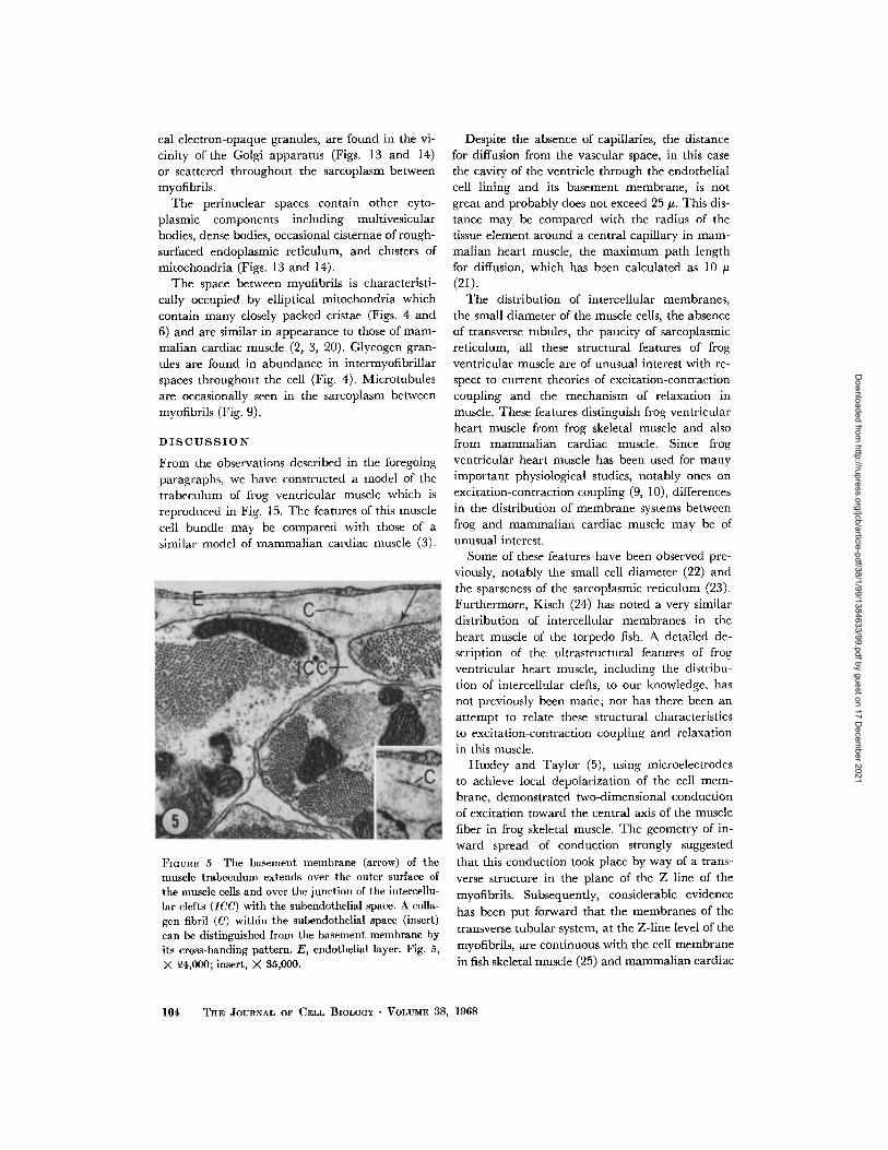
brane of the sarcolemma, in general, does notbecome a part of the lining of the intercellularclefts. At the junction of the clefts with the sarco-lemma, the basement membrane of the latter
separates the lumen of the intercellular clefts fromthe extracellular space beneath the endotheliallining of the ventricular cavity (Fig. 5). However,in some of the wider intercellular clefts, the base-ment membrane dips into the cleft lumen for ashort distance.
The intercellular clefts appear to be continuous
cell boundaries. In much of their course they arerelatively narrow, varying in width from 200 to400 A. They widen at points at which the curva-tures of the surfaces of the adjacent cells diverge(Fig. 4), and in these pockets wisps of basementmembrane-like material appear. At frequent butirregular intervals, along the intercellular clefts,fusiform, electron-opaque thickenings of the ad-jacent plasma membranes occur (Figs. 6 and 7).These thickenings are somewhat granular andhave the appearance of the desmosomes seen inthe intercalated disc of mammalian cardiac mus-
FIGoIrI. 3 Cross-section of a nerve fiber, within thesubendothelial extracellular space, which containsagranular (a) and granular (g) vesicles. No Schwanncell or specialized synaptic junction is present. E,endothelial layer. X 18,000.
cle, which occur in the portions of the disc parallelto the long axis of the myofibrils (17), and whichin turtle atrial muscle are present at the cellboundaries of adjacent myocardial cells (18).These structures often lie directly opposite the Zlines of the underlying myofibrils of one of theadjacent muscle cells, and the dense material ofthe desmosome is then continuous with the denseZ-line substance (Fig. 6).
The abundance of intercellular clefts gives the
cells relatively narrow diameters. The cell di-ameters, from outer cell membrane to cleft orfrom cleft to cleft, range from I to 5 . In cross-sections of a cell, one can see three to five myo-fibrils per cell (Fig. 4). In longitudinal section,one to two myofibrils are seen between one cleft
and the next (Figs. 2 and 6). Typically, in scanningacross a longitudinal section, one notes an arrange-ment of this type: cleft, myofibril, a row of mito-chondria, myofibril, cleft (Fig. 7).
Intercalated discs are numerous and have a
simple structure (Fig. 8). They run transverselyacross cells from one cleft to the next and crossmyofibrils in a plane where the Z line mightotherwise be found. They have the typical appear-ance of what has been termed the "interfibrillar
segment" of the disc (17); they are made up oftwo dense membranes separated by a clear spaceand surrounded by an irregular band of electron--opaque material. They lack the steplike course ofdiscs seen in mammalian cardiac muscle wherelongitudinal segments between myofibrils ("inter-sarcoplasmnic segments") alternate with transverse
segments intersecting myofibrils (interfibrillarsegments) (17). Tight junctions, similar to thoseof the intercalated discs of mammalian heartmuscle, were not seen.
For the most part, the myofibrils have the typicalappearance of striated muscle in general, withsarcomeres being made up of interdigitating thickand thin filaments (Figs. 9 and 10). As in mamma-lian cardiac muscle, the H zone, when present, isindistinct. The M line is seen only occasionally(Fig. 9). A curious, and as yet unexplained, find-ing in some preparations is the occurrence of
spherical electron-opaque bodies, approximately
45 m/p in diameter, on thin filaments. These bodies
form a row across the I band, the row being
parallel to the Z line and approximately 100-150
mpy from it in sarcomeres at or near rest length
(Figs. 9-11).We have seen no transverse tubules in frog
102 THE JOURNAL OF CELL BIOLOGY VOLUME 38, 1968
Dow
nloaded from http://rupress.org/jcb/article-pdf/38/1/99/1384633/99.pdf by guest on 17 D
ecember 2021

FIGtuE 4 Cross-section of one trabeculum. Intercellular clefts (arrows) widen irregularly betweenmuscle cells. Each cell contains three to five myofibrils separated by clusters of mitochondria (M) andglycogen. E, endothelial layer. X 13,000.
ventricular cardiac muscle. This tubular systemwhich is so prominent in mammalian cardiacmuscle (2, 3) appears to be absent from the cardiacmuscle of the frog. Furthermore, the sarcoplasmicreticulum, the longitudinal system of membrane-bounded tubules surrounding each myofibril infrog skeletal and mammalian cardiac muscle, isexceedingly sparse in the frog heart muscle wehave examined. It appears to consist of scatteredsmall, single, membrane-bounded vesicles bearingno consistent spatial relationship to sarcomeretopography (Figs. 6 and 12). In some places, ele-ments of the sarcoplasmic reticulum are closelyapposed to the cell membrane and intercellularclefts. Occasionally small vesicles may also be seen
in the intermyofibrillar spaces at the Z lines (Fig.11). These vesicles are lined by a single membraneand do not have the appearance of transversetubules but resemble the "circumferential Z linetubules" described by Simpson in mammaliancardiac muscle (19). Small pinocytotic vesiclesare often seen immediately beneath the cell mem-brane (Fig. 12).
Nuclei are numerous and contain prominentnucleoli; the nuclear envelope has many pores(Fig. 13). Often in the sarcoplasm near one poleof a nucleus a Golgi apparatus is seen; it is com-posed of three to four parallel cisternae withnumerous adjacent clear vesicles. Vesicles approxi-mately 100 m/p in diameter, and containing spheri-
N. A. STALEY AND E. S. BENSON Frog Ventricular Cardiac Muscle 103
Dow
nloaded from http://rupress.org/jcb/article-pdf/38/1/99/1384633/99.pdf by guest on 17 D
ecember 2021
cal electron-opaque granules, are found in the vi-
cinity of the Golgi apparatus (Figs. 13 and 14)
or scattered throughout the sarcoplasm betweenmyofibrils.
The perinuclear spaces contain other cyto-
plasmic components including multivesicular
bodies, dense bodies, occasional cisternae of rough-
surfaced endoplasmic reticulum, and clusters of
mitochondria (Figs. 13 and 14).
The space between myofibrils is characteristi-
cally occupied by elliptical mitochondria which
contain many closely packed cristae (Figs. 4 and
6) and are similar in appearance to those of mam-
malian cardiac muscle (2, 3, 20). Glycogen gran-
ules are found in abundance in intermyofibrillar
spaces throughout the cell (Fig. 4). Microtubules
are occasionally seen in the sarcoplasm between
myofibrils (Fig. 9).
DISCUSSION
From the observations described in the foregoing
paragraphs, we have constructed a model of the
trabeculum of frog ventricular muscle which is
reproduced in Fig. 15. The features of this muscle
cell bundle may be compared with those of a
similar model of mammalian cardiac muscle (3).
FIGuRE 5 The basement membrane (arrow) of themuscle trabeculum extends over the outer surface ofthe muscle cells and over the junction of the intercellu-lar clefts (ICC) with the subendothelial space. A colla-gen fibril (C) within the subendothelial space (insert)can be distinguished from the basement membrane byits cross-banding pattern. E, endothelial layer. Fig. 5,X 24,000; insert, X 35,000.
Despite the absence of capillaries, the distancefor diffusion from the vascular space, in this case
the cavity of the ventricle through the endothelialcell lining and its basement membrane, is notgreat and probably does not exceed 25 u. This dis-
tance may be compared with the radius of the
tissue element around a central capillary in mam-malian heart muscle, the maximum path length
for diffusion, which has been calculated as 10 u
(21).The distribution of intercellular membranes,
the small diameter of the muscle cells, the absence
of transverse tubules, the paucity of sarcoplasmicreticulum, all these structural features of frogventricular muscle are of unusual interest with re-
spect to current theories of excitation-contraction
coupling and the mechanism of relaxation in
muscle. These features distinguish frog ventricularheart muscle from frog skeletal muscle and alsofrom mammalian cardiac muscle. Since frogventricular heart muscle has been used for manyimportant physiological studies, notably ones on
excitation-contraction coupling (9, 10), differencesin the distribution of membrane systems between
frog and mammalian cardiac muscle may be of
unusual interest.Some of these features have been observed pre-
viously, notably the small cell diameter (22) and
the sparseness of the sarcoplasmic reticulum (23).
Furthermore, Kisch (24) has noted a very similar
distribution of intercellular membranes in the
heart muscle of the torpedo fish. A detailed de-
scription of the ultrastructural features of frog
ventricular heart muscle, including the distribu-
tion of intercellular clefts, to our knowledge, hasnot previously been made; nor has there been an
attempt to relate these structural characteristics
to excitation-contraction coupling and relaxationin this muscle.
Huxley and Taylor (5), using microelectrodes
to achieve local depolarization of the cell mem-
brane, demonstrated two-dimensional conduction
of excitation toward the central axis of the muscle
fiber in frog skeletal muscle. The geometry of in-
ward spread of conduction strongly suggested
that this conduction took place by way of a trans-
verse structure in the plane of the Z line of the
myofibrils. Subsequently, considerable evidence
has been put forward that the membranes of the
transverse tubular system, at the Z-line level of the
myofibrils, are continuous with the cell membrane
in fish skeletal muscle (25) and mammalian cardiac
104 THE JOURNAL OF CELL BIOLOGY VOLUME 38, 1968
Dow
nloaded from http://rupress.org/jcb/article-pdf/38/1/99/1384633/99.pdf by guest on 17 D
ecember 2021

FIGURE 6 Desmosomes (arrows) occur along the intercellular clefts, often at the Z line. Here the electron-opaque material of the desmosome appears continuous with the Z-line material. Sarcoplasmic reticulum(SR) is scant and shows no consistent relation to sarcomere structure. M, mitochondria. X 13,500.
FIGURE 7 Each muscle cell typically contains two or more myofibrils separated by a sarcoplasmic corecontaining mitochondria (M) or a nucleus (N). A Golgi apparatus (G), with associated electron-opaquegranules (g), is located near the nucleus. Desmosomes (D) occur along intercellular clefts. X 15,000.
105
Dow
nloaded from http://rupress.org/jcb/article-pdf/38/1/99/1384633/99.pdf by guest on 17 D
ecember 2021

FIGURE 8 Intercalated discs (ID) show no associated tight junctions. Within the sarcomnere, the I bandmay show an increased density adjacent to the Z line (arrows). N, nucleus. X 19,500.
muscle (2, 3). Huxley (1), using ferritin, clearlydemonstrated continuity of the lumen of the trans-verse tubules with the interstitial space in frogskeletal muscle.
These observations recalled earlier ones of Hill
(26, 27) who, in noting the speed of transition fromrest to full activity after excitation of frog skeletalmuscle fibers, suggested that this transition was
too rapid to be explained by diffusion of an acti-vating substance from the cell membrane. He pre-dicted that a process, not a substance, must bearthe signal for contraction inward to the core of thefiber.
A substantial body of evidence supports the con-tention that an increase in the concentration ofcalcium ion in the myofibrillar space is the finalstep in the activation of contraction. Followingthe observation of Heilbrunn and Wiercinski(28) that injection of calcium into frog skeletalmuscle cells induced contraction, the studies ofEbashi (6) and of Hasselbach and Makinose (29)
revealed that the membranes of the sarcoplasmicreticulum were able to accumulate calcium and
that this activity was the basis of their relaxing
effect on myofibrils. Weber and her collaborators(7) showed that the contractile activity of myo-fibrillar preparations was controlled by calciumion in the micromolar range of concentration(10-6-10 - 7 M).
From these observations, the following pictureof excitation-contraction coupling in skeletal mus-cle has been constructed. The effect of membraneexcitation is borne inward across the muscle celldiameter by the transverse tubular system whichtriggers a release of calcium from the membranesof the sarcoplasmic reticulum into the myofibrillarspace. Calcium saturates sites on the myofibrils toinduce contraction. It is then reaccumulated bythe sarcoplasmic reticulum and, as its concentra-tion falls in the myofibrillar space, calcium is with-drawn from the myofibrils and contraction comesto an end (30).
The observations we have described in thispaper suggest to us that this picture of the processof excitation-contraction coupling and activationcannot apply in every particular to frog ventricularheart muscle. The absence of transverse tubulesand the paucity of sarcoplasmic reticulum in these
106 THE JOURNAL OF CELL Bi.OGY VOI.U ME 38, 1968
Dow
nloaded from http://rupress.org/jcb/article-pdf/38/1/99/1384633/99.pdf by guest on 17 D
ecember 2021

FIGURE 9 Sarcomeres show an indistinct H zone (H) and M line (M). Spherical bodies (black arrows)attached to thin filaments form rows parallel to the Z lines. Microtubules (double-stemmed arrow) areinfrequently seen in the sarcoplasm. G, Golgi apparatus. X 13,500.
FIGURE 10 Cross-section through sarcomeres at various levels show spherical bodies (arrows) asso-ciated with Z lines (Z) and thin filaments in I bands (1). Translucent zones within myofibrils representareas of glycogen storage. Myofibrils in cross-section (insert) show hexagonal array of thick filaments withthin filaments at triagonal points. E, endothelial layer. X 23,000; insert, X 82,000.
Dow
nloaded from http://rupress.org/jcb/article-pdf/38/1/99/1384633/99.pdf by guest on 17 D
ecember 2021

FIGURE 11 Small vesicles of sarcoplasmic reticulum (SR) are occasionally seen at or near Z lines or
adjacent to the plasma membrane. Spherical bodies (arrow) associated with thin filaments are located
150 myn from the Z line. The I band shows an increased density within the region between the sphericalbodies and the Z line. X 23,500.
FIGunE 12 Sarcoplasmic reticulum (SR) is not well developed and consists of single membrane-boundedvesicles. Transverse tubules are absent. Pinocytotic vesicles (arrows) are often numerous. X 20,000.
108
Dow
nloaded from http://rupress.org/jcb/article-pdf/38/1/99/1384633/99.pdf by guest on 17 D
ecember 2021

FIGURE 13 The Golgi apparatus (G), located near the nucleus, is formed of two to three parallel cisternae.Adjacent to it are numerous 1000-A electron-opaque granules (g), multivesicular bodies (MVB) andan occasional profile of rough endoplasmic reticulum (ER). Nuclear pores are prominent (arrows).X 28,000.
FIGURE 14 Dense bodies (DB) and microtubules (arrow) are found in the perinuclear area associatedwith the Golgi apparatus. Electron-opaque granules (g) are seen within Golgi cisternae. X 31,500.
109
Dow
nloaded from http://rupress.org/jcb/article-pdf/38/1/99/1384633/99.pdf by guest on 17 D
ecember 2021

FIGUiE 15 A diagrammatic model of one trabeculum of frog ventricular heart muscle with its endothelial
covering (E) illustrates the relationship of the intercellular clefts (ICC) to the muscle cells. The inter-
cellular clefts are narrow extensions of extracellular space that form interconnecting channels around
muscle cells. Each muscle cell is approximately 5 , in diameter and contains three to five myofibrils
(Myo). A Golgi apparatus (G), associated electron-opaque granules and mitochondria (M) are present
adjacent to the nucleus (N). Desmosoines (D) occur along the intercellular clefts frequently at the Z line.
muscle cells make it necessary to consider alterna-tive mechanisms.
In experiments on frog ventricular muscle inwhich the movements of 4 5 Ca were studied,
Niedergerke (9) noted the effects of contracture-producing solutions (reduced external Na con-centration or increased external K concentration)on 45Ca uptake and loss from the muscle cells.These contracture solutions increased calcium up-take by the cells; the strength of the contracturewas related to the amount of calcium influx. Whenthese movements were studied in the beating heart,it was found that either increased extracellular Kconcentration or decreased Na concentration pro-duced both an increase in Ca influx and an in-creased strength of contraction (10). Both Ca up-take into and release from the cellular space were
increased during activity when compared to rest.
Niedergerke suggested that entry of calcium into
the cell during the action potential initiated con-
traction in frog ventricular muscle. In mammalian
cardiac muscle, on the other hand, Langer (31)
has presented evidence that the inotropic effect of
low extracellular sodium ion concentration is
associated with movement of calcium ion from an
intracellular space, presumably the sarcoplasmic
reticulum.
Calculations by Sandow (32), using data of
Bianchi and Shanes (33) on the amount of calcium
entry into skeletal muscle cells during contraction,
make it clear that the influx of extracellular cal-cium in these cells is much too small to account foractivation of the contractile elements. Similarobservations have been made and conclusions
110 THE JOURNAL OF CEIL BIOLOGY VOLUME 38, 1968
Dow
nloaded from http://rupress.org/jcb/article-pdf/38/1/99/1384633/99.pdf by guest on 17 D
ecember 2021

have been drawn by Winegrad (34) in experi-
ments with guinea pig atrial muscle. Using adifferent type of calculation, Winegrad concluded
that the amount of calcium influx during a twitchwas much too small to account for activation of
contraction. Recently, on the basis of the effect ofcontracture-inducing solutions and the magnitudeof the calcium influx during membrane depolariza-
tion, Edwards and Lorkovic (35) have renewed
the suggestion of Niedergerke (10) that contrac-tion in frog ventricle is initiated by entry of extra-cellular calcium into the muscle cell.
In frog ventricular muscle, Niedergerke (10)found an influx of Ca' per beat of about 3 X
10- 6 moles/liter of myocardium. In order to acti-vate the myofilaments and produce contraction,
the ionic calcium concentration of the sarcoplasmin the myofibrillar space must rise above that re-
quired for threshold activity, and the calcium-binding sites on the myofilaments also must beoccupied to an extent greater than that necessaryfor threshold activity (34). Weber and Herz (36)have shown that threshold activity of skeletalmyofibrils occurs at a Ca4 4 concentration of aboutI X 10- 7
M and that maximum activity is reached
at a concentration of about 1 X 10- 6 M. Weber etal. (37) furthermore have found that, over thisrange of Ca++ concentration, bound calcium ofmyofibrils goes from about 0.9 to 2.2 gumoles/g ofactomyosin. Although no data are available con-cerning the calcium-binding properties of frogcardiac myofibrils, Katz and Repke (38) havefound the Ca++ sensitivity of dog cardiac actomyo-
sin to be approximately the same as that of rabbitskeletal actomyosin. If we assume, therefore, that
the Ca4 + sensitivity and binding properties offrog cardiac myofibrils are roughly similar to
those of skeletal myofibrils and, furthermore, thatthe actomyosin content of frog heart muscle isapproximately the same as that of rabbit heartmuscle, an estimate of the amount of added Ca'4
needed to maximally activate resting frog cardiacmyofibrils may be made.
A liter of frog ventricular myocardium containsabout 650 ml of intrafiber water (9) and, if the
actomyosin concentration is approximately thesame as that of rabbit myocardium, 56 g of acto-
myosin (39). When the myofibrils go from the
inactive state to maximum activity, an increase of
0.6 Amoles of free Ca+4 and 72.8 gumoles of calcium
bound to actomyosin per liter of myocardium
would be required. The amount of calcium re-
quired to induce maximal activity (73.4 moles/
liter) is far in excess of the amount Niedergerkefound to be the maximum influx per contraction
(3 moles/liter).These calculations make it seem unlikely that
enough calcium enters the frog cardiac musclecell to activate the contractile elements in eachcontraction. Alternatively, calcium may be re-
leased by membrane depolarization from theinner surface of the cell membrane, including the
membranes of the intercellular clefts, may diffuse
to the sites of action on the contractile proteins
and may return to the cell membrane in relaxation.A mechanism such as this one, indeed, has been
suggested by Niedergerke (9).Hill (26, 27) considered activation on the basis
of a process of diffusion from the cell membrane. His
calculations, based on the diffusion constant ofcalcium or a similar substance, the time for diffu-
sion (the time from excitation to the peak of acti-vation) and the radius of diffusion (the radius of
the muscle fiber), indicated that in skeletal musclecells the time-distance relationships ruled outdiffusion from the cell membrane as the basis ofactivation. We have carried out similar calcula-
tions on frog ventricular muscle. If activation is asrapid in onset in this muscle as in frog skeletalmuscle, then the time for diffusion of an activatingsubstance would be 15 msec (27). At a maximumcell diameter of 5 pu (compared to 100 pt for frog
skeletal muscle), ample time for diffusion of cal-cium from the cell membrane is available for full
activation to take place by this mechanism.It would appear, thus, that important differ-
ences exist between the mechanism of activation ofcontractile elements in frog ventricular cardiacmuscle, on the one hand, and that in frog andmammalian skeletal muscle and mammalian
cardiac muscle, on the other. While in both groups,
undoubtedly, variation in calcium concentrationin the myofibrillar space is the immediate meansof activation and relaxation of the contractile
elements, the site of release and reaccumulation of
activating calcium differs between the two groupsof muscle types. In frog heart muscle, the acti-
vating calcium apparently comes from the cellmembrane and returns, in relaxation, to the cell
membrane location. In skeletal muscle and in
mamalian cardiac muscle, the activating calcium
is released from an intracellular compartment and
is reaccumulated by this compartment in relaxa-
tion. Considerable evidence has been advanced
N. A. STALEY AND E. S. BENSON Frog Ventricular Cardiac Muscle 111
Dow
nloaded from http://rupress.org/jcb/article-pdf/38/1/99/1384633/99.pdf by guest on 17 D
ecember 2021
that this compartment is the sarcoplasmic reticu-lum (8, 30) and that the stimulus of excitation isborne to it by the transverse tubular system (5,40).
The absence of transverse tubules in frog ven-tricular heart muscle, the paucity of sarcoplasmicreticulum, the distribution and continuities of theintercellular clefts, and the small cell diameterall favor a mechanism of activation in which aflux of calcium from a superficial site such as thecell membrane is the activating stimulus for con-traction and in which a return of calcium to thecell membrane is followed by relaxation. If thisformulation is correct, isolated preparations of cellmembrane from frog ventricular muscle might beexpected to possess some of the properties of iso-lated preparations of sarcoplasmic reticulum(vesicular relaxing factor) of skeletal muscle.Notably such preparations may have the ability(a) to inhibit contraction of isolated myofibrils,(6) to inhibit the ATPase activity of myofibrils,and (c) to accumulate calcium in the presence ofATP (30). Studies of such activity may be of someinterest.
It should be pointed out that Niedergerke (9)cited unpublished observations made by Huxleyand himself, using electron microscopy, in whichthe absence of a highly developed sarcoplasmicreticulum in frog ventricular muscle was noted.
The intercellular clefts are true cell boundaries.They form uninterrupted boundaries between ad-jacent cells. The absence of continuous and well-formed basement membranes, the frequent oc-currence of desmosomes along their course, andthe narrowness of the space within the cleft sug-gest that these boundaries, like the intercalateddiscs, may be low resistance spaces across whichexcitatory impulses readily travel (17).
It must be noted, however, that we have seenno true tight junctions, regions in which the op-posing cell membranes are fused along their outerlamellae, in the intercellular clefts of frog heartmuscle. Tight junctions (nexuses) have been de-scribed in the frog atrium after permanganatefixation (17). These structures are apparentlyquite labile and easily disrupted by hypertonicsolutions (17). Baldwin (41) found very occasionaltight junctions in frog atrium after glutaraldehydefixation; Revel et al. (42) noted 20-A gap junc-tions in mammalian heart muscle, liver, andsmooth muscle, also after glutaraldehyde fixation.It seems unlikely, therefore, that the absence of
tight junctions in the frog ventricular muscle weexamined in this study is due to a fixation artifact.
As we noted earlier, the heart muscle of torpedofish (24) bears a striking resemblance to frogventricular muscle. Boa constrictor heart muscle,described by Leak (43), resembles frog ventriclein that it shows the presence of intercellular clefts,small cell diameters, and a sparsity of sarcoplasmicreticulum. On the other hand, heart muscle cellsof the mantis shrimp have well-developed trans-verse tubules communicating with the interstitialspace and an abundant sarcoplasmic reticulumsurrounding each myofibril (44). Toad heartmuscle, in most respects, also resembles frog heartmuscle (45, 46).
We can do no more than speculate on the sig-nificance of the dense bodies on the thin filaments(Figs. 9 and 10). These bodies are inconstantlypresent. Perhaps they are an exaggerated equiva-lent of the N line irregularly seen in the middle ofthe I band in skeletal and mammalian cardiacmuscle. The significance of the N line is similarlyobscure. Gillis and Page (47) noted in skeletalmuscle that the position of the N line relative tothe Z line varies with sarcomere length and maybe caught up in the A band in contracted sar-comeres. In their preparations, I band ATPaseactivity tended to be localized along the N line.Are these bodies an accumulation of tropomyosin,known to be associated with actin and the thinfilaments (48)?
The electron-opaque granules (neurosecretory-like granules) observed in the Golgi region, inthese frog ventricular muscle cells closely resemblein structure similar granules found in atrial butnot ventricular muscle cells of a variety of mam-mals (49) and in the ventricular muscle of toads(46) and cyclostomes (50). These granules are verysimilar in appearance to the large, dense-corevesicles which are found in adrenal medulla (51),peripheral autonomic nerves (52), and in the cen-tral nervous system (53) and which are thought tocontain norepinephrine. In cyclostome hearts,Bloom has equated these "specific granularbodies" with chromaffin-positive, presumablycatecholamine-containing granules seen by lightmicroscopic examination of ventricular muscle(50). In rat atrium (49) similar but larger granulesfailed to give a positive chromaffin reaction orto incorporate dopamine-3H. The catecholaminecontent of these granules may be further investi-
112 THE JOURNAL OF CELL BIOLOGY VOLUME 38, 1968
Dow
nloaded from http://rupress.org/jcb/article-pdf/38/1/99/1384633/99.pdf by guest on 17 D
ecember 2021

gated by studying their uptake of tritium-labeled
catecholamine or catecholamine precursors and
subsequent depletion of their catecholamine con-
tent by administration of reserpine or alpha-
methyl metatyrosine.
This study was supported in part by the UnitedStates Public Health Service grants No. HE-01584and HTF-5222.
Received for publication 26 December 1967, and in revised
form 15 March 1968.
REFERENCES
1. HUXLEY, H. E. 1964. Evidence for continuity
between the central elements of the triads andextracellular space in frog sartorius muscle.Nature. 202:1067.
2. SIMPSON, F. O., and S. J. OERTELIS. 1962. Thefine structure of sheep myocardial cells; sar-colemmal invaginations and the transversetubular system. J. Cell Biol. 12:91.
3. NELSON, D. A., and E. S. BENSON. 1963. On the
structural continuities of the transverse tubularsystem of rabbit and human myocardial cells.J. Cell Biol. 16:297.
4. RAYNS, D. G., F. O. SIMPSON, and W. S.
BERTRAND. 1967. Transverse tubule apertures
in mammalian myocardial cells: surface array.Science. 156:656.
5. HUXLEY, A. F., and R. E. TAYLOR. 1958. Local
activation of striated muscle fibers. J. Physiol.(London). 144:426.
6. EBASHI, S. 1961. Calcium binding activity ofvesicular relaxing factor. J. Biochem. 50:236.
7. WEBER, A., R. HERZ, and I. REISS. 1963. On themechanism of the relaxing effect of fragmentedsarcoplasmic reticulum. J. Gen. Physiol. 46:679.
8. CONSTANTIN, L. L., and R. J. PODOLSKY. 1965.
Calcium localization and the activation ofstriated muscle fibers. Federation Proc. 24:1141.
9. NIEDERGERKE, R. 1963. Movements of calcium
in frog heart ventricles at rest and duringcontractures. J. Physiol. (London). 167:515.
10. NIEDERGERKE, R. 1963. Movements of calcium
in beating ventricles of the frog heart. J.Physiol. (London). 167:551.
11. SABATINI, D. D., K. BENSCH, and R. J. BARRNETT.
1963. Cytochemistry and electron microscopy.The preservation of cellular ultrastructure andenzymatic activity by aldehyde fixation. J.Cell Biol. 17:19.
12. KARNOVSKY, M. J. 1965. A formaldehyde-glutar-aldehyde fixative of high osmolality for use inelectron microscopy. J. Cell Biol. 27:137A.
13. CAULFIELD, J. B. 1957. Effects of varying the
vehicle for OsO in tissue fixation. J. Biophys.Biochem. Cytol. 3:827.
14. LUFT, J. H. 1961. Improvements in epoxy resinembedding methods. J. Biophys. Biochem. Cytol.9:409.
15. REYNOLDS, E. S. 1963. The use of lead citrate
at high pH as an electron-opaque stain inelectron microscopy. J. Cell Biol. 17:208.
16. GOMPERTZ, C. 1884. Ueber Herz und Blut-kreislauf bei nadsten Amphibien. Arch. Anat.Physiol. Abt. Physiol. 242.
17. BARR, L., M. M. DEWEY, and W. BERGER. 1965.
Propagation of action potentials and the struc-ture of the nexus in cardiac muscle. J. Gen.Physiol. 48:797.
18. FAWCETT, D. W. and C. C. SELBY. 1958. Ob-servations on the fine structure of turtle atrium.J. Biophys. Biochem. Cytol. 4:63.
19. SIMPSON, F. O. 1965. The transverse tubularsystem in mammalian myocardial cells. Am. J.Anat. 117:1.
20. STENGER, R. J., and D. SPIRO. 1961. The ultra-structure of mammalian cardiac muscle. J.Biophys. Biochem. Cytol. 9:325.
21. SCHAFER, D. E., and J. A. JOHNSON. 1964.Permeability of mammalian heart capillariesto sucrose and inulin. Am. J. Physiol. 206:985.
22. SCHEYER, S. C. 1960. Fibrillar and membranalrelationships in frog ventricular muscle. Anat.Record. 136:273.
23. KIscH, B. 1961. Electron microscopy of thefrog's heart. Exptl. Med. Surg. 19:104.
24. KisCH, B. 1966. The ultrastructure of the myo-cardium of fishes. Exptl. Med. Surg. 24:220.
25. FRANZINI-ARMsTRONG, C., and K. R. PORTER.
1964. Sarcolemmal invaginations and the T-
system in fish skeletal muscle. Nature. 202:355.
26. HILL, A. V. 1948. On the time required for
diffusion and its relation to processes in muscle.
Proc. Roy. Soc. (London) Ser. B. 135:446.27. HILL, A. V. 1949. The abrupt transition from
rest to activity in muscle. Proc. Roy. Soc.
(London) Ser. B. 136:399.28. HEILBRUNN, L. V., and F. J. WIERCINSKI. 1947.
The action of various cations on muscle
protoplasm. J. Cellular Comp. Physiol. 29:15.29. HASSELBACH, W., and M. MAKINOSE. 1960. Die
Calciumpumnpe der "Erschlaffungsgrana" des
Muskels und ihre Abhingigeit von der
ATP-Spaltung. Biochem. Z. 333:518.
30. HASSELBACH, W. 1964. Relaxing factor and the
relaxation of muscle. Progr. Biophys. Mol. Biol.
14:169.31. LANGER, G. A. 1964. Kinetic studies of calcium
N. A. STALEY AND E. S. BENSON Frog Ventricular Cardiac Muscle 113
Dow
nloaded from http://rupress.org/jcb/article-pdf/38/1/99/1384633/99.pdf by guest on 17 D
ecember 2021

distribution in ventricular muscle of the dog.Circulation Res. 15:393.
32. SANDOW, A. 1965. Excitation-contraction cou-pling in skeletal muscle. Pharmacol. Rev. 17:265.
33. BIANCHI, C. P., and A. M. SHANES. 1959. Calciuminflux in skeletal muscle at rest, during activity,and during potassium contracture. J. Gen.Physiol. 42:803.
34. WINEGRAD, S. 1961. The possible role of calciumin excitation-contraction coupling of heartmuscle. Circulation. 24:523.
35. EDWARDS, C., and H. LORKOVIC. 1967. The rolesof calcium in excitation-contraction couplingin various muscles of the frog, mouse andbarnacle. Am. Zool. 7:615.
36. WEBER, A., and R. HERZ. 1963. The binding ofcalcium to actomyosin systems in relation totheir biological activity. J. Biol. Chem. 238:599.
37. WEBER, A. V., R. HERZ, and I. REISS. 1964.
The regulation of myofibrillar activity bycalcium. Proc. Roy. Soc. (London) Ser. B. 160:489.
38. KATZ, A. M., and D. I. REPKE. 1966. Control of
myocardial contraction: the sensitivity of car-diac actomyosin to calcium ion. Science. 152:1242.
39. INCHIOSA, M., JR. 1964. Actomyosin content of
rabbit heart ventricle. Am. J. Physiol. 206:541.40. CONSTANTIN, L. L. and R. J. PODOLSKY. 1966.
Evidence for depolarization of the internalmembrane system in activation of frog semi-tendinosus muscle. Nature. 210:483.
41. BALDWIN, K. M. 1967. Fine structure and elec-trophysiology of heart muscle injury. J. CellBiol. 35:8A.
42. REVEL, J. P., W. OLSON, and M. J. KARNOVSKY.1967. A twenty-angstrom gap junction with ahexagonal array of subunits in smooth muscle.J. Cell Biol. 35:112A.
43. LEAK, L. V. 1967. The ultrastructure of myo-fibrils in a reptilian heart. Am. J. Anat. 120:553.
44. IRISAWA, A., and K. HAMA. 1965. Some ob-servations on the fine structure of the mantisshrimp heart. Z. Zellforsch. Mikroskop. Anat.68:674.
45. GRIMLEY, P. M., and G. A. EDWARDS. 1960. Theultrastructure of cardiac desmosomes in thetoad and their relationship to the intercalateddisc. J. Biophys. Biochem. Cytol. 8:305.
46. NAYLER, W. G., and N. C. R. MERRILLEES. 1964.
Some observations on the fine structure andmetabolic activity of normal and glycerinatedventricular muscle of toad. J. Cell Biol. 22:533.
47. GILLIS, J. M., and S. G. PAGE. 1967. Localizationof ATPase activity in striated muscle andprobable sources of artifact. J. Cell Scz. 2:113.
48. ENDO, M., Y. NONOMURA, T. MASAKI, I. OHTSUKI,
and S. EBAsHI. 1966. Localization of nativetropomyosin in relation to striation patterns.J. Biochem. 60:605.
49. JAMIESON, J. D., and G. E. PALADE. 1964. Specific
granules in atrial muscle cells. J. Cell Biol.23:151.
50. BLOOM, G. D. 1962. Fine structure of cyclostomecardiac muscle cells. Z. Zellforsch. Mikroskop.Anat. 57:213.
51. ELFVIN, L. D. 1965. The fine structure of thecell surface of chromaffin cells in the ratadrenal medulla. J. Ultrastruct. Res. 12:263.
52. BLOOM, F. E., and R. J. BARRNETT. 1966. Fine
structural localization of noradrenaline invesicles of autonomic nerve endings. Nature.210:599.
53. AGHAJANIAN, G. K., and F. E. BLOOM. 1966.Electron-microscopic autoradiography of rathypothalamus after intraventricular injectionof H3-norepinephrine. Science. 153:308.
114 THE JOURNAL OF CELL BIOLOGY VOLUME 38, 1968
Dow
nloaded from http://rupress.org/jcb/article-pdf/38/1/99/1384633/99.pdf by guest on 17 D
ecember 2021


![Practice For May: Cell Ultrastructure [114 marks]blogs.4j.lane.edu/.../2018/02/Cell-Ultrastructure-Test-1.pdfPractice For May: Cell Ultrastructure [114 marks]1. Which structure found](https://img.pdfslide.net/doc/110x75/5eda4db5b3745412b5711d9c/practice-for-may-cell-ultrastructure-114-marksblogs4jlaneedu201802cell-ultrastructure-test-1pdf.jpg)