Embed Size (px)
Citation preview
3
Dzambazova E., Bocheva A. The unique brain dipeptide kyotorphin …
Review
Elena B. Dzambazova,1
Adriana I. Bocheva
Department of Physiology and Clinical Physiology,Faculty of Medicine, Sofia University St. Kl. Ohridski, Sofia, Bulgaria1Department of Pathophysiology, Faculty of Medicine, Medical University, Sofia, Bulgaria
Corresponding Author:Elena DzhambazovaSofia University St. Kl. Ohridski Faculty of Medicine 1, Kozyak str.Sofia, 1407Bulgaria
e-mail:
Received: July 02, 2010Revision received: July 23, 2010Accepted: July 29, 2010
Summary
Endogenous opioid peptides take part in various functions as hormones or neuromodulators. In 1979, Takagi and his co-workers identified a new morphine-like substance - “kyotorphin” (Kyo) - a dipeptide synthesized in specific brain regions. The highest levels were found in the lower brain stem and dorsal spinal cord - areas closely associated with the pain regulatory system. The peptide binds to a specific receptor and induced Met-enkephalin release at rates of approximately four times the basal release. Literature data showed that Kyo receptor is identified in the membrane-preparations of the brain, which suggest that it plays a physiologically significant role in neurotransmission as a neurotransmitter/neuroregulator.
+It is also transported by H -coupled peptide transporter PEPT2 across the BBB. The majority of research associated with kyotorphin relates to modulation of pain mechanisms via its ability to directly excite cortical neurons, and indirectly exert μ- and δ-opioid receptors to produce potent naloxone-reversible and long-lasting analgesia by releasing methionine-enkephalin (Met-Enk) and β-endorpins. However, Kyo has shown a wide dynamic range of bell-shaped dose-response curves in peripheral pain experiments. The effects of Kyo have been demonstrated to depend on different factors such as environmental temperature, animal species, experimental conditions, etc. They fall into two clearly identifiable groups: the ones, mediated via opioid peptides, and the opioid peptide-independent ones. It is certain that this peptide is a potent neuromodulator and its extensive actions, might, hopefully, stimulate consideration of possible therapeutic applications.Key words: kyotorphin, synthesis, transport, receptors, metabolism, physiological effects
THE UNIQUE BRAIN DIPEPTIDE KYOTORPHIN – FROM DISCOVERY TO NOWADAYS
Isolation
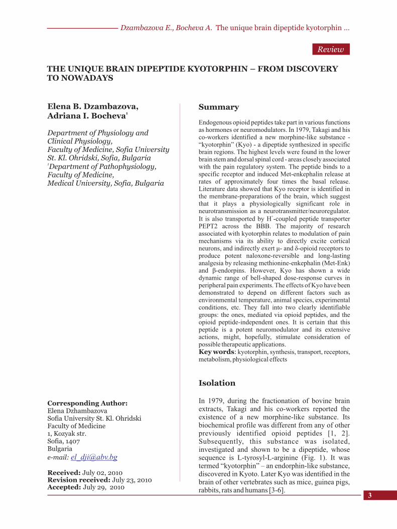
In 1979, during the fractionation of bovine brain extracts, Takagi and his co-workers reported the existence of a new morphine-like substance. Its biochemical profile was different from any of other previously identified opioid peptides [1, 2]. Subsequently, this substance was isolated, investigated and shown to be a dipeptide, whose sequence is L-tyrosyl-L-arginine (Fig. 1). It was termed “kyotorphin” – an endorphin-like substance, discovered in Kyoto. Later Kyo was identified in the brain of other vertebrates such as mice, guinea pigs, rabbits, rats and humans [3-6].
4
J Biomed Clin Res Volume 3 Number 1, 2010
Fig. 1. The neuroactive dipeptide Kyotorphin (L-tyrosyl-L-arginine)
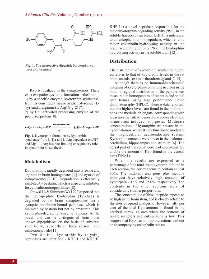
Kyo is localized in the synaptosomes. There exist two pathways for its formation in the brain: 1) by a specific enzyme, kyotorphin synthetase, from its constituent amino acids, L-tyrosine (L-Tyr) and L-arginine (L-Arg) (fig. 2) [7];
2+2) by Ca -activated processing enzyme of the precursor protein [8].
Fig. 2. Kyotorphin formation by kyotorphin synthetase from L-Tyr and L-Arg dependent on ATP
2+and Mg . L-Arg has rate-limiting or regulatory role in kyotorphin biosynthesis
Metabolism
Kyotorphin is rapidly degraded into tyrosine and arginine in brain homogenates [9] and cytosol of synaptosomes [7, 10]. Degradation is effectively inhibited by bestatin, which is a specific inhibitor for cytosolic aminopeptidases [8].
Orawski A & Simmons W (1992) reported that the neuropeptide kyotorphin (Tyr-Arg) is degraded by rat brain synaptosomes via a synaptic membrane-bound peptidase which is inhibited by bestatin but not by amastatin. This kyotorphin-degrading enzyme appears to be novel, and can be distinguished from other known dipeptidases on the basis of substrate specificity, subcellular localization, and inhibition profile [11].
Two distinct kyotorphin-hydrolysing peptidases are identified - KHP I and KHP II.
KHP I is a novel peptidase responsible for the major kyotorphin-degrading activity (95%) in the soluble fraction of rat brain. KHP II is indentical to an enkephalin aminopeptidase, which elicit a major enkephalin-hydrolysing activity in the brain, accounting for only 5% of the kyotorphin-hydrolysing activity in the soluble brain [12].
Distribution
The distribution of kyotorphin synthetase highly correlates to that of kyotorphin levels in the rat brain, and also exists in the adrenal gland [7, 13].
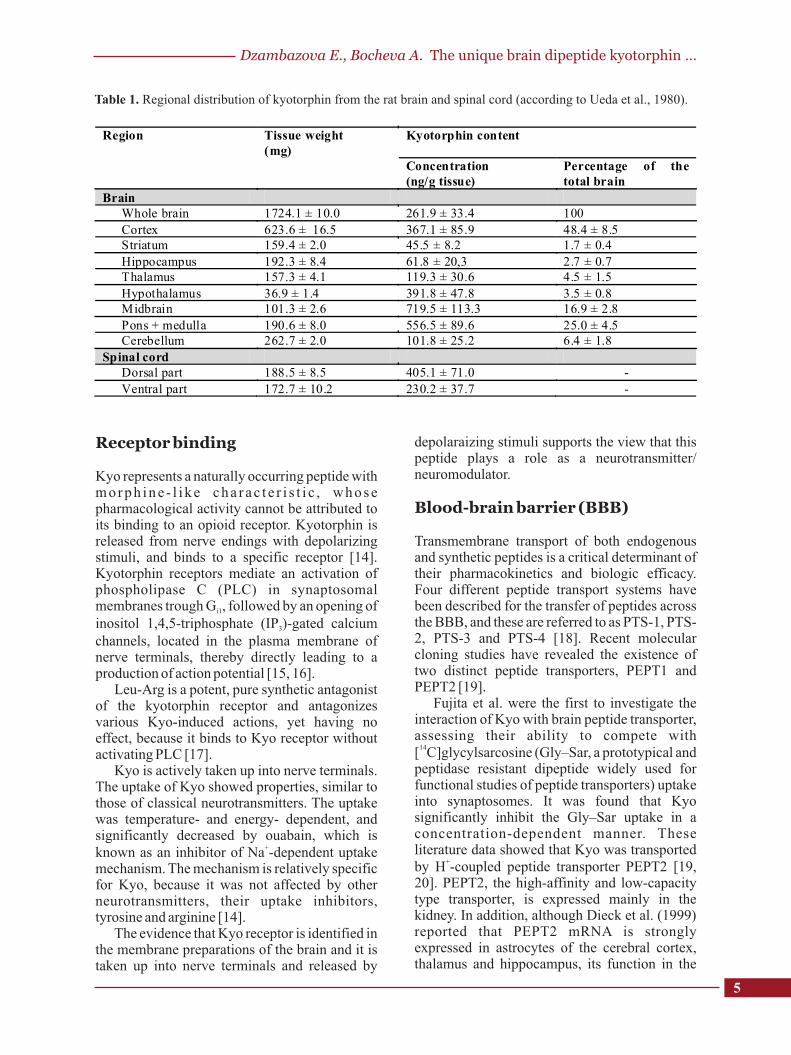
Although there is no immunohistochemical mapping of kyotorphin-containing neurons in the brain, a regional distribution of the peptide was measured in homogenates of rat brain and spinal cord tissues, using high performance liquid chromatography (HPLC). There is data reported, that the highest levels are found in the midbrain, pons and medulla oblongata, corresponding with areas most sensitive to morphine and/or electrical stimulation-induced analgesia. Moderate concentrations of kyotorphin are present in the hypothalamus, where it may function to modulate the magnocellular neuroendocrine system. Kyotorphin contents were lower in the thalamus, cerebellum, hippocampus and striatum [4]. The dorsal part of the spinal cord had approximately double the amount of Kyo found in the ventral part (Table 1).
When the results are expressed as a percentage of the total brain kyotorphin found in each section, the cortex seems to contain almost 50%. The midbrain and pons plus medulla oblongata have relatively high amounts of kyotorphin - 16.9 and 25.0%, respectively. The contents in the other sections were of considerably smaller proportions.
The concentration of this dipeptide appears to be high in the brain stem, and is closely related to the sites of opioid analgesia. However, fifty per cent of the total Kyo amount is found in the cerebral cortex, an area where the contents of opiate receptors and enkephalins is low. This suggest that Kyo has non-opioid actions without an accompanying enkephalin release.
5
Table 1. Regional distribution of kyotorphin from the rat brain and spinal cord (according to Ueda et al., 1980).
Receptor binding
Kyo represents a naturally occurring peptide with morph ine - l i ke cha rac t e r i s t i c , whose pharmacological activity cannot be attributed to its binding to an opioid receptor. Kyotorphin is released from nerve endings with depolarizing stimuli, and binds to a specific receptor [14]. Kyotorphin receptors mediate an activation of phospholipase C (PLC) in synaptosomal membranes trough G , followed by an opening of i1
inositol 1,4,5-triphosphate (IP )-gated calcium 3
channels, located in the plasma membrane of nerve terminals, thereby directly leading to a production of action potential [15, 16].
Leu-Arg is a potent, pure synthetic antagonist of the kyotorphin receptor and antagonizes various Kyo-induced actions, yet having no effect, because it binds to Kyo receptor without activating PLC [17].
Kyo is actively taken up into nerve terminals. The uptake of Kyo showed properties, similar to those of classical neurotransmitters. The uptake was temperature- and energy- dependent, and significantly decreased by ouabain, which is
+known as an inhibitor of Na -dependent uptake mechanism. The mechanism is relatively specific for Kyo, because it was not affected by other neurotransmitters, their uptake inhibitors, tyrosine and arginine [14].
The evidence that Kyo receptor is identified in the membrane preparations of the brain and it is taken up into nerve terminals and released by
depolaraizing stimuli supports the view that this peptide plays a role as a neurotransmitter/ neuromodulator.
Blood-brain barrier (BBB)
Transmembrane transport of both endogenous and synthetic peptides is a critical determinant of their pharmacokinetics and biologic efficacy. Four different peptide transport systems have been described for the transfer of peptides across the BBB, and these are referred to as PTS-1, PTS-2, PTS-3 and PTS-4 [18]. Recent molecular cloning studies have revealed the existence of two distinct peptide transporters, PEPT1 and PEPT2 [19].
Fujita et al. were the first to investigate the interaction of Kyo with brain peptide transporter, assessing their ability to compete with
14[ C]glycylsarcosine (Gly–Sar, a prototypical and peptidase resistant dipeptide widely used for functional studies of peptide transporters) uptake into synaptosomes. It was found that Kyo significantly inhibit the Gly–Sar uptake in a concentration-dependent manner. These literature data showed that Kyo was transported
+by H -coupled peptide transporter PEPT2 [19, 20]. PEPT2, the high-affinity and low-capacity type transporter, is expressed mainly in the kidney. In addition, although Dieck et al. (1999) reported that PEPT2 mRNA is strongly expressed in astrocytes of the cerebral cortex, thalamus and hippocampus, its function in the
Dzambazova E., Bocheva A. The unique brain dipeptide kyotorphin …
Region Tissue weight (mg)
Kyotorphin content Concentration (ng/g tissue)
Percentage of the total brain
Brain Whole brain 1724.1 ± 10.0 261.9 ± 33.4 100
Cortex 623.6 ± 16.5 367.1 ± 85.9 48.4 ± 8.5 Striatum 159.4 ± 2.0 45.5 ± 8.2 1.7 ± 0.4
Hippocampus 192.3 ± 8.4 61.8 ± 20,3 2.7 ± 0.7 Thalamus 157.3 ± 4.1 119.3 ± 30.6 4.5 ± 1.5
Hypothalamus 36.9 ± 1.4 391.8 ± 47.8 3.5 ± 0.8 Midbrain 101.3 ± 2.6 719.5 ± 113.3 16.9 ± 2.8
Pons + medulla 190.6 ± 8.0 556.5 ± 89.6 25.0 ± 4.5 Cerebellum 262.7 ± 2.0 101.8 ± 25.2 6.4 ± 1.8
Spinal cord Dorsal part 188.5 ± 8.5 405.1 ± 71.0 -
Ventral part 172.7 ± 10.2 230.2 ± 37.7 -
cerebral cortex is [21]. Recently, Berger and Hediger (1999) have shown that PEPT2 mRNA is expressed in the entire rat brain, and is localized in non-neuronal cells by means of in situ hybridization histochemistry [22].
Mechanism of kyotorphin-induced action
Kyotorphin is synthesized in specific brain regions where it may modulate synaptic transmission. Opioid systems may have mediated the effects of kyotorphin. The majority of research associated with kyotorphin relates to modulation of pain mechanisms via its ability to directly excite cortical neurons, and indirectly exert μ- and δ-opioid receptors to produce potent naloxone-reversible and long-lasting analgesia via releasing methionine-enkephalin (Met-Enk) and β-endorpins [1, 23, 24]. However, Kyo showed a wide dynamic range of bell-shaped dose-response curves in peripheral pain experiments [25].
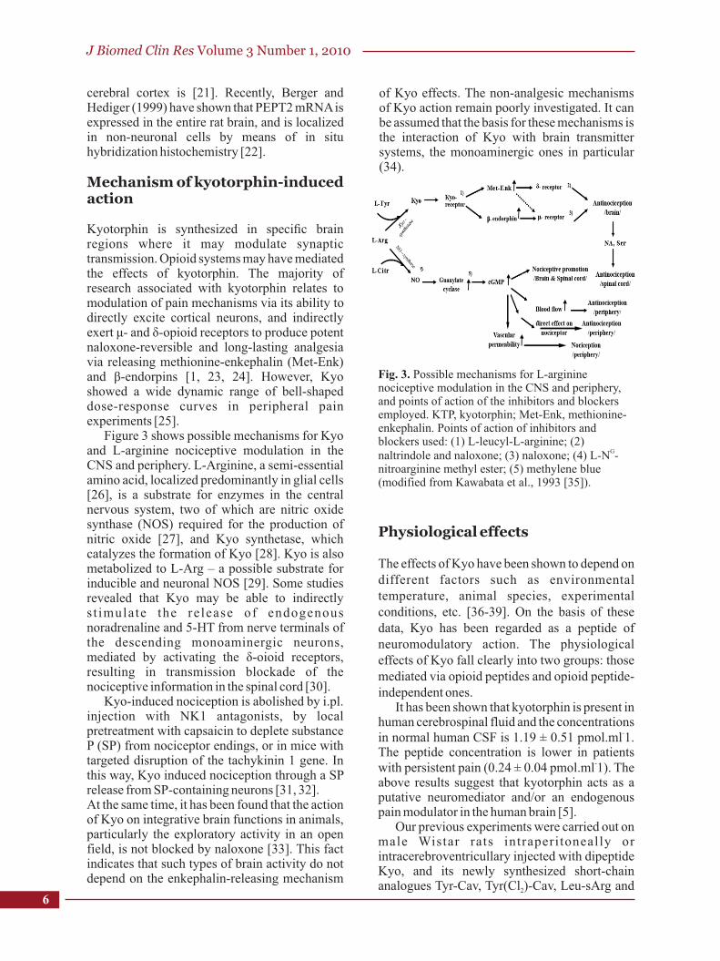
Figure 3 shows possible mechanisms for Kyo and L-arginine nociceptive modulation in the CNS and periphery. L-Arginine, a semi-essential amino acid, localized predominantly in glial cells [26], is a substrate for enzymes in the central nervous system, two of which are nitric oxide synthase (NOS) required for the production of nitric oxide [27], and Kyo synthetase, which catalyzes the formation of Kyo [28]. Kyo is also metabolized to L-Arg – a possible substrate for inducible and neuronal NOS [29]. Some studies revealed that Kyo may be able to indirectly s t imulate the re lease of endogenous noradrenaline and 5-HT from nerve terminals of the descending monoaminergic neurons, mediated by activating the δ-oioid receptors, resulting in transmission blockade of the nociceptive information in the spinal cord [30].
Kyo-induced nociception is abolished by i.pl. injection with NK1 antagonists, by local pretreatment with capsaicin to deplete substance P (SP) from nociceptor endings, or in mice with targeted disruption of the tachykinin 1 gene. In this way, Kyo induced nociception through a SP release from SP-containing neurons [31, 32]. At the same time, it has been found that the action of Kyo on integrative brain functions in animals, particularly the exploratory activity in an open field, is not blocked by naloxone [33]. This fact indicates that such types of brain activity do not depend on the enkephalin-releasing mechanism
of Kyo effects. The non-analgesic mechanisms of Kyo action remain poorly investigated. It can be assumed that the basis for these mechanisms is the interaction of Kyo with brain transmitter systems, the monoaminergic ones in particular (34).
Fig. 3. Possible mechanisms for L-arginine nociceptive modulation in the CNS and periphery, and points of action of the inhibitors and blockers employed. KTP, kyotorphin; Met-Enk, methionine-enkephalin. Points of action of inhibitors and blockers used: (1) L-leucyl-L-arginine; (2)
Gnaltrindole and naloxone; (3) naloxone; (4) L-N -nitroarginine methyl ester; (5) methylene blue (modified from Kawabata et al., 1993 [35]).
Physiological effects
The effects of Kyo have been shown to depend on different factors such as environmental temperature, animal species, experimental conditions, etc. [36-39]. On the basis of these data, Kyo has been regarded as a peptide of neuromodulatory action. The physiological effects of Kyo fall clearly into two groups: those mediated via opioid peptides and opioid peptide-independent ones.
It has been shown that kyotorphin is present in human cerebrospinal fluid and the concentrations
-in normal human CSF is 1.19 ± 0.51 pmol.ml 1. The peptide concentration is lower in patients
-with persistent pain (0.24 ± 0.04 pmol.ml 1). The above results suggest that kyotorphin acts as a putative neuromediator and/or an endogenous pain modulator in the human brain [5].
Our previous experiments were carried out on male Wistar rats intraperitoneally or intracerebroventricullary injected with dipeptide Kyo, and its newly synthesized short-chain analogues Tyr-Cav, Tyr(Cl )-Cav, Leu-sArg and 2
J Biomed Clin Res Volume 3 Number 1, 2010
6
7
Tyr-sArg. Antinociception was determined by test based on the use of mechanical stimuli (paw pressure test) and tests based on the use of thermal stimuli (tail flick and hot plate tests). The data obtained provide evidence that histamin- and nitricoxide-ergic systems and second messengers are involved in the antinociceptive action of the investigated dipeptides. Spinal and supraspinal regulatory mechanisms are also involved in the antinociceptive effects [40, 41]. The dipeptide kyotorphin has been shown to have pain inhibitory properties, besides a wide spectrum of physiological effects in mammals, amphibia, and fish [34, 42-45].
Recent results suggest that kyotorphin can also release opioid peptides from rat cardiac muscle and have an indirect regulatory role in β-adrenergic action through cross-talk with opioid receptors [46]. Studies of the possible role peptides play in the mechanism of hibernation showed that Kyo has inhibitory effect on respiration, heart rate, and body temperature regulation in animals [36, 42].
There are contradictory literature data on the role of Kyo in thermoregulatory processes. Central injection of Kyo into the third cerebral ventricle of unrestrained cats did not consistently alter body temperature at an ambient temperature
oof 22 C [47]. Some authors have found that intracerebroventricular (i.c.v.) administration of Kyo produced significant dose-dependent hypothermic responses in mice at an ambient
otemperature of 24 C. The hypothermic action of kyotorphin was much greater than that of Met-Enk. This action was slightly but not significantly reversed by administering naloxone - intraperitoneally. The hypothermia induced by kyotorphin and Met-Enk was prevented by a small dose of thyrotropin-releasing hormone, which by itself had little effect on body temperature [48]. Bronnikov et al., (1997, 2005) showed that Kyo abolished the stimulating effect of noradrenaline on proliferation of brown adipose tissue, which plays a major role in heat production. This suggests that pripheral tissue cells contain receptors for Kyo, and that Kyo is concerned with regulation of their mitogenic activity [50, 51].
Little is known about the behavioral effects of Kyo. The data on this point are rather conflicting. KTP is known to have no effect on open-field behavior of mice [33]. Similarly, subcutaneous (s.c.) or intraventricular injections of L-Kyo and D-Kyo induce no marked behavioral abnormalities such as catalepsy, ataxia or
hypermotility in rats at a dose of 0.1 g per rat. At the same time, Kyo was found to inhibit the extinction of pole-jumping avoidance response in the rat [51], to affect aversive pecking in chicks [52] and to induce sedation and reduce motor activity in mice when injected at high doses [48]. According to these data, Kyo induced no strong behavioral changes in a variety of species.
Our experiments showed that Kyo suppress one of the two basic components of exploratory behaviour – ambulance, and does not affect the emotional state, i.e. rearing [53].
In order to test some hypotheses for the mechanisms by which Kyo affects the behavior of higher animals, Kolaeva et al. (2000) carried out a number of experiments on fish, applying this substance onto the Mauthner neurons (MN) of the medulla oblongata, a double-cell motor center responsible for particular easily registered forms of behavior in fish and amphibia [45, 54, 55]. Like in higher animals, the application of Kyo mediated potentiation of the serotonergic transmission and exerted an inhibitory action on the neuronal activity at the level of single neurons in fish [34].
Application of Kyo itself was found to produce significant decreases in the functional activity of MN without inducing ultrastructural rearrangements, though it did have protective influence in conditions of prolonged stimulation, this being evident at both behavioral and ultrastructural level. This effect appeared to have some kind of relationship with the ability of Kyo to decrease the functional activity of MN. Data have been obtained showing that dipeptide Kyo can block calcium influx currents, as demonstrated in myocardium from cold-blooded (frogs) and warm-blooded (rats, ground squirrels) species [42, 56]. In addition, experiments addressing the redox properties of exogenous calcium channel regulators showed that KT can function both as an electron acceptor and as an electron donor which, according to previous data, can affect the ability of this neuropeptide to block potential-dependent calcium channels [57]. These data provide indirect evidence of the ability of Kyo to block
2+potential-dependent Ca channels in MN,as well as other neurons.
Many authors define stress as a state of threatened homeostasis or disharmony, which is counteracted by a complex repertoire of physiologic and behavioral responses that reestablish homeostasis (adaptive stress – response) [58, 59]. Literature data has shown that
Dzambazova E., Bocheva A. The unique brain dipeptide kyotorphin …
adaptive behaviour of young rats, treated with Kyo, is accompanied by activation of noradrenergic, dopaminergic and serotoninergic systems of the brain. Kyo affected the sleep-awake cycle by decreasing the relax awake and increasing the sleep time, while it increased the duration of grooming, decreased water and food intake the exploratory activity via activation of the serotonergic system. Preadaptation of the organism by Kyo administration was also achieved through increasing antioxidant system activity [60].
Literature data showed that administration of Kyo i.c.v. in conscious rats may change plasma levels of neurohypophysial hormones and blood pressure. Central administration of Kyo increased plasma levels of oxytocin (OT), regarded as a ''stress'' hormone in rodents, coincident with stimulating a pressor response and elevating plasma glucose levels, when administered i.c.v. to normally hydrated rats. On the other hand, an excess of L-Arg and Kyo within the CNS may mimic stress response by augmenting release of oxytocin and stimulating the sympathetic nervous system to increase blood pressure and plasma glucose levels. This profile of activity resembles a central activation of stress response, involving stimulation of both the neuroendocrine and autonomic nervous systems. Moderate concentrations of kyotorphin are present in the hypothalamus – the main structure responsible for stress response. Opioid peptides expressed in vasopressin (VP) and OT neurons [61, 62] are known to attenuate release of neurohypophysial hormones, and would be a potential mechanism by which Kyo could indirectly affect the magnocellular system [63].
In our recent studies we examined the ability of Kyo to modify the effects of morphine (Mo), L-NAME (L-Nitro-Arginine Methyl Ester, a non-selective inhibitor of nitric oxide synthase) and immobi l iza t ion s t ress - induced - antinociception in acute pain. It is known that analgesia, induced by stress is a phenomenon, often referred to as “stress-induced analgesia” (SIA) [64]. Literature data has also shown that anti-opioid peptides (AOP) could reverse morphine-induced analgesia and inhibit the expression of some forms of SIA in various species. Our results showed that Kyo significantly decreased the analgesic effect of L-NAME in both paw pressure and hot platе tests. The effect of Mo decreased significantly only in hot plate tests. When administered after stress, Kyo reduced stress-induced antinociception in
paw pressure tests. We suggest that these findings indicate that Kyo may act as an anti-opioid peptide [65].
It's known that stress activates the hypothalamic-pituitary-adrenal (HPA) axis by stimulating neuronal activity within the paraventricular nucleus of the hypothalamus [66]. Some workers have reported that HPA axis responses to neural stimuli, which are not dependent on immune factors, can be modulated by nitric oxide (NO). NO also plays an important role in regulating the response of the HPA axis to various stresses. Morphological studies have provided evidence for the existence of a signaling pathway between an opioid- and the nitric oxide ergic systems in the hypothalamus of rat brain. [67-69].
Our histochemical study on NADPH-d reactive neurons in rat hypothalamic paraventricular nucleus showed that Kyo, when administered in cold-exposed rats can increase NO activity. The results of our experiment suggest that Kyo and NO may play an important role in the continuity of homeostasis, acting as agonist substances. Further studies are needed to understand the exact role of Kyo in response to cold exposure [70]. Unpublshed data of ours also showed that Kyo may modulate opioid and non-opioid neurotransmitter networks and thus help the stressed organism to reach homeostasis, i.e. Kyo may have an anti-stressor effect, besides that, according to our results, it inhibited stress-induced increase in ACTH and corticosterone plasma levels.
Conclusions
This review is an attempt to provide a comprehensive summary of the neuropeptide kyotorphin. Its mechanisms of action and the wide range of biological effects make possible the assumption that this peptide is a potent neuromodulator. Most findings emphasize the effects on the CNS, including the spinal cord as well as the brain, but these effects also concern the peripheral body and communication across the BBB. Although fifty percent of the total Kyo amount is found in the cerebral cortex, an area where the contents of opiate receptors and enkephalins is low and its physiological effects are opioid and non-opioid mediated, this review has shown that Kyo actions are far more extensive, hopefully stimulating consideration of possible therapeutic applications.
J Biomed Clin Res Volume 3 Number 1, 2010
8
9
Dzambazova E., Bocheva A. The unique brain dipeptide kyotorphin …
References
1. Takagi H, Shiomi H, Ueda H, Amano H. A novel analgesic dipeptide from bovine brain is a possible Met-enkephalin releaser. Nature. 1979а;282(5737):410-412.
2. Takagi H, Shiomi H, Ueda H, Amano H. Morphine-like analgesia by a new dipeptide, L-tyrosyl-L-arginine (Kyotorphin) and its analo-gue. Eur J Pharmacol. 1979b;55(1):109-111.
3. Shiomi H, Ueda H, Takagi H. Isolation and identification of an analgesic opioid dipeptide kyotorphin (Tyr-Arg) from bovine brain. Neuropharmacol. 1981;20(7):633-638.
4. Ueda H, Shiomi H, Takagi H. Regional distribution of a novel analgesic dipeptide kyotorphin (Tyr-Arg) in the rat brain and spinal cord. Brain Res. 1980;198(2):460-464.
5. Nishimura K, Kaya K, Hazato T, Ueda H, Satoh M, Takagi H. Kyotorphin like substance in human cerebrospinal fluid of patients with persistent pain. Masui. 1991;40(11):1686-90.
6. Lopes SC, Fedorov A, Castanho MA. Chiral recognition of D-kyotorphin by lipidic membranes: relevance toward improved analgesic efficiency. Chem Med Chem. 2006a;1(7):723-8.
7. Ueda H, Yoshihara Y, Fukushima N, Shiomi H, Nakamura A, Takagi H. Kyotorphin (tyrosine-arginine) synthetase in rat brain synaptosomes. J Biol Chem., 1987;262(17):8165-73.
8. Yoshihara Y, Ueda H, Fujii N, Shide A, Yajima H, Satoh M. Purification of a novel type of calcium-activated neutral protease from rat brain. Possible involvement in production of the neuropeptide kyotorphin from calpastatin fragments. J Biol Chem. 1990;265(10):5809-5815.
9. Ueda H, Ming G, Hazato T, Katayama T, Takagi H. Degradation of kyotorphin by a purified membrane-bound-aminopeptidase from monkey brain: potentiation of kyotorphin-induced analgesia by a highly effective inhibitor, bestatin. Life Sci. 1985a;36(19):1865-1871.
10. Ueda H, Yoshihara Y, Nakamura A, Shiomi H, Satoh M, Takagi H. How is kyotorphin (Tyr-Arg) generated in the brain? Neuropeptides. 1985b;5(4-6):525-8.
11. Orawski AT, Simmons WH. Dipeptidase activities in rat brain synaptosomes can be distinguished on the basis of inhibition by bestatin and amastatin: identification of a kyotorphin (Tyr-Arg)-degrading enzyme. Neurochem Res. 1992;17(8):817-20.
12. Akasaki K, Nakamura A, Shiomi H, Tsuji H. Identification and characterization of two distinct kyotorphin-hydrolyzing enzymes in rat brain. Neuropeptides. 1991;20(2):103-107.
13. Kawabata A, Muguruma H, Tanaka M, Takagi H. Kyotorphin synthetase activity in rat adrenal
g l a n d s a n d s p i n a l c o r d . P e p t i d e s . 1996;17(3):407-11.
14. Ueda H, Yoshihara Y, Takagi H. A putative met-enkephalin releaser, kyotorphin enhances intracellular Ca2+ in the synaptosomes. B i o c h e m B i o p h y s R e s C o m m u n . 1986;137(2):897-902.
15. Ueda H, Miyamae T, Fukushima N, Watanabe S, Misu Y. Evidence for a metabostatic opioid kappa-receptor inhibiting pertussis toxin-sensitive metabotropic glutamate receptor-currents in Xenopus oocytes. FEBS Lett. 1995a;375(3):201-205.
16. Ueda H, Miyamae T, Hayashi C, Watanabe S, Fukushima N, Sasaki Y, Iwamura T, Misu Y. Protein kinase C involvement in homologous desensitization of delta-opioid receptor coupled to Gi1-phospholipase C activation in Xenopus oocytes. J Neurosci. 1995 b;15(11):7485-7499.
17. Ueda H, Yoshihara Y, Misawa H, Fukushima N, Katada T, Ui M, Takagi H, Satoh M. The kyotorphin (tyrosine-arginine) receptor and a selective reconstitution with purified Gi, measured with GTPase and phospholipase C assays. J Biol Chem. 1989;264(7):3732-3741.
18. Ganapathy V, Miyauchi S. Transport systems for opioid peptides in mammalian tissues. AAPS J. 2005;7(4):E852-856.
19. Fujita T, Kishida T, Wada M, Okada N, Yamamoto A, Leibach FH, Ganapathy V. Functional characterization of brain peptide transporter in rat cerebral cortex: identification of the high-affinity type H+/peptide transporter PEPT2. Brain Res. 2004;997(1):52-61.
20. Fujita T, Kishida T, Okada N, Ganapathy V, Leibach FH, Yamamoto A. Interaction of kyotorphin and brain peptide transporter in synaptosomes prepared from rat cerebellum: implication of high affinity type H+/peptide transporter PEPT2 mediated transport system. Neurosci Lett. 1999;271(2):117-120.
21. Dieck ST, Heuer H, Ehrchen J, Otto C, Bauer K. The peptide transporter PEPT2 is expressed in rat brain and mediates the accumulation of the fluorescent dipeptide derivative h-Ala-Lys-Nq-AMCA in astrocytes, Glia. 1999;25:10–20.
22. Berger U.V., M.A. Hediger, Distribution of peptide transporter PEPT2 mRNA in the rat nervous system, Anat Embryol. 1999; 199:439–449.
23. Vlaskovska M. Opioid peptides. In.: Mitzov V, Vlaskovska M, Kazakov L, edi tors . Neurotransmitters and neuromodulators. Sofia: State Publishing House “Medicina i Fizkultura”; 1989. p. 128-162
24. Kawabata A, Nishimura Y, Takagi H. L-leucyl-L-arginine, naltrindole and D-arginine block antinociception elicited by L-arginine in mice with carrageenin-induced hyperalgesia. Br J Pharmacol. 1992;107(4):1096-1101.
10
J Biomed Clin Res Volume 3 Number 1, 2010
25. Ueda H. In vivo molecular signal transduction of peripheral mechanisms of pain. Jpn J Pharmacol. 1999;79(3):263-8.
26. Aoki E, Semba R, Mikoshiba K, Kashiwamata S. Predominant localization in glial cells of free L-arginine. Immunocytochemical evidence. Brain Res. 1991;547(2):190-192.
27. Moncada S. The L-arginine: nitric oxide pathway. Acta Physiol Scand. 1992;145(3):201-227.
28. Takagi H, Ueda H. Kyotorphin in the brain may function as an endogenous pain modulator; In: Takagi H, Oomura Y, Ito M, Otsuka M, editors. Biowarning System in the Brain. Tokyo: A Naito Foundation Symposium, University of Tokyo Press; 1988. p. 139-153.
29. Arima T, Kitamura Y, Nishiya T, Takagi H, Nomura Y. Kyotorphin (L-tyrosyl-L-arginine) as a possible substrate for inducible nitric oxide synthase in rat glial cells. Neurosci Lett. 1996;212(1):1-4.
30. Ochi T, Ohkubo Y, Mutoh S. Blockade of the antinociceptive effect of spinally administered kyotorphin by naltrindole in mice. Neurosci Lett. 2002;322(2):95-98.
31. Inoue M, Yamada T, Ueda H. Low dose of kyotorphin (tyrosine-arginine) induces nociceptive responses through a substance P release from nociceptor endings. Brain Res Mol Brain Res. 1999;69(2):302-305.
32. Ueda H, Inoue M. In vivo signal transduction of nociceptive response by kyotorphin (tyrosine-arginine) through Galpha(i)- and inositol trisphosphate-mediated Ca(2+) influx. Mol Pharmacol. 2000;57(1):108-115.
33. Shi L, Ku B, Yao H. Studies on antidepressant effects of several overshort peptides. Yao Hsueh Hsueh pao. 1991;26:546–547.
34. Kolaeva SG, Semenova TP, Santalova IM, Moshkov DA, Anoshkina IA, Golozubova V. Effects of L-thyrosyl-L-arginine (kyotorphin) on the behavior of rats and goldfish. Peptides. 2000;21(9):1331-1336.
35. Kawabata A, Ueda N, Takagi H. L-arginine exerts a dual role in nociceptive processing in the brain: involvement of the kyotorphin-Met-enkephalin pathway and NO-cyclic GMP pathway. Br J Pharmacol. 1993;109(1):73-79.
36. Emelianova T, Usenko A, Ushakov V, Kononova L, Michaleva I. Effects of kyotorphin and neokyotorphin on thermoregulation in rats at different temperatures. In: Kolaeva S, Popova N, Solomonov N, Wahg L, editors. Ecologo-physiological characteristics of natural hypometabolic states. Puschino: ONTI SGBK; 1992. p. 132–137.
37. Laubia M, Schmitt H. Indication for central vagal endorphinergic control of heart rate in dogs. Eur J Pharmacol. 1981;71:401–409.
38. Shiomi H, Kuraishi Y, Ueda H, Harada Y, Amano
J, Takagi H. Mechanisma of kyotorhin-induced release of met-enkephalin from guinea pig striatum and spinal cord. Brain Res. 1981; 221:161–169.
39. Ueda H, Tatsumi K, Shiomi H, Takagi H. Analgesic dipeptide kyotorphin in highly concentrated in the synaptosomal fraction of the brain. Brain Res. 1982;231(1):222–224.
40. Bocheva, A., E. Dzambazova-Maximova, T. Pajpanova. Is nitric oxide involved in the antinociceptive effects of kyotorphin, Tyr-Cav and MIF's analogues in rats? Coll Symp Ser. 2003;6:1-3.
41. Dzambazova-Maximova, E., A. Bocheva, S. Todorov. Analgesic action of Kyotorphin and its canavanine analogues in rats: role of histamine- and nitric oxide- ergic systems. Compt Rend Bulg Acad Sci. 2003;56(8):75-80.
42. Ignat'ev DA, Vorob'ev VV, Ziganshin RKh. The effect of short peptides isolated from the brain of a hibernating suslik on the rat EEG and behavior. Zh Vyssh Nerv Deiat Im I P Pavlova. 1996;46(6):1049-1058.
43. Nazarenko IV, Zvrushchenko MSh, Volkov AV, Kamenskii AA, Zaganshin RKh. Functional-morphologic evaluation of the effect of the regulatory peptide kyotorphin on the status of the CNS in the post-resuscitation period. Patol Fiziol Eksp Ter. 1999; (2):31-33.
44. Santalova IM, Moshkov DA, Chailakhian LM. Effect of fractions of peptides (1-10 kDa) from tissues of hibernating squirrels and peptides kyotorphin and neokyotorphin on fish behavior. Dokl Akad Nauk. 1998;362(5):696-698.
45. Santalova IM, Moshkov DA. Smooth endoplasmic reticulum in fish Mauthner cells at different functional states. Neurosci . 1999;89(2):593–602.
46. Li Y, Saito Y, Suzuki M, Ueda H, Endo M, Maruyama K. Kyotorphin has a novel action on rat cardiac muscle. Biochem Biophys Res Commun. 2006;339(3):805-809.
47. Clark WG, Ponder SW. Thermoregulatory effects of (D-ala2)-methionine-enkephalinamide in the cat. Evidence for multiple naloxone-sensitive opioid receptors. Brain Res Bull. 1980;5(4):415-420.
48. Sakurada T, Sakurada S, Watanabe S, Matsumura H, Kisara K, Akutsu Y, Sasaki Y, Suzuki K. Actions of intracerebroventricular administration of kyotorphin and an analog on thermoregulation in the mouse. Peptides 1983;4(6):859-863.
49. Bronnikov G, Dolgacheva L, Zhang SJ, Galitovskaya E, Kramarova L, Zinchenko V. The effect of neuropeptides kyotorphin and neokyotorphin on proliferation of cultured brown preadipocytes. FEBS Lett. 1997; 407(1):73-77.
50. Bronnikov GE, Kolaeva SG, Dolgacheva LP,
11
Dzambazova E., Bocheva A. The unique brain dipeptide kyotorphin …
Kramarova LI. Kyotorphin suppresses proliferation and Ca2+ signaling in brown preadipocytes. Bull Exp Biol Med. 2006; 141(2):223-225.
51. Yamamoto M, Kawamuki K, Satoh M, Takagi H. Kyotorphin inhibits extinction of pole-jumping avoidance response in the rat. Neurosci Lett. 1982;31:175–179.
52. Kastin A, Honour L, Coy D. Kyotorphin effect aversive pecking in chicks. Physiol Behav. 1981;27:10173–10176.
53. Bocheva A., V.V. Petkov, T. Pajpanova, L. Kazakov, E. Golovinsky. Biological properties of novel kyotorphin analogues: in vivo and in vitro investigations. Compt Rend Acad Bulg Sci. 1994;47(4):77-80.
54. Confield YG, Rose GY. Activation of Mauthner neurons during prey capture. J Compar Phisiol. 1993;172:611–618.
55. Didomenico R, Nissanov J, Eaton RC. Lateralization and adaptation of a continuously variable behavior following lesions of a reticulospi-nal command neuron. Brain Res. 1988;473(1):15–28.
56. Alekseev A. E. Characteristics of the function of potential-dependent Ca channels in cardiocytes in hibernating animals and their regulation by endogenous peptides [dissertation]. Pushchino: Institute of Protein Research; 1994.
57. Marinov BS, Ziganshin RKh. Redox properties of peptides potentially regulating animal hibernation. Biofizika. 1997;42(1):147-153.
58. Pacák K, Palkovits M. Stressor specificity of central neuroendocrine responses: implications for stress-related disorders. Endocr Rev. 2001; 22:502-548.
59. Tsigos C, Kyrou I, Chrousos G. Stress, Endocrine Physiology and Pathophysiology. In: George Chrousos, editor. Adrenal Physiology and Diseases. 2004.
60. Lysenko AV, Mendzeritstky GL, Voloshina GL, Mikhaleva II. Neurochemical mechanisms of kyotorphin effects on adaptational reactions of rats with high level of anxiety. Neurochimia. 2004;21(3):217-224.
61. Martin R, Voigt KH. Enkephalins co-exist with oxytocin and vasopressin in nerve terminals of rat neurohypophysis. Nature. 1981; 289(5797): 502-504.
62. Watson SJ, Akil H, Fischli W, Goldstein A, Zimmerman E, Nilaver G, Van Wimersma Griedanus TB. Dynorphin and vasopressin: common localization in magnocellular neurons. Science. 1982;216(4541):85-87.
63. Summy-Long JY, Bui V, Gestl S, Koehler-Stec E, Liu H, Terrell ML, Kadekaro M. Effects of central injection of kyotorphin and L-arginine on oxytocin and vasopressin release and blood pressure in conscious rats. Brain Res Bull. 1998;45(4):395-403.
64. Butler RK, Finn DP. Stress-induced analgesia. Prog Neurobiol. 2009;88(3):184-202.
65. Dzambazova-Maximova E., A. Bocheva, Hr. Nocheva. Is kyotorphin an antiopioid peptide. Bulg Chem Commun. 2006;38(1):36-41.
66. Swain MG, Maric M. Impaired stress and interleukin-1beta-induced hypothalamic expression of the neuronal activation marker FOS in cholestatic rats. Hepatology. 1996; 24(4):914-918.
67. Gilinskii MA, Petrakova GM, Amstislavskaya TG, Maslova LN, Bulygina VV. Hypothalamic monoamines in cold stress on the background of changes in the activity of the nitric oxide system. Neurosci Behav Physiol. 2005;35(2):171-175.
68. Uribe RM, Lee S, Rivier C. Endocrinology 1999; 140(12):5971-5981.
69. Gupta V., A. Gupta, S. Saggu, H.M. Divekar, S.K. Grover, R. Kumar. Evid Based Complement Alternat Med. 2005;2(1):93-97.
70. Dzambazova E, Bocheva A, Landzhov B, Bozhilova-Pastirova A. Effects of kyotorphin on NADPH-d reactive neurons in rats after cold stress. Compt Rend Acad Bulg Sci. 2008; 61(5):661-666.