Embed Size (px)
Citation preview

The University of Toledo
Core Laboratories
The University of Toledo has invested substantial resources to develop,
maintain, and expand Core Laboratories. This brochure is designed to
make the UT Core Labs more visible in the community. The UT Core
Labs include the Advanced Microscopy and Imaging Center, Flow
Cytometry Core, Genomics Core (College of Medicine and Life
Sciences), Instrumentation Center, Nuclear Magnetic Resonance
Facility (College of Natural Sciences and Mathematics), Center for Drug
Design and Development (College of Pharmacy and Pharmaceutical
Sciences), and Center for Materials and Sensor Characterization
(College of Engineering). These Core Labs are equipped with state-of-
the-art instruments and offer cutting-edge technological services in
various research fields.
Major instruments include: multiphoton laser scanning microscope,
confocal microscopes, laser capture microdissection system, in vivo
imaging systems, multicolor high-speed cell sorter, microarray scanner,
MALDI-TOF/TOF mass spectrometers, scanning and transmission
electron microscopes, robotics for protein crystallization, nuclear
magnetic resonance spectroscopies, X-ray diffractometer, and a
confocal Raman spectrometer. The UT Core Labs are staffed with
experts in the fields, and can provide core users with basic and
advanced on-site training. Depending on user needs, they also can
process and analyze samples.
Please contact individual Core Labs for specific inquiries.
Page
Advanced Microscopy and Imaging Center (AMIC) 2-3 Center for Materials and Sensor Characterization (CMSC) 4 Center for Drug Design and Development (CD3) 5 Genomics Core 6 Flow Cytometry Core 7 Nuclear Magnetic Resonance Facility (NMR) 8 Instrumentation Center 9 Histology Core Facility 10

2
The University of Toledo Advanced Microscopy and Imaging Center on the
Health Science Campus is a 3,000 square foot facility designed to bring
together advanced light and fluorescence microscopy systems and “state-of-
the-art” image analysis software to perform biomedical research. The AMIC
consists of a 1,000 sq. ft. general microscopy laboratory that contains the
following instrumentation.
A state-of-the-art Electron Microscopy Laboratory is
part of the Advanced Microscopy & Imaging Center.
The EM facility is directed by Dr. William Gunning
who specializes in ultrastructural diagnosis of
human disease and also provides research support
to the University of Toledo. The EM lab is equipped
with two transmission electron microscopes, one
being used for clinical diagnostic purposes and the
other available for use by researchers.
Transmission Electron Microscopy
IVIS Spectrum whole
animal fluorescence imaging system developed by
Xenogen/Caliper Life Sciences. The IVIS Spectrum
is a multimodal bioluminescent and fluorescent
imaging system specifically designed for
noninvasive longitudinal imaging of cells and
tissues in small animals. This instrument facilitates
the study of biological processes via fluorescence
in small animals, including tumor growth, cancer
metastasis, bacterial infections, immune
responses and
inflammation,
and regulation
o f t i s s u e -
specific gene
expression.
Advanced Microscopy & Imaging Center (AMIC)
FluoView™ FV1000 Confocal
Microscope is a next -
generation imaging system designed for high-
resolution, confocal observation of both fixed and
living cells. The FV1000 offers advances in confocal
system performance while providing the speed and
sensitivity required for live cell imaging with minimal
risk of damage to living specimens.
TCS SP5 Laser Scanning
Confocal Microscope
with MP is equipped with both
conventional and high-speed resonance
scanners. This includes conventional lasers plus
multi-photon excitation (458, 488, 514, 561, 633,
and a tunable Ti-Sapphire MP laser 710-990nm).
This system is capable of collecting up to five colors
simultaneously for quantitative confocal image
analysis in both live cell and animal imaging, fixed
tissue and includes the capabilities for 3-D
reconstruction, FRAP, FRET, animation, stereo
imaging, single layer projection, time lapse
collection, and co-localization analysis.
TIRF (Total Internal Reflection Fluorescence)
Microscopy is available on an Olympus IX81 invert-
ed microscope/imaging system that allows the
visualization of fluorescent molecules either in
wide-field (conventional) or exclusively
at the cel l -g lass inter face
(TIRF). This latter capability allows
selective, real-time tracking of single
molecule or particle dynamics at the
ACUSON Sequoia C512 cardi-
ac ultrasound imaging system. This Echocardiog-
raphy system is a valuable tool for studying cardio-
vascular disease processes in small animals, includ-
ing ischemic heart disease, heart failure, cardiac
hypertrophy and remodeling, hypertension and
diabetic cardiomyopathy. Capabilities include high-
resolution imaging, tissue harmonic imaging, differ-
ential echo amplification, spectral Doppler (pulsed
and continuous wave), color Doppler (for measure-
ments of velocity energy and tissue Doppler imag-
ing) and color-Doppler M-mode imaging.
Contact Information:
Andrea L. Kalinoski, Ph.D.
Health Science Campus
Block Health Science Bldg., Room 057
Phone 419.383.4205
Email: [email protected]
Website: utoledo.edu/corelabs
3
Advanced Microscopy & Imaging Center (AMIC)
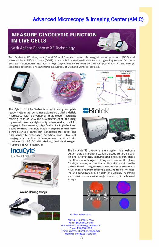
The Cytation™ 5 by BioTek is a cell imaging and plate
reader system that combines automated digital widefield
microscopy with conventional multi-mode microplate
reading. With 4X, 20X and 40X magnification, the imag-
ing module provides high-quality cellular and sub-cellular
imaging in fluorescence, brightfield, color brightfield and
phase contrast. The multi-mode microplate reader incor-
porates variable bandwidth monochromator optics and
high sensitivity filter-based detection optics. Live cell
imaging and multi-mode assays are optimized with
incubation to 65 °C with shaking, and dual reagent
injectors with Gen5 software.
The IncuCyte S3 Live-cell analysis system is a real-time
system that sits inside a standard tissue culture incuba-
tor and automatically acquires and analyzes HD, phase
and fluorescent images of living cells, around the clock,
for days, weeks, or months, while cells remain undis-
turbed. Kinetic, image-based measurements ensure you
never miss a relevant response allowing for cell monitor-
ing and surveillance, cell health and viability, migration
and invasion, plus a wide range of phenotypic cell-based
assays.
Two Seahorse XFe Analyzers (8 and 96-well format) measure the oxygen consumption rate (OCR) and
extracellular acidification rate (ECAR) of live cells in a multi-well plate to interrogate key cellular functions
such as mitochondrial respiration and glycolysis. The instruments perform compound addition and mixing,
label-free detection, and automatic calculation of OCR and ECAR in real time.
Contact Information:
Andrea L. Kalinoski, Ph.D.
Health Science Campus
Block Health Science Bldg., Room 057
Phone 419.383.4205
Email: [email protected]
Website: utoledo.edu/corelabs
Wound Healing Assays
4
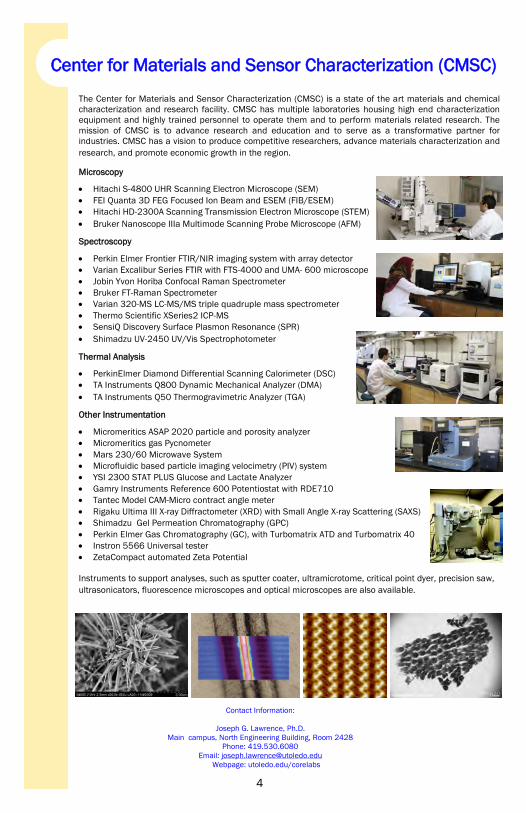
Center for Materials and Sensor Characterization (CMSC)
The Center for Materials and Sensor Characterization (CMSC) is a state of the art materials and chemical
characterization and research facility. CMSC has multiple laboratories housing high end characterization
equipment and highly trained personnel to operate them and to perform materials related research. The
mission of CMSC is to advance research and education and to serve as a transformative partner for
industries. CMSC has a vision to produce competitive researchers, advance materials characterization and
research, and promote economic growth in the region.
Microscopy
Hitachi S-4800 UHR Scanning Electron Microscope (SEM)
FEI Quanta 3D FEG Focused Ion Beam and ESEM (FIB/ESEM)
Hitachi HD-2300A Scanning Transmission Electron Microscope (STEM)
Bruker Nanoscope IIIa Multimode Scanning Probe Microscope (AFM)
Spectroscopy
Perkin Elmer Frontier FTIR/NIR imaging system with array detector
Varian Excalibur Series FTIR with FTS-4000 and UMA- 600 microscope
Jobin Yvon Horiba Confocal Raman Spectrometer
Bruker FT-Raman Spectrometer
Varian 320-MS LC-MS/MS triple quadruple mass spectrometer
Thermo Scientific XSeries2 ICP-MS
SensiQ Discovery Surface Plasmon Resonance (SPR)
Shimadzu UV-2450 UV/Vis Spectrophotometer
Thermal Analysis
PerkinElmer Diamond Differential Scanning Calorimeter (DSC)
TA Instruments Q800 Dynamic Mechanical Analyzer (DMA)
TA Instruments Q50 Thermogravimetric Analyzer (TGA)
Other Instrumentation
Micromeritics ASAP 2020 particle and porosity analyzer
Micromeritics gas Pycnometer
Mars 230/60 Microwave System
Microfluidic based particle imaging velocimetry (PIV) system
YSI 2300 STAT PLUS Glucose and Lactate Analyzer
Gamry Instruments Reference 600 Potentiostat with RDE710
Tantec Model CAM-Micro contract angle meter
Rigaku Ultima III X-ray Diffractometer (XRD) with Small Angle X-ray Scattering (SAXS)
Shimadzu Gel Permeation Chromatography (GPC)
Perkin Elmer Gas Chromatography (GC), with Turbomatrix ATD and Turbomatrix 40
Instron 5566 Universal tester
ZetaCompact automated Zeta Potential
Instruments to support analyses, such as sputter coater, ultramicrotome, critical point dyer, precision saw,
ultrasonicators, fluorescence microscopes and optical microscopes are also available.
Contact Information:
Joseph G. Lawrence, Ph.D.
Main campus, North Engineering Building, Room 2428
Phone: 419.530.6080
Email: [email protected]
Webpage: utoledo.edu/corelabs
5
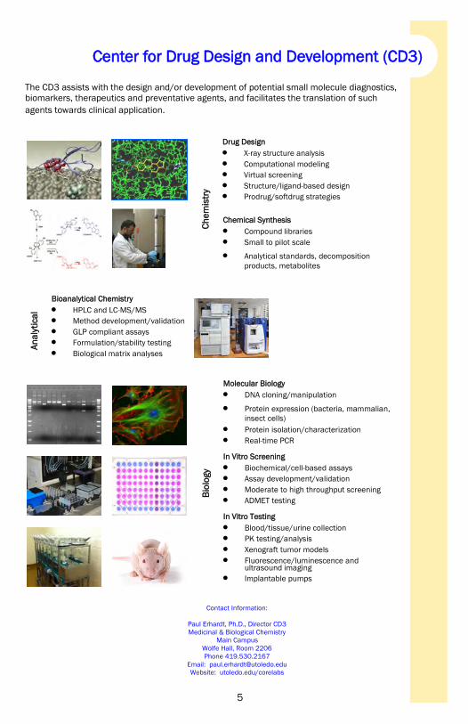
The CD3 assists with the design and/or development of potential small molecule diagnostics,
biomarkers, therapeutics and preventative agents, and facilitates the translation of such
agents towards clinical application.
Drug Design
X-ray structure analysis
Computational modeling
Virtual screening
Structure/ligand-based design
Prodrug/softdrug strategies C
he
mis
try
Chemical Synthesis
Compound libraries
Small to pilot scale
Analytical standards, decomposition
products, metabolites
Bioanalytical Chemistry
HPLC and LC-MS/MS
Method development/validation
GLP compliant assays
Formulation/stability testing
Biological matrix analyses
An
aly
tica
l
Contact Information:
Paul Erhardt, Ph.D., Director CD3
Medicinal & Biological Chemistry
Main Campus
Wolfe Hall, Room 2206
Phone 419.530.2167
Email: [email protected]
Website: utoledo.edu/corelabs
Molecular Biology
DNA cloning/manipulation
Protein expression (bacteria, mammalian,
insect cells)
Protein isolation/characterization
Real-time PCR In Vitro Screening
Biochemical/cell-based assays
Assay development/validation
Moderate to high throughput screening
ADMET testing In Vitro Testing
Blood/tissue/urine collection
PK testing/analysis
Xenograft tumor models
Fluorescence/luminescence and ultrasound imaging
Implantable pumps
Bio
logy
Center for Drug Design and Development (CD3)

6
The Genomics Core Laboratory (GCL) provides researchers and students with advanced
analytical tools and approaches for biomedical research utilizing microarray technology.
Microarrays can track tens of thousands of molecular reactions in parallel to detect specific
genes or to measure the activity of genes. The massive amounts of data produced from
these studies require “mining” or the systematic application of statistics to determine
significant findings.
Hybridization Oven 640
Fluidics Station 450 3000 Scanner (6G)
Aff
yme
trix
Ge
ne
Ch
ip®
S
yste
m
Microarray Scanning
Pe
rkin
Elm
er
Sca
nA
rra
y®
Sys
tem
ScanArray 4000
Microarray Data Analysis
There is high and increasing demand for analysis of data from microarray experiments. Statistical analysis
is available by Dr. David Weaver and in some cases, consultation with Dr. Sadik Khuder, statistician in the
Department of Medicine. This includes data from experiments done on the Affymetrix or PerkinElmer
systems here at UT or on Illumina Bead-Array equipment through our contract with the Cleveland Clinic
Foundation. Analysis can also be accomplished with data from third-party vendors or other laboratories.
Affymetrix GeneChip® System
Users will provide biotin-labeled, fragmented cRNAs and arrays. The GCL will be responsible for
hybridization, washing/staining and scanning of arrays.
PerkinElmer ScanArray® System
Hybridized slides, in a microscope slide array format, can be brought to the GCL for scanning. The
PerkinElmer system uses lasers with 543 nm and 633 nm excitation intensities for generation of the image.
Contact Information:
David A. Weaver, D.D.S., Ph.D.
Health Science Campus
Heath Education Building, Room 200
Phone 419.383.6105
Email: [email protected]
Website: utoledo.edu/corelabs
Genomics Core
7
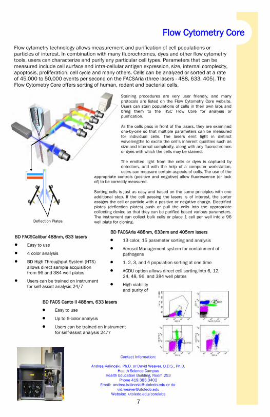
Flow cytometry technology allows measurement and purification of cell populations or
particles of interest. In combination with many fluorochromes, dyes and other flow cytometry
tools, users can characterize and purify any particular cell types. Parameters that can be
measured include cell surface and intra-cellular antigen expression, size, internal complexity,
apoptosis, proliferation, cell cycle and many others. Cells can be analyzed or sorted at a rate
of 45,000 to 50,000 events per second on the FACSAria (three lasers - 488, 633, 405). The
Flow Cytometry Core offers sorting of human, rodent and bacterial cells.
Staining procedures are very user friendly, and many
protocols are listed on the Flow Cytometry Core website.
Users can stain populations of cells in their own labs and
bring them to the HSC Flow Core for analysis or
purification.
As the cells pass in front of the lasers, they are examined
one-by-one so that multiple parameters can be measured
for individual cells. The lasers emit light in distinct
wavelengths to excite the cell’s inherent qualities such as
size and internal complexity, along with any fluorochromes
or dyes with which the cells may be stained.
The emitted light from the cells or dyes is captured by
detectors, and with the help of a computer workstation,
users can measure certain aspects of cells. The use of the
appropriate controls (positive and negative) allow fluorescence (or lack
of) to be correctly measured.
Sorting cells is just as easy and based on the same principles with one
additional step. If the cell passing the lasers is of interest, the sorter
assigns the cell or particle with a positive or negative charge. Electrified
plates (deflection plates) push or pull the cells into the appropriate
collecting device so that they can be purified based various parameters.
The instrument can collect bulk cells or place 1 cell per well into a 96
well plate for cloning.
BD FACSCalibur 488nm, 633 lasers
Easy to use
4 color analysis
BD High Throughput System (HTS)
allows direct sample acquisition
from 96 and 384 well plates
Users can be trained on instrument
for self-assist analysis 24/7
Flow Cytometry Core
Contact Information:
Andrea Kalinoski, Ph.D. or David Weaver, D.D.S., Ph.D.
Health Science Campus
Health Education Building, Room 253
Phone 419.383.3402
Email: [email protected] or da-
Website: utoledo.edu/corelabs
Flow Cytometry Core
BD FACSAria 488nm, 633nm and 405nm lasers
13 color, 15 parameter sorting and analysis
Aerosol Management system for containment of
pathogens
1, 2, 3, and 4 population sorting at one time
ACDU option allows direct cell sorting into 6, 12,
24, 48, 96, and 384 well plates
High viability
and purity of
BD FACS Canto II 488nm, 633 lasers
Easy to use
Up to 6-color analysis
Users can be trained on instrument
for self-assist analysis 24/7
Deflection Plates

8
NMR spectroscopy is a powerful tool for the determination of molecular structure, the study of molecular
dynamics, and the characterization of materials at the molecular level. The NMR Facility mission is to
support research and teaching at the University of Toledo. Instrumentation training and consultation are
available to companies that use the NMR spectrometers. The NMR Facility is located in the basement of
Bowman-Oddy Laboratories. It houses 4 NMR spectrometers: Bruker Avance 600MHz, Varian Inova
600MHz, VXRS 400MHz and Gemini 200MHz.
Varian Unity Inova 600 Mhz with a Penta, 1H{13C,15N,31P,2D} probe
It is an indirect detection probes designed for versatility in biomolecular
applications. It is tuned to allow decoupling of up to four different nuclei
including 2H lock.
Other probes:
Triple Resonance, 1H{13C,15N} indirect detection probe with triple axis
(XYZ) gradients for superior solvent suppression
Double Resonance Indirect, 1H{15N - 31P} probe, outer coil is tunable
over the frequency range (15N - 31P)
Dual Broadband (15N - 31P){ 1H }5mm and 10mm probes Multinuclear
probes optimized for superior sensitivity for nuclei in the typical fre-
quency range of 15N - 31P
Bruker Avance 600 MHz with a Dual resonance 5mm Cryoprobe, DCH with
Z gradient. CryoProbes. While it is optimized for 13C detection, the 1H
sensitivity is also very good. The Cryoprobe delivers the single largest
increase in NMR sensitivity in the last few decades. This enables an
increase sample throughput by up to 16- fold.
Other probes
4 mm Top-loading DVT Multinuclear Double Resonance MAS probe
tunable from 15N to 31P, with 50μL active volume and 15 kHz
maximum spinning speed. VT range -50oC to +120oC
5 mm SMARTProbe™ sample diameter with actively shielded Z-
gradient and
digital tuning for observation over the range from 15N to 31P as
well as 19F with 1H decoupling
Varian Vxrs 400MHz with versatile AutoSwitchable (13C/31P){1H/19F}probes
Other probes: Dual Broadband (15N - 31P){ 1H } 5mm and 10mm probes
Varian Gemini 200MHz with versatile AutoSwitchable (13C/31P){1H/19F}probes.
Esquire-LC (Bruker-HP) routinely configured with ESI source and manual
injection. This system combines Hewlett Packard’s HP1100 series HPLC with
Bruker’s multipole ion trap MS and MSn analyzer.
Other resources:
Atmospheric Pressure Chemical Ionization (APCI)
Nanospray
Contact Information:
Yong-Wah Kim, Ph.D.
Main Campus
Chemistry - Mail Stop #602,
Bowman-Oddy Laboratories Room 187,
Phone (419) 530-2563,
fax - (419) 530-4033
Email: [email protected]
Nuclear Magnetic Resonance Facility (NMR)

9
Robots for protein
crystallization
High brilliance
macromolecular
diffractometer
Combustion analysis
(CHN Analysis)
UV/Vis/NIR Spectrometer
also available for use.
Contact Information:
Kristin Kirschbaum, Ph.D.
Main Campus
Bowman-Oddy Laboratories, Room 200A
Phone 419.530.7847
Email: [email protected]
Website: utoledo.edu/corelabs
SEM with a STEM detector,
EDS, and EBIC applications
Thermal gravimetric
differential thermal
analyzer (TG-DTA)
655.310
657.302
0
100
200
300
400
500
600
Inte
ns. [
a.u.
]
652 653 654 655 656 657 658 659 660m/z
In 1985 the state of Ohio appropriated money for the creation of the Instrumentation Center
at the University of Toledo. The purpose of the center is to support faculty research, provide
access and training for graduate students in the use of advanced instrumentation and provide
a scientific support base for local industries through technical advice and sophisticated
problem solving capabilities. The Center also offers outreach programs that allow cyber-
access to instruments. Areas of advanced technologies include scanning electron microscopy
(SEM), mass spectrometry (MS), and crystallography.
MALDI/TOF/TOF MS
with a PROTEINEER fc
for LC-MALDI and
ImagePrep system
Three small molecule
diffractometers
Two powder X-ray
diffractometers (P-XRD)
Instrumentation Center

10
Histology Core Facility
The Histology Core provides services for Faculty, Staff and Students in the
University of Toledo Research Enterprise. We offer histological pro-
cessing, sectioning and staining of frozen or formalin-fixed, paraffin-embedded tissues. We routinely perform hematoxylin and eosin, Mas-
son’s Trichrome, PAS (Periodic Acid Schiff), and Oil Red O staining. The
Core produces slides for immunofluorescence, immunohistochemistry or
whole tissue IDISCO staining. Troubleshooting new antibodies to optimize
staining protocols and customization of services is available to accommo-
date specific needs of individual researchers.
Instrumentation
Leica automatic tissue processor
Leica EG 1160 embedding station
Reichert-Jung 2030 microtome
Microm HM550 Cryostat
Olympus VS120 Slide Scanner slide analyzer
Contact Information:
Allen Schroering
Block Health Science Bldg
Lab 1 , Room 007
Phone 419-383-6131
Email: [email protected]

11

The University of Toledo
Core Laboratories
The Core Research Facilities at The University
of Toledo are comprised of multiple facilities
located on both Main Campus and the Health
Science Campus of The University of Toledo.
Individual core facilities can be found on the
website at:
http://www.utoledo.edu/corelabs/