Embed Size (px)
Citation preview

The ventricles are structures that
produce cerebrospinal fluid, and transport it around the
cranial cavity. They are lined by ependymal cells, which
form a structure called the choroid plexus. It is within
the choroid plexus that CSF is produced.
Ventricuar system consists of four ventricles; - right and
left lateral ventricles,
- third ventricle
- fourth ventricle
- cerebral aqueduct of Sylvius.

The lateral ventricles communicate with the third
ventricle through interventricular foramens, and the third
ventricle communicates with the fourth ventricle through
the cerebral aqueduct.

CHOROID PLEXUS
Tufts of capillaries invaginate the roofs of ventricles, forming the
choroid plexuses of the ventricles. Cerebrospinal fluid (CSF) is
secreted by the choroid plexuses, filling the ventricular system.
CSF flows out of the fourth ventricle through the 3 apertures
formed at the roof of the fourth ventricle
One median aperture (Magendie) and two lateral apertures
(Luschka)
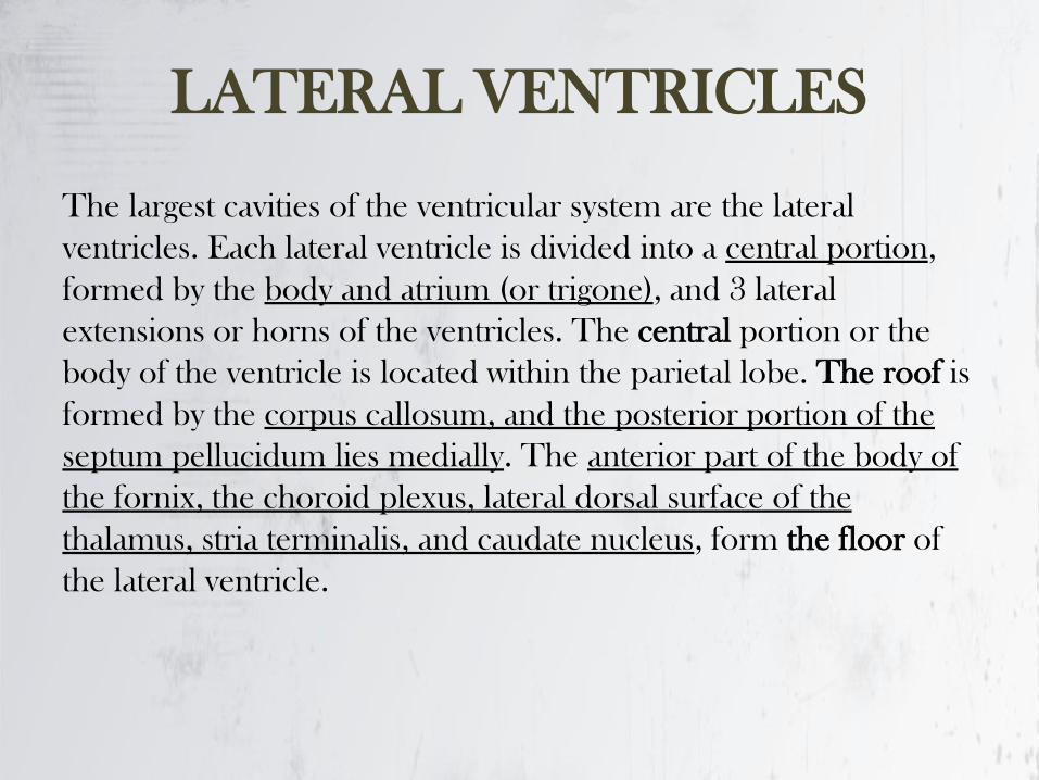
LATERAL VENTRICLES
The largest cavities of the ventricular system are the lateral
ventricles. Each lateral ventricle is divided into a central portion,
formed by the body and atrium (or trigone), and 3 lateral
extensions or horns of the ventricles. The central portion or the
body of the ventricle is located within the parietal lobe. The roof is
formed by the corpus callosum, and the posterior portion of the
septum pellucidum lies medially. The anterior part of the body of
the fornix, the choroid plexus, lateral dorsal surface of the
thalamus, stria terminalis, and caudate nucleus, form the floor of
the lateral ventricle.

-The interventricular foramen is located between the thalamus
and anterior pillar of the fornix, at the anterior margin of the body.
The 2 interventricular foramens (or foramina of Monro) connect
the lateral ventricles with the third ventricle. The body of the
lateral ventricle is connected with the occipital and temporal horns
by a wide area named the atrium.
-The anterior or frontal horn is located anterior to the
interventricular foramen. The floor and the lateral wall are formed
by the head of the caudate nucleus, the corpus callosum
constitutes the roof and anterior border, and the septum
pellucidum delineates the medial wall. The posterior or occipital
horn is located within the occipital lobe. The fibers of the corpus
callosum and the splenium form the roof. The forceps major is
located on the medial side and forms the bulb of the occipital
horn.

-The inferior or temporal horn is located within the temporal lobe. The
roof is formed by the fibers of the temporal lobe; the medial border
contains the stria terminalis and tail of the caudate. The medial wall and
the floor are formed by the hippocampus and its associated structures.
The amygdaloid complex is located at the anterior end of the inferior
horn.
-Capillaries of the choroid arteries from the pia mater project into the
ventricular cavity, forming the choroid plexus of the lateral ventricle. The
choroid plexus is attached to the adjacent brain structures by a double
layer of pia mater called the tela choroidea. The choroid plexus extends
from the lateral ventricle into the inferior horn. The anterior and
posterior horn have no choroid plexus.
-The choroid plexus of the lateral ventricle is connected with the
choroid plexus of the contralateral ventricle and the third ventricle
through the interventricular foramen. The anterior choroidal arteries
(branch of internal carotid artery) and lateral posterior choroidal arteries
(branch of the posterior cerebral artery) form the choroid plexus.
Venous supply from the choroidal veins drain into the cerebral veins.
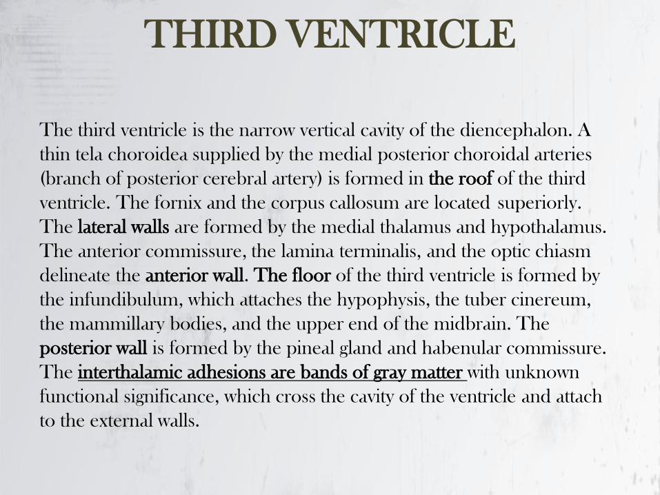
THIRD VENTRICLE
The third ventricle is the narrow vertical cavity of the diencephalon. A
thin tela choroidea supplied by the medial posterior choroidal arteries
(branch of posterior cerebral artery) is formed in the roof of the third
ventricle. The fornix and the corpus callosum are located superiorly.
The lateral walls are formed by the medial thalamus and hypothalamus.
The anterior commissure, the lamina terminalis, and the optic chiasm
delineate the anterior wall. The floor of the third ventricle is formed by
the infundibulum, which attaches the hypophysis, the tuber cinereum,
the mammillary bodies, and the upper end of the midbrain. The
posterior wall is formed by the pineal gland and habenular commissure.
The interthalamic adhesions are bands of gray matter with unknown
functional significance, which cross the cavity of the ventricle and attach
to the external walls.

FOURTH VENTRICLE
The fourth ventricle is connected to the third ventricle by a narrow cerebral
aqueduct. The fourth ventricle is a diamond-shaped cavity located posterior to
the pons and upper medulla oblongata and anterior-inferior to the cerebellum.
The superior cerebellar peduncles and the anterior and posterior medullary
vela form the roof of the fourth ventricle. The apex or fastigium is the extension
of the ventricle up into the cerebellum. The floor of the fourth ventricle is
named the rhomboid fossa. The lateral recess is an extension of the ventricle on
the dorsal inferior cerebellar peduncle.
Inferiorly, it extends into the central canal of medulla. The fourth ventricle
communicates with the subarachnoid space through the lateral foramen of
Luschka, located near the flocculus of the cerebellum, and through the median
foramen of Magendie, located in the roof of the ventricle. Most of the CSF
outflow passes through the medial foramen. The cerebral aqueduct contains no
choroid plexus. The tela choroidea of the fourth ventricle, which is supplied by
branches of the posterior inferior cerebellar arteries, is located in the posterior
medullary velum

- Cerebellomedullary cistern (largest)
- Pontocerebellar cistern (pontine cistern)
- Interpeduncular cistern (basal cistern)
- Chiasmatic cistern
- Quadrigeminal cistern (cistern of great cerebral vein)
- Cisterna ambiens
SUBARACHNOID CISTERNS
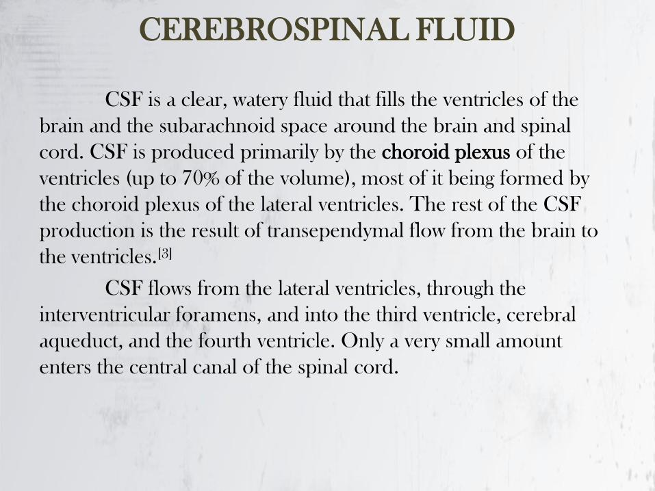
CEREBROSPINAL FLUID
CSF is a clear, watery fluid that fills the ventricles of the
brain and the subarachnoid space around the brain and spinal
cord. CSF is produced primarily by the choroid plexus of the
ventricles (up to 70% of the volume), most of it being formed by
the choroid plexus of the lateral ventricles. The rest of the CSF
production is the result of transependymal flow from the brain to
the ventricles.[3]
CSF flows from the lateral ventricles, through the
interventricular foramens, and into the third ventricle, cerebral
aqueduct, and the fourth ventricle. Only a very small amount
enters the central canal of the spinal cord.
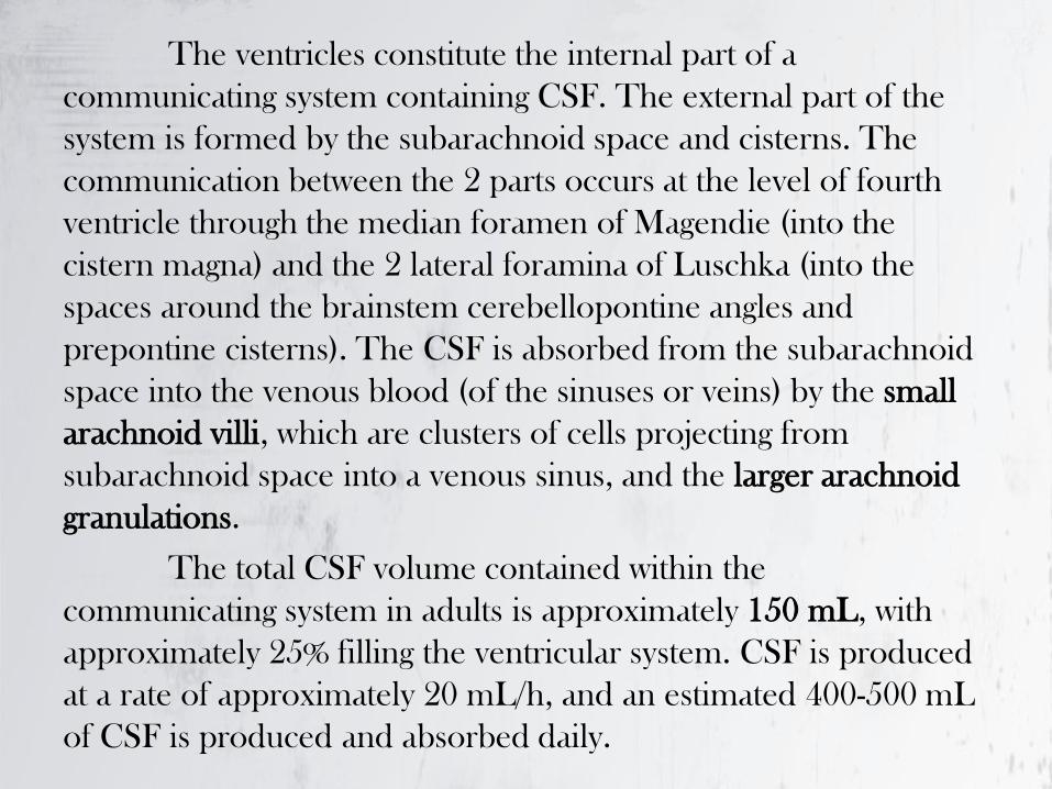
The ventricles constitute the internal part of a
communicating system containing CSF. The external part of the
system is formed by the subarachnoid space and cisterns. The
communication between the 2 parts occurs at the level of fourth
ventricle through the median foramen of Magendie (into the
cistern magna) and the 2 lateral foramina of Luschka (into the
spaces around the brainstem cerebellopontine angles and
prepontine cisterns). The CSF is absorbed from the subarachnoid
space into the venous blood (of the sinuses or veins) by the small
arachnoid villi, which are clusters of cells projecting from
subarachnoid space into a venous sinus, and the larger arachnoid
granulations.
The total CSF volume contained within the
communicating system in adults is approximately 150 mL, with
approximately 25% filling the ventricular system. CSF is produced
at a rate of approximately 20 mL/h, and an estimated 400-500 mL
of CSF is produced and absorbed daily.

CEREBRAL MENINGES
The meninges are the three membranes that
envelop the brain and spinal cord. The meninges
are the dura mater, the arachnoid mater, and
the pia mater. Cerebrospinal fluid is located in
the subarachnoid space between the arachnoid
mater and the pia mater.The primary function of
the meninges is to protect the central nervous
system.

DURA MATER
The dura mater (Latin: tough mother) is a thick, durable
membrane, closest to the skull and vertebrae. The dura
mater, the outermost part. It consists of two layers:
the endosteal layer, which lies closest to
the calvaria (skullcap), and the inner meningeal layer,
which lies closer to the brain. It contains larger blood
vessels that split into the capillaries in the pia mater. The
dura mater is a sac that envelops the arachnoid mater
and surrounds and supports the large dural
sinuses carrying blood from the brain toward the heart.

DURA INFOLDINGS
The dura has four areas of infolding:
-Falx cerebri, the largest, sickle-shaped; separates the cerebral hemispheres.
Starts from the frontal crest of frontal bone and the crista galli running to
the internal occipital protuberance.
- Tentorium cerebelli, the second largest, crescent-shaped; separates
the occipital lobes from cerebellum. The falx cerebri attaches to it giving a
tentlike appearance.
-Falx cerebelli, vertical infolding; lies inferior to the tentorium cerebelli,
separating the cerebellar hemispheres.
- Diaphragma sellae, smallest infolding; covers the pituitary gland and sella
turcica.
ARACHNOID MATER
The middle element of the meninges is the arachnoid mater, so
named because of its spider web-like appearance. It cushions the central nervous system.
The shape of the arachnoid does not follow the convolutions of
the surface of the brain and so looks like a loosely fitting sac. In
particular, in the region of the brain a large number of fine
filaments called arachnoid trabeculae pass from the arachnoid
through the subarachnoid space to blend with the tissue of the pia
mater.
PIA MATER
The pia mater (Latin: tender mother) is a very delicate
membrane. It is the meningeal envelope that firmly
adheres to the surface of the brain and spinal cord,
following all of the brain's contours (the gyri and sulci). It
is a very thin membrane composed of fibrous tissue
covered on its outer surface by a sheet of flat cells
thought to be impermeable to fluid. The pia mater is
pierced by blood vessels to the brain and spinal cord,
and its capillaries nourish the brain.

MENINGEAL SPACES
The subarachnoid space is the space that normally exists
between the arachnoid and the pia mater, which is filled
with cerebrospinal fluid.
The dura mater is attached to the skull, whereas in the spinal cord,
the dura mater is separated from the bone (vertebrae) by a space
called the epidural space, which contain fat and blood vessels. The
arachnoid is attached to the dura mater, while the pia mater is
attached to the central nervous system tissue. When the dura
mater and the arachnoid separate through injury or illness, the
space between them is the subdural space. There is a subpial
space underneath the pia mater that separates it from the glia
limitans.

MEDULLA SPINALIS
The spinal cord is the most important structure between the body and the brain. The spinal cord extends from the foramen magnum where it is continuous with the medulla to the level of the first or second lumbar vertebrae. It is a vital link between the brain and the body, and from the body to the brain. The spinal cord is 40 to 50 cm long and 1 cm to 1.5 cm in diameter. Two consecutive rows of nerve roots emerge on each of its sides. These nerve roots join distally to form 31 pairs of spinal nerves. The spinal cord is a cylindrical structure of nervous tissue composed of white and gray matter, is uniformly organized and is divided into four regions:
cervical (C),
thoracic (T),
lumbar (L)
and sacral (S).
Each of them is comprised of several segments. The spinal nerve contains motor and sensory nerve fibers to and from all parts of the body. Each spinal cord segment innervates a dermatome.

GENERAL FEATURES
Similar cross-sectional structures at all spinal cord levels.
-It carries sensory information (sensations) from the body and some from the head to the central nervous system (CNS) via afferent fibers, and it performs the initial processing of this information.
-Motor neurons in the ventral horn project their axons into the periphery to innervate skeletal and smooth muscles that mediate voluntary and involuntary reflexes.
-It contains neurons whose descending axons mediate autonomic control for most of the visceral functions.
-It is of great clinical importance because it is a major site of traumatic injury and the locus for many disease processes.
Although the spinal cord constitutes only about 2% of the central nervous system (CNS), its functions are vital. Knowledge of spinal cord functional anatomy makes it possible to diagnose the nature and location of cord damage and many cord diseases.

SEGMENTAL AND LONGITUDINAL
ORGANIZATION
The spinal cord is divided into four different regions: the cervical, thoracic, lumbar and sacral regions. The different cord regions can be visually distinguished from one another. Two enlargements of the spinal cord can be visualized: The cervical enlargement, which extends between C3 to T1; and the lumbar enlargements which extends between L1 to S2.
The cord is segmentally organized. There are 31 segments, defined by 31 pairs of nerves exiting the cord. These nerves are divided into 8 cervical, 12 thoracic, 5 lumbar, 5 sacral, and 1 coccygeal nerve .
Dorsal and ventral roots enter and leave the vertebral column respectively through intervertebral foramen at the vertebral segments corresponding to the spinal segment.
The cord is sheathed in the same three meninges
as is the brain: the pia, arachnoid and dura. The
dura is the tough outer sheath, the arachnoid lies
beneath it, and the pia closely adheres to the
surface of the cord. The spinal cord is attached to
the dura by a series of lateral denticulate
ligaments emanating from the pial folds.
DEVELOPMENT
During the initial third month of embryonic development, the spinal cord extends the entire length of the vertebral canal and both grow at about the same rate. As development continues, the body and the vertebral column continue to grow at a much greater rate than the spinal cord proper. This results in displacement of the lower parts of the spinal cord with relation to the vertebrae column. The outcome of this uneven growth is that the adult spinal cord extends to the level of the first or second lumbar vertebrae, and the nerves grow to exit through the same intervertebral foramina as they did during embryonic development. This growth of the nerve roots occurring within the vertebral canal, results in the lumbar, sacral, and coccygeal roots extending to their appropriate vertebral levels.

All spinal nerves, except the first, exit below their corresponding
vertebrae. In the cervical segments, there are 7 cervical vertebrae and
8 cervical nerves. C1-C7 nerves exit above their vertebrae whereas the
C8 nerve exits below the C7 vertebra. It leaves between the C7
vertebra and the first thoracic vertebra. Therefore, each subsequent
nerve leaves the cord below the corresponding vertebra. In the
thoracic and upper lumbar regions, the difference between the
vertebrae and cord level is three segments. Therefore, the root
filaments of spinal cord segments have to travel longer distances to
reach the corresponding intervertebral foramen from which the spinal
nerves emerge. The lumbosacral roots are known as the cauda
equina.
Each spinal nerve is composed of nerve fibers that are related to the
region of the muscles and skin that develops from one body somite
(segment). A spinal segment is defined by dorsal roots entering and
ventral roots exiting the cord, (i.e., a spinal cord section that gives rise
to one spinal nerve is considered as a segment.)
DERMATOME
A dermatome is an area of skin supplied by peripheral nerve fibers originating from a single dorsal root ganglion. If a nerve is cut, one loses sensation from that dermatome. Because each segment of the cord innervates a different region of the body, dermatomes can be precisely mapped on the body surface, and loss of sensation in a dermatome can indicate the exact level of spinal cord damage in clinical assessment of injury.
TERMINOLOGY
Four different terms are often used to describe bundles of axons such as those found in the white
matter: funiculus, fasciculus, tract, and pathway.
Funiculus is a morphological term to describe a large group of nerve fibers which are located in a
given area (e.g., posterior funiculus).
Within a funiculus, groups of fibers from diverse origins, which share common features, are
sometimes arranged in smaller bundles of axons called fasciculus, (e.g., fasciculus proprius).
Fasciculus is primarily a morphological term whereas tracts and pathways are also terms applied to
nerve fiber bundles which have a functional connotation. A tract is a group of nerve fibers which
usually has the same origin, destination, and course and also has similar functions. The tract name is
derived from their origin and their termination (i.e., corticospinal tract - a tract that originates in the
cortex and terminates in the spinal cord; lateral spinothalamic tract - a tract originated in the lateral
spinal cord and ends in the thalamus).
A pathway usually refers to the entire neuronal circuit responsible for a specific function, and it
includes all the nuclei and tracts which are associated with that function. For example, the
spinothalamic pathway includes the cell bodies of origin (in the dorsal root ganglia), their axons as
they project through the dorsal roots, synapses in the spinal cord, and projections of second and
third order neurons across the white commissure, which ascend to the thalamus in the
spinothalamic tracts.
INTERNAL STRUCTURE OF THE
SPINAL CORD
A transverse section of the adult spinal cord shows white matter in the periphery, gray matter inside, and a tiny central canal filled with CSF at its center. Surrounding the canal is a single layer of cells, the ependymal layer. Surrounding the ependymal layer is the gray matter – a region containing cell bodies – shaped like the letter “H” or a “butterfly”. The two “wings” of the butterfly are connected across the midline by the dorsal gray commissure and below the white commissure.
The gray matter mainly contains the cell bodies of neurons and glia and is divided into four main columns: dorsal horn, intermediate column, lateral horn and ventral horn column. The dorsal horn is found at all spinal cord levels and is comprised of sensory nuclei that receive and process incoming somatosensory information.
The root cells are situated in the ventral and lateral gray horns and
vary greatly in size. The root cells contribute their axons to the
ventral roots of the spinal nerves and are grouped into two major
divisions: 1) somatic efferent root neurons, which innervate the
skeletal musculature; and 2) the visceral efferent root neurons, also
called preganglionic autonomic axons, which send their axons to
various autonomic ganglia.
- The column or tract cells and their processes are located mainly
in the dorsal gray horn and are confined entirely within the CNS.
-The axons of the column cells form longitudinal ascending tracts
that ascend in the white columns and terminate upon neurons
located rostrally in the brain stem, cerebellum or diencephalon. - -
-Some column cells send their axons up and down the cord to
terminate in gray matter close to their origin and are known as
intersegmental association column cells.

- Other column cell axons terminate within the segment in which
they originate and are called intrasegmental association column
cells. Still other column cells send their axons across the midline
to terminate in gray matter close to their origin and are called
commissure association column cells.
The propriospinal cells are spinal interneurons whose axons do
not leave the spinal cord proper. Propriospinal cells account for
about 90% of spinal neurons. Some of these fibers also are found
around the margin of the gray matter of the cord and are
collectively called the fasciculus proprius or the propriospinal
tract.

Spinal Cord Nuclei and Laminae
Spinal neurons are organized into nuclei and
laminae.

NUCLEI
The prominent nuclear groups of cell columns within the spinal
cord from dorsal to ventral are the marginal zone, substantia
gelatinosa, nucleus proprius, dorsal nucleus of Clarke,
intermediolateral nucleus and the lower motor neuron nuclei.

Marginal zone nucleus – lateral spinothalamic tract – pain and
temperature to the thalamus.
Substantia gelatinosa – ventral and lateral spinothalamic tract –
relays pain, temperature and mechanical (light touch) information.
Nucleus proprius- ventral spinothalamic tract and spinocerebellar
tract – mechanical and temperature sensation
Dorsal nucleus of Clark – through lateral funiculus – dorsal
spinocerebellar tract – unconcious proprioception from muscle
spindles and Golgi tendon organs to the cerebellum (C8-L3).
Intermediolateral and intermediomedial cell columns
Lower motor neuron nuclei are located in the ventral horn of the
spinal cord. They contain predominantly motor nuclei consisting
of α, and motor neurons and are found at all levels of the
spinal cord--they are root cells.
REXED LAMINAE
The distribution of cells and fibers within the gray matter of the spinal cord exhibits a pattern of lamination. The cellular pattern of each lamina is composed of various sizes or shapes of neurons (cytoarchitecture) which led Rexed to propose a new classification based on 10 layers (laminae).
Laminae I to IV, in general, are concerned with exteroceptive sensation and comprise the dorsal horn, whereas laminae V and VI are concerned primarily with proprioceptive sensations. Lamina VII is equivalent to the intermediate zone and acts as a relay between muscle spindle to midbrain and cerebellum, and laminae VIII-IX comprise the ventral horn and contain mainly motor neurons. The axons of these neurons innervate mainly skeletal muscle. Lamina X surrounds the central canal and contains neuroglia.

WHITE MATTER
Surrounding the gray matter is white matter containing myelinated
and unmyelinated nerve fibers. These fibers conduct information
up (ascending) or down (descending) the cord. The white matter is
divided into the dorsal (or posterior) column (or funiculus), lateral
column and ventral (or anterior) column. The anterior white
commissure resides in the center of the spinal cord, and it contains
crossing nerve fibers that belong to the spinothalamic tracts,
spinocerebellar tracts, and anterior corticospinal tracts.
WHITE MATTER
Three general nerve fiber types can be distinguished in the spinal
cord white matter:
1) long ascending nerve fibers originally from the column cells,
which make synaptic connections to neurons in various
brainstem nuclei, cerebellum and dorsal thalamus,
2) long descending nerve fibers originating from the cerebral
cortex and various brainstem nuclei to synapse within the
different Rexed layers in the spinal cord gray matter, and
3) shorter nerve fibers interconnecting various spinal cord levels
such as the fibers responsible for the coordination of flexor
reflexes. Ascending tracts are found in all columns whereas
descending tracts are found only in the lateral and the anterior
columns.
ASCENDING TRACTS
The spinal cord white matter contains ascending and descending tracts.
Ascending tracts . The nerve fibers comprise the ascending tract emerge from
the first order (1°) neuron located in the dorsal root ganglion (DRG). The
ascending tracts transmit sensory information from the sensory receptors to
higher levels of the CNS. The ascending gracile and cuneate fasciculi occupy the
POSTERIOR COLUMN, and sometimes are named the dorsal funiculus.
These fibers carry information related to tactile, two point discrimination of
simultaneously applied pressure, vibration, position, and movement sense and
conscious proprioception.
In the LATERAL COLUMN lateral spinothalamic tract is located more
anteriorly and laterally, and carries pain, temperature and crude touch
information from somatic and visceral structures. Nearby laterally, the dorsal
and ventral spinocerebellar tracts carry unconscious proprioception information
from muscles and joints of the lower extremity to the cerebellum.


![Comunicación interventricular [Ventricular Septal Defect]](https://img.pdfslide.net/doc/110x75/559262691a28ab33128b4573/comunicacion-interventricular-ventricular-septal-defect.jpg)