Embed Size (px)
Citation preview

-~.
CHAPTER IX
THE WINGS OF THE NEUROPTERA
(a) THE MORE GENERAL FEATURES OF THE WINGS OF THE NEUROPTERA
The wings of the Neuroptera are membranous and usually furnished with many wing-veins. The two pairs of wings are similar in texture and usually in outliner in some, the fore wings are slightly larger than the hind wings, in others the two pairs of wings are of the same size. The anal area is small in both fore and hind wings; it is rarely folded, and then only slightly so. A distinct anal furrow is rarely developed. Definitive accessory veins are usually present, and, as a rule, there are many marginal accessory veins. Intercalary veins are never developed. When at rest, with few exceptions, the wings are folded roof-like over the abdomen. In 5OII1e cases organs for uniting the fore and hind wings are present.
In most of the families the radial sector is pectinately branched. In nearly all cases, media is obviously not more than two branched, if accessory veins be not counted; and in those cases where it appears to be three or four branched it may be that this condition is due to a secondary splitting of one or of both of the two primitive branches. In a few members of the order the hind wings are very small and in some they are wanting in one sex. In the family Nemopteridre the two pairs of wings differ greatly in form, the hind wings being very long and narrow; in some members of this family the hind wings are thread-like.
In the following discussion of the wings of the Neuroptera figures are given of the wings of representatives of all of the recognized families of the order. It was neces~ary to figure a large proportion of these in order to illustrate the various methods of specialization that are found within the order; and when this was done it seemed worth while to add figures of representatives of the remaining families, for the sake of completeness, although they presented no difficulties in the application of the uniform terminology of the wing-veins.
For convenience in making generalizations the families Sisyridre, Sympherobiidre, Hemerobiidse, and Dilaridse are referred to as thc hemerobiid group of families; and the Nymphidre, Myrmeleonidse, Ascalaphidse, Nemopteridre, and Apochrysidec, as the myrmeleonid group.
This study of the wings of the Neuroptera has been greatly facilitated by the presence in the entomological museum of Cornell University of a large collection of Neuroptera, which has been brought together by Dr. Needham.
(145)
146 THE WINGS OF NEUROPTERA
(b) THE SPECIAL FEATURES OF THE WINGS OF THE NEUROPTERA
In the order Neuroptera there are several very distinct types of specialization of the wings. Some of these are described in the accounts of the wings of the families in which they occur; but there are others that are characteristic of more than one family; these are described here in order to avoid repetition.
The accessory veins.-In some Neuroptera the development of accessory veins has progressed to a very limited extent; but in a much larger
Fig. 137.-Wings of Osmylus hyalinatus.
number of representatives of this order the most striking features of the wings are produced by accessory veins. It is here that the development of accessory veins, both definitive and marginal, reaches its most perfect condition, and wings of wonderful beauty of form are the result.
The definitive accessory veins of the Neuroptera are produced in two ways; in some cases they are added distally by successive splittings of the tip of a principal vein, thus forming a regular series; in other cases, the number of accessory veins is increased in an irregular manner, by the splitting of branches of the principal veins. Wings illustrating each of these methods of increase in the number of accessory veins are figured iater.

THE WINGS OF NEUROPTERA 147
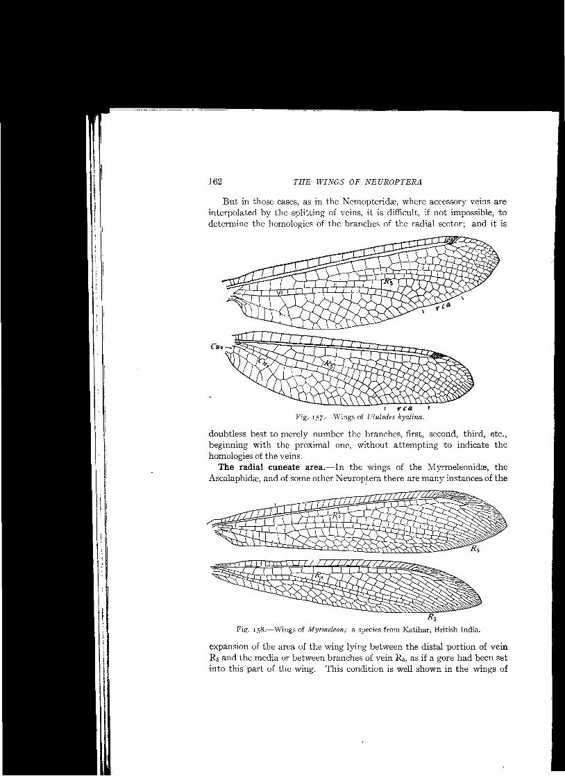
The marginal accessory veins in many of the Neuroptera differ remarkably from those of the other orders of insects in which this type of veins is found in the fact that they form a regular border of quite uniform width. This is illustrated by the veins of the outer margin of the wings of Osmylus hyalinatus (Fig. 13 7).
In some cases there is a splitting of one or both forks of a vein that has been split, thus forming a second rank of marginal accessory veins; this is the case in the wings of Polystachotes, figured later (Fig. 141).
The distinctive characteristic of marginal accessory veins is their instability; they vary in number and in length in different individuals of the same species, and even in the wings of the two sides of the same individual. But when the splitting of a vein has progressed sufficiently far it becomes fixed, and what at first, phylogenetically, was merely a marginal accessory vein become transformed into a definitive accessory vein.
The beautiful symmetry of the borders of marginal accessory veins in certain members of this order is due not merely to the nearly equal length of these veins but also to the fact that the two forks of each forked vein occupy bilaterally symmetrical positions. It follows that neither fork can be regarded as accessory to the other, they are sister veins, both accessory to the stem from which they were derived.
The suppression of the dichotomy of the Tadial sector.-Correlatedwith the extensive dvelopment of accessory veins in the Neuroptera, there has resulted in nearly all of the families of this order the production of a pectinately branched radial sector; that is, this vein is so modified that it consists of a supporting stem upon which are borne a greater or less number of parallel branches. This is shown in most of the figures of wings illustrating this chapter. This is a distinctive characteristic of this order; in no one of the other orders of living insects in which accessory veins occur is a welldeveloped pectinately branched radial sector found. Such a sector existed, however, in many of the Palreodictyoptera.
The typical radial sector is dichotomously branched, being divided into two chief branches, veins R 2+3, and R 4+5, and each of these in turn is divided into two branches, the former into veins R 2 and R 3 and the latter into veins R 4 and R 5•
The transformation of a dichotomously branched radial sector into one that is pectinately branched was discussed by Comstock and Needham, and the process by which this transformation is attained was termed by us the suppression of the dichotomy of the radial sector. In the course of our discussion, we pointed out that there are three ways in which this result may have been attained: first, by the splitting apart of veins R 4 and R 5
so that they arise separately from the supporting stem of the pectinate vein thus formed; second, by the switching of the base of vein R, to vein R 2+3,
following a cross-vein, this also resulting in the two veins arising separately

Flig. I38.-Diagrams of several types of radius.
THE WINGS OF NEUROPTERA
.. R3
R4
R s
-s,
C~~R'S R..
R3
- ~
~
-R
-R
148
n.---R,
3 ~~R'~ R", R3 R4
Rs
from the supporting stem of the pectinate vein; and third, by the coalescence of veins R4 and R5, which would obliterate the forking of these veins. The accompanying series of diagrams (Fig. 138) illustrates these changes.
The first diagram (Fig. 138, I) represents the manner of branching of the typical radius in which the radial sector is dichotomously branched. The
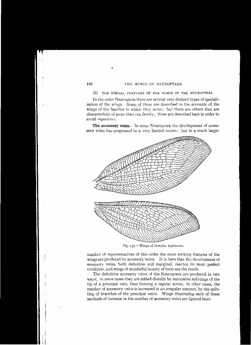
second diagram represents a radius in which the radial sector has become pectinate by the splitting apart of veins R4 and R5, so that they arise separately from the supporting stem of the pectinate vein thus formed. This represents the simplest type of a pectinately branched radial sector. An illustration of this simplest type is afforded by the wings of Sisyra (Fig. -139).
In comparatively few cases is a pectinately
4 ~ s, branched radial sector so . <~ ~ simple as that of Sisyra;
<--'< <: """'" ........ R. usually a pectinate sector bears more than four branches; this is the result of the development of accessory veins on the posterior side of vein R2•
The third diagram (Fig. 138, 3) represents a radial sector in which vein R 2
bears two accessory veins, labeled R2a and R2b ; this condition exists in the wing
of Chauliodes (Fig. 140); in the wings of Polystcechotes (Fig. 141) the addition of accessory veins to vein R2 has been carried to a much greater extent.
The second method of suppression of the dichotomy of the radial sector occurs in the wings of certain insects in which there are many crossveins. In these cases the base of vein R 4 has become connected with a cross-vein, perhaps by splitting back until the cross-vein is reached, and
.5-R'
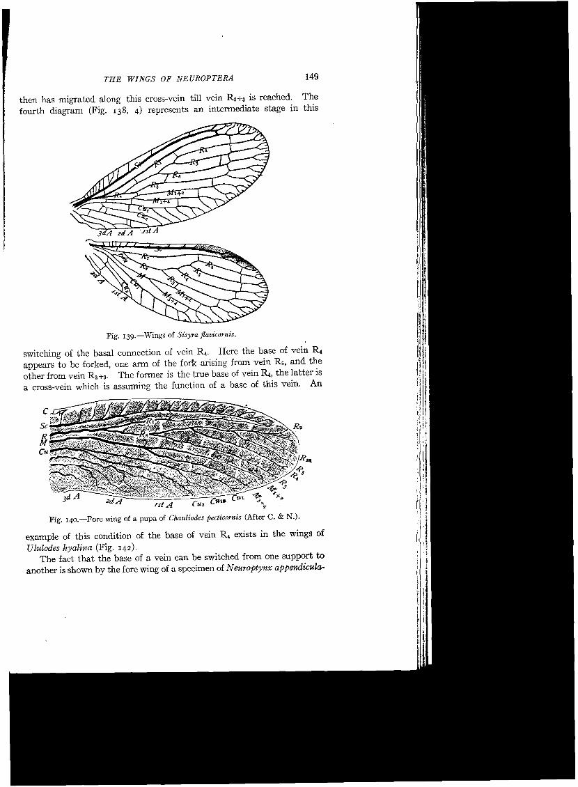
Fig. 139.-Wings of Sisyra flavicornis.
then has migrated along this cross-vein till vein R2+3 is reached. The fourth diagram (Fig. 138, 4) represents an intermediate stage in this
149THE WINGS OF NEUROPTERA
IslA
Fig. 140.-Fore wing of a pupa of Chauliodes pecticornis (After C. & N.).
example of this condition of the base of vein R 4 exists in the wings of Ululodes hyalina (Fig. 142).
The fact that the base of a vein can be switched from one support to another is shown by the fore wing of a specimen of N europtynx appendicula
switching of the basal connection of vein R 4• Here the base of vein R, appears to be forked, one arm of the fork arising from vein Rs, and the other from vein R 2+S• The former is the true base of vein Ra, the latter is a cross-vein which is assuming the function of a base of this vein. An

150 THE WINGS OF NEUROPTERA
Fig. 14I.-Wings of Polystcechotes punctatus,
I rca Fig. 142.-Wings of Ululodes hyalina.

THE WINGS OF NEUROPTERA 151
tus (Fig. 143) that I collected in Florida. In this wing the base of vein R2+3+4 has been switched along a cross-vein to vein RI, so that there appear to be two radial sectors.
It was the existence of this abnormal wing that suggested the probability that in wings like those of the Myrmeleonidse, where there are many crossveins, the suppression of the dichotomy had been attained by the switching of the base of vein R 4 to vein R 2 +3 along a cross-vein; and it was thought that an examination of a larger series of wings would reveal the existence of examples of the successive stages of this switching.
Very many examples were found of wings in which the base of vein R 4
appears to be at a halfway station in its journey along a cross-vein from
rca I
Fig. 143.-Wings of Neuroptynx appendiculatus.
vein R 5 to vein R2+3• This condition is very common in the Ascalaphidee (Fig. 142).
It is only recently that I have been able to find examples of the successive stages of the switching of the base of vein R 4 from vein R 5 to vein R 2+3• These were found in the osmyli dse, a family in which the dichot i:'
r<omy of the radial sector is normally suppressed. I'Ii 11Figure 144 represents the wings of an atavistic individual of Osmylus
; ~
hyalinatus, in which the primitive dichotomy of the radial sector has been '/' retained; the homologies of the wing-veins are indicated in Figure 145.
That such cases may be common in the Osmylidse is indicated by the JII:fact that in the small collection of osmylids to which I now have access two ij ,
individuals present this condition. In one Osmylus hyalinatus the radial sector is dichotomously branched in both fore wings and in one hind wing. I' In an Osmylus tessellatus the radial sector is dichotomously branched in the ~ I fore wings; but in the hind wings the dichotomy has been suppressed.

152 THE WINGS OF NEUROPTERA
Fig. 144·-Wings of Osmylus hyalinatus in which the dichotomy of the radial sector has not been suppressed.
Sc
IM~~RI R 2 •
R3
R 4
Rs kf'+2
M 3+4,.....l.,....JT-+--+-+-...,....,......-.........CUt~~:r-L--+J~I.>-JL..,.L4-L..-J...,--L+--+T\~C1l2
Fig. 14S·-Base of the fore wing shown in Figureraa, enlarged.

153 THE WINGS OF NEUROPTERA
Figures 146 and 147 represent the wings of a normal Osmylus hyalinatus. An important fact bearing on this question of the switching of the base
of a vein from one support to another is that in the family Sympherobiidse the base of vein R2+a has been transferred to vein R l .
The third possible method of suppression of the dichotomy of the radial sector would produce the result shown diagrammatically in the fifth diagram (Fig. 138, 5), where veins R 4 and R 5 are represented as coalescing completely. I know of no case where the suppression of the dichotomy has
Fig. 146.-Wings of Osmylus hyalinatus with the radial sector pectinately branched.
been attained by this method. I am now convinced that in the supposed examples of this method, cited by Comstock and Needham, that is in Corydalus and Chauliodes, the result has been attained by the splitting apart of veins R 4 and R 5, instead of by their coalescence.
The earlier conclusion was based on the fact that occasionally the first branch of the radial sector in Corydalus is forked, as is the case in the wing represented by Figure 148, 1. The forked condition of this first branch was believed to indicate that it is a compound vein in which the coalescence of its two elements is not quite complete. My reasons for abandoning this view are the following.

154 THE WINGS OF NEUROPTERA
A study of a series of wings of Corydalus cornutus has shown that the forking of the first branch of the radial sector in this species is of very
~JJIlIfr~~ R2&
R3
R4
Rs
M'+2
CU2
Fig, 147.-Base of the fore wing shown in Figure 146, enlarged.
irregular occurrence, and that there is a similar irregular forking of other veins in this region of the wing. In the specimen represented in Figure 148 vein M1 is forked in a similar manner, so too is the fourth branch of the sector; in the latter case one division of the forked vein is again forked.
In a collection of nineteen wings of Corydalus cornutus, that had been mounted for laboratory use and which had been taken at random, I find the first branch of the radial sector forked .n only two specimens; the third branch, in one; the fourth branch, in one; the fifth branch, in seven; the sixth branch, in twelve; and the seventh branch in four.
The evidence presented by this species indicates that the short branches near the margin of the wing in the area of the radial sector are merely marginal accessory veins; and that the forking of the first branch of the
Fig. 148.-Fore wing of Corydalus cornutus in which the first branch of the radial sector is forked.

THE WINGS OF NEUROPTERA 155
sector does not indicate that this branch is composed of two coalesced veins; it merely shows that the first branch of the sector is occasionally split at the tip as are other veins in this region of the wing.
This evidence is confirmed by a study of the wings of representatives of nearly all of the genera of this family. Fortunately we are able to make such a study, thanks to the labors of Dr. H.W. van derWeele. This author in his Monographic Revision oj the Megaloptera (Weele, 1910) has given photographic illustrations of representatives of nearly all of the genera of the Sialidse,
The most important, for the purposes of the present discussion, of the figures in this work is that of Corydalus primitivus, a species found in the Argentine Republic. The entire insect is figured, but I copy only the wings (Fig. 149).
In this individual the dichotomy of the radial sector is not suppressed in the right hind wing; but it is in the other three wings. That the first and
Fig. 149.-Wings of Corydalus primitivus (After van der Weele).
second branches of the sector in the left hind wing and in both fore wings correspond to the forked first branch in the right hind wing is shown by the fact that counting the forked first branch of the right hind wing as two branches, the sector is eight-branched in all wings. The splitting apart of veins R 4 and R 5 in the right hind wing so that they arise separately, would make the sector of this wing eight-branched like those of the other wings.
I regard the structure of the right hind wing of this insect as an example of atavism, similar to those found in the osmylids described above.
Another remarkable insect figured by van der Weele, in the same monograph, is Protohermes davidi from China. I copy a part of his figure somewhat enlarged (Fig. ISO), he figures the entire insect.
Excepting the right hind wing of Corydalus primitivus discussed above, the deepest forking of the first branch of the radial sector shown in the figures given by van der Weele occurs in this insect. That this forking is due to a splitting of the tip of this vein is evident by a comparison of it
156 THE WINGS OF NEUROPTERA
with the tips of the branches of media in which there is a similar splitting of the tips of veins.
Finally, excepting those cases in which the development of a pectinate radial sector has not progressed very far, as in Sisyra (Fig. I39), the most
Oh\M\ JV!1+2 \ Rs-, 3+4
Fig. Iso.-Wings of Protohermes davidi (After van der Weele).
striking feature of this vein, when its dichotomy has been suppressed and a pectinate form attained, is the presence of a greater or less number of supernumerary veins. These are accessory veins; and it has been shown that veins of this type are produced and their number increased by the splitting of veins.
It is evident, therefore, that the force that makes for the development of the pectinate type of radial sector acts by the splitting of veins; and it is not probable that this force should cause veins R4 and Rr. to coalesce in the Sialidse, instead of splitting apart as we know they do in the Hernerobiidse.
The numbering of the branches of the radial sector when it is peetinately branehed.-In most of the Neuroptera in which the radial sector is pectinately branched, the number of the branches of this vein is more than four, the number of branches of the typical radial sector. It is evident that when this is the case, some of the branches are accessory veins. The question arises, therefore, which of the branches are the primitive ones and which are

THE WINGS OF NEUROPTERA 157
accessory veins? In other words, to which end of the series of branches of the radial sector are the accessory veins added?
Comstock and Needham concluded from their studies of the tracheation of the wings of pupse of Chauliodes (Fig. 140) and Corydalus (Fig. 151) that in the case of these insects the accessory veins are added to the distal end of the series. The presence of fine twigs at the tip of trachea R2 indicated to us the method of increase, which we concluded to be as follows:
The fine tracheal twigs at the tip of trachea R 2 are the beginnings of accessory branches, which in the course of phylogenetic development become larger and, moving towards the base of the wing, make room for the addition of other branches.
From this it follows that vein R2 forms the terminal portion of the stem of the pectinate vein and that veins R 5, ~, and R, are the proximal branches. It also follows that of the accessory veins that have been developed on vein R2 the proximal one is the oldest, and according to our system should be labeled R 2a , as is done in Figures 140 and lSI.
My studies of the venation of the wings of insects during the twenty years that have elapsed since these conclusions were reached have only served to confirm them; and I am now convinced that what we showed to be the case in Chauliodes and Corydalus is true primarily of all of those neuropterous insects in which the radial sector is pectinately branched. In some cases, however, to be discussed later, accessory veins are interpolated in the primary series by the splitting of members of this series.
I am led to make this statement because some of the writers on the Neuroptera have not accepted these conclusions. Thus Enderlein ('10) in his figures of the wings of Mantispidre numbers the branches of the radial
Fig. ISI.-Tracheation of a wing of a pupa of Corydalus cornutus (after C. & N.).
sector from the apex of the wing towards the base; and Tillyard ('16, p. 277) violently attacks the view of Comstock and Needham. For this reason it seems incumbent upon me to give the additional data that have confirmed my belief in this view.

158 THE WINGS OF NEUROPTERA
Fig. 152.-Wings of Sisyra fiavicornis.
Fig. 153.-Wings of Hemerobius humuli.

Fig. 155.-Tracheation of the wings of a pupa : of Hemerobius humuli.
159THE WINGS OF NEUROPTERA
M J+4
Fig. 154.-Base of a hind wing of Hemerobius humuli.
hind wings of this species there are only four definitive branches of the radial sector, although there are many marginal accessory veins. The
In Sisyra flaoicornis (Fig. 152) definitive accessory veins have not been developed upon the radial sector, although there are marginal accessory veins upon its branches. In each wing the sector is four-branched. These
branches are doubtless veins R2, Ra, R4, and R 5• The dichotomy of the sector has been suppressed by the splitting back of vein R5• Two stages in this splitting process are represented by the two wings, it having been carried much farther in the fore wing than in the hind wing. Attention is called to the wings of this insect to show that the veins that are split back in the hemerobiid group of families are primitive branches of the radial sector and not accessory veins.
In the wings of Hemerobius humuli (Fig. 153), the splitting back of branches of the radial sector is carried much farther than in Sisyra. In the

160 THE WINGS OF NEUROPTERA
four definitive branches are doubtless veins Rs, R 3, R4, and R 5, as indicated in the figure. Vein R 5 is split back nearly to the base of the sector and the remainder of the sector, after the separation of vein R5, anastomoses with vein R 1. (Figure 154 represents this part of the wing greatly magnified.)
A study of the tracheation of the hind wing of a pupa of this species (Fig. ISS) confirms the interpretation of the venation of this part of the wing given in Figures 153 and 154. The trachea of the radial sector is typical except that trachea R 5 is split back from trachea R 4 ; and this splitting has been carried so far that the undivided stem of the radial sector is very short; the tracheation of the pupal wing and the venation of the adult wing correspond very closely in this respect. But in the pupal wing there is no indication of the anastomosis of veins R 1 and R Z+3+4• It is evident, however, that these two tracheas come together at the point marked a in Figure ISS in the course of the later development of the wing.
This anastomosis of vein R Z+3+4 with vein R 1 is carried much farther in the fore wing, where the two veins coalesce for a considerable distance, with the result that two of the branches of the radial sector are stranded on the main stem of the radius before the separation of the remainder of the radial sector from vein R 1. The first of these two stranded branches is doubtless vein R 5, as it is obviously serially homologous with vein R 5 of the hind wing, in which there are no definitive accessory veins to cast a doubt on the determination of the homologies of the branches of radius. From this it follows that the extra branch of the radial sector in this wing has not been added to the proximal end of the series but to vein R z, and is the one labeled R Za in the figure.
According to the view that the accessory veins are added to the proximal end of the series we must believe that the first fork of the radial sector has been pushed out to near the apex of the wing by the development of the accessory veins, and that the three short veins next to vein R z are veins R 3,
R 4, and R 5. 1£ this be true, either the size of the wing has been greatly increased or veins R3, R 4, and R 5 have been greatly shortened during the specialization of the wing. Either of these alternatives may be true, but neither seems probable.
In many cases the branches at the tip of the radial sector are merely short twigs, which differ in size, number, or arrangement in the wings of the two sides of the same insect; in such cases it is obviously impracticable to begin the numbering at the apex of the sector. The fact is, these terminal branches in cases of this kind are merely marginal accessory veins, that have not attained a definitive form and position.
Finally, while there are many cases, like those of Chauliodes and Corydalus, in which there appear to be taking place the development of additional branches at the distal end of the series of branches of the radial

THE WINGS OF NEUROPTERA 161
sector, no instance has been observed in the Neuroptera in which there is such an appearance at the proximal end of the series. The only possible exception to this statement is that it is conceivable that in the development of a radial cuneate area vein R, may be split to its base and in this wayan addition to the number of branches of the radial sector be made at the proximal end of the series.
As a rule, in the development of a radial cuneate area vein R 5 is not split back far enough to warrant this suggestion; but in Rapisma (Fig. 171) and in Megalomus (Fig. 178) the splitting back of vein R, has progressed nearly to the base of this vein.
I know of no near allies of Rapisma, but M egalomus can be compared with more generalized members of its family, as Hemerobius, in which there
Fig. 156.-Wings of Acanthaclisis; a species from the Seychelles Islands.
is no indication of accessory veins being added to the proximal end of the series.
I t seems worth while to make this extended discussion of this question, for the chief object of the uniform terminology is to apply the same term to homologous veins, in order that affinities between different groups of insects and differences in methods of specialization can be more clearly indicated. This end would be defeated, so far as the evidence presented by the radial sector is concerned, if the numbering of its branches begins at the wrong end of the series, that is at the distal end.
In those families in which the accessory veins of the radial sector are added only distally, thus forming a regular series, and this appears to be the case in most of the families, the first branch of the radial sector is vein R s, and following this in regular order are veins R 4, Ra, R 2a , R2b , and so on to the end of the series of definitive accessory veins.
162 THE WINGS OF NEUROPTERA
But in those cases, as in the Nemopteridse, where accessory veins are interpolated by the splitting of veins, it is difficult, if not impossible, to determine the homologies of the branches of the radial sector; and it is
I rca Fig. IS7.-Wings of Ululodes hyalina.
doubtless best to merely number the branches, first, second, third, etc., beginning with the proximal one, without attempting to indicate the homologies of the veins.
The radial cuneate area.-In the wings of the Myrmeleonidse, the Ascalaphida-, and of some other Neurop.tera there are many instances of the
Rs Fig. Is8.-Wings of Myrmeleon; a species from Katihar, British India.
expansion of the area of the wing lying between the distal portion of vein R D and the media or between branches of vein R D, as if a gore had been set into this part of the wing. This condition is well shown in the wings of

THE WINGS OF NEUROPTERA 163
insects of the genus Acanthaclisis (Fig. 156, rca). This area may be termed the radial cuneate area. The size of this area and the arrangement of the veins within it afford characters of considerable taxonomic importance, which will doubtless be used in the future.
In many cases, especially in the Myrmeleonidre, but less frequently in the Ascalaphidre, this area is bounded by two forks of the first branch of the radial sector (Fig. 156); in others it is irregular in form and appears to be behind the terminal portion of this branch of the radial sector (Fig. 157).
In many of the Myrmeleonidse the only indication of the expansion of this area is a splitting back of the tip of vein R 5 to a somewhat greater distance than are split the tips of other veins in this region of the wing (Fig. 158).
All intergrades between a slight splitting of the tip of vein R 5 and a broadly expanded radial cuneate area exist in the Myrmeleonidse. When the splitting of vein R 5 has progressed to any considerable extent, accessory veins have been developed on one or on both branches of vein R 5 (Fig. 156).
Some students of the Myrmeleonidse in our laboratory have suggested that when the first branch of the radial sector is forked the two divisions of this branch represent R 4 and Rsrespectively, thus indicating that the suppression of the dichotomy of the radial sector is by a coalescence of these two veins, which in these cases is not quite complete.
It is quite important to determine if possible, which of these two views is correct; as the terminology of the branches of the radial sector depends on the view adopted. If the suppression of the dichotomy is by the switching of the base of vein R 4 from vein R 5 to vein R 2 +a, the first branch of the sector is vein R 5 ; the second, R 4 ; the third, R a; and the following branches are accessory veins. If the suppression of the dichotomy is by the coalescence of veins R 4 and R 5, the first branch of the sector is vein R 4+5 ; the second, R a; and the series of accessory veins begins with the third branch.
The most conclusive evidence that the former view is the correct one is given in the earlier section of this chapter referred to above; but there are other considerations that support this view.
All of the data so far observed on the suppression of the dichotomy of the radial sector indicate that the forces that make for this suppression act to spread apart veins R 4 and R 5 so that they become separate and parallel branches of the sector. This has been shown in the case of the Sialidee and of the Hemerobiidse, In the Osmylidre we have seen that the switching of vein R 4 to vein R 2 +a is merely a modification of this process; veins R 4 and R 5 split apart for a short distance and then the base of vein R 4 takes a short cut to vein R 2+a via a cross-vein. The suppression of the dichotomy by the coalescence of veins R 4 and R 5 would be the result of a force acting in the opposite direction, one that tends to bring veins R 4 and R 5 together instead of splitting them apart. That such a force has acted seems improbable

164 THE WINGS OF NEUROPTERA
when the data presented by the Sialidse, Hemerobiidse, and Osmylidre are considered.
If the suppression of the dichotomy of the sector were taking place by the coalescence of veins R 4 and R 5 one would expect to find the incomplete
I rca I
Fig. 159.-Wings of Pal-pares aschnoides 'var, libelluloides.
stages of this coalescence, if they still exist, in the more generalized wings of this family, and to find it completely attained in the most specialized wings. The fact is the radial cuneate area is largest in the more highly modified wings of the Myrmelionidse, as in the Acanthaclisinre (Fig. 156) and in the Palparinre (Fig. 159), while it is lacking only in wings that are much less modified (Fig. 160).
That the radial cuneate area is a secondary development is indicated by the fact that in the wings where it is the largest similar areas have been developed in other parts of the wing, as in area behind the serial vein CUt
& M 3+4 of the fore wing of Pal-pares (Fig. 159)' In this connection compare the splitting back of the tip of vein R5 in the myrmeleonid wings represented by Figure 158 with similar splitting back of the tips of accessory veins near the apex of the same wings.
The secondary nature of the radial cuneate area is also shown in the Hemerobiidre. In the generalized Hemerobius humuli (Fig. 175) there is little if any indication of a greater expansion of the tip of vein R 5 than there is of other branches of the radial sector; but in the highly specialized Megalomus mcestus (Fig. 178, rca) the radial cuneate area is very prominent.

THE WINGS OF NEUROPTERA 165
In Megalomus the radial cuneate area is distinctly between branches of vein R 5 ; but in most cases where this area has become large it does not appear to be between two forks of vein R 5 but to be between the tip of vein R, and the tip of media; this is especially the case in the Ascalaphidse (Fig. 157).
The secondary cubital fork.-In the hemerobiid and myrmeleonid groups of families, vein CUI is usually more or less bent at the point where its first accessory vein, vein CUla, is given off and this accessory is quite prominent. In this way a fork is formed that frequently appears to be the chief fork of vein Cu; and, for this reason, vein Cu., is liable to be mistaken for vein CU2.
In those cases where a prominent fork of cubitus is formed in this way, vein CU2 separates from the stem of cubitus near the base of the wing and in many cases is greatly reduced or even lost; but in other cases this vein is well preserved.
Tillyard, who recognized the nature of this fork and the position of vein CU2 in the Hemerobiidse and in some of the Myrmeleonidse, termed the
Fig. 160.-Wings of a myrmeleonid from Persia. The base of the fore wing is broken in the specimen figured.
fork between veins CUI and Cu., the secondary cubital fork (cuf'), and that between veins CUI and CU2 the primary cubital fork (cuf) (Tillyard '16,
p. 29 1 ) .
The secondary cubital fork can be easily recognized in most of the figures of wings illustrating the accounts of the wings of the families named above.

166 THE WINGS OF NEUROPTERA
Gradate veins.-In many Neuroptera one or more series of cross-veins extend across the wing and form with sections of the longitudinal veins that they connect a very regular zigzag line; such cross-veins are termed gradate veins; there is a well-developed series of gradate veins parallel with the outer margin in each of the wings of Osmylus hyalinatus (Fig. 161), and a second, less perfect series proximad of this one.
The recurrent vein.-In many families of the Neuroptera the humeral cross-vein curves back toward the base of the wing and bears branches,
Fig. 16r.-Wings of Osmylus hyalinatus, showing gradate veins.
when of this form it is designated as the recurrent vein. The statement often made in descriptions of Neuroptera that there is no recurrent vein does not indicate the absence of the humeral cross-vein, but merely that this vein is not recurved and branched.
The coalescence of veins Sc and RI.-In many families, veins Sc and RI come together in the outer part of the wing and appear to be continued as a single vein, which is designated as vein Sc-]-R I. This apparent coalescence of these two veins, or the lack of it, is commonly considered an important characteristic, and is frequently mentioned in diagnoses of families.
I believe, however, that in some cases, where there appears to be a coalescence of these two veins, vein Sc ends upon vein R I at the point

THE WINGS OF NEUROPTERA 167
where the coalescence appears to begin. Examples of this condition are to be found in the Berothidee and are discussed later. In Megalomus mcestas, of the Hemerobiidse, vein Sc ends upon vein R] in the fore wing, while the two end separately in the hind wing.
Marginal dots or dashes.-While intercalary veins are never developed in the Neuroptera, there are present in the wings of many members of the order small thickened areas alternating with the tips of veins. The areas in some cases are small dots, in others they are slightly elongate. They are probably sense organs, as they bear one or more groups of setas, like those borne by the veins of the wing. These thickened areas are never long enough to be termed veins; for this reason I designate them as marginal dots or dashes. In some cases they are mere dots in one part of the margin of the wing and dashlike in another part.
The marginal dots and dashes are present quite commonly in the Ithonidse, Berothidse, Polystoechotidre, Psychopsidse, the hemerobiid group of families, Osmylidze, and Nymphidse.
Although they are present in Nymphes, I have not observed them in any other member of the myrmeleonid group of families.
In some cases they are present throughout the entire margin of the w:ng in other cases they are limited to a portion of the margin.
The first radio-medial cross-vein of the hind wings.-One of the most characteristic features of the venation of the hind wings of several families of neuropterous insects is the form of the first radio-medial cross-vein, which extends longitudinally in a sigmoid curve instead of transversely (Fig. 162, ist r-m). It arises from the radial sector and extends towards the base of the wing, joining media near its base.
The fact that this peculiar feature is found in the so-called Megaloptera as well as in the "True Neuroptera" may have some bearing on the proposed separation of the Megaloptera from other Neuroptera as a distinct order.
Frequently there is a fold in the wing which causes the base of the radius to overlap the base of the media, covering the point where this ct:ossvein joins media. A result of this is that in photographs of such wings the proximal end of the first radio-medial cross-vein appears to be attached to radius. This is the case in the wings of Osmylus hyalinatus represented by Figure 144, which is an accurate copy of a photograph.
It was probably this condition that led Tillyard to mistake this crossvein for the base of the radial sector in the Hemerobiidse and to regard the stalk of the radial sector as a cross-vein; this is indicated by his figure of the venation of Hemerobius humuli (Tillyard '16, p. 285). This matter is of considerable importance as "the presence of at least one false or secondary origin for the radial sector in the hind wing" is given as a distinctive characteristic of the Hemerobiidre as restricted by him.
Polystoschotidse, Sisyridse, Syrnpherobiidre, Hernerobiidse, Dilaridse, and Osmylidre.
THE WINGS OF NEUROPTERA168
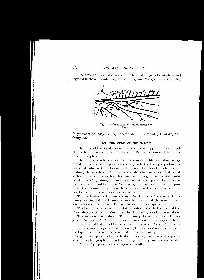
The first radio-medial cross-vein of the hind wings is longitudinal and sigmoid in the subfamily Corydalinse, the genus Ithone, and in the families
M3+4
Fig. I62.-Base of hind wing of Hemerobius humuli.
(c) THE WINGS OF THE SIALIDJE
The wings of the Sialidre form an excellent starting point for a study of the methods of specialization of the wings that have been evolved in the order Neuroptera.
The most character.stic feature of the more highly specialized wings found in this order is the presence of a very perfectly developed pectinately branched radial sector. In one of the two subfamilies of this family the Sialinse, the modification of the typical dichotomously branched radial sector into a pectinately branched one has not begun; in the other subfamily, the Corydalinre, this modification has taken place; but in some members of this subfamily, as Chauliodes, the modification has not progressed far, consisting merely in the suppression of the dichotomy and the development of one or two accessory veins.
The tracheation of the wings of nymphs of three of the genera of this family was figured by Comstock and Needham and the result of our studies leaves no doubt as to the homologies of the principal veins.
The family includes two quite distinct subfamilies, the Sialinre and the Corydalinse, which are distinguished by different types of wing-venation.
The wings of the Sialinee.e-The subfamily Sialinre includes only two genera, Sialis and Protosialis. These resemble each other very closely in the more general features of the venation of the wings. As we were able to study the wings of pupas of Sialis infumata, this species is used to illustrate the type of wing venation characteristic of the subfamily.
Figure 163 represents the tracheation of a wing of a pupa of this species which was photographed when the fanning veins appeared as pale bands; and Figure 164 represents the wings of an adult.

169 THE WINGS OF SIALIDAi:
It is unnecessary to describe these wings in detail, as the lettering of the figures indicates the homologies of the wing-veins. Some of the more
Cu. CU,a rooLU,
Fig. 163.-Wing of a pupa of Sialis infumata (After C. & N.).
striking features are the following: the prominence of the accessory branches of the subcosta; the coalescence of the tips of veins Sc and R 1 ;
the partial atrophy of the intermediate portion of media in the fore wing
Fig. 164.-Wings of Sialis infumata.
and of the greater part of the stem of this vein in the hind wing; the anastomosis of veins M and CUI in the fore wing; and the manner of branching of the radial sector.

radial sector has been developed, while in Sialis this vein has retained the dichotomous type of branching, modified only by the addition of a limited number of marginal accessory veins.
»r~~
, ~'~.1 " ~ <t
. ~ $ ~ ~x
.Ix ~ rIStA
THE WINGS OF NEUROPTERA
Fig. r6s.-Wing of a pupa of Chauliodes pectinicornis (After C. & N.).
Sc
170
c
The last mentioned feature is the one to which I wish to call especial attention. The radial sector in this genus is nearly typical in form; the only modification being the development of one or more marginal accessory veins upon it. These accessory veins, however, are ina quite different position than that occupied by the accessory veins borne by the radial sector in the Corydalinse, where a pectinately branched radial sector has been developed.
In Sialis the accessory veins are borne by vein R3, instead of vein R2 as in the Corydialinse, and they extend toward the costal margin of the wing. The method of specialization is in a quite different direction than that which produces the pectinately branched radial sector of the Corydalinze.
The accessory veins borne by vein R 3 in Sialis have not attained a permanent form; they vary in number in different individuals of the same species and in being either simple or forked; for this reason they are not numbered; they are not definitive accessory veins, but are of the type termed marginal accessory veins. Occasionally there are accessory veins on other branches of the radial sector; there is one on vein R4 in the fore wing represented in Figure 164.
The dichotomously branched radial sector of Sialis, which closely resembles the radial sector of the hypothetical primitive type of wing venation, shows conclusively that the pectinately branched radial sector of the allied genera and of most other Neuroptera, has been derived from the dichotomously branched type.
The wings of the Corydalirue.-The wings of the members of the subfamily Corydalinse present a very different appearance from those of Sialis; this is due largely to the fact that in the Corydalinre a pectinately branched

171 J'HE WING OF RAPHIDIID/E
Figure 165 represents a wing of a pupa of Chauliodes pectinicornis, taken at the stage when the fonning wing-veins appear as pale bands; and Figure 166 represents the tracheation of a wing of a pupa of Corydalus cornutus.
Fig. 166.-Tracheation of a wing of a pupa of Corydalus cornutus (After C. & N.).
These figures are from Comstock and Needham, but with a change in the lettering of the branches of the radial sector. The homologies of the wingveins are indicated by the lettering of the figures and require no discussion except in the case of the branches of the radial sector, where there may be a difference of opinion, depending on the view held regarding the method by which the dichotomy of the sector has been suppressed. This has been discussed on an earlier page.
(d) THE WINGS OF THE RAPHIDIIDlE
As no account of the tracheation of the wings of any pupa belonging to the Raphidiida has been published, we are forced to base our conclusions regarding the homologies of the wing-veins in this family upon a study of the wings of adults.
Of the two genera belonging to this family, Inocellia appears to be the more generalized, as regards the structure of the wings. The wings of Inocellia longicornis are figured here (Fig. 167).
In both genera, the subcosta ends in the costal margin of the wing a short distance before the pterostigma; the pterostigma is definitely limited, and conspicuous; the humeral vein is not recurrent, but sometimes it is forked; and the costal area of the wing is more or less widened in the proximal half of the wing.
In Inocellia there is no indication of a tendency to form a pectinately branched radial sector, the dichotomous branching of this vein being obvious. The forks of veins R 2+3 and R 4+5 are nearly opposite each other; and both are quite near the margin of the wing. The tip of vein R1 and of some of the branches of the radial sector bear short marginal accessory veins; but these differ in number in the wings of the two sides of the same individual.

172 THE WINGS OF NEUROPTER~
Media coalesces with radius for a considerable distance at the base of the wing. It is four-branched; and veins M, and M 4 each bears an accessory vein. A similar accessory vein is borne by vein CUI.
The fore and hind wings are very similar as regards the features mentioned above; but the course of vein CUI is markedly different in the two wings. In the fore wing, the basal part of vein CUI extends directly forward
R.
R.
Fig. 167.-Wings of Inocellia longicornis; CV, CV, CV, cross-veins.
from the cubital fork until it reaches media, with which its coalesces for a short distance. In the hind wing, the cubital fork is much nearer the base of the wing than it is in the fore wing, and vein CUI extends directly to the margin of the wing, being connected with media only by two cross-veins, the first of which is oblique. .
Vein CU2 in the fore wing is connected with the first anal vein by a very short cross-vein; in the hind wing these two veins are closely approximate but they do not anastomose.
In Inocellia the radial sector has moved out towards the apex of the wing, which has resulted in the shortening of veins R 2, R a, R 4, and R, so that they appear like the accessory veins that have been developed upon veins M a, M 4, andCu..
The wings of Raphidia adnixa may be taken as illustrating the type of wing-venation characteristic of the genus Raphidia (Fig. 168). In this species, the wings present two marked differences from those of I nocellia longicornis; these are the presence of a larger number of accessory veins and the fact that the radial sector, although still dichotomously branched, shows the beginning of the development of the pectinately branched type.
:1
ill 1.1
n l
I

THE WINGS OF RAPHIDIID/E 173
In Raphidia adnixa the forking of vein R 4 +5 is in about the same position as in Inoceliia, but the tips of one or of both of the veins R 4 and R 5 are split; vein R, is split back a considerable distance; and the forking of vein R2 has progressed so far that what is probably a definitive accessory vein, vein R 2a, has been developed. This development of a definitive accessory vein upon vein R 2 is of considerable interest as it indicates the beginning of the development of a pectinate radial sector.
In certain other species of Raphidia, the wings of which are figured by Albarda ('91), the forks of veins R2+3 and R 4+5 are more nearly opposite than they are in the wings figured here.
Returning to the wings of Raphidia adnixa, we find that the tips of all of the branches of media are split and that the number of accessory veins borne by vein CUI is greater than in I nocellia. In fore wing of the Raphidia, vein CUI not only anastomoses with media at its base but there is also an anastomosis of this vein with vein MS+4• The fact of this last anastomosis is made evident by comparison with the hind wing where the two veins are connected by a short cross-vein. In the hind wing veins CU2 and rst A anastomose along the region where they are closely parallel in I nocellia.
An obvious distinction between I nocellia and Raphidia, pointed out by A1barda ('91), is that in Raphidia the pterostigma is traversed by a branch of vein Rl, which is lacking in I nocellia.
Inboth genera the cross-veins are greatly reduced in number; and there is a marked tendency towards the alignment of those in the outer part of the wing in a gradate series; this is well shown in thehindwing ofRaphidia adnixa.

174 THE WINGS OF NEUROPTERA
(e) THE WINGS OF THE MANTISPlDlE
. The wings of Climaciella brunnea (Fig. 169) can be taken as an illustration of the type of wings found in this family. The two pairs of wings are similar in fonn and agree in the more general features of their venation.
R3
Fig. 169.-Wings of Climaciella brunnea.
They are long and narrow; a pterostigma is present, but the limits of it are not sharply defined; the sub costa extends midway between the costa and the radius to near the end of the pterostigma, where it is forked; the radial sector is pectinately branched; most of its branches are divided at the tip; the base of media coalesces with radius for a short distance at the base of the wing; in the fore wing, media after separating from radius and extending down for a short distance bends up sharply and anastomoses with radius, thus forming a small triangular cell.
The media is apparently four-branched in both wings; but the divisions of veins M1+2 and M 3+ 4 are short and may be merely accessory veins, corresponding with the divisions of the branches of the radial sector.
A large cell R1 is a characteristic feature of the wings of insects of this family; as a rule this cell is divided by three cross-veins, but in the wings figured here the third cross-vein has been eliminated by the anastomosing of veins R1 and R 2 near the apex of the wing.
The number of the branches of the radial sector that are given off opposite the 1St cell R 1 has been used as a diagnostic character; but this number

THE WINGS OF ITHONID/E 175
is not constant; it sometimes varies in the wings of the two sides of the same individual.
The wings of the Mantispidre resemble in some respects those of Raphidia; the outlines of the wings are similar; and the two resemble each other and differ from other neuropterous wings in the transverse bracing of the basal part of the fore wing in each by striking modifications of the courses of principal veins, although this result is brought about in very different ways in the two families.
The wings of the Mantispidre are much more highly specialized than are those of the Raphidiidee ; the discussion of them has been taken up at this point merely because there seems to be no better place in the series to include it.
In the Mantispidee the pectinate condition of the radial sector is very .. perfect, as is also the development of a gradate series of cross-veins, and a comparatively regular margin of accessory veins.
(f) THE WINGS OF THE ITHONIDJE
Newman in r853 established the family Ithonesidre for the reception of his genus Iihone (Zoologist, vol XI, Appendix p. CCll). Recently
•
Fig. r70.-Wings of Ithone fusca.
Tillyard ('r6) reestablished the family under the name of Ithonidee. As the name proposed by Tillyard conforms to the modern rules of nomenclature it is adopted here.

THE WINGS OF NEUROPTERA
..YQ'" "'7 <'d
...., ./,1'/ ' ....,
The type of the genus Iihone is the species described by Newman under the specific namefusca; the wings of this insect are figured here (Fig. 170).
The wings of Ithone fusca bear a superficial resemblance to those of the Corydalinre; but in Ithone the humeral vein of the fore wings is recurved and branched and veins Sc and R I do not coalesce at their tips.
In this species there is a single radial sector, which is pectinately branched; and most of the branches of this vein are forked at the tip. Media is two-branched in both wings; and vein CUI is very stout.
'~rca
Fig. 171.-Wings Rapisma viridipennis; rca, radial cuneate area.
This species is found in Australia and in Tasmania, and has been until recently the only species of the genus Ithone known.
Tillyard ('16) describes another Australian insect under the name Ithone fulva. But although the general appearance of the wings of this insect, as figured by Tillyard, is similar to that of Ithone, the differences in structure are so great that it is doubtful if the two are congeneric. The most important difference is that in the fore wings of Ithone fulva the first and second branches of the radial sector arise separately from the main stem of radius, the radius having three sectors. In the hind wings veins Sc and R 1 coalesce throughout the distal half of their length.
The limits and distinguishing characteristics of the Ithonidse must be determined by a study of other characters as well as those presented by the
176
...
1]
;1
!i ~1
il ~I
I; ,. ~
I: , l'
I,"

THE WINGS OF SISYRlD~ 177
wings, and does not fall within the scope of this essay. Until this is done, I suggest the inclusion in this family provisionally of another remarkable insect; this is Rapisma viridipennis.
The genus Rapisma was established by McLachlan to receive a species from the East Indies that had been described by Walker under the name H emerobius viridipennis. Dr. Needham has obtained this species from the East Himalayas and I give here a figure of its wings (Fig. 171).
In Rapisma the costal area of the fore wings is more expanded than in Ithone; but the courses of the terminal portions of veins Sc and R 1 are similar in the two genera, as are also most of the other more general features of the wings.
The chief reason for giving a figure of the wings of Rapisma in this discussion of the wings of the Neuroptera is to call attention to one feature of the structure of the radial sector. This is the fact that the primitive dichotomy of this sector appears to have been retained although the development of its other pectinate features has progressed far. This appearance is especially striking in the fore wing where what appears to be the forks of veins R 2+3 and R4+oare opposite.
A little consideration will suggest that it is not probable that the position of the fork of the first branch of the radial sector, vein R 4+5, would remain so nearly in its primitive position while the other features of the pectinate vein were being developed.
The explanation of the condition in question is that in each of the wings of Rapisma there has been developed a radial cuneate area; and that in the fore wing the first fork of vein R 5 has extended nearly to its base. In the hind wing this forking of vein R 5 has not extended so far.
The extent to which the development of a radial cuneate area has progressed in the wings of Rapisma and the transverse position of the first radio-medial cross-vein of the hind wings are the most striking differences between these wings and those of Ithone.
(g) THE WINGS OF THE SISYRIDJE
Until quite recently the members of this family have been included in the family Hemerobiidse; but several recent wri ters have separated them as a distinct family. The structure of their wings warrants this separation, as the distinctive characteristic of the hemerobiid wings is not exhibited by them.
The type of wing-venation characteristic of the Sisyridre is well illustrated by the wings of Sisyra fiavicornis, a species found in British India (Fig. 172). The more striking characteristics of these wings are the following: The costal area of the fore wings is not greatly broadened. The humeral vein is not recurrent and is not branched. Veins Sc and R 1
coalesce near the apex of the wing. The dichotomy of the radial sector has

178 THE WINGS OF NEUROPTERA
been suppressed in both wings, evidently by the splitting back of vein R, so that it arises from the main stem of the radial sector distinct from vein R 4• The splitting back of vein R 5 has progressed farther in the fore wing than it has in the hind wing.
The condition of the radial sector in Sisyra is much more generalized than it is in the wings of the Hemerobiidse, for in Sisyra it is only fourbranched, no definitive accessory veins having been developed, although marginal accessory veins are present. In fact the radial sector in these wings differs from the typical form only in the fact that the dichotomy has been suppressed.
Fig. 172.-Wings of Sisyra flavicornis.
Media is two-branched in both fore and hind wings, and in each case the tips of the branches are forked.
The beginning of the development of a secondary cubital fork is clearly indicated, especially in the fore wing; but vein CU2 is well preserved.
Marginal dashes are present in the outer margin of the wing. The extended migration of the base of vein R, of the fore wing towards
the base of the wing and the beginning of the formation of a secondary cubital fork are specializations that are carried much farther in the Hemerobiidse,
(h) THE WINGS OF THE SYMPHEROBIIDJE
Even after the separation of the Sisyridre and the Dilaridse from the Hemerobiidse, as is now commonly done, there remains in this family

179 THE WINGS OF SYMPHEROBIID./E
several genera that do not exhibit the method of specialization of the wings that is distinctively characteristic of. the Hemerobiidse, but on the other hand exhibit a type of specialization distinctively characteristic of this group of genera. To this group belong the American genera Sym.pherobius and Psectra, the genus A nnandalia from Calcutta, and the Australian genus N otiobiella. I suggest that these genera and such other genera as may be found to exhibit the same type of specialization of the wings be grouped in a separate family to be known as the Sympherobiidre.
Fig. In.-Wings of Sympherobius amiculus.
The distinctive characteristic of the Sympherobiidce is that vein R 2+S
of the fore wings has become separated from the remainder of the radial sector and is attached separately to vein R 1. This results in the radius of the fore wing having two sectors each of which is forked (Fig. 173).
In each case where this switching of the base of vein R2+S from the stem of the pectinate radial sector to vein R 1 has occurred there is a crossvein extending from vein R2 +S to the stem of the radial sector. It is probable that the switching occurred by the way of this cross-vein, and that after vein R 1 was reached by vein R 2+S the base of the latter vein migrated a short distance towards the base of the wing.