Embed Size (px)
Citation preview
AIMTo prepare a stained, temporary mount of onion peel and to studyits cells.
THEORYAll living organisms are composed of cells. New cells arise by the divisionof pre-existing cells. Cell is the structural and functional unit of life. Inplants, cells have an outermost rigid cell wall beneath which is a cellmembrane. The cell membrane encloses cytoplasm, cell organelles, anda nucleus.
MATERIALS REQUIREDAn onion bulb, slides, cover slips, two watch glasses, needle, brush, forceps,razor blade, compound microscope, blotting paper, methylene blue (orsafranin) solution, glycerine, and water.
PROCEDURE1. Take one fleshy scale leaf of an onion. Break it into two and using a
forcep pull out a thin membranous peel adhering to the inner surfaceof the leaf. This is the epidermal peel.
2. Place the peel in a watch glass containing water and cut it into smallrectangular pieces.
� � � � � � � � � � 18
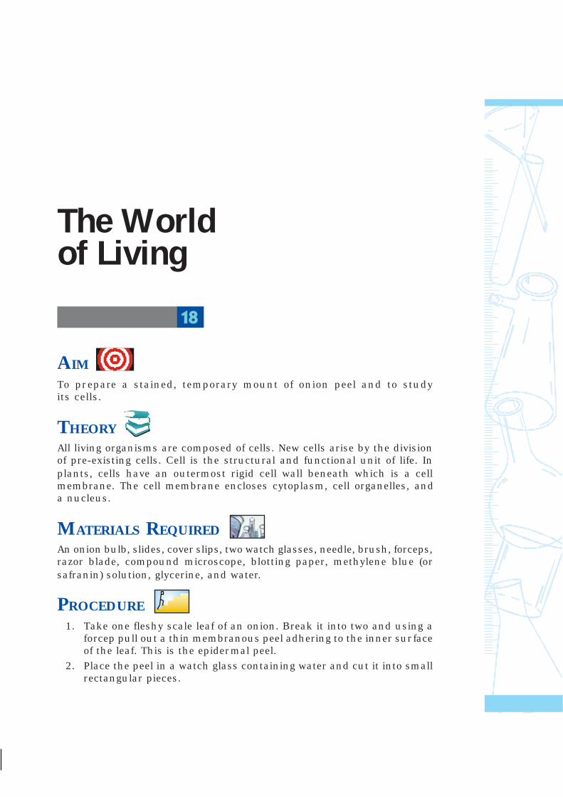
���� ���� ���� ���� ���� ����������The Worldof Living
Laboratory Manual – Science
70
3. Mix 1 or 2 drops of methylene blue or safrarin in a small quantity ofwater taken in another watch glass and transfer the peels into it.Leave the peels for about 3 minutes. Dip the peel in water to removeexcess stain.
4. Take a clean slide with a drop of glycerine in the middle and using abrush transfer the washed and stained peel on to it.
5. Place a cover slip over it by slowly lowering it with a needle. Avoidentry of air bubbles.
6. Remove excess glycerine from the edges of cover slip with the helpof a piece of blotting paper.
7. Observe the slide under the microscope, first in low power and thenin high power.
8. Draw a labelled diagram of the cells as seen under microscope.
9. Note the features listed in the observation table.
Fig. 18.1 : (b)Staining andmounting the
onion peel
Fig. 18.1 : (a)Method of
removing anepidermal peelfrom onion leaf
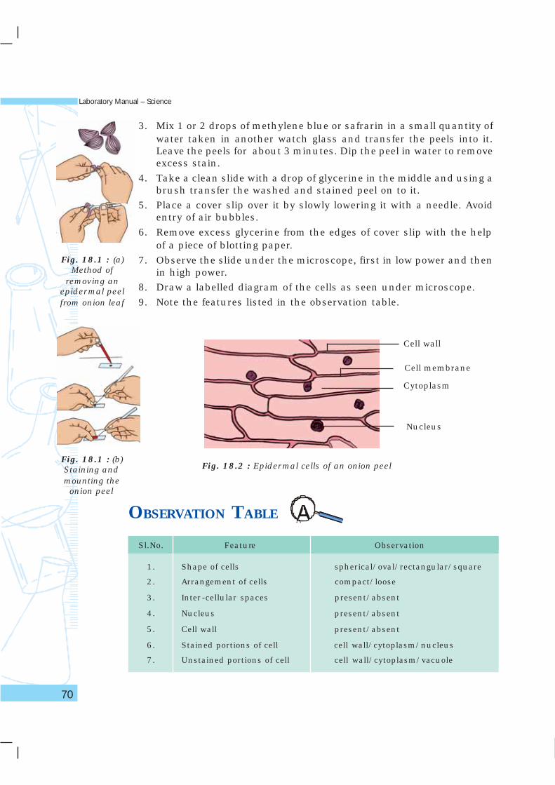
Fig. 18.2 : Epidermal cells of an onion peel
OBSERVATION TABLE
Sl.No. Feature Observation
1. Shape of cells spherical/oval/rectangular/square
2. Arrangement of cells compact/loose
3. Inter-cellular spaces present/absent
4. Nucleus present/absent
5. Cell wall present/absent
6. Stained portions of cell cell wall/cytoplasm/nucleus
7. Unstained portions of cell cell wall/cytoplasm/vacuole
Cell wall
Cytoplasm
Nucleus
Cell membrane
The World of Living
71
RESULTS AND DISCUSSIONThe cells that form the peel are rectangular in shape, compactly arrangedand without any intercellular spaces. Each cell has a distinct cell wall, aprominent nucleus and a vacuole. The cells form the outer layer of the leafknown as epidermis.
PRECAUTIONS• Staining of the peel must be appropriate. Excess stain can be removed
by rinsing the peel with water taken in a watch glass.
• Use a brush to transfer the peel on to the slide.
• While placing the cover slip care should be taken to avoid air bubbles.
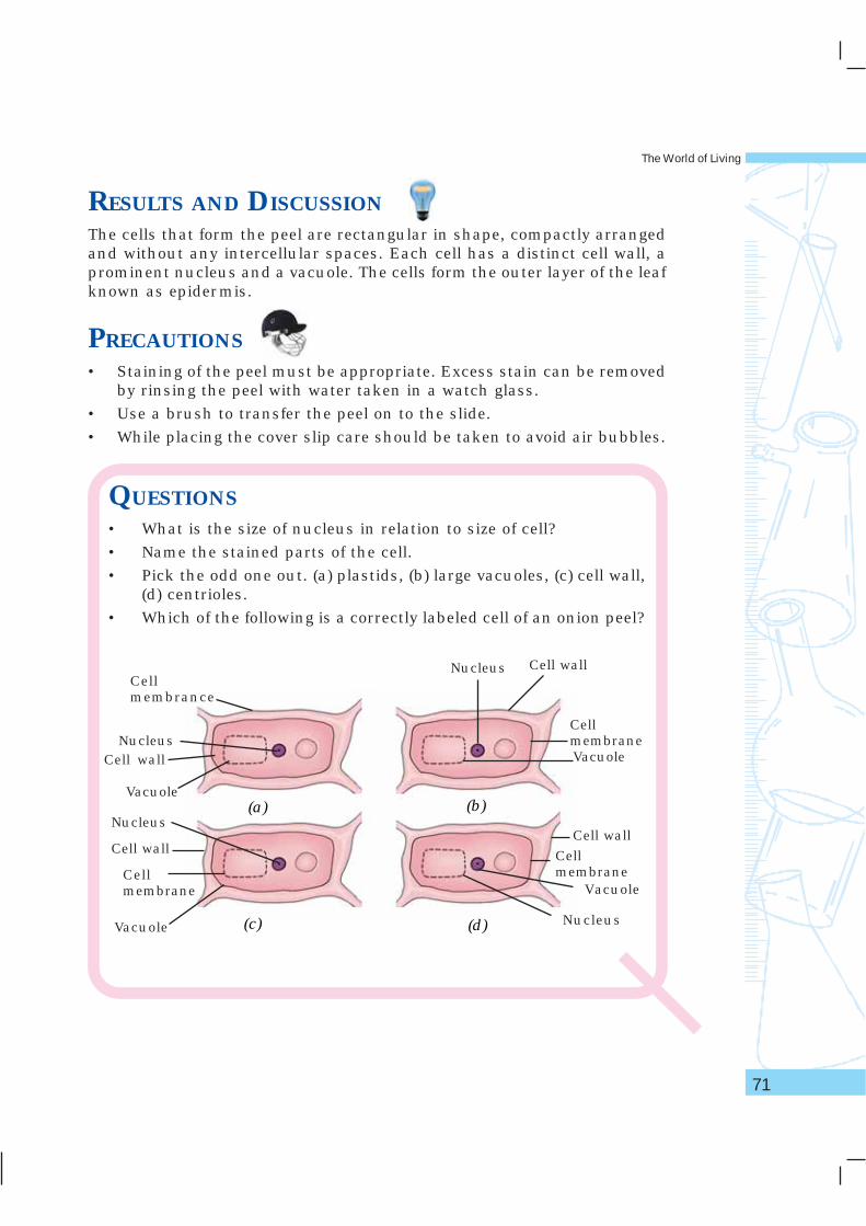
QUESTIONS• What is the size of nucleus in relation to size of cell?
• Name the stained parts of the cell.
• Pick the odd one out. (a) plastids, (b) large vacuoles, (c) cell wall,(d) centrioles.
• Which of the following is a correctly labeled cell of an onion peel?
(a) (b)
(d)(c)
Nucleus
Vacuole
Cell wall
Cellmembrane
Cell wall
Cellmembrane
Vacuole
Nucleus
NucleusCell wall
Nucleus
Cellmembrance
Vacuole
Vacuole
Cell wall
Cellmembrane
Laboratory Manual – Science
72
AIMTo prepare a temporary mount of human cheek epithelial cells, and tostudy its characteristics.
THEORYLike plants, the body of all animals including humans is composed of cells.Unlike plant cells, animal cells do not have cell wall. The outermost coveringof an animal cell is a cell membrane. The cytoplasm, nucleus and othercell organelles are enclosed in it. Epithelial tissue is the outermost coveringof most organs and cavities of an animal body.
MATERIALS REQUIREDMethylene blue stain, glycerine, a compound microscope, slide, cover slip,a clean spatula or a toothpick, a brush, a needle, and a piece of blottingpaper.
PROCEDURE1. Rinse your mouth with fresh water.
2. With the help of a clean spatula or a toothpick, gently scrap the innerside of your cheek.
3. Transfer the scrapped material into a drop of water taken on a cleanslide.
� � � � � � � � � � 19
The World of Living
73
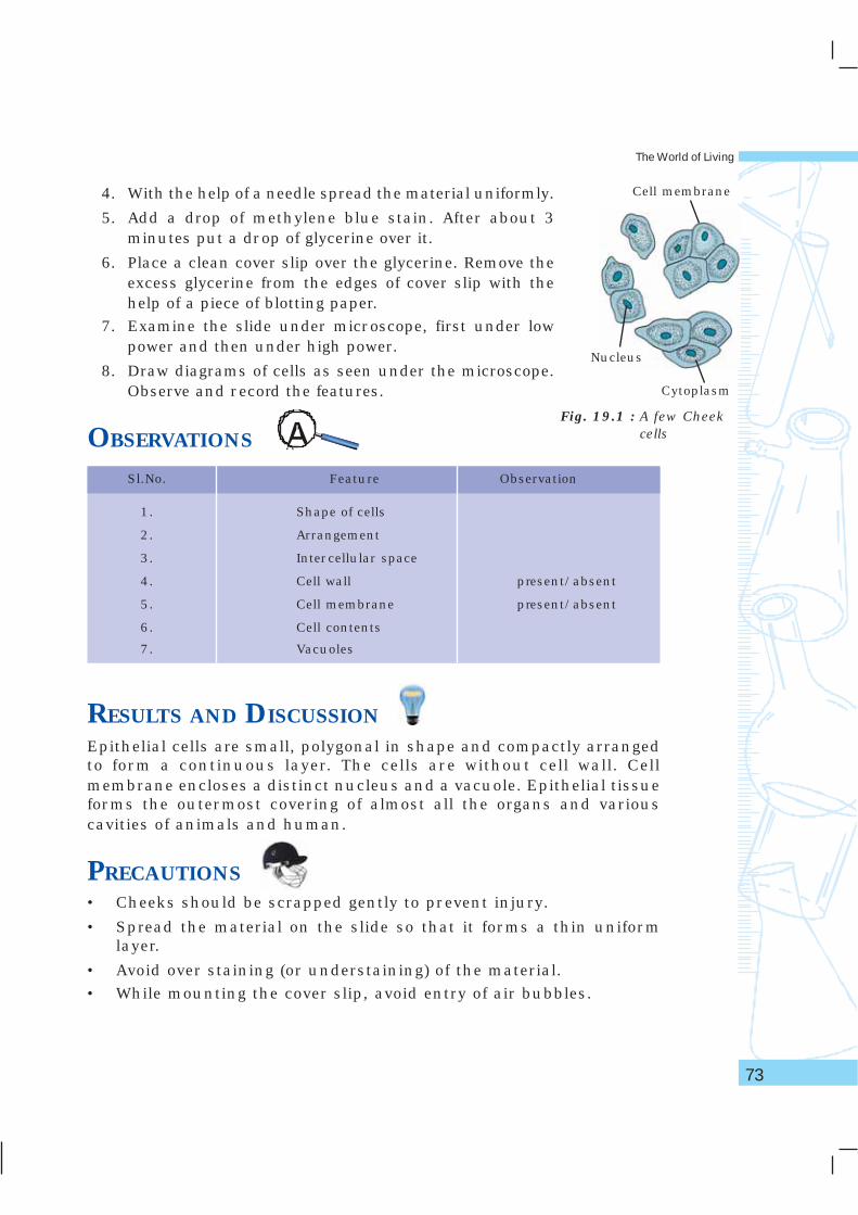
Fig. 19.1 : A few Cheekcells
4. With the help of a needle spread the material uniformly.
5. Add a drop of methylene blue stain. After about 3minutes put a drop of glycerine over it.
6. Place a clean cover slip over the glycerine. Remove theexcess glycerine from the edges of cover slip with thehelp of a piece of blotting paper.
7. Examine the slide under microscope, first under lowpower and then under high power.
8. Draw diagrams of cells as seen under the microscope.Observe and record the features.
OBSERVATIONS
Nucleus
Cytoplasm
Cell membrane
Sl.No. Feature Observation
1. Shape of cells
2. Arrangement
3. Intercellular space
4. Cell wall present/absent
5. Cell membrane present/absent
6. Cell contents
7. Vacuoles
RESULTS AND DISCUSSIONEpithelial cells are small, polygonal in shape and compactly arrangedto form a continuous layer. The cells are without cell wall. Cellmembrane encloses a distinct nucleus and a vacuole. Epithelial tissueforms the outermost covering of almost all the organs and variouscavities of animals and human.
PRECAUTIONS• Cheeks should be scrapped gently to prevent injury.
• Spread the material on the slide so that it forms a thin uniformlayer.
• Avoid over staining (or understaining) of the material.
• While mounting the cover slip, avoid entry of air bubbles.
Laboratory Manual – Science
74
NOTE FOR THE TEACHERIn such temporary preparations, cytoplasmic organelles are notvisible because they are too small and are not stained bymethylene blue.
QUESTIONS• Arrange the following steps in correct sequence–
(i) Putting a drop of glycerine on the cheek cells on a slide,
(ii) Scrapping the inner side of cheek,
(iii) Adding methylene blue stain, and
(iv) Placing the cover slip over the material.
(a) i, ii, iii, iv; (b) ii, i, iv, iii; (c) iv, ii, iii, i; (d) ii, iii, i, iv.
• Which one of the following is absent in animal cells–
(a) Cell membrane, (b) Nucleolus, (c) Cell wall, (d) Cytoplasm
• Cheek epithelial cells are an example of
(a) squamous epithelial cells, (b) cuboidal epithelial cells,
(b) columnar epithelial cells, (d) all of these.
• Why are cheek epithelial cells always moist?
• Name two structures which you would see in cheek cells if youwere using a very high magnifying power of microscope?
The World of Living
75
AIMTo study the phenomenon of osmosis.
THEORYEvery living cell has an extremely thin, elastic cell membrane, also calledplasma membrane, which separates cell contents from the externalenvironment. It is the outermost covering of animal cells. In plant cells,the membrane is present below the cell wall. It is selectively permeable asit allows solvent molecules and only selected solute molecules to passthrough it. It differs from a permeable membrane which allows all types ofmolecules to pass through it. Movement of molecules of water or solventfrom a region of its higher concentration to the region of its lowerconcentration across a selectively permeable membrane is called osmosis.It is of two types – endosmosis and exosmosis. Endosmosis is the entry ofwater into the cell while exosmosis is the movement of water out of the cellinto the external solution. Endosmosis takes place, when the cell is placedin a hypotonic solution. Exosmosis takes place when the cell is placed in ahypertonic solution.
MATERIALS REQUIREDTwo raw eggs, dil. hydrochloric acid, a salt (or sugar) solution of about25% concentration in water (dissolve about 25 g salt in 100 mL water),beakers and petri dishes.
� � � � � � � � � � 20
Laboratory Manual – Science
76
PROCEDURE1. Dissolve the shells of two eggs by placing them in two separate
beakers containing dil. HCl. Hydrochloric acid dissolves thecalcium chloride of the egg shells. The eggs will become de-shelled.
2. Carefully drain off the acid from the beakers and wash the eggsthoroughly with water while they are still in the beakers. Repeatthis process several times till all traces of acid are completelyremoved.
3. Observe the de-shelled eggs.
4. Fill one beaker containing one de-shelled egg with water and theother beaker with another de-shelled egg with the concentratedsalt (or sugar) solution.
5. Leave the set up for about four hours and observe the two de-shelled eggs.
OBSERVATIONSObserve the de-shelled eggs before and after placing them in water andconcentrated salt (or sugar) solution respectively. And answer thefollowing–
(i) What has happened to the de-shelled egg placed in water?
(ii) What has happened to the de-shelled egg placed in salt (or sugar)solution?
RESULTS AND DISCUSSIONThe de-shelled egg when placed in water swells because the concentrationof water molecules outside the egg is much higher than the concentrationof water molecules inside the egg, as a result of which endosmosis takesplace and water from the beaker enters into the egg. Exosmosis takesplace when the de-shelled egg is placed in a sugar (or salt) solution. Thewater comes out from the de-shelled egg into the (or salt) sugar solution.The loss of water results in the shrinkage of the egg.
PRECAUTIONS
• While washing the de-shelled eggs, care should be taken to prevent
the damage of egg membrane.
• Use dil. hydrochloric acid only lest the egg membrane gets damaged.
The World of Living
77
QUESTIONS• What is the difference between endosmosis and exosmosis?
• What will happen if a de-shelled egg is placed in a solution withthe same osmotic concentration as in the egg?
• Why did the egg swell when placed in water?
• Movement of water during osmosis takes place across–
(a) cell wall, (b) cell membrane, (c) cytoplasm, (d) protoplasm.
• The plasma membrane which selectively allows solvent moleculesand solute molecules to pass through it is–
(a) a permeable membrane, (b) a selectively permeable membrane,(c) an impermeable membrane, (d) a semi-permeable membrane.
Laboratory Manual – Science
78
AIMTo study plasmolysis in leaf epidermal peels of Rhoeo or Tradescantia.
THEORYLiving cells generally contain plenty of water due to which they areturgid. Turgidity is an important attribute of cells as it gives shape tocells. When turgid cell is placed in salt (or sugar) solution, water movesout of the cell across its membrane into the external solution. As aresult, the volume of protoplast decreases and the cell membranewithdraws from the cell wall creating an apparent colourless spacewithin the cell. This shrinkage of protoplast inside a cell is termedplasmolysis.
MATERIALS REQUIREDCompound microscope, fresh leaves of Rhoeo or Tradescantia, a sugar (orsalt) solution of about 10 per cent concentration in water(dissolve about 10 g sugar in 100 mL water), a new razor blade, slide, coverslip, needle, forceps, brush, and a piece of blotting paper.
PROCEDURE1. Using a new razor blade, take out three or four small peels from
the lower epidermis of leaf of Rhoeo or T radescantia.
� � � � � � � � � � 21
The World of Living
79
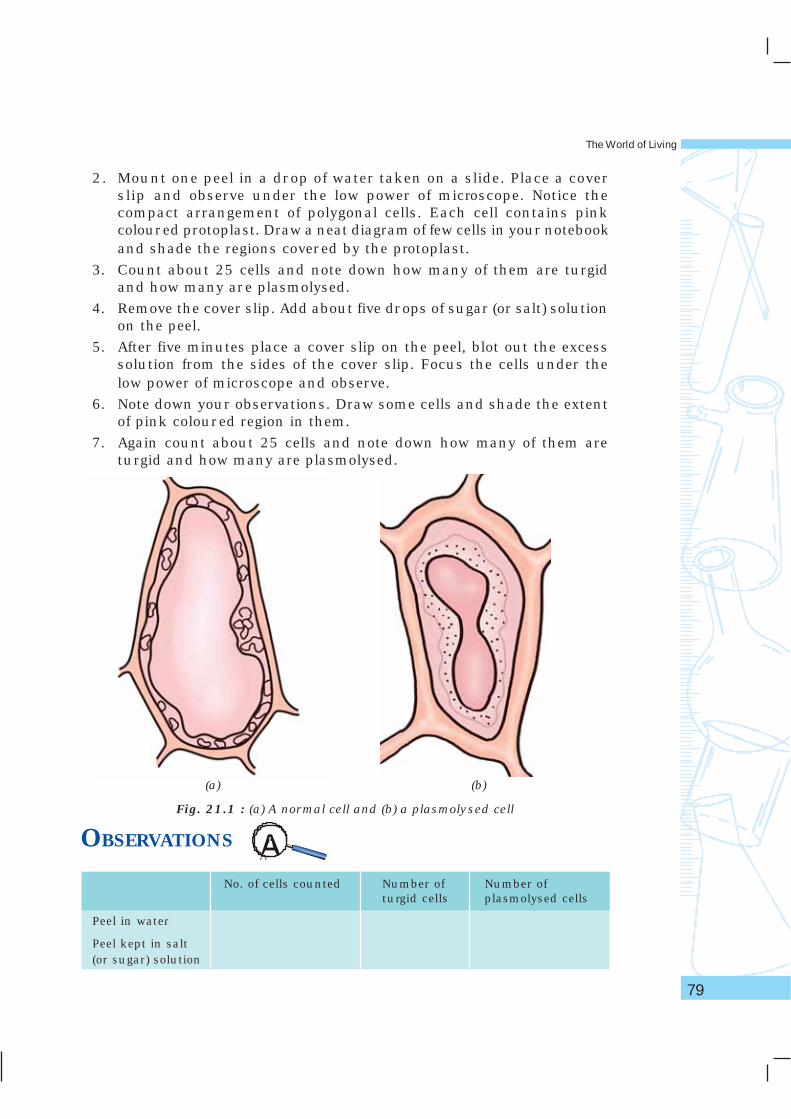
2. Mount one peel in a drop of water taken on a slide. Place a coverslip and observe under the low power of microscope. Notice thecompact arrangement of polygonal cells. Each cell contains pinkcoloured protoplast. Draw a neat diagram of few cells in your notebookand shade the regions covered by the protoplast.
3. Count about 25 cells and note down how many of them are turgidand how many are plasmolysed.
4. Remove the cover slip. Add about five drops of sugar (or salt) solutionon the peel.
5. After five minutes place a cover slip on the peel, blot out the excesssolution from the sides of the cover slip. Focus the cells under thelow power of microscope and observe.
6. Note down your observations. Draw some cells and shade the extentof pink coloured region in them.
7. Again count about 25 cells and note down how many of them areturgid and how many are plasmolysed.
OBSERVATIONS
No. of cells counted Number of Number ofturgid cells plasmolysed cells
Peel in water
Peel kept in salt(or sugar) solution
(a) (b)
Fig. 21.1 : (a) A normal cell and (b) a plasmolysed cell
Laboratory Manual – Science
80
RESULTS AND DISCUSSIONBased on the observations, analyse and reason out the causesfor plasmolysis.
PRECAUTIONS• Perform the experiment using coloured leaf samples like those of Rhoeo,
Tradescantia, Coleus, etc.
• Use concentrated sugar (or salt) solution.
• Ensure complete immersion of peels in the solution.
NOTE FOR THE TEACHERIn the experimental peel, water from the cells moves out into theexternal solution by a process called exosmosis. This happensdue to the fact that the cell sap is a weaker solution as comparedto the external (salt or sugar) solution. Consequently, it has morewater molecules than that in external solution. Due to thisdifference in the concentration of water inside the cell and theexternal solution, a concentration gradient is established. Dueto this gradient, water moves out of the cell into the externalsolution. This results in a reduction in volume of protoplasminside the cell hence the pink region appears shrunk in thesecells. The phenomenon of losing water from cells leading toshrinkage of protoplast is called plasmolysis.
QUESTIONS• What moves out from the cells in this experiment? Why?
• Why pigments and other cell contents do not move out of thecells?
• Why are living cells always turgid?
• What will happen if the cells are kept for a very long time in thesalt (or sugar) solution? Explain.
• Between the cell sap and solution (salt or sugar) in the experiment,which is the hypertonic solution?
• Will plasmolysis occur when cells are placed in isotonic solution?
The World of Living
81
AIMTo test the presence of starch in a given food sample and metanil yellow inpigeon pea.
THEORYThe presence of starch in a given food sample (say in potato) can bedetermined using iodine solution. Starch is a carbohydrate that producesblue colour when brought in contact with the iodine solution.
Food products are often adulterated for economic gains. For example,metanil yellow is used to adulterate pigeon pea (arhar dal). Metanil yellowturns into pink colour when it reacts with the concentrated hydrochloricacid. Whereas the pieces of unadultrated sample does not exhibit such achange.
MATERIALS REQUIREDPotato, pieces of pigeon pea (arhar dal), iodine solution, concentratedhydrochloric acid, petridish, test tube, knife, and a dropper.
PROCEDUREA. To test the presence of starch in potato–
1. Wash a potato alongwith its skin.
2. Take a thin slice of potato in a petridish.
� � � � � � � � � � 22
Laboratory Manual – Science
82
3. Add a few drops of iodine solution on the surface of the thin potatoslice.
4. Observe the change in colour in the area of slice where iodine solutonwas added.
B. To test the presence of metanil yellow in pigeon pea–
1. Take a few dry pieces of pigeon pea sample in a dry test tube.
2. Add a few drops of conc. HCl to these dry pieces.
3. Does the colour of the reaction mixture change?
4. Conclude whether the sample under test is adulterated or not?
OBSERVATIONS(a) The colour of potato slice changes into _________ on addition of iodine
solution.
(b) The colour of reaction mixture of pigeon pea sample when reactedwith conc. HCl changes into _________ .
RESULTS AND DISCUSSIONOn the basis of observations comment on (a) presence of starch in potatoand (b) whether the pigeon pea sample is adulterated with metanil yellowor not.
NOTE FOR THE TEACHER• The starch test may also be performed on samples of rice, wheat
flour etc.
• The presence of metanil yellow can also be detected in other foodsamples such as turmeric powder.
QUESTIONS• In what form the food is stored in plants?
• Which is the common adulterant of arhar dal?
• What are the efects of adulteration of food items.
• Why do the old stock of potato taste sweet?
• What are the different adulterants commonly used in foods?
The World of Living
83
AIMTo study parenchyma and sclerenchyma tissues in plants by preparingtemporary slides.
THEORYFlowering plants are structurally complex as they are made up of differentparts like roots, stem, leaves, flowers, fruits, etc. Each part is in turnan assembly of different types of tissues. Each tissue type has specificstructure and performs a particular function. Plant tissues are broadlyclassified into meristematic and permanent tissues. Permanent tissuesmay be simple, permanent tissues like parenchyma, collenchyma andsclerenchyma. Complex permanent tissues are xylem and phloem. Thestructural features of tissues like wall characteristics, cell size, lumensize, and cytoplasmic contents are different in different tissues.
MATERIALS REQUIREDTender stem of a herb (balsam/Tridax/Petunia/any cultivatedornamental herb or wild plant), safranin stain solution, dilute glycerine,chart of transverse section of stem, compound microscope, razor blade,slide, cover slip, brush, petri dish, and a piece of blotting paper.
� � � � � � � � � � 23
Laboratory Manual – Science
84
PROCEDUREA. Making a temporary slide
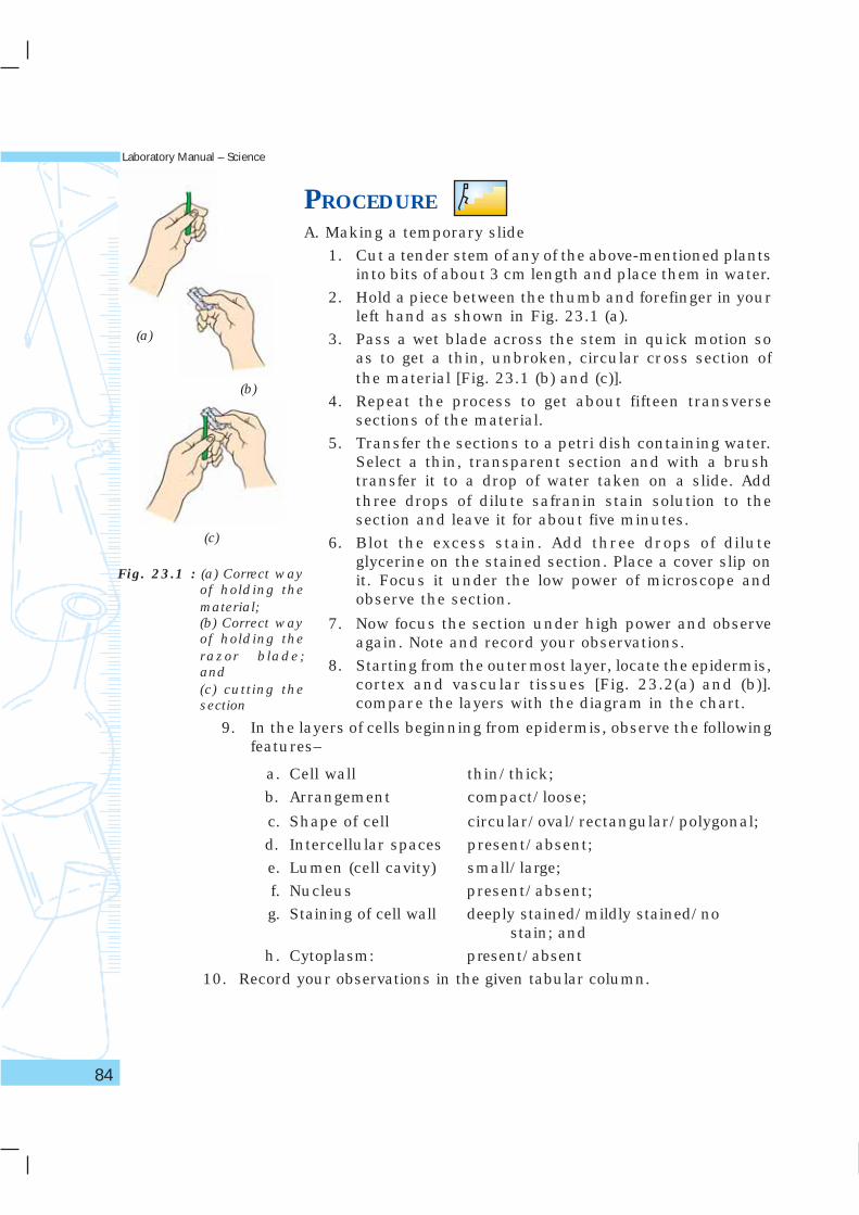
1. Cut a tender stem of any of the above-mentioned plantsinto bits of about 3 cm length and place them in water.
2. Hold a piece between the thumb and forefinger in yourleft hand as shown in Fig. 23.1 (a).
3. Pass a wet blade across the stem in quick motion soas to get a thin, unbroken, circular cross section ofthe material [Fig. 23.1 (b) and (c)].
4. Repeat the process to get about fifteen transversesections of the material.
5. Transfer the sections to a petri dish containing water.Select a thin, transparent section and with a brushtransfer it to a drop of water taken on a slide. Addthree drops of dilute safranin stain solution to thesection and leave it for about five minutes.
6. Blot the excess stain. Add three drops of diluteglycerine on the stained section. Place a cover slip onit. Focus it under the low power of microscope andobserve the section.
7. Now focus the section under high power and observeagain. Note and record your observations.
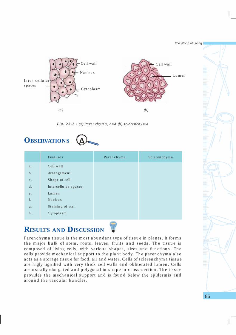
8. Starting from the outermost layer, locate the epidermis,cortex and vascular tissues [Fig. 23.2(a) and (b)].compare the layers with the diagram in the chart.
Fig. 23.1 : (a) Correct wayof holding thematerial;(b) Correct wayof holding therazor blade;and(c) cutting thesection
(a)
(b)
(c)
9. In the layers of cells beginning from epidermis, observe the followingfeatures–
a. Cell wall thin/thick;
b. Arrangement compact/loose;
c. Shape of cell circular/oval/rectangular/polygonal;
d. Intercellular spaces present/absent;
e. Lumen (cell cavity) small/large;
f. Nucleus present/absent;
g. Staining of cell wall deeply stained/mildly stained/nostain; and
h. Cytoplasm: present/absent
10. Record your observations in the given tabular column.
The World of Living
85
OBSERVATIONS
Features Parenchyma Sclerenchyma
a. Cell wall
b. Arrangement
c. Shape of cell
d. Intercellular spaces
e. Lumen
f. Nucleus
g. Staining of wall
h. Cytoplasm
RESULTS AND DISCUSSIONParenchyma tissue is the most abundant type of tissue in plants. It formsthe major bulk of stem, roots, leaves, fruits and seeds. The tissue iscomposed of living cells, with various shapes, sizes and functions. Thecells provide mechanical support to the plant body. The parenchyma alsoacts as a storage tissue for food, air and water. Cells of sclerenchyma tissueare higly lignified with very thick cell walls and obliterated lumen. Cellsare usually elongated and polygonal in shape in cross-section. The tissueprovides the mechanical support and is found below the epidermis andaround the vascular bundles.
Fig. 23.2 : (a) Parenchyma; and (b) sclerenchyma
Inter cellularspaces
Lumen
Cell wall
Cytoplasm
Nucleus
Cell wall
(a) (b)
Laboratory Manual – Science
86
PRECAUTIONS• For sectioning, select soft, tender herbaceous stem only. Avoid stems
that are hard and woody.
• Take care not to injure your finger while sectioning.
• Always keep the plant materials and sections in water.
• Use dilute safranin stain solution.
• The trachea and tracheids of xylem tissue appear to be very muchsimilar to sclerenchyma and may be erroneously identified assclerenchyma tissue. Xylem is always confined to the vascular bundleand is generally not seen in the cortex or pith.
NOTE FOR THE TEACHER• Features of parenchyma and sclerenchyma are given below for
purpose of their identification
Parenchyma• Parenchyma constitutes the major type of tissue in plants.
• Parenchyma cells have very thin walls, may be circular,rectangular, oval or polygonal in shape, loosely arranged in mostcases with intercellular spaces between cells. In some plantsintercellular spaces are absent and the cells are compactlyarranged.
• Parenchyma is composed of living cells with large internal space(lumen). Under the high power of microscope nucleus may bevisible.
• Epider mis, cortex and pith are essentially composed ofparenchyma tissue.
Sclerenchyma• Sclerenchyma cells are generally found below the epidermis or
just above the vascular bundles.
• The cells have very thick walls and they stain deep red whenstained with safranin.
• Lumen is reduced and there is no nucleus [Fig. 25.3(b)].
• Cells are generally elongated in vertical section and polygonal incross section; compactly arranged without any intercellular spaces.
The World of Living
87
QUESTIONS• In the transverse section of stem which tissue occupies larger
space-parenchyma or sclerenchyma?
• Draw an outline of the section of stem and indicate the regionswhere parenchyma and sclerenchyma are situated.
• Which tissue, when matured, has dead cells?
• Mention the main function of sclerenchyma tissue?
• You can bite fruits like guava, grapes, banana etc. but not a pieceof wood. Why?

Laboratory Manual – Science
88
AIMTo identify and study striated muscle fibre and nerve fibre in animals.
THEORYAnimal body is made up of groups of similar cells which performspecific function. Such groups of identical cells are called tissues. Thereare four basic types of tissues: epithelial, connective, muscular, and neural.These tissues vary from each other not only in their structure but also intheir functions.
MATERIALS REQUIREDPermanent slides of striated muscle fibre and nerve fibre, charts ofanimal tissues with straited muscle fibre and nerve cell (neuron), andcompound microscope.
PROCEDURE1. Place a permanent slide of straited muscle fibre under a compound
microscope.
2. Observe it first under low power and then under high power. Do yousee an alternate arrangement of dark and light bands? Do you alsofind some nuclei along the fibre?
� � � � � � � � � � 24
The World of Living
89
3. Identify the tissues with the help of charts. Draw diagrams of thetissues as seen under the microscope.
4. Replace the permanent slide of straited muscle fibre by the permanentslide of a nerve fibre. Identify different parts of a cell with the help ofcharts. Draw diagrams.
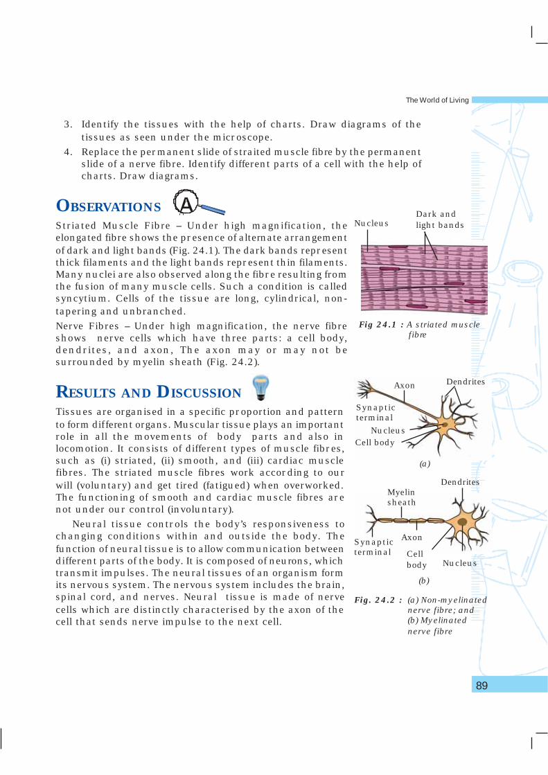
OBSERVATIONSStriated Muscle Fibre – Under high magnification, theelongated fibre shows the presence of alternate arrangementof dark and light bands (Fig. 24.1). The dark bands representthick filaments and the light bands represent thin filaments.Many nuclei are also observed along the fibre resulting fromthe fusion of many muscle cells. Such a condition is calledsyncytium. Cells of the tissue are long, cylindrical, non-tapering and unbranched.
Nerve Fibres – Under high magnification, the nerve fibreshows nerve cells which have three parts: a cell body,dendrites, and axon, The axon may or may not besurrounded by myelin sheath (Fig. 24.2).
RESULTS AND DISCUSSIONTissues are organised in a specific proportion and patternto form different organs. Muscular tissue plays an importantrole in all the movements of body parts and also inlocomotion. It consists of different types of muscle fibres,such as (i) striated, (ii) smooth, and (iii) cardiac musclefibres. The striated muscle fibres work according to ourwill (voluntary) and get tired (fatigued) when overworked.The functioning of smooth and cardiac muscle fibres arenot under our control (involuntary).
Neural tissue controls the body’s responsiveness tochanging conditions within and outside the body. Thefunction of neural tissue is to allow communication betweendifferent parts of the body. It is composed of neurons, whichtransmit impulses. The neural tissues of an organism formits nervous system. The nervous system includes the brain,spinal cord, and nerves. Neural tissue is made of nervecells which are distinctly characterised by the axon of thecell that sends nerve impulse to the next cell.
Fig 24.1 : A striated musclefibre
NucleusDark andlight bands
Fig. 24.2 : (a) Non-myelinatednerve fibre; and(b) Myelinatednerve fibre
(b)
(a)
Nucleus
Dendrites
Nucleus
Cellbody
Axon
Myelinsheath
DendritesAxon
Cell body
Synapticterminal
(b)
Synapticterminal
Laboratory Manual – Science
90
QUESTIONS• What are the features of striated muscle fibre? Where do we find
these in our body?
• Mention the function of skeletal muscles in our body.
• What are the features observed in a neuron?
The World of Living
91
AIMTo study the characteristics of Spirogyra, Agaricus, moss, fern, Pinus andan angiosperm plant.
THEORYWe know that plants of different groups exhibit different characteristics.Thallophytes, bryophytes, pteridophytes, gymnosperms, and angiospermsare the five major groups of plants. This classification is essentially basedon the structure of their bodies and methods of reproduction. Thallophyteshave the simplest structure. The plant body becomes more complex frombryophytes onwards, and reaches its highest complexity in angiosperms.
MATERIALS REQUIREDPermanent slides of Spirogyra, specimen of Agaricus, moss, fern, Pinus,and an angiosperm such as Petunia, balsam, Amaranthus, Chenopodium,Tridax or other locally available plants, compound microscope.
PROCEDURE1. Observe the permanent slide of Spirogyra under low power of
microscope and record your observations. Draw the diagram ofSpirogyra and label the parts.
2. Likewise, observe and record the characters of Agaricus, moss, fern,Pinus and an angiosperm plant. Draw their diagrams.
� � � � � � � � � � 25
Laboratory Manual – Science
92
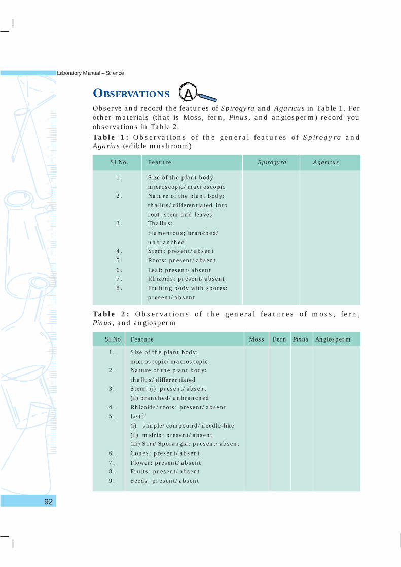
OBSERVATIONSObserve and record the features of Spirogyra and Agaricus in Table 1. Forother materials (that is Moss, fern, Pinus, and angiosperm) record youobservations in Table 2.
Table 1: Observations of the general features of Spirogyra andAgarius (edible mushroom)
Sl.No. Feature Spirogyra Agaricus
1. Size of the plant body:
microscopic/macroscopic2. Nature of the plant body:
thallus/dif ferentiated into
root, stem and leaves3. Thallus:
filamentous; branched/
unbranched4. Stem: present/absent
5. Roots: present/absent
6. Leaf: present/absent7. Rhizoids: present/absent
8. Fruiting body with spores:
present/absent
Table 2: Observations of the general features of moss, fern,Pinus, and angiosperm
Sl.No. Feature Moss Fern Pinus Angiosperm
1. Size of the plant body:
microscopic/macroscopic2. Nature of the plant body:
thallus/differentiated3. Stem: (i) present/absent
(ii) branched/unbranched
4. Rhizoids/roots: present/absent5. Leaf:
(i) simple/compound/needle-like
(ii) midrib: present/absent(iii) Sori/Sporangia: present/absent
6. Cones: present/absent
7. Flower: present/absent8. Fruits: present/absent
9. Seeds: present/absent
NOTE FOR THE TEACHER
The World of Living
93
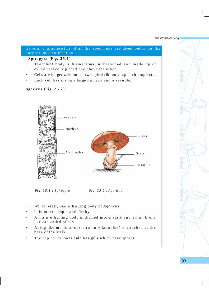
Fig. 25.2 : AgaricusFig. 25.1 : Spirogyra
• We generally see a fruiting body of Agaricus.
• It is macroscopic and fleshy.
• A mature fruiting body is divided into a stalk and an umbrellalike cap called pileus.
• A ring like membranous structure (annulus) is attached at thebase of the stalk.
• The cap on its lower side has gills which bear spores.
Nucleus
Chloroplast
Vacuole
Pileus
Stalk
Annulus
General characteristics of all the specimens are given below for thepurpose of identification.
Spirogyra (Fig. 25.1)• The plant body is filamentous, unbranched and made up of
cylindrical cells placed one above the other.
• Cells are longer with one or two spiral ribbon shaped chloroplasts.
• Each cell has a single large nucleus and a vacuole.
Agaricus (Fig. 25.2)
Laboratory Manual – Science
94
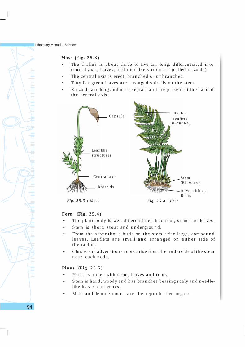
Moss (Fig. 25.3)• The thallus is about three to five cm long, differentiated into
central axis, leaves, and root-like structures (called rhizoids).
• The central axis is erect, branched or unbranched.
• Tiny flat green leaves are arranged spirally on the stem.
• Rhizoids are long and multiseptate and are present at the base ofthe central axis.
Fig. 25.3 : Moss Fig. 25.4 : Fern
Fern (Fig. 25.4)• The plant body is well differentiated into root, stem and leaves.
• Stem is short, stout and underground.
• From the adventitous buds on the stem arise large, compoundleaves. Leaflets are small and arranged on either side ofthe rachis.
• Clusters of adventitous roots arise from the underside of the stemnear each node.
Pinus (Fig. 25.5)• Pinus is a tree with stem, leaves and roots.
• Stem is hard, woody and has branches bearing scaly and needle-like leaves and cones.
• Male and female cones are the reproductive organs.
Rhizoids
Capsule
Central axis
Leaf likestructures
Rachis
Leaflets
(Pinnules)
Stem(Rhizome)
AdventitiousRoots
The World of Living
95
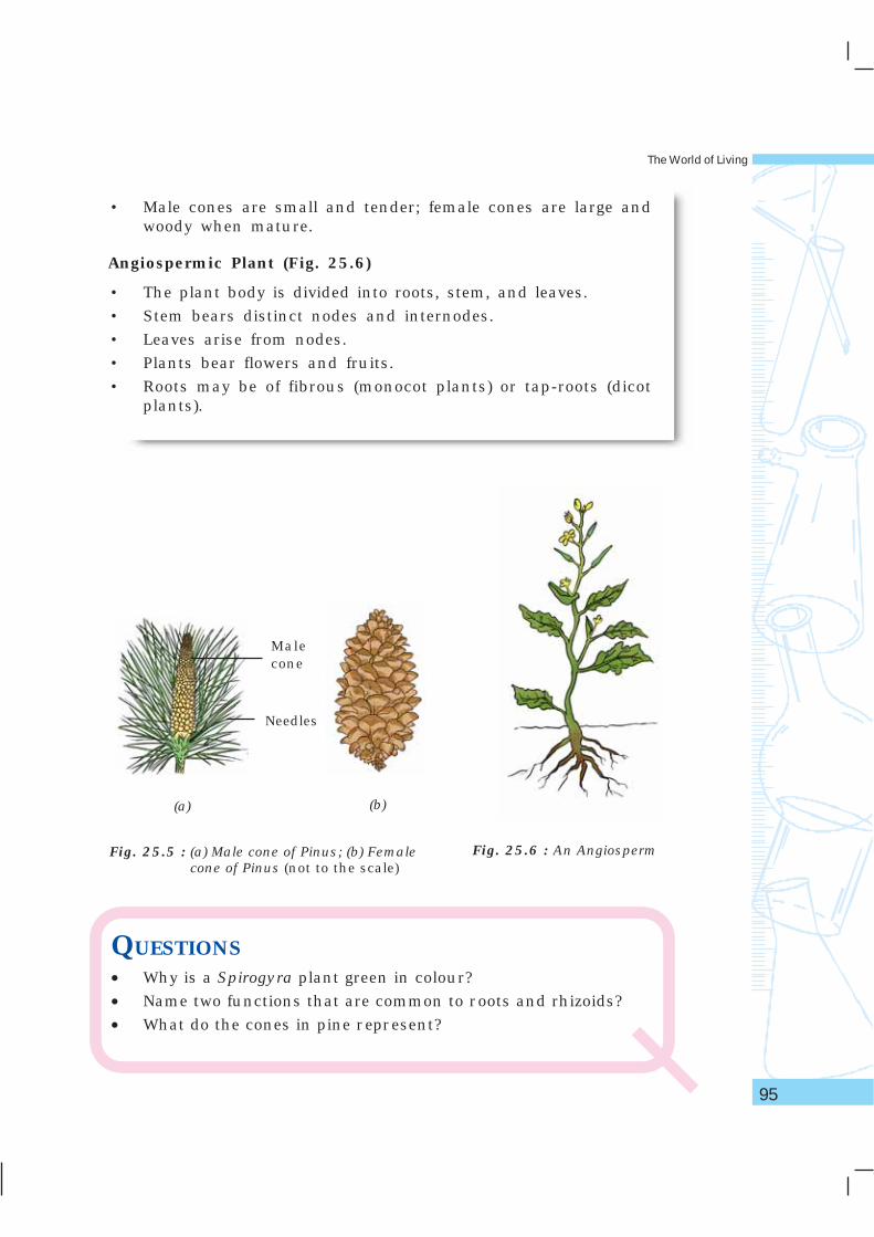
• Male cones are small and tender; female cones are large andwoody when mature.
Angiospermic Plant (Fig. 25.6)
• The plant body is divided into roots, stem, and leaves.
• Stem bears distinct nodes and internodes.
• Leaves arise from nodes.
• Plants bear flowers and fruits.
• Roots may be of fibrous (monocot plants) or tap-roots (dicotplants).
Fig. 25.5 : (a) Male cone of Pinus; (b) Femalecone of Pinus (not to the scale)
Fig. 25.6 : An Angiosperm
(a) (b)
QUESTIONS• Why is a Spirogyra plant green in colour?
• Name two functions that are common to roots and rhizoids?
• What do the cones in pine represent?
Malecone
Needles
Laboratory Manual – Science
96
AIMTo prepare herbarium sheet of a flowering plant.
THEORYHerbarium sheets are generally prepared by botanists and storedsystematically in a laboratory for an immediate reference. It consists of athick white sheet of a specific dimension on which a dried plant specimenis mounted. The mounted specimen must have leaves, flowers and fruits(optional). Only one plant specimen is mounted on a herbarium sheet.Herbarium sheets have to be carefully preserved to prevent insectinfestation. The term Herbarium refers to the place (such as a laboratory)where herbarium sheets are preserved systematiclly and are made availablefor reference.
MATERIALS REQUIREDPlant specimen or a twig of a plant (20 – 25 cm long) with leaves andflowers, a thick white sheet (card sheet) of dimension 40 × 28 cm,old news papers or blotting sheets, adhesive, field press with a longrope or a heavy mass (such as a brick or a book), sewing needle,and thread.
� � � � � � � � � � 26
The World of Living
97
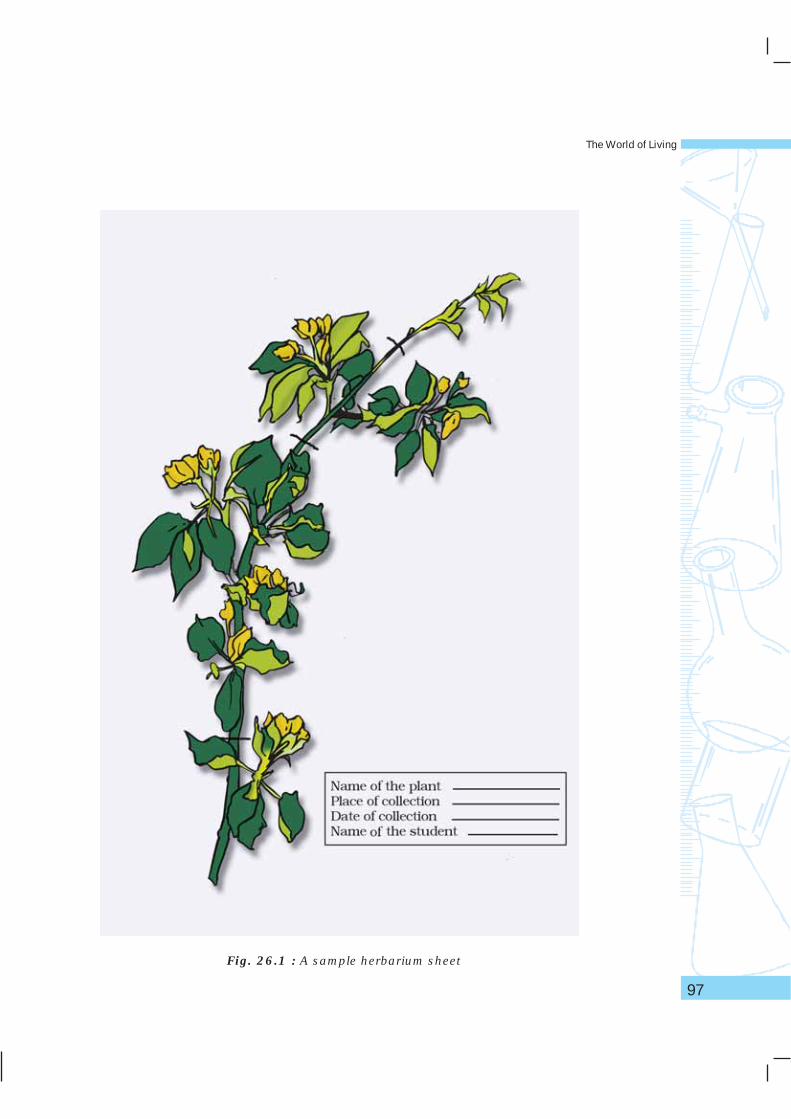
Fig. 26.1 : A sample herbarium sheet
Laboratory Manual – Science
98
PROCEDURE1. Collect a plant or a twig with leaves and flowers.
2. Place it inside the folds of a newspaper (or a blotting sheet) and spreadthe leaves and flowers gently without damaging them.
3. Turn one of the leaves so that its ventral surface faces upwards.
4. Cover the plant with the other half of the newspaper; place a fewmore sheets containing plants and a few newspaper sheets one abovethe other and keep a heavy mass (such as a brick or a book) on thepile. If a field press is available, the sheets of newspaper containingplant specimen may be stacked one above the other and the fieldpress should be tied tightly using a long rope.
5. Next day, transfer the plant to a fresh set of dry newspapers and repeatstep 4.
6. Repeat this process for four to five days till the plant becomes dry.
7. Smear a small quantity of adhesive at a few places on the stem orbranches and leaves. Mount the plant on the card sheet as shown inFig. 26.1.
8. Stich the twig or stem at a few places using a sewing needle and thread.
9. Keep this sheet in a dry newspaper (or a blotting sheet). Keep the heavymass on the newspaper for two to three hours to allow the dried plantto stick to the card sheet.
10. At the bottom right corner, write your name, name of plant, place anddate of collection. Now the herbarium sheet is ready (Fig. 26.1).
PRECAUTIONS• Do not prepare herbarium sheets of aquatic plants, succulents and
plants with thorns. (Why?)
• Select plants with small leaves and flowers as they are easier to handle.
• Spreading the differernt parts of plant on newspaper sheet has to bedone very carefully before pressing them using a heavy mass.
• Mount the plant specimen after all the moisture and water has beencompletely removed from the plant.
• Apply a small quantity of the adhesive only at a few places on theherbarium sheet. Use of cellophane adhesive tape to stick the plant onthe herbarium sheet must be avoided.
NOTE FOR THE TEACHER• Professional herbarium keepers treat plants with 1 per cent mercuric
chloride or 4 per cent formalin before mounting them on the
The World of Living
99
herbarium sheet. This process is called poisoning the specimen.As these chemicals are dangerous, and keeping in mind the age ofstudents, this step has not been mentioned in the procedure here.Fumigants and naphthalene balls are placed in the cupboards whereherbarium sheets are stored to prevent insect attack.
• Students may also be advised to find information about any fiveimportant international and national herbaria. They may also beencouraged to use internet or magazines or other informationsources for this purpose.
QUESTIONS• What are the advantages of a herbarium sheet?
• Why are water plants not suitable for preparing herbarium sheets?
• Why are plants treated with mercuric chloride or formalin beforemounting on the herbarium sheet?
• What is the difference between a Herbarium and a herbariumsheet?

Laboratory Manual – Science
100
AIMTo study the features and draw diagrams of earthworm, cockroach, bonyfish and bird.
THEORYAnimals are variously adapted to different kinds of habitat and environment.Adaptation is an inherent quality of living organisms which enable them tosurvive in specific habitats. Adaptation of organisms are due to certainmodifications that are observed in the organisms at the morphological,anatomical as well as physiological levels. In this experiment, fourorganisms are consider ed for the study. For each organism,characteristics of the phylum to which it belongs to and a few adaptivefeatures are studied and correlated with the habitat (or environment)in which they live.
MATERIALS REQUIREDPreserved specimens of earthworm, cockroach, bony fish, a stuffed bird,charts showing detailed diagrams of animals under study, and a handlens.
PROCEDURE1. Observe the given specimens and for each specimen, record one
specific feature of the group (phylum/class) to which it belongs.
� � � � � � � � � � 27
The World of Living
101
2. Write down one adaptive feature of each specimen with reference toits habitat.
3. Draw diagrams of the specimens, using the chart(s). Identify thevarious parts of organisms observed.
OBSERVATIONSTable 1
Sl. No. Organism Phylum/Class Features of Adaptive Features Habitatthe phylum observed
observed
1. Earthworm
2. Cockroach
3. Bony fish
4. Bird
Based on your observations, record some more features of the specimens(at least five for each specimen).
Table 2
Sl.No. Earthworm Cockroach Bony Fish Bird
1.
2.
3.
4.
5.
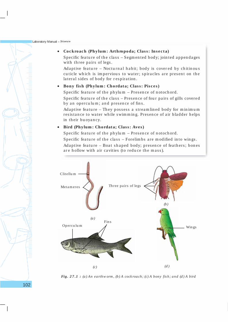
NOTE FOR THE TEACHERSome characteristic features of the phylum (or class) and adaptivefeatures of organisms are given for easy identification.
• Earthworm (Phylum: Annelida)Specific feature of the phylum – Body surface is characterised bydistinct annular segments or metameres; body is not differentiated.
Adaptive features – Moist and slimy skin, presence of clitellum(merger of 14 - 17 metameric segments).
Laboratory Manual – Science
102
• Cockroach (Phylum: Arthropoda; Class: Insecta)Specific feature of the class – Segmented body; jointed appendageswith three pairs of legs.
Adaptive feature – Nocturnal habit; body is covered by chitinouscuticle which is impervious to water; spiracles are present on thelateral sides of body for respiration.
• Bony fish (Phylum: Chordata; Class: Pisces)Specific feature of the phylum – Presence of notochord.
Specific feature of the class – Presence of four pairs of gills coveredby an operculum; and presence of fins.
Adaptive feature – They possess a streamlined body for minimumresistance to water while swimming. Presence of air bladder helpsin their buoyancy.
• Bird (Phylum: Chordata; Class: Aves)Specific feature of the phylum – Presence of notochord.
Specific feature of the class – Forelimbs are modified into wings.
Adaptive feature – Boat shaped body; presence of feathers; bonesare hollow with air cavities (to reduce the mass).
Fig. 27.1 : (a) An earthworm, (b) A cockroach; (c) A bony fish; and (d) A bird
(a)
(d)(c)
(b)
Clitellum
Metameres Three pairs of legs
WingsOperculumFins
The World of Living
103
APPLICATIONS• Earthworms are known as ‘Farmer’s Friend’ because of their role in
enhancing the fertility of soil. As they burrow into the soil, they ingestsoil along with the organic matter present in it. They excrete the samesoil and bring it on the top as casts. This way, it is able to loosen thesoil. Earthworms are also used to prepare vermicompost that is usedas manure to enhance the fertility of soil.
QUESTIONS• Name the phyla to which earthworm, cockroach, bony fish, and
bird belong.
• What is an adaptation?
• In which body segments of an earthworm is the clitellum found?
• How does a cockroach adapt itself to a wide range of habitats?
• Mention two adaptive characters of a bony fish besides thepossession of a streamlined body and air bladder.
• Feathers are an adaptive feature of birds. How are they helpful tothem?
Laboratory Manual – Science
104
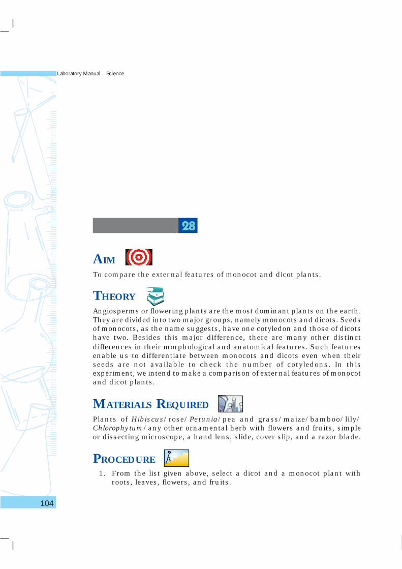
AIMTo compare the external features of monocot and dicot plants.
THEORYAngiosperms or flowering plants are the most dominant plants on the earth.They are divided into two major groups, namely monocots and dicots. Seedsof monocots, as the name suggests, have one cotyledon and those of dicotshave two. Besides this major difference, there are many other distinctdifferences in their morphological and anatomical features. Such featuresenable us to differentiate between monocots and dicots even when theirseeds are not available to check the number of cotyledons. In thisexperiment, we intend to make a comparison of external features of monocotand dicot plants.
MATERIALS REQUIREDPlants of Hibiscus/rose/Petunia/pea and grass/maize/bamboo/lily/Chlorophytum/any other ornamental herb with flowers and fruits, simpleor dissecting microscope, a hand lens, slide, cover slip, and a razor blade.
PROCEDURE1. From the list given above, select a dicot and a monocot plant with
roots, leaves, flowers, and fruits.
� � � � � � � � � � 28
The World of Living
105
2. Observe the differences in the external features of stem, leaf, roots,flowers, and seeds. To study the root system, wash the roots carefullyand spread them on a sheet of paper and study their nature.
3. Study the leaves for their shape and venation.
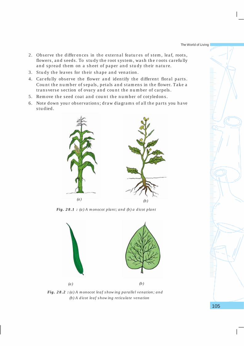
4. Carefully observe the flower and identify the different floral parts.Count the number of sepals, petals and stamens in the flower. Take atransverse section of ovary and count the number of carpels.
5. Remove the seed coat and count the number of cotyledons.
6. Note down your observations; draw diagrams of all the parts you havestudied.
Fig. 28.1 : (a) A monocot plant; and (b) a dicot plant
(a) (b)
Fig. 28.2 : (a) A monocot leaf showing parallel venation; and
(b) A dicot leaf showing reticulate venation
(a) (b)
Laboratory Manual – Science
106
(b)
Fig. 28.3 : (a) A monocot flower and (b) A dicot flower
Fig. 28.4 : Transverse section of ovary (a) bicarpellary and (b) tricarpellary
(a)
(b)(a)
Ovules
Fig. 28.5 : (a) A seed with one cotyledon and (b) a seed with two cotyledons
(b)(a)
Cotyledon
The World of Living
107
OBSERVATIONSSome important features that distinguish a monocot and a dicot plant arelisted in the table given below. You may observe some more features ofdifference between them. Record your observations.
Sl.No. Feature Monocot Dicot
1. Roots: fibrous/tap root
2. Leaf shape: broad/narrow
3. Leaf venation: parallel/reticulate
4. Floral parts: multiple of 3 or 5
5. Sepals: number and colour
6. Petals: number and colour
7. Stamen: number
8. Pistil: number of carpels
9. Cotyledon: one or two
RESULTS AND DISCUSSION• The study reveals many differences between dicot and monocot plants.
The distinctive features are consistently seen in most other plantsbelonging to these groups.
QUESTIONS• How do we differentiate betwen fibrous root system and
tap-root system?
• A plant has leaves with reticulate venation and floral partsconsisting of 5 sepals, 5 petals, 5 stamens, and 5 carpels. Inwhich group of angiosperms would you place this plant? Givereasons.
• In a plant, name two features which you would examine tocategorise it into a monocot or a dicot plant.
• Do all flowers have all the floral parts? Explore.
Laboratory Manual – Science
108
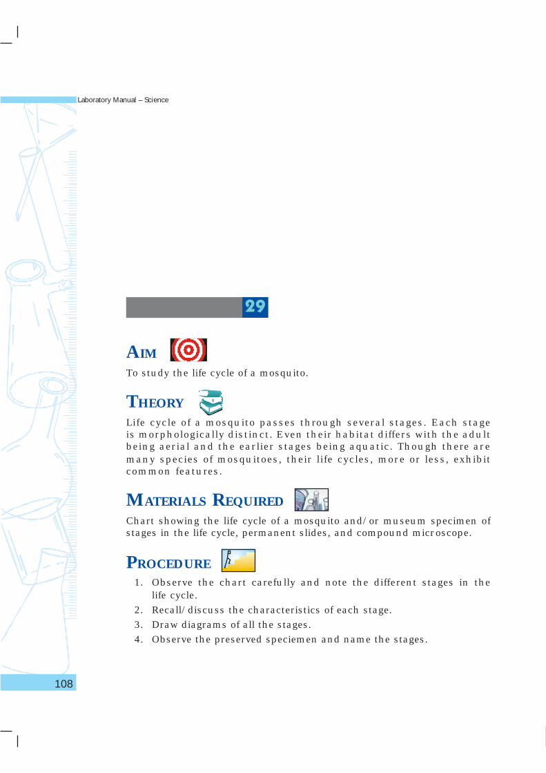
AIMTo study the life cycle of a mosquito.
THEORYLife cycle of a mosquito passes through several stages. Each stageis morphologically distinct. Even their habitat differs with the adultbeing aerial and the earlier stages being aquatic. Though there aremany species of mosquitoes, their life cycles, more or less, exhibitcommon features.
MATERIALS REQUIREDChart showing the life cycle of a mosquito and/or museum specimen ofstages in the life cycle, permanent slides, and compound microscope.
PROCEDURE1. Observe the chart carefully and note the different stages in the
life cycle.
2. Recall/discuss the characteristics of each stage.
3. Draw diagrams of all the stages.
4. Observe the preserved speciemen and name the stages.
� � � � � � � � � � 29
The World of Living
109
DISCUSSIONThe eggs of various species of mosquito are deposited on stagnant waterbodies like ponds, ditches, cess pools, lakes etc. Any container withstagnant water is a potential breeding place for mosquitoes.
The larvae hatch out from the eggs within a few hours and beginfeeding on decaying plant matter. They float on the surface of water andbreath through a specialised siphon tube. The larval stage lasts for afew days during which several layers of skin are shed. This stage lastsfor a few days to a few weeks. The larval stage is followed by the pupalstage.
Pupae do not feed but gradually metamorphose or change into adults.Pupal stage lasts for a few days. From the pupa an adult mosquitoemerges. Before the adult starts flying it rests for a few days duringwhich its outer cuticle hardens.
After about a week, adult female mosquito begins searching for ahost. It generally feeds on blood, which is a rich source of protein thatis helpful to make a fresh batch of eggs, Eggs are deposited on or nearwater. The male mosquito does not seek a blood meal, but prefers asugar meal which it obtains by feeding on the nectar of flowers. Thefemale adults also feed on nectar in between blood meals. Adultmosquitoes live for several weeks.
Fig. 29.1 : Stages in the life cycle of a mosquito
Adult
Eggs
Pupa
Larva
Laboratory Manual – Science
110
NOTE FOR THE TEACHER• The mosquito goes through four distinct stages during its life cycle
(Fig. 29.1)–
(i) egg: deposited in water; hatches in water;
(ii) larva (plural; larvae): lives on the surface of water; moults severaltimes;
(iii) pupa (plural; pupae): A stage just prior to the adult stage; pupaedo not feed; and
(iv) adult emerges from the pupae, body parts harden and startsflying.
• Permanent slides of different stages in the life cycle of a mosquitomay be focussed under a compound microscope and shown to thestudents. This will enable students to have a better understandingof life cycle of a mosquito.
QUESTIONS• Why is it important to study the life cycle of mosquito?
• At which stage in the life cycle of a mosquito, moulting takes place?
• Why does only the female mosquito require a blood meal?
• What are the conditions that are helpful for breeding of mosquitoes?
• Suggest three measures to check the breeding of mosquitoes.
The World of Living
111
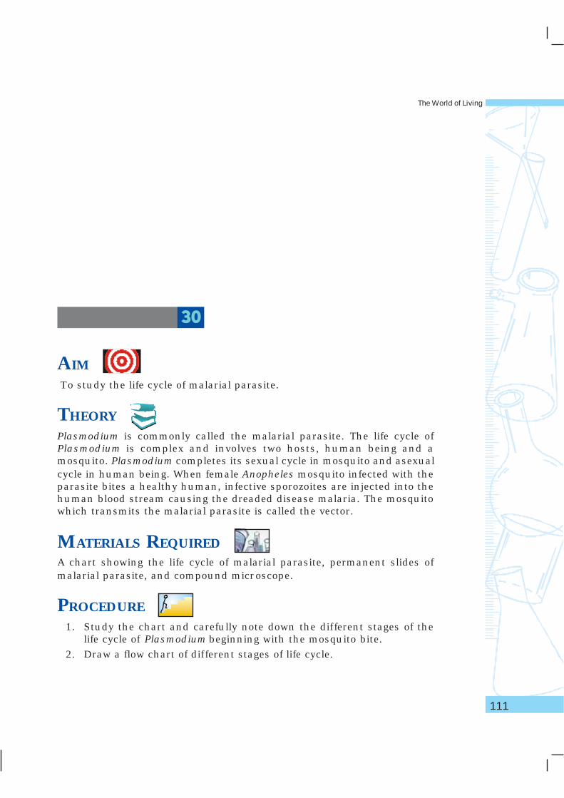
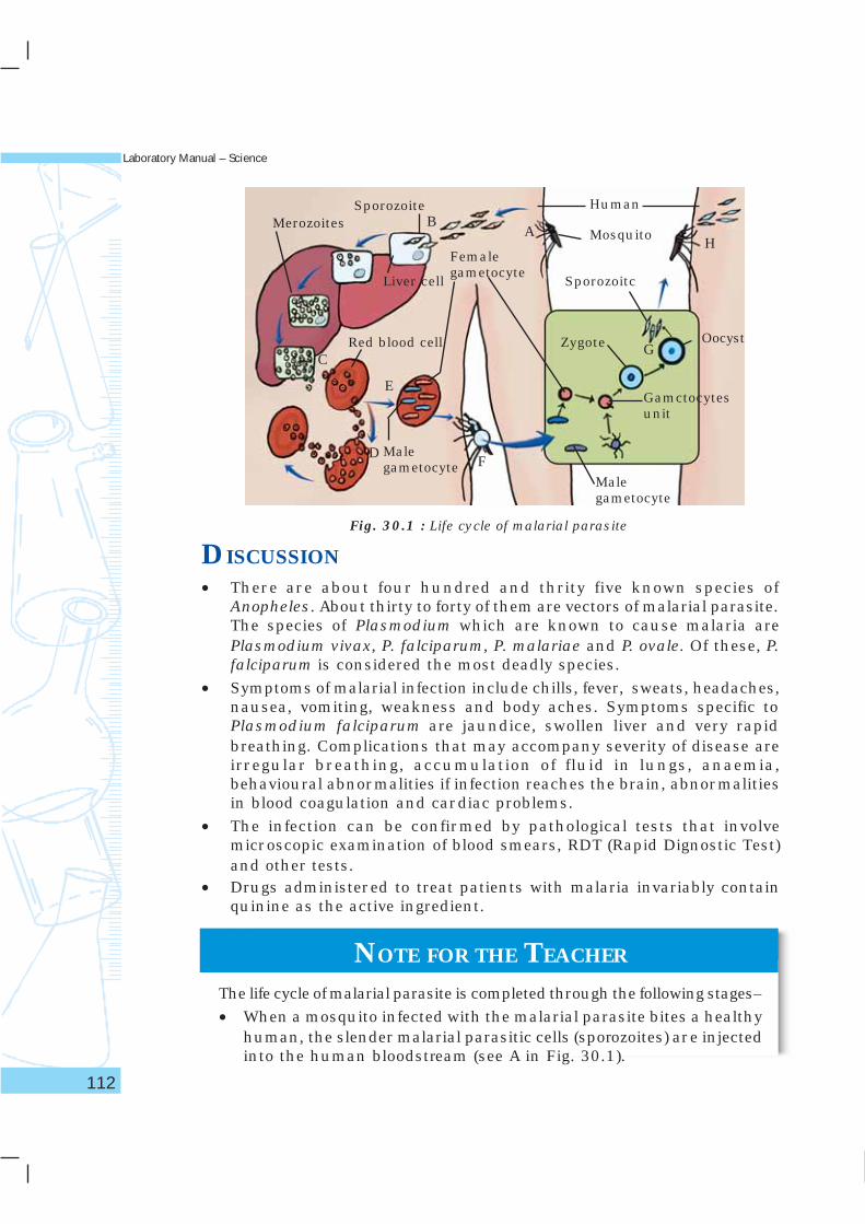
AIM To study the life cycle of malarial parasite.
THEORYPlasmodium is commonly called the malarial parasite. The life cycle ofPlasmodium is complex and involves two hosts, human being and amosquito. Plasmodium completes its sexual cycle in mosquito and asexualcycle in human being. When female Anopheles mosquito infected with theparasite bites a healthy human, infective sporozoites are injected into thehuman blood stream causing the dreaded disease malaria. The mosquitowhich transmits the malarial parasite is called the vector.
MATERIALS REQUIREDA chart showing the life cycle of malarial parasite, permanent slides ofmalarial parasite, and compound microscope.
PROCEDURE1. Study the chart and carefully note down the different stages of the
life cycle of Plasmodium beginning with the mosquito bite.
2. Draw a flow chart of different stages of life cycle.
� � � � � � � � � � 30
Laboratory Manual – Science
112
DISCUSSION• There are about four hundred and thrity five known species of
Anopheles. About thirty to forty of them are vectors of malarial parasite.The species of Plasmodium which are known to cause malaria arePlasmodium vivax, P. falciparum, P. malariae and P. ovale. Of these, P.falciparum is considered the most deadly species.
• Symptoms of malarial infection include chills, fever, sweats, headaches,nausea, vomiting, weakness and body aches. Symptoms specific toPlasmodium falciparum are jaundice, swollen liver and very rapidbreathing. Complications that may accompany severity of disease areirregular breathing, accumulation of fluid in lungs, anaemia,behavioural abnormalities if infection reaches the brain, abnormalitiesin blood coagulation and cardiac problems.
• The infection can be confirmed by pathological tests that involvemicroscopic examination of blood smears, RDT (Rapid Dignostic Test)and other tests.
• Drugs administered to treat patients with malaria invariably containquinine as the active ingredient.
NOTE FOR THE TEACHER
The life cycle of malarial parasite is completed through the following stages–
• When a mosquito infected with the malarial parasite bites a healthyhuman, the slender malarial parasitic cells (sporozoites) are injectedinto the human bloodstream (see A in Fig. 30.1).
Fig. 30.1 : Life cycle of malarial parasite
Red blood cell
Liver cell
Human
Mosquito
Sporozoitc
SporozoiteMerozoites
Femalegametocyte
Malegametocyte
Gamctocytesunit
Malegametocyte
Zygote Oocyst
AB
C
D
E
F
G
H
The World of Living
113
• Within thirty minutes, the parasite invades the human liver throughthe blood and lymphatic system. It infects the liver cells(hepatocytes), where it multiplies producing thousands of parasiticcells within a week (See B in Fig. 30.1).
• The parasitic cells re-enter the blood stream and infect red bloodcells (see C in Fig. 30.1).
• They grow in the red blood cells and undergo another phase ofmultiplication, eventually causing rupture of red blood cells andreleasing more parasitic cells along with their toxins. This leads tomanifestation of symptoms of malaria like chills and fever (see Din Fig. 30.1).
• Some parasitic cells form gametocytes (sex cells). These are of twotypes: (i) male gametocytes and (ii) female gametocytes (see E inFig. 30.1).
• When another mosquito bites the infected human, it ingests thesporozoites along with blood (see F in Fig. 30.1).
• In the stomach of mosquito (midgut), the gametocytes mature andfertilization occurs resulting in the formation of zygote. Zygotedevelops an outer covering and becomes the oocyst. Within theoocyst thousands of sporozoites are formed. The oocysts ruptureand release the sporozoites into the body cavity from where theymigrate to the salivary glands of mosquito (see G in Fig. 30.1).
• When this mosquito bites another human, along with the salivathe parasites are injected and the life cycle continues.
• Permanent slide of blood smear of persons suffering from malariacan be shown to the students.
QUESTIONS• What are the different species of malarial parasite that cause
malaria?
• When do the symptoms of malaria such as fever and chills appear?
• When a mosquito bites a person infected with Plasmodium, whichstage of the parasite will the mosquito ingest?
• How does the malarial parasite reproduce in the red blood cells?
• Why are people suffering from malaria anaemic?
Laboratory Manual – Science
114
AIMTo collect and study symptoms of diseases in locally available crop plants.
THEORYMicrobes like fungi, bacteria and virus are capable of causing seriousdiseases in plants. Such parasitic microbes af fect many of ourcommercial crops like cereals, pulses, vegetables, fruits etc. Generallya parasitic infection is specific, that is, it infects a specific plant. Somemicrobes are capable of infecting plant species belonging to a particulargroup. The infected plant is called the host and the infecting organismis called the parasite. Parasitic microbes require a living host forcompleting their life cycle. They absorb nutrients from the host plantsand may even kill the host. If the disease is not checked, it is capable ofspreading rapidly to other plants causing severe loss. Bacterial blights,smuts, white rust, black rust, tobacco mosaic are a few common diseasesof crop plants in our country.
MATERIALS REQUIREDTwo or three diseased crop plants or ornamental plants or weeds, compoundmicroscope, permanent slides of some diseased plants, a hand lens, slides,cover slips, needle, and a brush,
� � � � � � � � � � 31
The World of Living
115
PROCEDURE1. Collect two to three different kinds of diseased or infected plants.
2. Carefully observe each plant part for visible disease symptomssuch as decolouration, infection spots, coloured patches, soft anddecaying parts.
3. Observe if the entire plant is infected or only some parts like leaves,flowers or stem are infected.
4. Observe the infected parts and the physical characteristics of infectionand record in the observations table.
5. Scrape the infected spot with a needle/blade and transfer it to a dropof water on a slide. Place a cover slip and observe under the microscope.
6. Under the low power of microscope, observe the presence of spores/hyphae and damage caused to plant tissues (or cells).
7. Draw a diagram of the infected part and show the disease symptoms.
OBSERVATIONS
Sl.No. Observation Plant 1 Plant 2
1. Infected part is: (Yes/No) (Yes/No)
Stem
RootLeaf
Flower
Fruit2. Extent of infection:
LocalisedEnitre plant
3. Infection spot
Soft patchDry patch
4. Are spores visible?
5. Are hyphae visible?
RESULTS AND DISCUSSIONDraw the diagrams of the infected parts and label the diseased parts. Alsodraw diagrams of spore and hyphae.
Laboratory Manual – Science
116
NOTE FOR THE TEACHER • This is a group activity. Students may be asked to work in a
team.• The task of collecting infected plants may be assigned to students
in advance.• It is advised to facilitate students in identifying diseased plants
by observing the external features like stunted or abnormalgrowth, discolourd spots on plant parts, etc.
• All weak plants are not necessarily diseased plants. Poor growthmay also be due to a deficiency of certain minerals.
• Permanent slides of diseased plant parts may be focussed underthe compound microscope.
• Careful observation of a slide may reveal the presence of sporesor hyphae or both. Hyphae are generally filamentous, colourlessand appear as branched and delicate strands.
• Students may also be advised to find out five diseases of cropplants which lead to extensive loss of revenue in our country. Itmay also be suggested to use internet or magazines or otherinformation sources for this purpose.
QUESTIONS• What are parasites? How do they obtain food?
• How do parasitic microbes spread from plant to plant?
• How do farmers check the spread of diseases in their fields?