Embed Size (px)
Citation preview
/ J of IMAB. 2013, vol. 19, issue 4/ http://www.journal-imab-bg.org 309
THERAPEUTIC DIFFICULTIES IN ACHIEVEMENTOF OPTIMAL ROOT COVERAGE AND AESTHETICIN CLASS III GINGIVAL RECESSION
Christina Popova, Antoaneta Mlachkova, Velichka Dosseva-PanovaDepartment of Periodontology, Faculty of Dental Medicine,Medical University - Sofia, Bulgaria
Journal of IMAB - Annual Proceeding (Scientific Papers) 2013, vol. 19, issue 4ISSN: 1312-773X (Online)
SUMMARYThe width of the attached gingiva is defined as a
distance between the depth of the gingival sulcus or gingival/periodontal pocket to the mucogingival junction. Authorssuggest that a minimal amount of attached gingiva isnecessary to ensure the gingival health. When the buccal boneplate and gingival tissues are thin and the position of the toothis too vestibular gingival margin often displaces apically, andgingival recession develops. In the presence of gingivalrecession and reduced vestibular depth oral hygieneprocedures are embarrassed.
The definition of class III gingival recession ismarginal lack of tissue extended to/or beyond themucogingival junction with bone and soft tissue lossinterdentally or malpositioning of the tooth.
Prognosis for class III and IV gingival recessionis that only partial coverage can be expected after rootcoverage procedures - FGG (free gingival graft) or connectivetissue graft (CTG). Adjunctive surgical techniques would behelpful to achieve better aesthetic outcomes.
Key words: gingival recession, FGG - free gingivalgraft, CTG - connective tissue graft.
INTRODUCTION:It is well accepted that deficiency or lack of attached
gingiva in areas with loss of alveolar bone and progressiverecessions as well as positive tension test due to frenulum areindicators for procedure of enlarging the keratinized gingivaltissues. Free gingival graft is the suitable surgical approachfor soft tissue augmentation [5, 9, 10, 11].
The aim of this presentation is to show the outcomesof FGG surgical procedures in the management of soft tissuesdeficiency, and the deeper and larger gingival recession incircumstances of interdentally bone loss or root caries.
MATERIALS AND METHODS:Two different clinical cases are presented with
complete lack of attached gingival tissues, vestibular bonedeficiency, inconvenient occlusal relationships, and radicularcaries. The technique of free gingival graft was applied forkeratinized gingival tissue creation [1, 2, 8]. After firstsurgical step another surgical procedure was added to achieve
tooth root covering - coronally positioned flap (Fig. 1, 2, 3,4, 5) and subepithelial connective tissue graft (Fig. 6, 7, 8,9, 10 and 11). [3, 4, 6].
RESULTS:After first surgery partial root coverage and successful
and adequate increasing of the gingival tissues was obtained.After the second surgical step a great reduction of the gingivalrecessions, satisfactory root coverage, and acceptableaesthetic view in the frontal area was achieved.
CONCLUSION:Although limited aesthetic results the free gingival
graft procedure is an appropriate surgical method in thetreatment of shallow vestibule, insufficient width of attachedtissues and leads to enlargement of the keratinized gingiva,improvement of the plaque control, stabilization of theperiodontal attachment, and play an important role for alimitation of the alveolar bone loss progression. In class IIIand class IV gingival recessions the FGG procedures resultin a partially coverage of the previously denuded root surfaceand a second surgical procedure as a subepithelial CTG orcoronally positioned flap are recommended [3, 5, 6, 7].
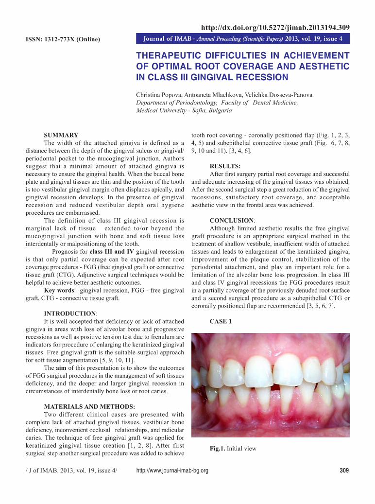
CASE 1
Fig.1. Initial view
http://dx.doi.org/10.5272/jimab.2013194.309

310 http://www.journal-imab-bg.org / J of IMAB. 2013, vol. 19, issue 4/
Fig. 6. Case 2 - orthopantomography
Fig. 2. Case 1 - intraoral x-ray Fig.4. Second surgery - flap elevation
Fig. 3. 5 months after first surgery Fig. 5. 6 months after second surgery – almostcomplete root coverage of #41 and partial root coverage of#31, and attached gingiva augmentation is achieved
CASE 2

/ J of IMAB. 2013, vol. 19, issue 4/ http://www.journal-imab-bg.org 311
Fig.7. Initial view – Class III gingival recession
Fig. 8.5 months after first surgery and root caries of#31
Fig. 9.Second surgery - flap elevation
Fig. 10. Second surgery - CTG in place
Fig. 11. 6 months after second surgery – acceptableresult with significant root coverage and stabile amount ofattached gingiva is achieved

312 http://www.journal-imab-bg.org / J of IMAB. 2013, vol. 19, issue 4/
Address for correspondence:Professor Ch. Popova, PhD, DM;Department of Periodontology, Faculty of Dental Medicine,1, Georgi Sofiiski str, 1431 Sofia, BulgariaE-mail: [email protected]
1. Popova Chr, AlloDerm®(AlloDerm® Regenerative TissueMatrix) in attached gingival tissueaugmentation. Continued education.2012; 11(2):28-32 [in Bulgarian]
2. Popova Chr. Treatment of classII Miller localized gingival recessionwith free gingival graft: case reports.Problems of stomatology. 2005;31(2):45-46. [in Bulgarian]
3. Bernimoulin JP, LuscherB, Muhlemann HR. Coronallyrepositioned periodontal flap. J ClinPeriodontol. 1975 Feb;2(1):1-13.[PubMed]
4. Bruno JF. Connective tissue grafttechnique assuring wide root coverage.Int J of Periodontics and RestorativeDentistry. 1994 Apr;14(2):127- 137.[PubMed]
5. Holbrook T, Ochsenbein C.Complete coverage of denuded rootsurface with a one stage gingival graft.Int J Periodontics Restorative Dent.1983; 3(3): 8-27. [PubMed]
6. Langer B, Langer L.Subepithelial connective tissue grafttechnique for root coverage. JPeriodontol. 1985 Dec;56(12):715-720. [PubMed] [CrossRef]
7. Laney JB, Saunders VG, GarnickJJ. A comparison of two techniques forattaining root coverage. J Periodontol.1992 Jan;63(1):19-23. [PubMed][CrossRef]
8. Miller PD Jr. Root coverageusing the free soft tissue autograftfollowing citric acid application.² ²². Asuccessful and predictable procedure inareas of deep-wide recession. Int J
Restorative Dent. 1985; 5(2):15-37.[PubMed]
9. Popova Chr, Boyarova Tsv. Freegingival autograft for augmentation ofkeratinized tissue and stabilization ofgingival recessions. J of IMAB. 2008;14(2):19-25.
10. Popova Chr, Dosseva V.Gingival tissue augmentation inconjunction with regenerativeperiodontal procedures. J of IMAB.2007; 13(2):16-18.
11. Takkei HH, Azzi RR, Han TJ.Periodontal Plastic and EstheticSurgery; Newman MG, Takei HH,Klokkevold PR, Carranza FA.Carranza’s Clinical Periodontology,Tenth Edition, Saunders and ElsevierInc., p: 1005-1009, 2006.
REFERENCES: