Embed Size (px)
Citation preview

Tissue Types
There are four basic tissue types:
Epithelial - covering and lining membranes o Type of epithelium is according to its function o Glands can be of both exocrine and endocrine type
Connective - numerous varieties, including bone, cartilage, ligaments, tendons, blood, areolar and adipose types
Muscular is very cellular with very little intercellular substance. Has powers of conduction and movement- three types: o Skeletal - posture and movement o Smooth - movement of substances through hollow tubes: gut, blood vessels, bladder o Cardiac - pumps blood
Nervous - has developed the ability to conduct impulses and secrete neurotransmitters
Epithelial tissue
Epithelial tissue is largely cellular tissue with very little intercellular substance. Epithelia are divided into the major divisions of covering and lining membranes, which can have the ability to absorb, and the various glands which have the ability to secrete. They are also named according to the cell shape they have in their side view.
Epithelial tissues has three possible functions, sometimes only one and occasionally all three. The functions are:
1. Protection 2. Secretion 3. Absorption
Types of epithelium:
Simple epithelium– this is one layer of cells on a basement membrane. The basement membrane is non-living intercellular substance which allows materials to pass in and out of the cells.
Simple squamous epithelium (squamous = scale-like) Figure 1 - Simple Squamous epithelium
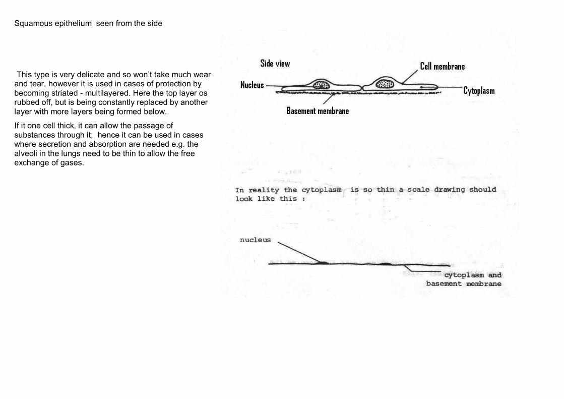
Squamous epithelium seen from the side
This type is very delicate and so won’t take much wear and tear, however it is used in cases of protection by becoming striated - multilayered. Here the top layer os rubbed off, but is being constantly replaced by another layer with more layers being formed below.
If it one cell thick, it can allow the passage of substances through it; hence it can be used in cases where secretion and absorption are needed e.g. the alveoli in the lungs need to be thin to allow the free exchange of gases.
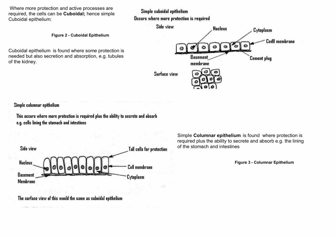
Where more protection and active processes are required, the cells can be Cuboidal; hence simple Cuboidal epithelium: Figure 2 - Cuboidal Epithelium
Cuboidal epithelium is found where some protection is needed but also secretion and absorption, e.g. tubules of the kidney.
Simple Columnar epithelium is found where protection is required plus the ability to secrete and absorb e.g. the lining of the stomach and intestines Figure 3 - Columnar Epithelium
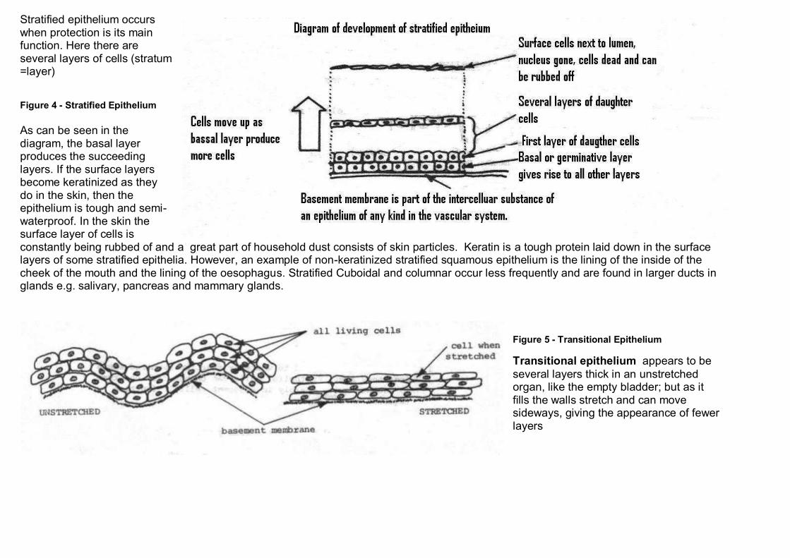
Stratified epithelium occurs when protection is its main function. Here there are several layers of cells (stratum =layer)
Figure 4 - Stratified Epithelium As can be seen in the diagram, the basal layer produces the succeeding layers. If the surface layers become keratinized as they do in the skin, then the epithelium is tough and semi-waterproof. In the skin the surface layer of cells is constantly being rubbed of and a great part of household dust consists of skin particles. Keratin is a tough protein laid down in the surface layers of some stratified epithelia. However, an example of non-keratinized stratified squamous epithelium is the lining of the inside of the cheek of the mouth and the lining of the oesophagus. Stratified Cuboidal and columnar occur less frequently and are found in larger ducts in glands e.g. salivary, pancreas and mammary glands.
Figure 5 - Transitional Epithelium
Transitional epithelium appears to be
several layers thick in an unstretched organ, like the empty bladder; but as it fills the walls stretch and can move sideways, giving the appearance of fewer layers

Figure 6 - Pseudostratified Epithelium
Pseudostratified epithelium is not truly stratified. Only one layer of cells sits on the
basement membrane, but some do not reach the top surface.
Figure 7 - Ciliated Epithelium
Epithelia are sometimes ciliated, where ciliary action wafts something along a tube e.g. particles trapped in mucous are wafted up by ciliary action up the trachea; or an oocyte wafted by ciliated columnar epithelium along a fallopian tube.
Epithelia can also have a ‘brush border’. This is not ciliated as such they don’t have the power of movement; here they have the function of increasing the surface area for absorption e.g. the columnar epithelium of the small intestine for absorption of foodstuffs; proximal convoluted tubule of the kidney for reabsorption of glucose, water and other substances.
Endothelium
All blood and lymph vessels are lined with simple squamous epithelium, here known as endothelium. Here, though, there is no absorption through the walls except in the capillaries. The larger blood vessels have blood vessels in their walls (in the tunica adventitia) to proved oxygen and nutrients to the vessel walls themselves. They are called the vasa vasorum.
The endothelium of the blood vessels needs to very smooth to prevent blood sticking to it and clotting; this could be fatal e.g. coronary blood vessels, cerebral blood vessels.
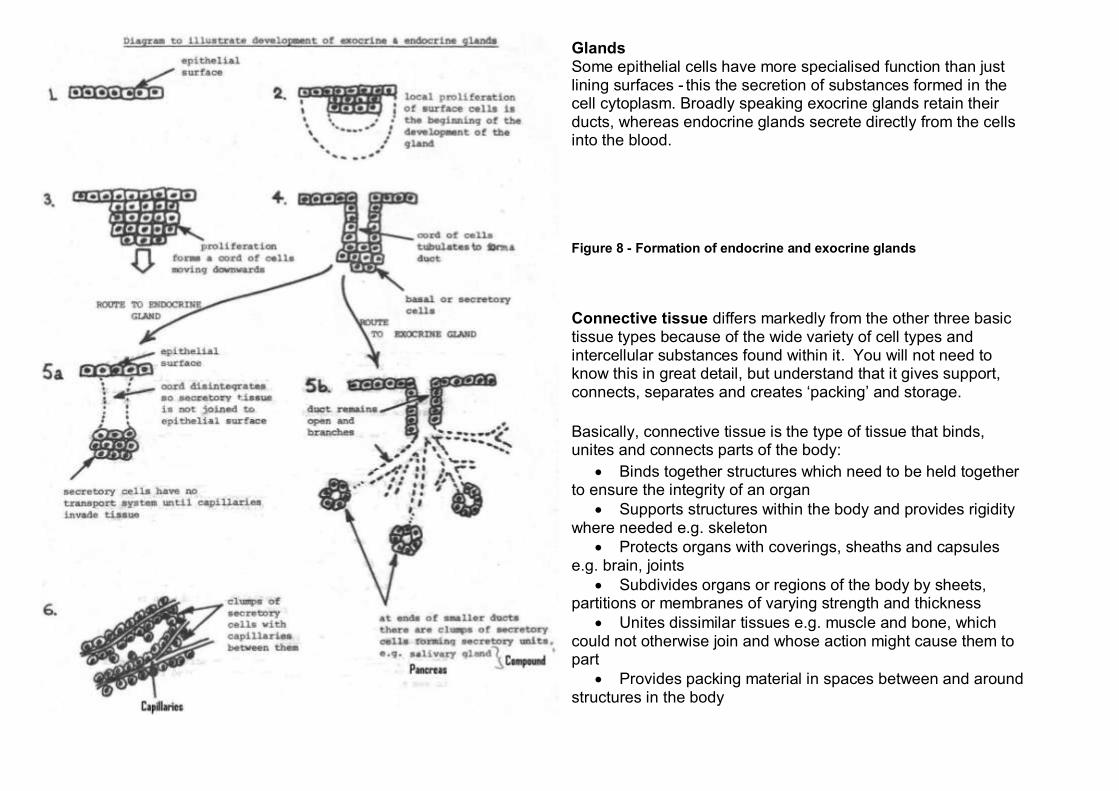
Glands Some epithelial cells have more specialised function than just lining surfaces - this the secretion of substances formed in the cell cytoplasm. Broadly speaking exocrine glands retain their ducts, whereas endocrine glands secrete directly from the cells into the blood. Figure 8 - Formation of endocrine and exocrine glands
Connective tissue differs markedly from the other three basic tissue types because of the wide variety of cell types and intercellular substances found within it. You will not need to know this in great detail, but understand that it gives support, connects, separates and creates ‘packing’ and storage.
Basically, connective tissue is the type of tissue that binds, unites and connects parts of the body:
Binds together structures which need to be held together to ensure the integrity of an organ
Supports structures within the body and provides rigidity where needed e.g. skeleton
Protects organs with coverings, sheaths and capsules e.g. brain, joints
Subdivides organs or regions of the body by sheets, partitions or membranes of varying strength and thickness
Unites dissimilar tissues e.g. muscle and bone, which could not otherwise join and whose action might cause them to part
Provides packing material in spaces between and around structures in the body
Types of connective tissue:
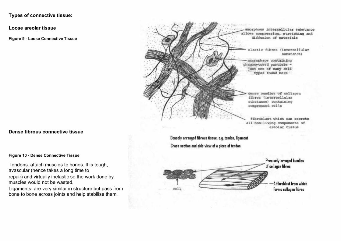
Loose areolar tissue Figure 9 - Loose Connective Tissue
Dense fibrous connective tissue
Figure 10 - Dense Connective Tissue
Tendons attach muscles to bones. It is tough, avascular (hence takes a long time to
repair) and virtually inelastic so the work done by muscles would not be wasted.
Ligaments are very similar in structure but pass from bone to bone across joints and help stabilise them.
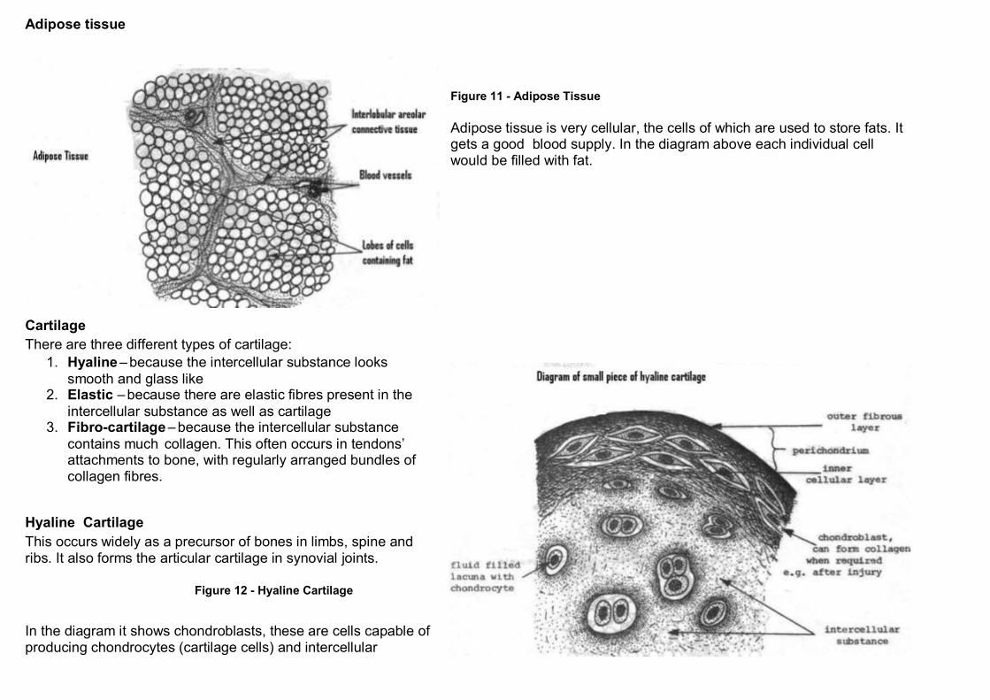
Adipose tissue
Figure 11 - Adipose Tissue
Adipose tissue is very cellular, the cells of which are used to store fats. It gets a good blood supply. In the diagram above each individual cell would be filled with fat.
Cartilage
There are three different types of cartilage:
1. Hyaline – because the intercellular substance looks
smooth and glass like 2. Elastic – because there are elastic fibres present in the
intercellular substance as well as cartilage 3. Fibro-cartilage – because the intercellular substance
contains much collagen. This often occurs in tendons’ attachments to bone, with regularly arranged bundles of collagen fibres.
Hyaline Cartilage
This occurs widely as a precursor of bones in limbs, spine and ribs. It also forms the articular cartilage in synovial joints.
Figure 12 - Hyaline Cartilage
In the diagram it shows chondroblasts, these are cells capable of producing chondrocytes (cartilage cells) and intercellular
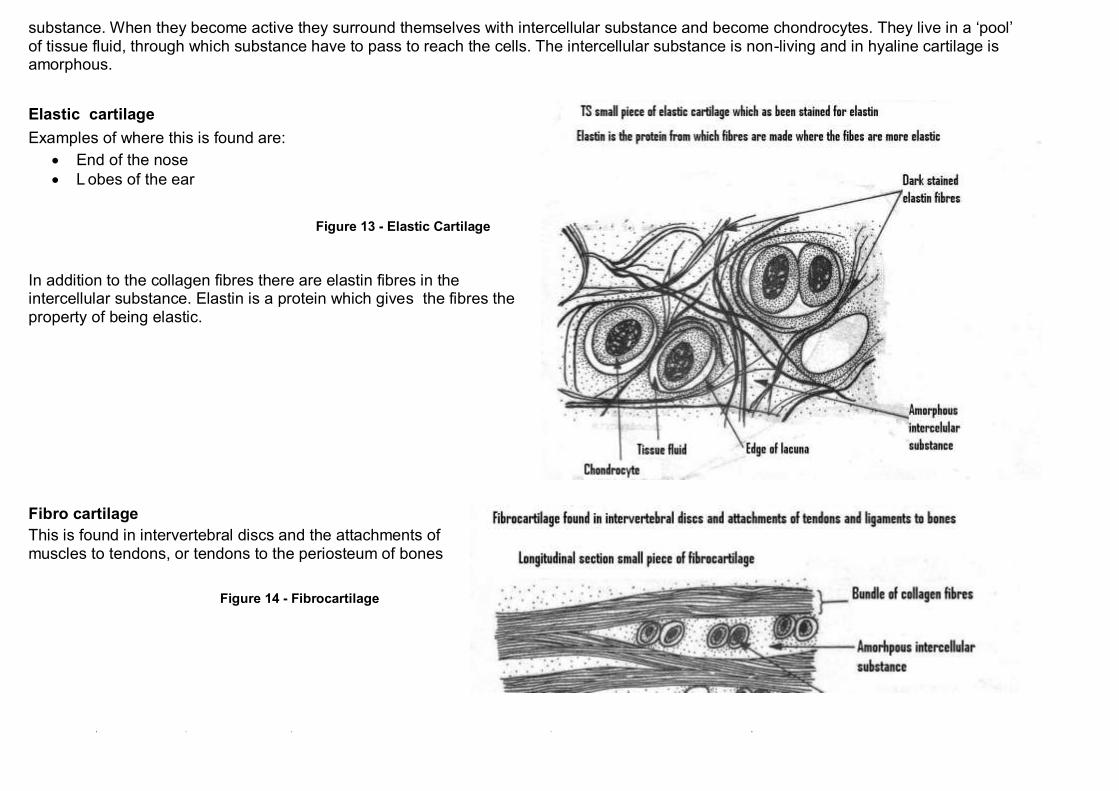
substance. When they become active they surround themselves with intercellular substance and become chondrocytes. They live in a ‘pool’ of tissue fluid, through which substance have to pass to reach the cells. The intercellular substance is non-living and in hyaline cartilage is amorphous.
Elastic cartilage
Examples of where this is found are:
End of the nose
L obes of the ear
Figure 13 - Elastic Cartilage
In addition to the collagen fibres there are elastin fibres in the intercellular substance. Elastin is a protein which gives the fibres the property of being elastic.
Fibro cartilage
This is found in intervertebral discs and the attachments of muscles to tendons, or tendons to the periosteum of bones
Figure 14 - Fibrocartilage
The importance of the amorphous intercellular substance
Cartilage cells are not in close proximity to blood vessels and it therefore called avascular. It can seen, therefore that substances like oxygen, carbon dioxide and nutrition and waste products have to diffuse through the water of the amorphous intercellular substance in the lacunae. Remember that the intercellular substance is non-living; therefore the survival of the cartilage cells depends upon the water in the amorphous intercellular substance. If it is reduced or calcification occurs (as in bone formation) then the cartilage dies. This is why calcified cartilage cannot be a permanent tissue. Bone, on the other hand, has a highly organised blood supply.
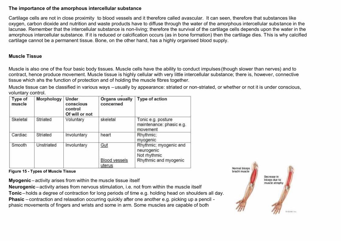
Muscle Tissue
Muscle is also one of the four basic body tissues. Muscle cells have the ability to conduct impulses(though slower than nerves) and to contract, hence produce movement. Muscle tissue is highly cellular with very little intercellular substance; there is, however, connective tissue which ahs the function of protection and of holding the muscle fibres together.
Muscle tissue can be classified in various ways – usually by appearance: striated or non-striated, or whether or not it is under conscious, voluntary control.
Figure 15 - Types of Muscle Tissue
Myogenic – activity arises from within the muscle tissue itself
Neurogenic – activity arises from nervous stimulation, i.e. not from within the muscle itself
Tonic – holds a degree of contraction for long periods of time e.g. holding head on shoulders all day.
Phasic – contraction and relaxation occurring quickly after one another e.g. picking up a pencil -
phasic movements of fingers and wrists and some in arm. Some muscles are capable of both

Skeletal muscle
Skeletal muscle is under voluntary control and must have a nerve supply to work; without adequate nerve stimulation, the muscles do not work e.g. polio and other forms or paralysis.
A skeletal muscle contains hundreds of muscle fibres and each muscle fibre has its own nerve supply. The point of contact between the nerve and the muscle is usually a motor end-plate.
Each muscle fibre obeys an ‘all or none’ law; that is the whole fibre will respond and contract to a nerve stimulus, and not just any small part of it.
Figure 16 - Nerve Fibre and its Motor End Plate
Figure 17 - Diagram of a Muscle Fibre
This diagram shows why skeletal muscle is termed striated – because it looks like it. You will see later that the striations create great organisation within the muscle and hence creates great power when it contracts.
The muscle fibres are complex, elongated and multinucleate (unlike ‘ordinary cells’ which have one nucleus each (mononucleate). They also have a large number of mitochondria (for energy production). Because of this a long muscle fibre acts (and contracts) as a single unit, known as a syncitium. The appearance of the striations is formed by the overlap of microscopic filaments:
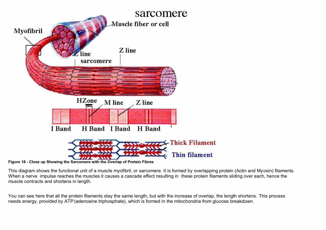
Figure 18 - Close up Showing the Sarcomere with the Overlap of Protein Fibres
This diagram shows the functional unit of a muscle myofibril, or sarcomere. It is formed by overlapping protein (Actin and Myosin) filaments. When a nerve impulse reaches the muscles it causes a cascade effect resulting in these protein filaments sliding over each, hence the muscle contracts and shortens in length.
You can see here that all the protein filaments stay the same length, but with the increase of overlap, the length shortens. This process needs energy, provided by ATP(adenosine triphosphate), which is formed in the mitochondria from glucose breakdown.
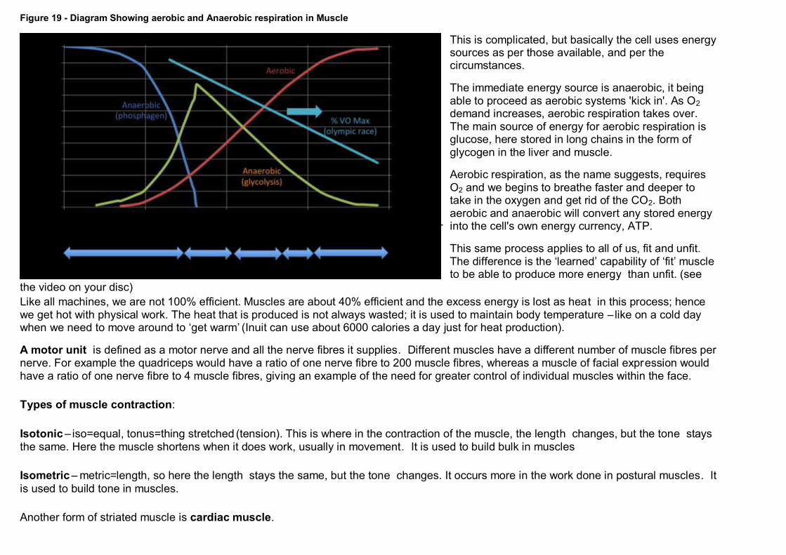
Figure 19 - Diagram Showing aerobic and Anaerobic respiration in Muscle
This is complicated, but basically the cell uses energy sources as per those available, and per the circumstances.
The immediate energy source is anaerobic, it being able to proceed as aerobic systems 'kick in'. As O2 demand increases, aerobic respiration takes over. The main source of energy for aerobic respiration is glucose, here stored in long chains in the form of glycogen in the liver and muscle.
Aerobic respiration, as the name suggests, requires O2 and we begins to breathe faster and deeper to take in the oxygen and get rid of the CO2. Both aerobic and anaerobic will convert any stored energy into the cell's own energy currency, ATP.
This same process applies to all of us, fit and unfit. The difference is the ‘learned’ capability of ‘fit’ muscle to be able to produce more energy than unfit. (see
the video on your disc)
Like all machines, we are not 100% efficient. Muscles are about 40% efficient and the excess energy is lost as heat in this process; hence we get hot with physical work. The heat that is produced is not always wasted; it is used to maintain body temperature – like on a cold day when we need to move around to ‘get warm’ (Inuit can use about 6000 calories a day just for heat production).
A motor unit is defined as a motor nerve and all the nerve fibres it supplies. Different muscles have a different number of muscle fibres per nerve. For example the quadriceps would have a ratio of one nerve fibre to 200 muscle fibres, whereas a muscle of facial expression would have a ratio of one nerve fibre to 4 muscle fibres, giving an example of the need for greater control of individual muscles within the face.
Types of muscle contraction:
Isotonic – iso=equal, tonus=thing stretched (tension). This is where in the contraction of the muscle, the length changes, but the tone stays the same. Here the muscle shortens when it does work, usually in movement. It is used to build bulk in muscles
Isometric – metric=length, so here the length stays the same, but the tone changes. It occurs more in the work done in postural muscles. It
is used to build tone in muscles.
Another form of striated muscle is cardiac muscle.
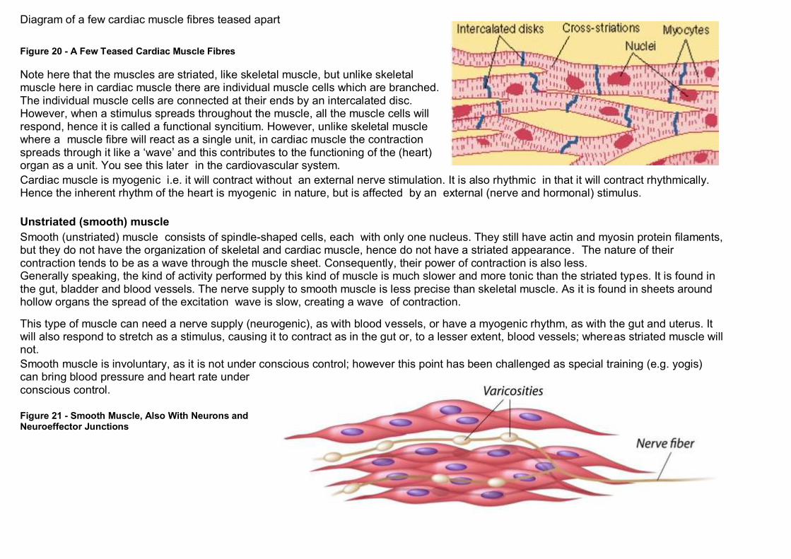
Diagram of a few cardiac muscle fibres teased apart
Figure 20 - A Few Teased Cardiac Muscle Fibres
Note here that the muscles are striated, like skeletal muscle, but unlike skeletal muscle here in cardiac muscle there are individual muscle cells which are branched. The individual muscle cells are connected at their ends by an intercalated disc. However, when a stimulus spreads throughout the muscle, all the muscle cells will respond, hence it is called a functional syncitium. However, unlike skeletal muscle where a muscle fibre will react as a single unit, in cardiac muscle the contraction spreads through it like a ‘wave’ and this contributes to the functioning of the (heart) organ as a unit. You see this later in the cardiovascular system.
Cardiac muscle is myogenic i.e. it will contract without an external nerve stimulation. It is also rhythmic in that it will contract rhythmically. Hence the inherent rhythm of the heart is myogenic in nature, but is affected by an external (nerve and hormonal) stimulus.
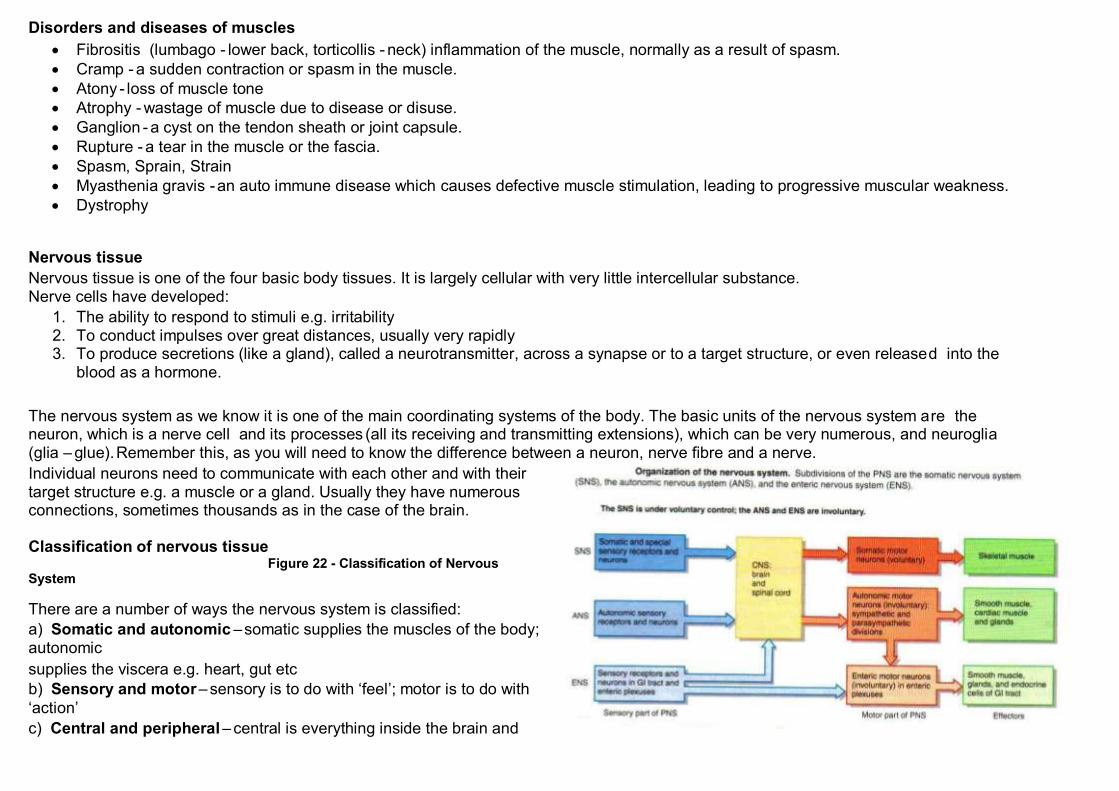
Unstriated (smooth) muscle
Smooth (unstriated) muscle consists of spindle-shaped cells, each with only one nucleus. They still have actin and myosin protein filaments, but they do not have the organization of skeletal and cardiac muscle, hence do not have a striated appearance. The nature of their contraction tends to be as a wave through the muscle sheet. Consequently, their power of contraction is also less. Generally speaking, the kind of activity performed by this kind of muscle is much slower and more tonic than the striated types. It is found in the gut, bladder and blood vessels. The nerve supply to smooth muscle is less precise than skeletal muscle. As it is found in sheets around hollow organs the spread of the excitation wave is slow, creating a wave of contraction.
This type of muscle can need a nerve supply (neurogenic), as with blood vessels, or have a myogenic rhythm, as with the gut and uterus. It will also respond to stretch as a stimulus, causing it to contract as in the gut or, to a lesser extent, blood vessels; whereas striated muscle will not.
Smooth muscle is involuntary, as it is not under conscious control; however this point has been challenged as special training (e.g. yogis) can bring blood pressure and heart rate under conscious control.
Figure 21 - Smooth Muscle, Also With Neurons and Neuroeffector Junctions
Disorders and diseases of muscles
Fibrositis (lumbago - lower back, torticollis - neck) inflammation of the muscle, normally as a result of spasm.
Cramp - a sudden contraction or spasm in the muscle.
Atony - loss of muscle tone
Atrophy - wastage of muscle due to disease or disuse.
Ganglion - a cyst on the tendon sheath or joint capsule.
Rupture - a tear in the muscle or the fascia.
Spasm, Sprain, Strain
Myasthenia gravis -an auto immune disease which causes defective muscle stimulation, leading to progressive muscular weakness.
Dystrophy
Nervous tissue
Nervous tissue is one of the four basic body tissues. It is largely cellular with very little intercellular substance. Nerve cells have developed:
1. The ability to respond to stimuli e.g. irritability 2. To conduct impulses over great distances, usually very rapidly 3. To produce secretions (like a gland), called a neurotransmitter, across a synapse or to a target structure, or even released into the
blood as a hormone.
The nervous system as we know it is one of the main coordinating systems of the body. The basic units of the nervous system are the neuron, which is a nerve cell and its processes (all its receiving and transmitting extensions), which can be very numerous, and neuroglia (glia – glue).Remember this, as you will need to know the difference between a neuron, nerve fibre and a nerve.
Individual neurons need to communicate with each other and with their target structure e.g. a muscle or a gland. Usually they have numerous connections, sometimes thousands as in the case of the brain. Classification of nervous tissue Figure 22 - Classification of Nervous System
There are a number of ways the nervous system is classified:
a) Somatic and autonomic –somatic supplies the muscles of the body; autonomic
supplies the viscera e.g. heart, gut etc
b) Sensory and motor– sensory is to do with ‘feel’; motor is to do with
‘action’
c) Central and peripheral – central is everything inside the brain and
spinal cord; peripheral is everything outside the brain and spinal cord.
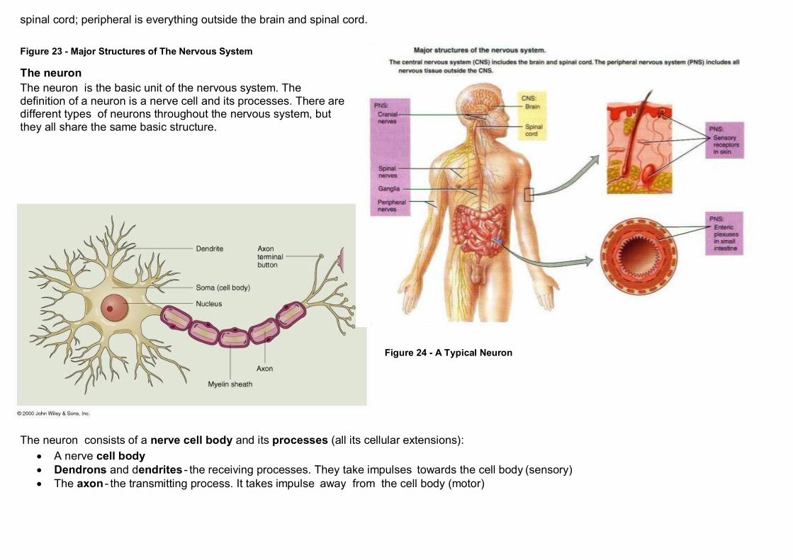
Figure 23 - Major Structures of The Nervous System
The neuron
The neuron is the basic unit of the nervous system. The definition of a neuron is a nerve cell and its processes. There are different types of neurons throughout the nervous system, but they all share the same basic structure.
Figure 24 - A Typical Neuron
The neuron consists of a nerve cell body and its processes (all its cellular extensions):
A nerve cell body
Dendrons and dendrites - the receiving processes. They take impulses towards the cell body (sensory)
The axon - the transmitting process. It takes impulse away from the cell body (motor)
Hence:
A nerve fibre is one of those processes (towards or away)
A nerve is seen anatomically as a single unit but consists of hundreds of nerve fibres, both sensory and motor (some myelinated, some not – see later)
A collection of cell bodies outside the brain or spinal cord is called a ganglion (pleural –ganglia); inside the brain or spinal cord they are called a nucleus (pleural –nuclei).
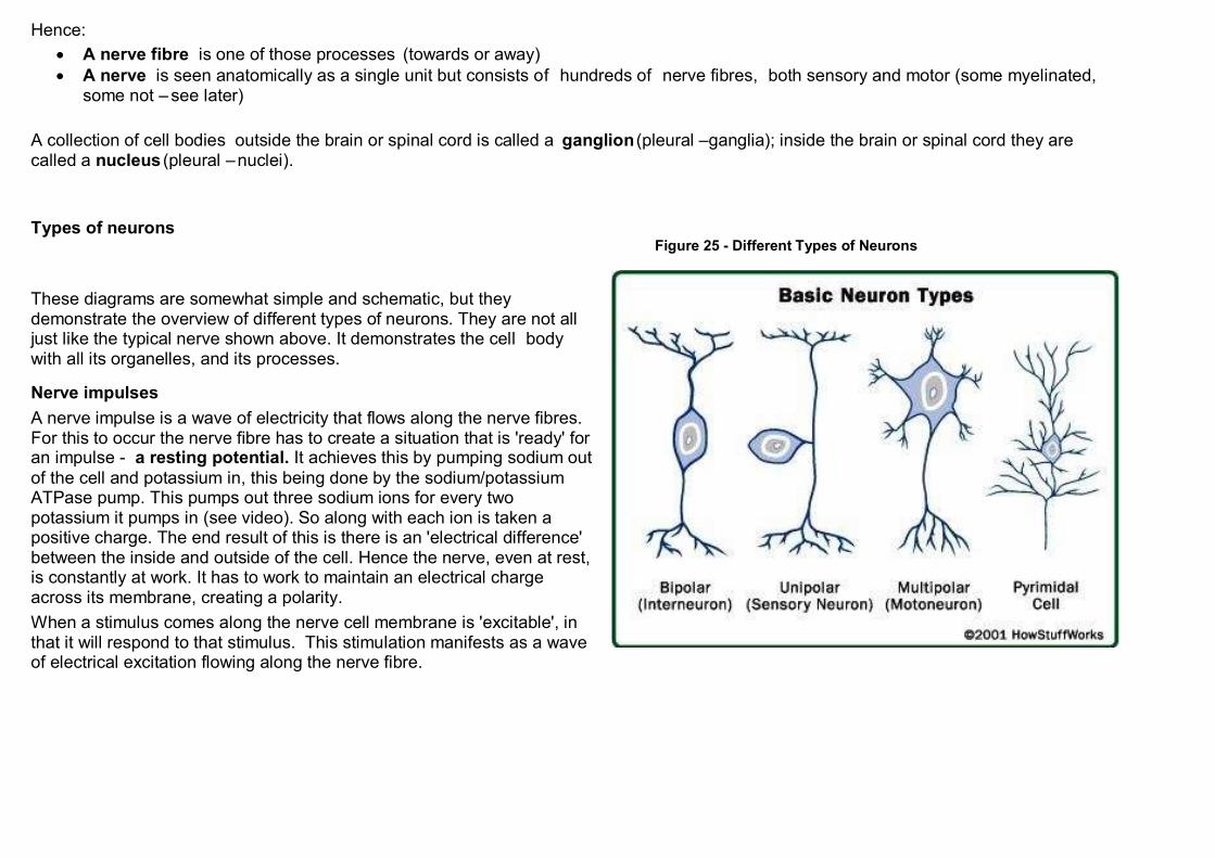
Types of neurons Figure 25 - Different Types of Neurons
These diagrams are somewhat simple and schematic, but they demonstrate the overview of different types of neurons. They are not all just like the typical nerve shown above. It demonstrates the cell body with all its organelles, and its processes.
Nerve impulses
A nerve impulse is a wave of electricity that flows along the nerve fibres. For this to occur the nerve fibre has to create a situation that is 'ready' for an impulse - a resting potential. It achieves this by pumping sodium out
of the cell and potassium in, this being done by the sodium/potassium ATPase pump. This pumps out three sodium ions for every two potassium it pumps in (see video). So along with each ion is taken a positive charge. The end result of this is there is an 'electrical difference' between the inside and outside of the cell. Hence the nerve, even at rest, is constantly at work. It has to work to maintain an electrical charge across its membrane, creating a polarity.
When a stimulus comes along the nerve cell membrane is 'excitable', in that it will respond to that stimulus. This stimulation manifests as a wave of electrical excitation flowing along the nerve fibre.
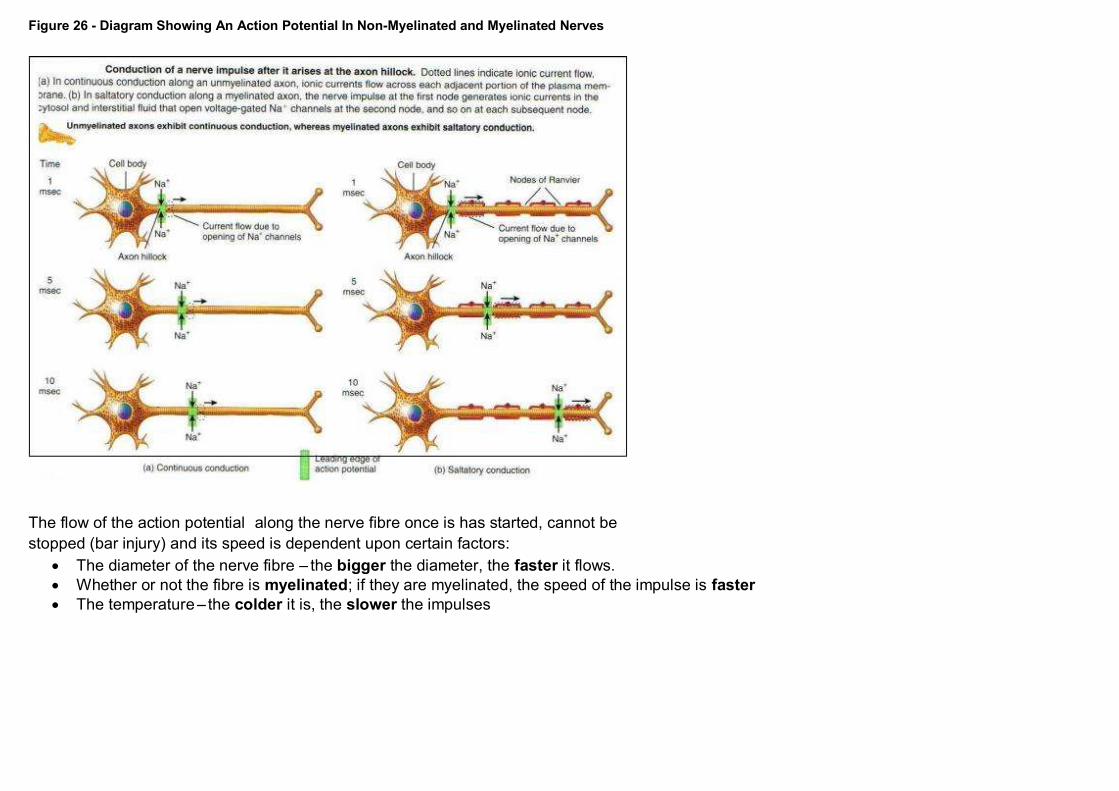
Figure 26 - Diagram Showing An Action Potential In Non-Myelinated and Myelinated Nerves
The flow of the action potential along the nerve fibre once is has started, cannot be
stopped (bar injury) and its speed is dependent upon certain factors:
The diameter of the nerve fibre – the bigger the diameter, the faster it flows.
Whether or not the fibre is myelinated; if they are myelinated, the speed of the impulse is faster
The temperature – the colder it is, the slower the impulses
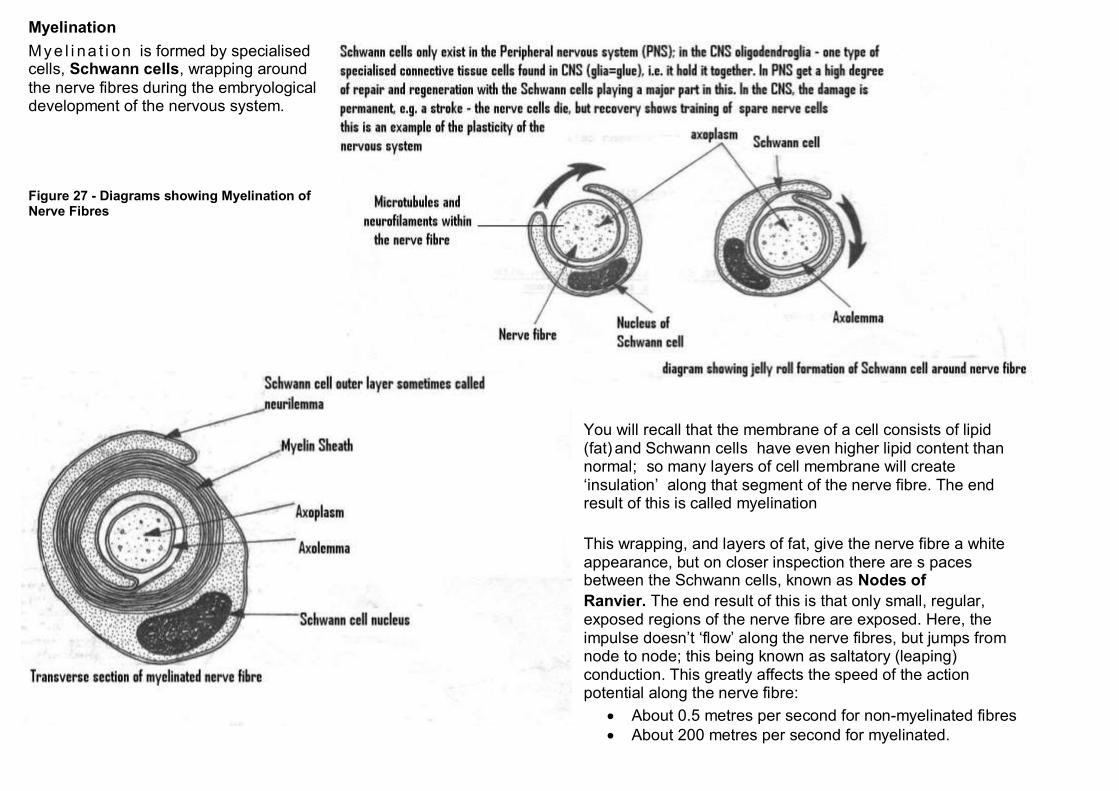
Myelination
Mye l i na t i on is formed by specialised cells, Schwann cells, wrapping around
the nerve fibres during the embryological development of the nervous system. Figure 27 - Diagrams showing Myelination of Nerve Fibres
You will recall that the membrane of a cell consists of lipid (fat) and Schwann cells have even higher lipid content than normal; so many layers of cell membrane will create ‘insulation’ along that segment of the nerve fibre. The end result of this is called myelination
This wrapping, and layers of fat, give the nerve fibre a white appearance, but on closer inspection there are s paces between the Schwann cells, known as Nodes of
Ranvier. The end result of this is that only small, regular, exposed regions of the nerve fibre are exposed. Here, the impulse doesn’t ‘flow’ along the nerve fibres, but jumps from node to node; this being known as saltatory (leaping) conduction. This greatly affects the speed of the action potential along the nerve fibre:
About 0.5 metres per second for non-myelinated fibres
About 200 metres per second for myelinated.
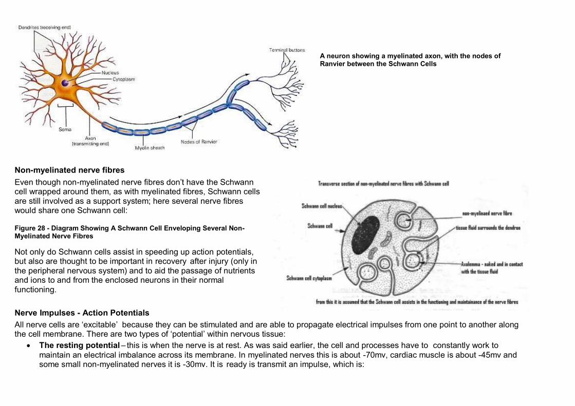
A neuron showing a myelinated axon, with the nodes of Ranvier between the Schwann Cells
Non-myelinated nerve fibres
Even though non-myelinated nerve fibres don’t have the Schwann cell wrapped around them, as with myelinated fibres, Schwann cells are still involved as a support system; here several nerve fibres would share one Schwann cell: Figure 28 - Diagram Showing A Schwann Cell Enveloping Several Non-Myelinated Nerve Fibres
Not only do Schwann cells assist in speeding up action potentials, but also are thought to be important in recovery after injury (only in the peripheral nervous system) and to aid the passage of nutrients and ions to and from the enclosed neurons in their normal functioning.
Nerve Impulses - Action Potentials
All nerve cells are ‘excitable’ because they can be stimulated and are able to propagate electrical impulses from one point to another along the cell membrane. There are two types of ‘potential’ within nervous tissue:
The resting potential – this is when the nerve is at rest. As was said earlier, the cell and processes have to constantly work to
maintain an electrical imbalance across its membrane. In myelinated nerves this is about -70mv, cardiac muscle is about -45mv and some small non-myelinated nerves it is -30mv. It is ready is transmit an impulse, which is:
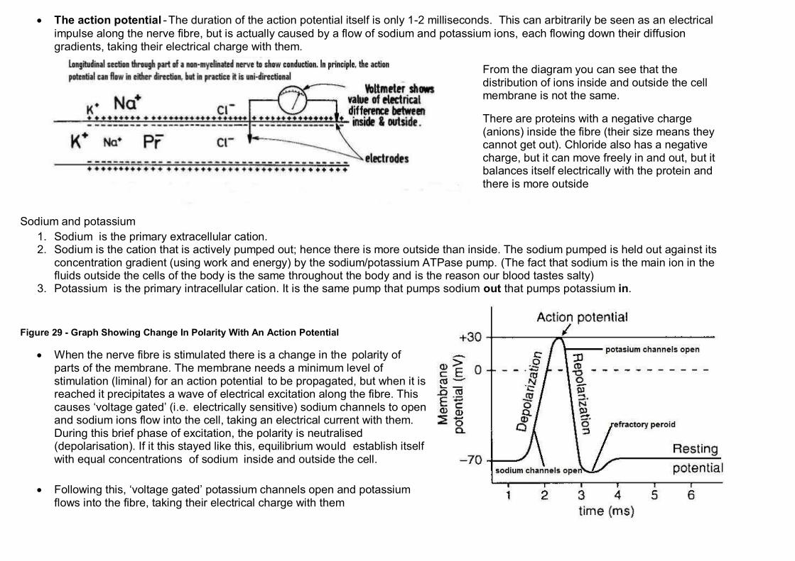
The action potential -The duration of the action potential itself is only 1-2 milliseconds. This can arbitrarily be seen as an electrical
impulse along the nerve fibre, but is actually caused by a flow of sodium and potassium ions, each flowing down their diffusion gradients, taking their electrical charge with them.
From the diagram you can see that the distribution of ions inside and outside the cell membrane is not the same.
There are proteins with a negative charge (anions) inside the fibre (their size means they cannot get out). Chloride also has a negative charge, but it can move freely in and out, but it balances itself electrically with the protein and there is more outside
Sodium and potassium
1. Sodium is the primary extracellular cation. 2. Sodium is the cation that is actively pumped out; hence there is more outside than inside. The sodium pumped is held out against its
concentration gradient (using work and energy) by the sodium/potassium ATPase pump. (The fact that sodium is the main ion in the fluids outside the cells of the body is the same throughout the body and is the reason our blood tastes salty)
3. Potassium is the primary intracellular cation. It is the same pump that pumps sodium out that pumps potassium in.
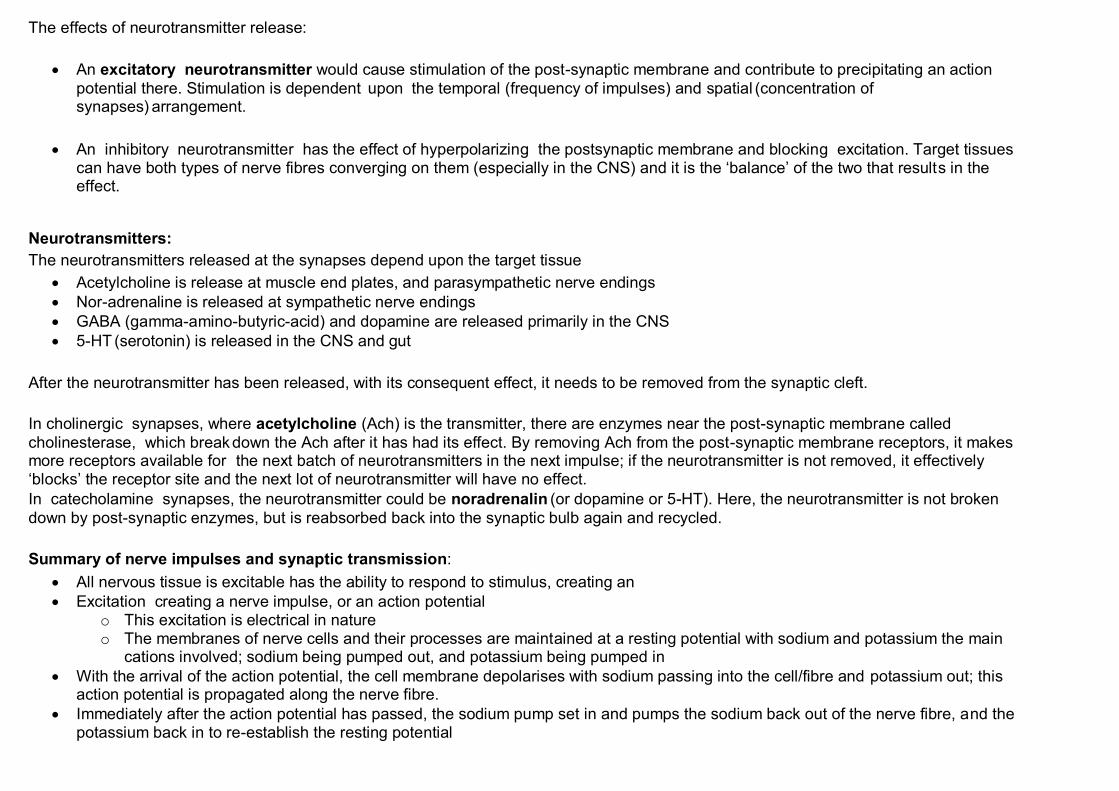
Figure 29 - Graph Showing Change In Polarity With An Action Potential
When the nerve fibre is stimulated there is a change in the polarity of parts of the membrane. The membrane needs a minimum level of stimulation (liminal) for an action potential to be propagated, but when it is reached it precipitates a wave of electrical excitation along the fibre. This causes ‘voltage gated’ (i.e. electrically sensitive) sodium channels to open and sodium ions flow into the cell, taking an electrical current with them. During this brief phase of excitation, the polarity is neutralised (depolarisation). If it this stayed like this, equilibrium would establish itself with equal concentrations of sodium inside and outside the cell.
Following this, ‘voltage gated’ potassium channels open and potassium flows into the fibre, taking their electrical charge with them

For the brief period that the action potential is there, the inside of the fibre changes from negative to positive but the sodium pump kicks in again and pumps the sodium back out and the potassium back in. just after the area of depolarisation is a region of hyperpolarisation (just before ‘normal polarisation is re-established) and non-responsive to an ordinary stimulation. So where the action potential is and the area of recovery, including the period of hyperpolarisation, the nerve is unresponsive to stimuli and is therefore known as the refractory period. It follows that the number of impulses along the nerve fibre is limited e.g. if the refractory period was 2msec then the cell cannot fire at more than 1000/2 = 500 impulses per second.
In myelinated fibres, the same principle applies except that the ionic exchange only occurs at the sites of the nodes of Ranvier.
Synapses Figure 30 - A Synapse
Nerve fibres do not attach directly to each other or to target tissues; they come into close apposition with them, but with a definite gap between the two called a synapse. The term synapse includes
The end of the nerve fibre,
The target tissue and
The gap – the synaptic cleft.
1. When the action potential reaches the end of the axon, the pre-synaptic bouton, it affects the permeability of the membrane.
2. It then also stimulates the release of a chemical, a neurotransmitter substance from vesicles within the synaptic end bulb 3. This chemical diffuse across the synaptic cleft ( the distance here is measured in thousandths of a millimetre) 4 . The neurotransmitter then combines with receptor sites on the post-synaptic membrane ( they occur all over the post-synaptic cell, but
are concentrated at the synaptic sites)
The effects of neurotransmitter release:
An excitatory neurotransmitter would cause stimulation of the post-synaptic membrane and contribute to precipitating an action
potential there. Stimulation is dependent upon the temporal (frequency of impulses) and spatial (concentration of synapses) arrangement.
An inhibitory neurotransmitter has the effect of hyperpolarizing the postsynaptic membrane and blocking excitation. Target tissues can have both types of nerve fibres converging on them (especially in the CNS) and it is the ‘balance’ of the two that results in the effect.
Neurotransmitters:
The neurotransmitters released at the synapses depend upon the target tissue
Acetylcholine is release at muscle end plates, and parasympathetic nerve endings
Nor-adrenaline is released at sympathetic nerve endings
GABA (gamma-amino-butyric-acid) and dopamine are released primarily in the CNS
5-HT (serotonin) is released in the CNS and gut
After the neurotransmitter has been released, with its consequent effect, it needs to be removed from the synaptic cleft.
In cholinergic synapses, where acetylcholine (Ach) is the transmitter, there are enzymes near the post-synaptic membrane called
cholinesterase, which break down the Ach after it has had its effect. By removing Ach from the post-synaptic membrane receptors, it makes more receptors available for the next batch of neurotransmitters in the next impulse; if the neurotransmitter is not removed, it effectively ‘blocks’ the receptor site and the next lot of neurotransmitter will have no effect.
In catecholamine synapses, the neurotransmitter could be noradrenalin (or dopamine or 5-HT). Here, the neurotransmitter is not broken
down by post-synaptic enzymes, but is reabsorbed back into the synaptic bulb again and recycled.
Summary of nerve impulses and synaptic transmission:
All nervous tissue is excitable has the ability to respond to stimulus, creating an
Excitation creating a nerve impulse, or an action potential o This excitation is electrical in nature o The membranes of nerve cells and their processes are maintained at a resting potential with sodium and potassium the main
cations involved; sodium being pumped out, and potassium being pumped in
With the arrival of the action potential, the cell membrane depolarises with sodium passing into the cell/fibre and potassium out; this action potential is propagated along the nerve fibre.
Immediately after the action potential has passed, the sodium pump set in and pumps the sodium back out of the nerve fibre, and the potassium back in to re-establish the resting potential

When the action potential reaches the synapse at its target tissue, it stimulates the release of a neurotransmitter which diffuses across the synaptic cleft; effecting a change on the post-synaptic membrane in terms of its stimulation of inhibition
The overall effect on the post-synaptic membrane is a summation of temporal and spatial stimulation
After the end effect at the post-synaptic membrane, the neurotransmitter is removed from the synaptic cleft by enzyme breakdown, or pre-synaptic reabsorption.
Disorders of the nervous system
See later in the neurological system