Embed Size (px)
Citation preview
After careful study of this chapter, you should be able to:
1. Name the four main groups of tissues and give the location andgeneral characteristics of each
2. Describe the difference between exocrine and endocrine glandsand give examples of each
3. Give examples of circulating, generalized, and structuralconnective tissues
4. Describe three types of epithelial membranes5. List several types of connective tissue membranes6. Show how word parts are used to build words related to tissues,
glands, and membranes (see Word Anatomy at the end of thechapter)
The following terms andother boldface terms inthe chapter are definedin the Glossary
adipose
areolar
cartilage
collagen
endocrine
epithelium
exocrine
fascia
histology
matrix
membrane
mucosa
myelin
neuroglia
neuron
parietal
serosa
visceral
PASSportto Success
Visit thePoint or see theStudent Resource CD inthe back of this book fordefinitions and pronun-
ciations of key terms as well as a pretest
for this chapter.
äBen’s Case: How a Tissue Failure Affects the Entire Body
59
does he taste saltier than what you might ex-pect?” The doctor wasn’t surprised whenAlison answered yes. “I need to run a fewmore tests before I can make a diagnosis,” hesaid. “In the meantime, let’s start Ben onsome oral antibiotics for his chest infection.”
A few days later, Ben’s doctor reviewedhis chart and the lab test results. The sweattest revealed that Ben’s sweat glands ex-creted abnormally high concentrations ofsalt. Chest and sinus radiography showed ev-idence of bacterial infection and thickeningof the epithelial membrane lining Ben’s res-piratory passages. The blood test also indi-cated that Ben was deficient in severalfat-soluble vitamins. With the evidence hehad, the doctor was ready to make his diag-nosis. Ben had cystic fibrosis.
Cystic fibrosis is caused by a mutation ina gene that codes for a channel protein in theplasma membrane of certain types of cells.Although the disease affects certain cells ofonly one of the four types of tissue (epithe-lium), its consequences are seen in many dif-ferent organs and systems—especially therespiratory and digestive systems. We willlearn more about the implications of this tis-sue disease later in the chapter.
“Cough. Cough. Cough.” Alison awokewith a start. Not again, she thoughtas she stumbled out of bed toward
the baby’s room. For the past few days, Alison’s2-year-old was sick with what appeared to be anasty chest infection. This wasn’t unusual forBen—he had come down with several lung in-fections in the past and often seemed con-gested, but Alison had chalked this up tonormal childhood illnesses. Lately though,Alison had become more worried, especiallyafter taking Ben to their community center,where she noticed that he seemed smallerthan the rest of the children his age and hadtrouble keeping up with them as they played inthe gym. I’ll take him in to see the doctor tomor-row, Alison thought as she sat down in therocking chair beside Ben’s crib and began pat-ting his back.
At the medical center, Ben’s doctor exam-ined him carefully. Ben was smaller andweighed less than most boys his age, despitehis mom’s observation that he had a good ap-petite. His recurrent respiratory infectionswere also cause for worry. In addition, Alisonreported that Ben had frequent bowel move-ments with stools that were often foul-smellingand greasy. The doctor’s next question caughtAlison off guard. “When you kiss your son,
60 Unit I The Body as a Whole
Tissues are groups of cells similar in structure,arranged in a characteristic pattern, and specializedfor the performance of specific tasks. The study of
tissues is known as histology (his-TOL-o-je). This studyshows that the form, arrangement, and composition ofcells in different tissues account for their properties.
The tissues in our bodies might be compared with thedifferent materials used to construct a building. Think fora moment of the great variety of building materials usedaccording to need—wood, stone, steel, plaster, insulation,and others. Each of these has different properties, but to-gether they contribute to the building as a whole. Thesame may be said of tissues. To read about the origin ofthe different tissues, see Box 4-1, Stem Cells: So MuchPotential.
Tissue ClassificationThe four main tissue groups are the following:
n Epithelial (ep-ih-THE-le-al) tissue covers surfaces, linescavities, and forms glands.
n Connective tissue supports and forms the frameworkof all parts of the body.
n Muscle tissue contracts and produces movement.
n Nervous tissue conducts nerve impulses.
This chapter concentrates mainly on epithelial andconnective tissues; muscle and nervous tissues receive moreattention in later chapters.
Epithelial TissueEpithelial tissue, or epithelium (ep-ih-THE-le-um), forms aprotective covering for the body. It is the main tissue of theskin’s outer layer. It also forms membranes, ducts, and thelining of body cavities and hollow organs, such as the or-gans of the digestive, respiratory, and urinary tracts.
Structure of Epithelial TissueEpithelial cells are tightly packed to better protect underly-ing tissue or form barriers between systems. The cells varyin shape and arrangement according to their function.Epithelial tissue is classified on the basis of these character-istics. In shape, the cells may be described as follows:
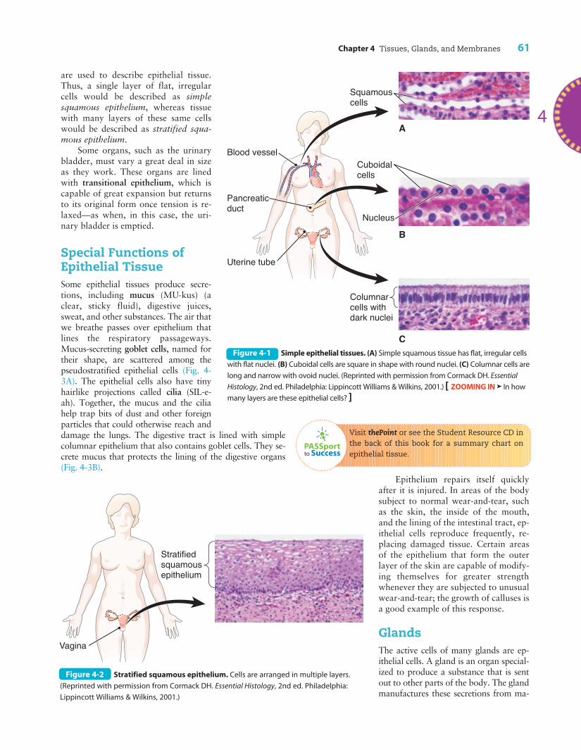
n Squamous (SKWA-mus)—flat and irregular
n Cuboidal—square
n Columnar—long and narrow
The cells may be arranged in a single layer, in whichcase it is described as simple (Fig. 4-1). Simple epitheliumfunctions as a thin barrier through which materials canpass fairly easily. For example, simple epithelium allowsfor absorption of materials from the lining of the digestivetract into the blood and allows for passage of oxygen fromthe blood to body tissues. Areas subject to wear-and-tearthat require protection are covered with epithelial cells inmultiple layers, an arrangement described as stratified(Fig. 4-2). If the cells are staggered so that they appear tobe in multiple layers but really are not, they are termedpseudostratified. Terms for both shape and arrangement
Box 4-1
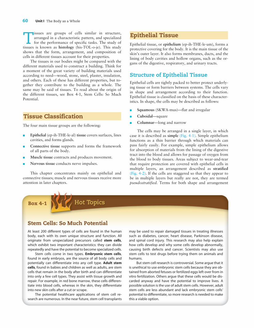
Stem Cells: So Much PotentialAt least 200 different types of cells are found in the humanbody, each with its own unique structure and function. Alloriginate from unspecialized precursors called stem cells,which exhibit two important characteristics: they can dividerepeatedly and have the potential to become specialized cells.
Stem cells come in two types. Embryonic stem cells,found in early embryos, are the source of all body cells andpotentially can differentiate into any cell type. Adult stemcells, found in babies and children as well as adults, are stemcells that remain in the body after birth and can differentiateinto only a few cell types. They assist with tissue growth andrepair. For example, in red bone marrow, these cells differen-tiate into blood cells, whereas in the skin, they differentiateinto new skin cells after a cut or scrape.
The potential healthcare applications of stem cell re-search are numerous. In the near future, stem cell transplants
may be used to repair damaged tissues in treating illnessessuch as diabetes, cancer, heart disease, Parkinson disease,and spinal cord injury. This research may also help explainhow cells develop and why some cells develop abnormally,causing birth defects and cancer. Scientists may also usestem cells to test drugs before trying them on animals andhumans.
But stem cell research is controversial. Some argue that itis unethical to use embryonic stem cells because they are ob-tained from aborted fetuses or fertilized eggs left over from invitro fertilization. Others argue that these cells would be dis-carded anyway and have the potential to improve lives. Apossible solution is the use of adult stem cells. However, adultstem cells are less abundant and lack embryonic stem cells’potential to differentiate, so more research is needed to makethis a viable option.
Chapter 4 Tissues, Glands, and Membranes 61
4
are used to describe epithelial tissue.Thus, a single layer of flat, irregularcells would be described as simplesquamous epithelium, whereas tissuewith many layers of these same cellswould be described as stratified squa-mous epithelium.
Some organs, such as the urinarybladder, must vary a great deal in sizeas they work. These organs are linedwith transitional epithelium, which iscapable of great expansion but returnsto its original form once tension is re-laxed—as when, in this case, the uri-nary bladder is emptied.
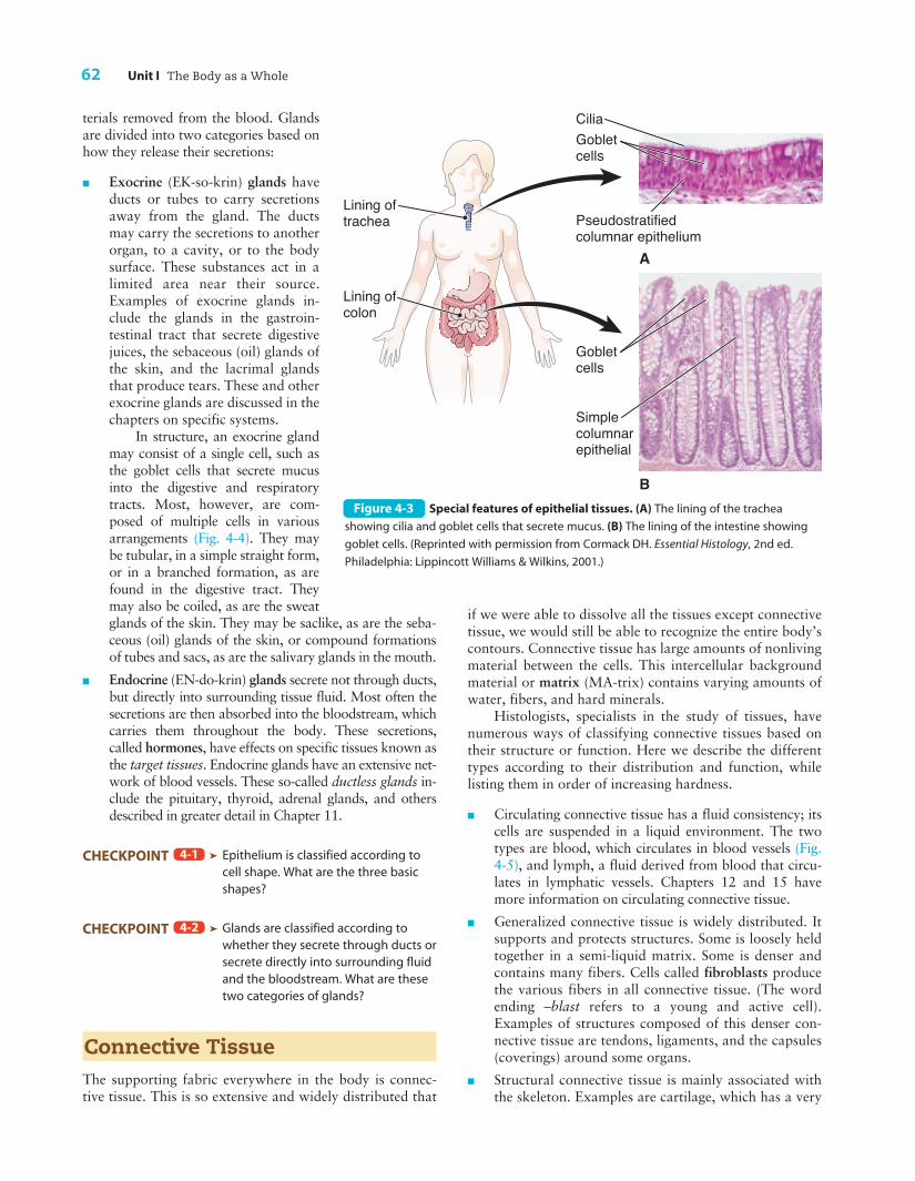
Special Functions ofEpithelial TissueSome epithelial tissues produce secre-tions, including mucus (MU-kus) (aclear, sticky fluid), digestive juices,sweat, and other substances. The air thatwe breathe passes over epithelium thatlines the respiratory passageways.Mucus-secreting goblet cells, named fortheir shape, are scattered among thepseudostratified epithelial cells (Fig. 4-3A). The epithelial cells also have tinyhairlike projections called cilia (SIL-e-ah). Together, the mucus and the ciliahelp trap bits of dust and other foreignparticles that could otherwise reach anddamage the lungs. The digestive tract is lined with simplecolumnar epithelium that also contains goblet cells. They se-crete mucus that protects the lining of the digestive organs(Fig. 4-3B).
PASSportto Success
Visit thePoint or see the Student Resource CD inthe back of this book for a summary chart onepithelial tissue.
Epithelium repairs itself quicklyafter it is injured. In areas of the bodysubject to normal wear-and-tear, suchas the skin, the inside of the mouth,and the lining of the intestinal tract, ep-ithelial cells reproduce frequently, re-placing damaged tissue. Certain areasof the epithelium that form the outerlayer of the skin are capable of modify-ing themselves for greater strengthwhenever they are subjected to unusualwear-and-tear; the growth of calluses isa good example of this response.
GlandsThe active cells of many glands are ep-ithelial cells. A gland is an organ special-ized to produce a substance that is sentout to other parts of the body. The glandmanufactures these secretions from ma-
Blood vessel
Pancreaticduct
Uterine tube
A
B
C
Squamouscells
Columnarcells withdark nuclei
Cuboidalcells
Nucleus
Figure 4-1 Simple epithelial tissues. (A) Simple squamous tissue has flat, irregular cells
with flat nuclei. (B) Cuboidal cells are square in shape with round nuclei. (C) Columnar cells are
long and narrow with ovoid nuclei. (Reprinted with permission from Cormack DH. Essential
Histology, 2nd ed. Philadelphia: Lippincott Williams & Wilkins, 2001.) [ ZOOMING IN ä In how
many layers are these epithelial cells? ]
Vagina
Stratified squamous epithelium
Figure 4-2 Stratified squamous epithelium. Cells are arranged in multiple layers.
(Reprinted with permission from Cormack DH. Essential Histology, 2nd ed. Philadelphia:
Lippincott Williams & Wilkins, 2001.)
62 Unit I The Body as a Whole
terials removed from the blood. Glandsare divided into two categories based onhow they release their secretions:
n Exocrine (EK-so-krin) glands haveducts or tubes to carry secretionsaway from the gland. The ductsmay carry the secretions to anotherorgan, to a cavity, or to the bodysurface. These substances act in alimited area near their source.Examples of exocrine glands in-clude the glands in the gastroin-testinal tract that secrete digestivejuices, the sebaceous (oil) glands ofthe skin, and the lacrimal glandsthat produce tears. These and otherexocrine glands are discussed in thechapters on specific systems.
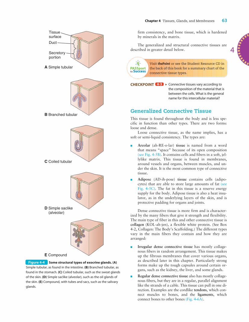
In structure, an exocrine glandmay consist of a single cell, such asthe goblet cells that secrete mucusinto the digestive and respiratorytracts. Most, however, are com-posed of multiple cells in variousarrangements (Fig. 4-4). They maybe tubular, in a simple straight form,or in a branched formation, as arefound in the digestive tract. Theymay also be coiled, as are the sweatglands of the skin. They may be saclike, as are the seba-ceous (oil) glands of the skin, or compound formationsof tubes and sacs, as are the salivary glands in the mouth.
n Endocrine (EN-do-krin) glands secrete not through ducts,but directly into surrounding tissue fluid. Most often thesecretions are then absorbed into the bloodstream, whichcarries them throughout the body. These secretions,called hormones, have effects on specific tissues known asthe target tissues. Endocrine glands have an extensive net-work of blood vessels. These so-called ductless glands in-clude the pituitary, thyroid, adrenal glands, and othersdescribed in greater detail in Chapter 11.
CHECKPOINT ä Epithelium is classified according tocell shape. What are the three basicshapes?
CHECKPOINT ä Glands are classified according towhether they secrete through ducts orsecrete directly into surrounding fluidand the bloodstream. What are thesetwo categories of glands?
Connective TissueThe supporting fabric everywhere in the body is connec-tive tissue. This is so extensive and widely distributed that
4-2
4-1
if we were able to dissolve all the tissues except connectivetissue, we would still be able to recognize the entire body’scontours. Connective tissue has large amounts of nonlivingmaterial between the cells. This intercellular backgroundmaterial or matrix (MA-trix) contains varying amounts ofwater, fibers, and hard minerals.
Histologists, specialists in the study of tissues, havenumerous ways of classifying connective tissues based ontheir structure or function. Here we describe the differenttypes according to their distribution and function, whilelisting them in order of increasing hardness.
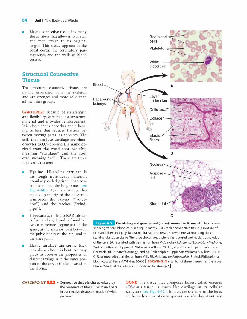
n Circulating connective tissue has a fluid consistency; itscells are suspended in a liquid environment. The twotypes are blood, which circulates in blood vessels (Fig.4-5), and lymph, a fluid derived from blood that circu-lates in lymphatic vessels. Chapters 12 and 15 havemore information on circulating connective tissue.
n Generalized connective tissue is widely distributed. Itsupports and protects structures. Some is loosely heldtogether in a semi-liquid matrix. Some is denser andcontains many fibers. Cells called fibroblasts producethe various fibers in all connective tissue. (The wordending –blast refers to a young and active cell).Examples of structures composed of this denser con-nective tissue are tendons, ligaments, and the capsules(coverings) around some organs.
n Structural connective tissue is mainly associated withthe skeleton. Examples are cartilage, which has a very
Lining of trachea
Lining of colon
CiliaGoblet cells
Pseudostratifiedcolumnar epithelium
Simplecolumnarepithelial
Gobletcells
A
BFigure 4-3 Special features of epithelial tissues. (A) The lining of the trachea
showing cilia and goblet cells that secrete mucus. (B) The lining of the intestine showing
goblet cells. (Reprinted with permission from Cormack DH. Essential Histology, 2nd ed.
Philadelphia: Lippincott Williams & Wilkins, 2001.)
Chapter 4 Tissues, Glands, and Membranes 63
4
Tissue surface
Duct
Secretory portion
A Simple tubular
C Coiled tubular
D Simple saclike (alveolar)
E Compound
B Branched tubular
Figure 4-4 Some structural types of exocrine glands. (A)
Simple tubular, as found in the intestine. (B) Branched tubular, as
found in the stomach. (C) Coiled tubular, such as the sweat glands
of the skin. (D) Simple saclike (alveolar), such as the oil glands of
the skin. (E) Compound, with tubes and sacs, such as the salivary
glands.
PASSportto Success
firm consistency, and bone tissue, which is hardenedby minerals in the matrix.
The generalized and structural connective tissues aredescribed in greater detail below.
Visit thePoint or see the Student Resource CD inthe back of this book for a summary chart of theconnective tissue types.
CHECKPOINT ä Connective tissues vary according tothe composition of the material that isbetween the cells. What is the generalname for this intercellular material?
Generalized Connective TissueThis tissue is found throughout the body and is less spe-cific in function than other types. There are two forms:loose and dense.
Loose connective tissue, as the name implies, has asoft or semi-liquid consistency. The types are:
n Areolar (ah-RE-o-lar) tissue is named from a wordthat means “space” because of its open composition(see Fig. 4-5B). It contains cells and fibers in a soft, jel-lylike matrix. This tissue is found in membranes,around vessels and organs, between muscles, and un-der the skin. It is the most common type of connectivetissue.
n Adipose (AD-ih-pose) tissue contains cells (adipo-cytes) that are able to store large amounts of fat (seeFig. 4-5C). The fat in this tissue is a reserve energysupply for the body. Adipose tissue is also a heat insu-lator, as in the underlying layers of the skin, and isprotective padding for organs and joints.
Dense connective tissue is more firm and is character-ized by the many fibers that give it strength and flexibility.The main type of fiber in this and other connective tissue iscollagen (KOL-ah-jen), a flexible white protein. (See Box4-2, Collagen: The Body’s Scaffolding.) The different typesvary in the main fibers they contain and how they arearranged:
n Irregular dense connective tissue has mostly collage-nous fibers in random arrangement. This tissue makesup the fibrous membranes that cover various organs,as described later in this chapter. Particularly strongforms make up the tough capsules around certain or-gans, such as the kidney, the liver, and some glands.
n Regular dense connective tissue also has mostly collage-nous fibers, but they are in a regular, parallel alignmentlike the strands of a cable. This tissue can pull in one di-rection. Examples are the cordlike tendons, which con-nect muscles to bones, and the ligaments, whichconnect bones to other bones (Fig. 4-6A).
4-3
64 Unit I The Body as a Whole
n Elastic connective tissue has manyelastic fibers that allow it to stretchand then return to its originallength. This tissue appears in thevocal cords, the respiratory pas-sageways, and the walls of bloodvessels.
Structural ConnectiveTissueThe structural connective tissues aremainly associated with the skeletonand are stronger and more solid thanall the other groups.
CARTILAGE Because of its strengthand flexibility, cartilage is a structuralmaterial and provides reinforcement.It is also a shock absorber and a bear-ing surface that reduces friction be-tween moving parts, as at joints. Thecells that produce cartilage are chon-drocytes (KON-dro-sites), a name de-rived from the word root chondro,meaning “cartilage” and the rootcyto, meaning “cell.” There are threeforms of cartilage:
n Hyaline (HI-ah-lin) cartilage isthe tough translucent material,popularly called gristle, that cov-ers the ends of the long bones (seeFig. 4-6B). Hyaline cartilage alsomakes up the tip of the nose andreinforces the larynx (“voice-box”) and the trachea (“wind-pipe”).
n Fibrocartilage (fi-bro-KAR-tih-laj)is firm and rigid, and is found be-tween vertebrae (segments) of thespine, at the anterior joint betweenthe pubic bones of the hip, and inthe knee joint.
n Elastic cartilage can spring backinto shape after it is bent. An easyplace to observe the properties ofelastic cartilage is in the outer por-tion of the ear. It is also located inthe larynx.
CHECKPOINT ä Connective tissue is characterized bythe presence of fibers. The main fibersin connective tissue are made of whatprotein?
4-4 BONE The tissue that composes bones, called osseous(OS-e-us) tissue, is much like cartilage in its cellularstructure (see Fig. 4-6C). In fact, the skeleton of the fetusin the early stages of development is made almost entirely
Blood
Fat aroundkidneys
Layer under skin
Collagen
Cells
B
C
A
Elasticfibers
Red bloodcellsPlatelets
White blood cell
Nucleus
Adipose cell
Stored fat
Figure 4-5 Circulating and generalized (loose) connective tissue. (A) Blood smear
showing various blood cells in a liquid matrix. (B) Areolar connective tissue, a mixture of
cells and fibers in a jellylike matrix. (C) Adipose tissue shown here surrounding dark-
staining glandular tissue. The slide shows areas where fat is stored and nuclei at the edge
of the cells. (A, reprinted with permission from McClatchey KD. Clinical Laboratory Medicine,
2nd ed. Baltimore: Lippincott Williams & Wilkins, 2001; B, reprinted with permission from
Cormack DH. Essential Histology, 2nd ed. Philadelphia: Lippincott Williams & Wilkins, 2001;
C, Reprinted with permission from Mills SE. Histology for Pathologists, 3rd ed. Philadelphia:
Lippincott Williams & Wilkins, 2006.) [ ZOOMING IN ä Which of these tissues has the most
fibers? Which of these tissues is modified for storage? ]
Chapter 4 Tissues, Glands, and Membranes 65
contract by conscious thought. The cells in skeletalmuscle are very large and are remarkable in havingmultiple nuclei and a pattern of dark and light band-ing described as striations. This type of muscle is alsocalled striated muscle. Chapter 7 has more details onskeletal muscles.
n Cardiac muscle, which forms the bulk of the heartwall and is known also as myocardium (mi-o-KAR-de-um) (see Fig. 4-7B). This is the muscle that pro-duces the regular contractions known as heartbeats.Cardiac muscle is described as involuntary muscle be-cause it typically contracts independently of thought.Most of the time we are not aware of its actions at all.Cardiac muscle has branching cells and specializedmembranes between the cells that appear as dark linesunder the microscope. Their technical name is interca-lated (in-TER-cal-a-ted) disks. The heart and cardiacmuscle are discussed in Chapter 13.
n Smooth muscle is also involuntary muscle (see Fig. 4-7C). It forms the walls of the hollow organs in theventral body cavities, including the stomach, intes-tines, gallbladder, and urinary bladder. Togetherthese organs are known as viscera (VIS-eh-rah), sosmooth muscle is sometimes referred to as visceralmuscle. Smooth muscle is also found in the walls ofmany tubular structures, such as the blood vesselsand the tubes that carry urine from the kidneys. Asmooth muscle is attached to the base of each bodyhair. Contraction of these muscles causes the condi-tion of the skin that we call gooseflesh. Smoothmuscle cells are of a typical size and taper at eachend. They are not striated and have only one nu-
4
of cartilage. This tissue gradually becomes impregnatedwith salts of calcium and phosphorus that make bonecharacteristically solid and hard. The cells that formbone are called osteoblasts (OS-te-o-blasts), a name thatcombines the root for bone (osteo) with a root (blast)that means an immature cell. As these cells mature, theyare referred to as osteocytes (OS-te-o-sites). Within theosseous tissue are nerves and blood vessels. A specializedtype of tissue, the bone marrow, is enclosed withinbones. The red bone marrow contained in certain regionsproduces blood cells. Chapter 6 has more information onbones.
CHECKPOINT ä Connective tissue is the supportiveand protective material foundthroughout the body. What are someexamples of circulating, generalized,and structural connective tissue?
Muscle TissueMuscle tissue is designed to produce movement by con-traction of its cells, which are called muscle fibers becausemost of them are long and threadlike. If a piece of well-cooked meat is pulled apart, small groups of these musclefibers may be seen. Muscle tissue is usually classified asfollows:
n Skeletal muscle, which works with tendons and bonesto move the body (Fig. 4-7A). This type of tissue is de-scribed as voluntary muscle because it can be made to
4-5
Box 4-2
Collagen: The Body’s ScaffoldingThe most abundant protein in the body, making up about25% of total protein, is collagen. Its name, derived from aGreek word meaning “glue,” reveals its role as the main struc-tural protein in connective tissue.
Fibroblasts secrete collagen molecules into the surround-ing matrix, where the molecules are then assembled into fibers.These fibers give the matrix its strength and its flexibility.Collagen fibers’ high tensile strength makes them strongerthan steel fibers of the same size, and their flexibility confers re-silience on the tissues that contain them. For example, collagenin skin, bone, tendons, and ligaments resists pulling forces,whereas collagen found in joint cartilage and between verte-brae resists compression. Based on amino acid structure, thereare at least 19 types of collagen, each of which imparts a differ-ent property to the connective tissue containing it.
The arrangement of collagen fibers in the matrix revealsmuch about the tissue’s function. In the skin and membranescovering muscles and organs, collagen fibers are arranged irreg-ularly, with fibers running in all directions. The result is a tissuethat can resist stretching forces in many different directions. Intendons and ligaments, collagen fibers have a parallel arrange-ment, forming strong ropelike cords that can resist longitudinalpulling forces. In bone tissue, collagen fibers’ meshlike arrange-ment promotes deposition of calcium salts into the tissue,which gives bone strength while also providing flexibility.
Collagen’s varied properties are also evident in thepreparation of a gelatin dessert. Gelatin is a collagen extractmade by boiling animal bones and other connective tissue. Itis a viscous liquid in hot water but forms a semisolid gel oncooling.
66 Unit I The Body as a Whole
cleus per cell. Structures containing smooth muscleare discussed in the chapters on the various bodysystems.
Muscle tissue, like nervous tissue, repairs itself onlywith difficulty or not at all once an injury has been sus-tained. When injured, muscle tissue is frequently replacedwith connective tissue.
CHECKPOINT ä What are the three types of muscletissue?
Nervous TissueThe human body is made up of countless structures, eachof which contributes to the action of the whole organism.This aggregation of structures might be compared to alarge corporation. For all the workers in the corporationto coordinate their efforts, there must be some centralcontrol, such as the president or CEO. In the body, thiscentral agent is the brain (Fig. 4-8A). Each structure is in
4-6
direct communication with the brainby means of its own set of “wires,”called nerves (see Fig. 4-8B). Nervesfrom even the most remote parts ofthe body come together and feed intoa great trunk cable called the spinalcord, which in turn leads into the cen-tral switchboard of the brain. Here,messages come in and orders go out24 hours a day. Some nerves, the cra-nial nerves, connect directly with thebrain and do not communicate withthe spinal cord. This entire controlsystem, including the brain, is made ofnervous tissue.
The NeuronThe basic unit of nervous tissue is theneuron (NU-ron), or nerve cell (see Fig.4-8C). A neuron consists of a nerve cellbody plus small branches from the cellcalled fibers. One type of fiber, the den-drite (DEN-drite), is generally shortand forms treelike branches. This typeof fiber carries messages in the form ofnerve impulses to the nerve cell body. Asingle fiber, the axon (AK-son), carriesimpulses away from the nerve cellbody. Neurons may be quite long; theirfibers can extend for several feet. Anerve is a bundle of such nerve cellfibers held together with connective tis-sue (see Fig. 4-8B).
Just as wires are insulated to keepthem from being short-circuited, someaxons are insulated and protected by a
coating of material called myelin (MI-eh-lin). Groups ofmyelinated fibers form “white matter,” named for thecolor of the myelin, which is much like fat in appearanceand consistency.
Not all neurons have myelin, however; some axonsare unmyelinated, as are all dendrites and all cell bodies.These areas appear gray in color. Because the outer layerof the brain has large collections of cell bodies and un-myelinated fibers, the brain is popularly termed gray mat-ter, even though its interior is composed of white matter(see Fig. 4-8A).
NeurogliaNervous tissue is supported and protected by specializedcells known as neuroglia (nu-ROG-le-ah) or glial (GLI-al)cells, which are named from the Greek word glia meaning“glue.” Some of these cells protect the brain from harmfulsubstances; others get rid of foreign organisms and cellulardebris; still others form the myelin sheath around axons.They do not, however, transmit nerve impulses.
Fibroblasts
Collagen
Chondrocytes(cartilage cells)
Spaces forosteocytes(bone cells)Channel(for nervesand bloodvessels)
CartilageTendon
Bone
Matrix
A B
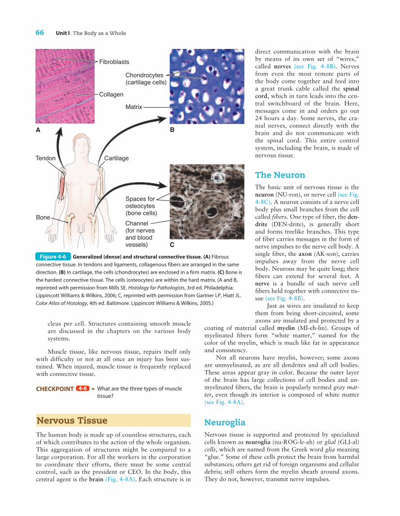
CFigure 4-6 Generalized (dense) and structural connective tissue. (A) Fibrous
connective tissue. In tendons and ligaments, collagenous fibers are arranged in the same
direction. (B) In cartilage, the cells (chondrocytes) are enclosed in a firm matrix. (C) Bone is
the hardest connective tissue. The cells (osteocytes) are within the hard matrix. (A and B,
reprinted with permission from Mills SE. Histology for Pathologists, 3rd ed. Philadelphia:
Lippincott Williams & Wilkins, 2006; C, reprinted with permission from Gartner LP, Hiatt JL.
Color Atlas of Histology, 4th ed. Baltimore: Lippincott Williams & Wilkins, 2005.)
Chapter 4 Tissues, Glands, and Membranes 67
A more detailed discussion of nervous tissue and thenervous system can be found in Chapters 8 and 9.
CHECKPOINT ä What is the basic cellular unit of thenervous system and what is itsfunction?
CHECKPOINT ä What are the nonconducting supportcells of the nervous system called?
4-8
4-7
MembranesMembranes are thin sheets of tissue.Their properties vary: some are fragile,others tough; some are transparent,others opaque (i.e., they cannot be seenthrough). Membranes may cover a sur-face, may be a dividing partition, mayline a hollow organ or body cavity, ormay anchor an organ. They may con-tain cells that secrete lubricants to easethe movement of organs, such as theheart and lung, and the movement ofjoints. Epithelial membranes and con-nective tissue membranes are describedbelow.
Epithelial MembranesAn epithelial membrane is so namedbecause its outer surface is made of ep-ithelium. Underneath, however, thereis a layer of connective tissue thatstrengthens the membrane, and insome cases, there is a thin layer ofsmooth muscle under that. Epithelialmembranes are made of closely packedactive cells that manufacture lubricantsand protect the deeper tissues from in-vasion by microorganisms. Epithelialmembranes are of several types:
n Serous (SE-rus) membranes line thewalls of body cavities and are foldedback onto the surface of internal or-gans, forming their outermost layer.
n Mucous (MU-kus) membranes linetubes and other spaces that open tothe outside of the body.
n The cutaneous (ku-TA-ne-us) mem-brane, commonly known as the skin,has an outer layer of epithelium.This membrane is complex and isdiscussed in detail in Chapter 5.
SEROUS MEMBRANES Serous mem-branes line the closed ventral body cavities and do notconnect with the outside of the body. They secrete a thin,watery lubricant, known as serous fluid, that allows or-gans to move with a minimum of friction. The thin epithe-lium of serous membranes is a smooth, glistening kind oftissue called mesothelium (mes-o-THE-le-um). The mem-brane itself may be referred to as the serosa (se-RO-sah).
There are three serous membranes:
n The pleurae (PLU-re), or pleuras (PLU-rahs), line thethoracic cavity and cover each lung.
4
A
B
C
Striations
Connectivetissue
Nuclei
Nuclei
Nuclei
Intercalateddisks
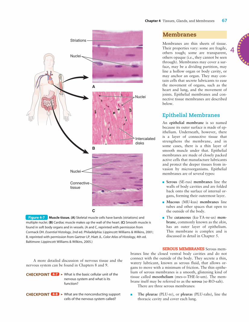
Figure 4-7 Muscle tissue. (A) Skeletal muscle cells have bands (striations) and
multiple nuclei. (B) Cardiac muscle makes up the wall of the heart. (C) Smooth muscle is
found in soft body organs and in vessels. (A and C, reprinted with permission from
Cormack DH. Essential Histology, 2nd ed. Philadelphia: Lippincott Williams & Wilkins, 2001;
B, reprinted with permission from Gartner LP, Hiatt JL. Color Atlas of Histology, 4th ed.
Baltimore: Lippincott Williams & Wilkins, 2005.)
68 Unit I The Body as a Whole
n The serous pericardium (per-ih-KAR-de-um) formspart of a sac that encloses the heart, which is locatedin the chest between the lungs.
n The peritoneum (per-ih-to-NE-um) is the largestserous membrane. It lines the walls of the abdominalcavity, covers the abdominal organs, and forms sup-porting and protective structures within the abdomen(see Fig. 17-3 in Chapter 17).
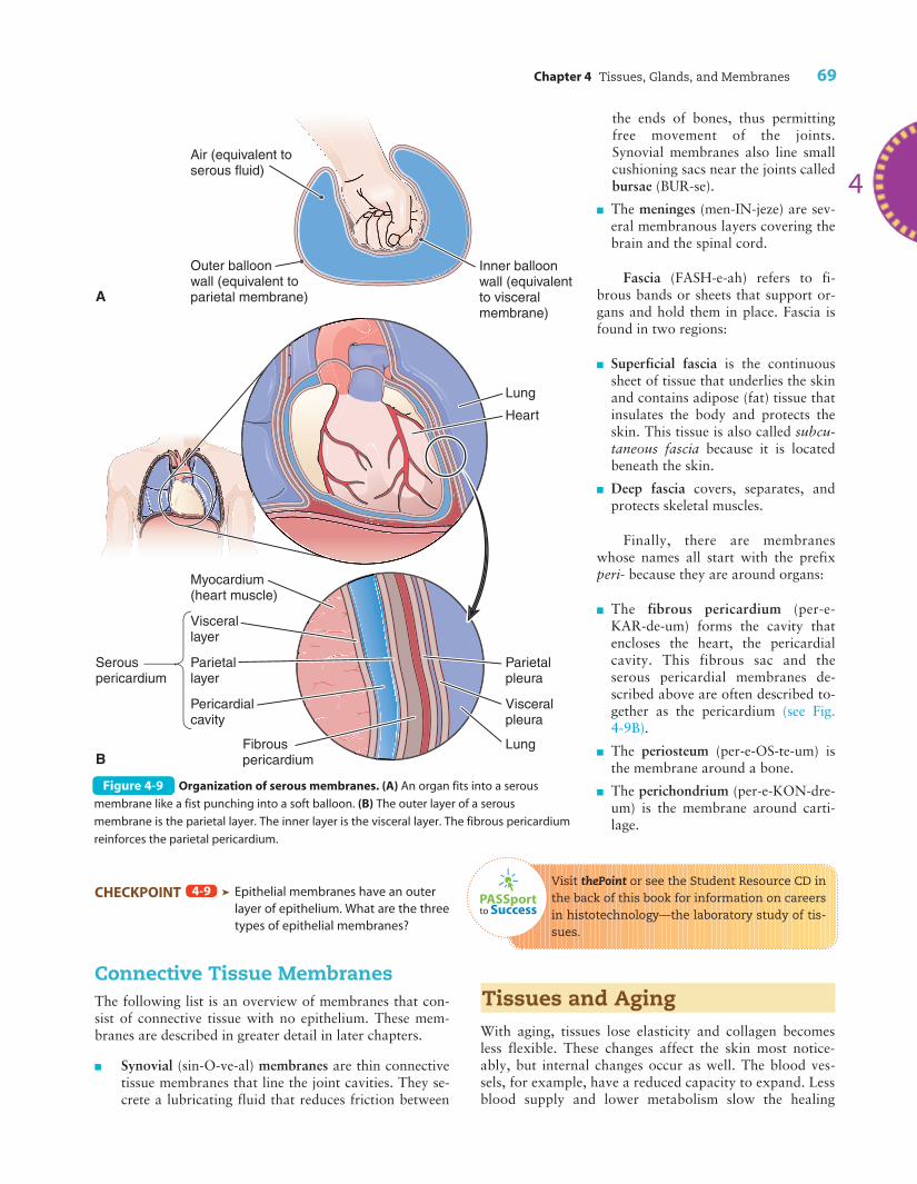
Serous membranes are arranged so that one portionforms the lining of a closed cavity, while another part foldsback to cover the surface of the organ contained in thatcavity. The relationship between an organ and the serousmembrane around it can be visualized by imagining yourfist punching into a large, soft balloon (Fig. 4-9). Your fistis the organ and the serous membrane around it is in twolayers, one against your fist and one folded back to forman outer layer. Although in two layers, each serous mem-brane is continuous.
The portion of the serous membrane attached to thewall of a cavity or sac is known as the parietal (pah-RI-eh-tal) layer; the word parietal refers to a wall. In the exampleabove, the parietal layer is represented by the outermostlayer of the balloon. Parietal pleura lines the thoracic(chest) cavity, and parietal pericardium lines the fibrous
sac (the fibrous pericardium) that en-closes the heart (see Fig. 4-9).
Because internal organs are calledviscera, the portion of the serous mem-brane attached to an organ is the vis-ceral layer. Visceral pericardium is onthe surface of the heart, and each lungsurface is covered by visceral pleura.Portions of the peritoneum that coverorgans in the abdomen are named ac-cording to the particular organ in-volved. The visceral layer in ourballoon example is in direct contactwith your fist.
A serous membrane’s visceral andparietal layers normally are in directcontact with a minimal amount of lu-bricant between them. The area be-tween the two layers forms a potentialspace. That is, it is possible for a spaceto exist there, although normally onedoes not. Only if substances accumu-late between the layers, as when in-flammation causes the production ofexcessive amounts of fluid, is there anactual space.
MUCOUS MEMBRANES Mucousmembranes are so named because theyproduce a thick and sticky substancecalled mucus (MU-kus). (Note that theadjective mucous contains an “o,”whereas the noun mucus does not.)These membranes form extensive con-
tinuous linings in the digestive, respiratory, urinary, andreproductive systems, all of which are connected with theoutside of the body. These membranes vary somewhat inboth structure and function. The cells that line the nasalcavities and the respiratory passageways are suppliedwith tiny, hairlike extensions called cilia, described inChapter 3. The microscopic cilia move in waves thatforce secretions outward. In this way, foreign particles,such as bacteria, dust, and other impurities trapped inthe sticky mucus, are prevented from entering the lungsand causing harm. Ciliated epithelium is also found incertain tubes of both the male and the female reproduc-tive systems.
The mucous membranes that line the digestive tracthave special functions. For example, the stomach’s mucousmembrane protects its deeper tissues from the action ofpowerful digestive juices. If for some reason a portion ofthis membrane is injured, these juices begin to digest a partof the stomach itself—as happens in cases of peptic ulcers.Mucous membranes located farther along in the digestivesystem are designed to absorb nutrients, which the bloodthen transports to all cells.
The noun mucosa (mu-KO-sah) refers to the mucousmembrane of an organ.
C
B
A
Gray matter
White matter
Neuron fibers
Nucleolusin nucleus
Bundles ofneuron fibers
Cell body
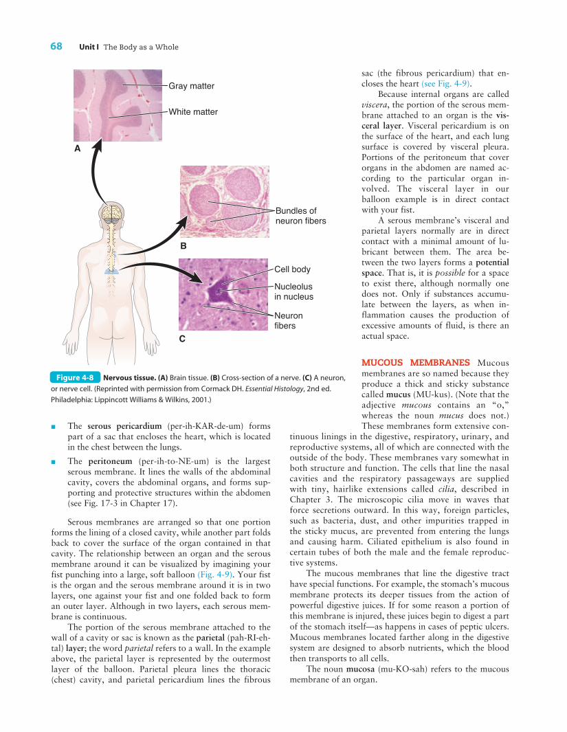
Figure 4-8 Nervous tissue. (A) Brain tissue. (B) Cross-section of a nerve. (C) A neuron,
or nerve cell. (Reprinted with permission from Cormack DH. Essential Histology, 2nd ed.
Philadelphia: Lippincott Williams & Wilkins, 2001.)
Chapter 4 Tissues, Glands, and Membranes 69
CHECKPOINT ä Epithelial membranes have an outerlayer of epithelium. What are the threetypes of epithelial membranes?
Connective Tissue MembranesThe following list is an overview of membranes that con-sist of connective tissue with no epithelium. These mem-branes are described in greater detail in later chapters.
n Synovial (sin-O-ve-al) membranes are thin connectivetissue membranes that line the joint cavities. They se-crete a lubricating fluid that reduces friction between
4-9
4
PASSportto Success
the ends of bones, thus permittingfree movement of the joints.Synovial membranes also line smallcushioning sacs near the joints calledbursae (BUR-se).
n The meninges (men-IN-jeze) are sev-eral membranous layers covering thebrain and the spinal cord.
Fascia (FASH-e-ah) refers to fi-brous bands or sheets that support or-gans and hold them in place. Fascia isfound in two regions:
n Superficial fascia is the continuoussheet of tissue that underlies the skinand contains adipose (fat) tissue thatinsulates the body and protects theskin. This tissue is also called subcu-taneous fascia because it is locatedbeneath the skin.
n Deep fascia covers, separates, andprotects skeletal muscles.
Finally, there are membraneswhose names all start with the prefixperi- because they are around organs:
n The fibrous pericardium (per-e-KAR-de-um) forms the cavity thatencloses the heart, the pericardialcavity. This fibrous sac and theserous pericardial membranes de-scribed above are often described to-gether as the pericardium (see Fig.4-9B).
n The periosteum (per-e-OS-te-um) isthe membrane around a bone.
n The perichondrium (per-e-KON-dre-um) is the membrane around carti-lage.
Visit thePoint or see the Student Resource CD inthe back of this book for information on careersin histotechnology—the laboratory study of tis-sues.
Tissues and AgingWith aging, tissues lose elasticity and collagen becomesless flexible. These changes affect the skin most notice-ably, but internal changes occur as well. The blood ves-sels, for example, have a reduced capacity to expand. Lessblood supply and lower metabolism slow the healing
Figure 4-9 Organization of serous membranes. (A) An organ fits into a serous
membrane like a fist punching into a soft balloon. (B) The outer layer of a serous
membrane is the parietal layer. The inner layer is the visceral layer. The fibrous pericardium
reinforces the parietal pericardium.
70 Unit I The Body as a Whole
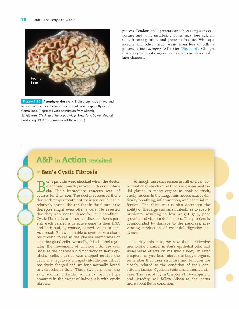
process. Tendons and ligaments stretch, causing a stoopedposture and joint instability. Bones may lose calciumsalts, becoming brittle and prone to fracture. With age,muscles and other tissues waste from loss of cells, aprocess termed atrophy (AT-ro-fe) (Fig. 4-10). Changesthat apply to specific organs and systems are described inlater chapters.
Frontal lobe
Figure 4-10 Atrophy of the brain. Brain tissue has thinned and
larger spaces appear between sections of tissue, especially in the
frontal lobe. (Reprinted with permission from Okazaki H,
Scheithauer BW. Atlas of Neuropathology. New York: Gower Medical
Publishing, 1988. By permission of the author.)
äBen’s Cystic Fibrosis
Ben’s parents were shocked when the doctordiagnosed their 2-year-old with cystic fibro-sis. Their immediate concern was, of
course, for their son. The doctor reassured themthat with proper treatment their son could lead arelatively normal life and that in the future, newtherapies might even offer a cure. He assertedthat they were not to blame for Ben’s condition.Cystic fibrosis is an inherited disease—Ben’s par-ents each carried a defective gene in their DNAand both had, by chance, passed copies to Ben.As a result, Ben was unable to synthesize a chan-nel protein found in the plasma membranes ofexocrine gland cells. Normally, this channel regu-lates the movement of chloride into the cell.Because the channels did not work in Ben’s ep-ithelial cells, chloride was trapped outside thecells. The negatively charged chloride ions attractpositively charged sodium ions normally foundin extracellular fluid. These two ions form thesalt, sodium chloride, which is lost in highamounts in the sweat of individuals with cysticfibrosis.
Although the exact reason is still unclear, ab-normal chloride channel function causes epithe-lial glands in many organs to produce thick,sticky mucus. In the lungs, this mucus causes dif-ficulty breathing, inflammation, and bacterial in-fection. The thick mucus also decreases theability of the large and small intestines to absorbnutrients, resulting in low weight gain, poorgrowth, and vitamin deficiencies. This problem iscompounded by damage to the pancreas, pre-venting production of essential digestive en-zymes.
During this case, we saw that a defectivemembrane channel in Ben’s epithelial cells hadwidespread effects on his whole body. In laterchapters, as you learn about the body’s organs,remember that their structure and function areclosely related to the condition of their con-stituent tissues. Cystic fibrosis is an inherited dis-ease. The case study in Chapter 21, Developmentand Heredity, will follow Alison as she learnsmore about Ben’s condition.
Chapter 4 Tissues, Glands, and Membranes 71
Word Anatomy
Medical terms are built from standardized word parts (prefixes, roots, and suffixes). Learning the meanings of these parts can help you remember words and interpret unfamiliar terms.
WORD PART MEANING EXAMPLE
hist/o tissue Histology is the study of tissues.
Epithelial Tissue
epi- on, upon Epithelial tissue covers body surfaces.pseud/o- false Pseudostratified epithelium appears to be in multiple layers
but is not.
Connective Tissue
-blast immature cell, early A fibroblast is a cell that produces fibers.stage of cell
chondr/o cartilage A chondrocyte is a cartilage cell.oss, osse/o bone, bone tissue Osseous tissue is bone tissue.oste/o bone, bone tissue An osteocyte is a mature bone cell.
Muscle Tissue
my/o muscle The myocardium is the heart muscle. cardi/o heart See above example.
Nervous Tissue
neur/o nerve, nervous system A neuron is a nerve cell.
Membranes
pleur/o side, rib The pleurae are membranes that line the chest cavity.peri- around The peritoneum wraps around the abdominal organs.
4
I. TISSUE CLASSIFICATION—epithelial tissue, connectivetissue, muscle tissue, nervous tissue
II. EPITHELIAL TISSUE—covers surfaces; lines cavities,organs, and ductsA. Structure of epithelial tissue
1. Cells—squamous, cuboidal, columnar2. Arrangement—simple or stratified
B. Special functions1. Produces secretions (e.g., mucus, digestive
juices, sweat)2. Filters impurities using cilia
C. Glands—active cells are epithelial cells1. Exocrine
a. Secrete through ductsb. Examples: digestive glands, tear glands,
sweat and oil glands of skin
2. Endocrinea. Secrete into body fluids and bloodstreamb. Produce hormones
III. CONNECTIVE TISSUE—supports, binds, forms frame-work of bodyA. Circulating—fluid matrix; travels in vessels
1. Blood2. Lymph
B. Generalized—widely distributed; not specialized1. Loose—cells and fibers in semiliquid matrix
a. Areolar—in membranes, around vesselsand organs, under skin
b. Adipose—stores fat; insulation, padding,energy reserve
2. Dense—has many fibers (e.g. collagenous,elastic) made by fibroblasts
Summary
72 Unit I The Body as a Whole
a. Irregular—fibers not organized; in mem-branes, capsules
b. Regular—fibers in parallel alignment; intendons, ligaments
c. Elastic—fibers can stretch and return toshape; in vocal cords, respiratory passage-ways, blood vessel walls
C. Structural—mainly associated with skeleton1. Cartilage
a. Strong and flexibleb. Cushions and absorbs shockc. Produced by chondrocytesd. Types
(1) Hyaline—covers ends of bones, makesup tip of nose, reinforces larynx andtrachea
(2) Fibrocartilage—in certain joints(3) Elastic—in outer ear, larynx
2. Bonea. Matrix contains mineral saltsb. Cells
(1) Osteoblasts—produce bone(2) Osteocytes—mature bone cells
IV. MUSCLE TISSUE—contracts to produce movementA. Skeletal muscle—voluntary; moves skeletonB. Cardiac muscle—involuntary; forms main part of
heartC. Smooth muscle—involuntary; forms visceral
organsV. NERVOUS TISSUE
A. Neuron—nerve cell1. Cell body—contains nucleus2. Dendrite—fiber carrying impulses toward cell
body
3. Axon—fiber carrying impulses away from cellbodya. Myelin—fatty material that insulates some
axons(1) Myelinated fibers—make up white
matter(2) Unmyelinated cells and fibers—make
up gray matterB. Neuroglia—support and protect nervous tissue
VI. MEMBRANES—thin sheets of tissueA. Epithelial membranes—outer layer epithelium
1. Serous membrane—secretes watery fluida. Parietal layer—lines body cavityb. Visceral layer—covers internal organsc. Examples—pleurae, pericardium, peri-
toneum2. Mucous membrane
a. Secretes mucusb. Lines tube or space that opens to the out-
side (e.g., respiratory, digestive, reproduc-tive tracts)
3. Cutaneous membrane—skinB. Connective tissue membranes
1. Synovial membrane—lines joint cavity2. Meninges—around brain and spinal cord3. Fascia—under skin and around muscles4. Pericardium—around heart; periosteum—
around bone; perichondrium—around cartilage
VII. TISSUES AND AGING
Questions for Study and Review
BUILDING UNDERSTANDING
Fill in the blanks
1. A group of similar cells arranged in a characteristic pat-tern is called a(n) ______.
2. Glands that secrete their products directly into the bloodare called ______ glands.
3. Tissue that supports and forms the framework of thebody is called ______ tissue.
4. Skeletal muscle is also described as ______ muscle.
5. Nerve tissue is supported by specialized cells known as______.
Chapter 4 Tissues, Glands, and Membranes 73
4
Matching > Match each numbered item with the most closely related lettered item.
___ 6. Membrane around the heart
___ 7. Membrane around each lung
___ 8. Membrane around bone
___ 9. Membrane around cartilage
___ 10. Membrane around abdominal organs
a. perichondrium
b. pericardium
c. peritoneum
d. periosteum
e. pleura
Multiple Choice
___ 11. Epithelium composed of a single layer of long andnarrow cells is called
a. simple cuboidal epitheliumb. simple columnar epitheliumc. stratified cuboidal epitheliumd. stratified columnar epithelium
___ 12. Tendons and ligaments are examples of
a. areolar connective tissueb. loose connective tissuec. regular, dense connective tissued. cartilage
___ 13. A tissue composed of long striated cells with multi-ple nuclei is
a. smooth muscle tissueb. cardiac muscle tissuec. skeletal muscle tissued. nervous tissue
___ 14. A bundle of nerve cell fibers held together with con-nective tissue is called a(n)
a. dendriteb. axonc. nerved. myelin
___ 15. All of the following are types of epithelial mem-branes except
a. cutaneous membraneb. mucous membranec. serous membraned. synovial membrane
UNDERSTANDING CONCEPTS
16. Explain how epithelium is classified and discuss at leastthree functions of this tissue type.
17. Compare the structure and function of exocrine and en-docrine glands and give two examples of each type.
18. Describe the functions of connective tissue. Name twokinds of fibers found in connective tissue and discusshow their presence affects tissue function.
19. Compare and contrast the three different types of muscletissue.
20. Compare serous and mucous membranes.
CONCEPTUAL THINKING
21. Prolonged exposure to cigarette smoke causes damage tociliated epithelium that lines portions of the respiratorytract. Discuss the implications of this damage.
22. The middle ear is connected to the throat by a tube calledthe eustachian (auditory) tube. All are lined by a continu-ous mucous membrane. Using this information, describewhy a throat infection (pharyngitis) may lead to an earinfection (otitis media).
23. In cystic fibrosis, the production of abnormally thick stickymucus results in lung and digestive disorders. What aresome of the normal functions of mucus in the body?
24. In Ben’s case, an abnormal epithelial channel protein hadwidespread effects. Another hereditary disease, osteogene-sis imperfecta, is characterized by abnormal collagen fibersynthesis. Which tissue type would be most affected by thisdisorder? List some possible symptoms of this disease.