Embed Size (px)
Citation preview
Thesis for the Master of Science Degree in Molecular Biology Androgen and glucocorticoid receptor mediated changes in histone acetylation at the MMTV promoter Martina Tesikova
Department of Molecular Biosciences
The Faculty of Mathematics and Natural Sciences UNIVERSITY OF OSLO
August 2008

TABLE OF CONTENTS
ACKNOWLEDGEMENTS……………………………………………………...............4
ABBREVIATIONS…………………………………………………………….................5
GENERAL INTRODUCTION..……………………………………………………….. 6 1. Androgens……………….………………………………………………...................6
2. The Androgen Receptor………………………………………………………….7
2.1. Structural features of AR……………………………………………….........8 2.1.1. Domain structure…………………………………………………………...8
The NTD or A/B domain….………………………………………………..8
The DBD domain….…..………………………….………………………10
The hinge region….…….…………………………………………….......10
The LBD domain………………………………………………………….10
2.1.2. Post-translational modifications…………………………………………. 11
2.2. AR transcriptional activation and regulation…………………………... 12 2.2.1. AR coregulators….………………………………………………………..14
Coactivators...……………………………………………………………16
Corepressors…………………………………………………………….. 17
2.2.2. AR and specific transcription factors……………………………………..18
3. Glucocorticoid Receptor……………………………………...............................19
3.1. GR transcriptional activation and regulation…………………………….19
4. Nuclear receptor dynamics…….………………………………………………..20
4.1. Transcriptional activation of NRs…………………………………………20 4.1.1. The classic model………....……………………………………………….21
4.1.2. The dynamic model……………………………………………………….22
4.1.3. The “static” versus “dynamic” view………...……………………………25
4.2. Mehanisms contributing to NR mobility…………………………………25 4.2.1. Ligand-specific dynamics of NRs…………………………………………26

TABLE OF CONTENTS
4.2.2. Chromatin remodelling and NRs dynamics……………………………….26
ATP-dependent chromatin remodelling complexes………………………27
Histone modifications……………………………………………………..28
Histone acetylation ……………………………………………………...29
Histone deacetylation and HDAC inhibitors………………………………...30
5. Aim of the study…………………………………………………………………...31
MANUSCRIPT…………………………………………………………………………...32
1. Summary…..……………..………………………………………………………….32
2. Introduction………………………………………………………………………...33
3. Materials and Methods…………………………………………………………..36
3.1. Materials………………………………………………………………………..36
3.2. Methods………………………………………………………………………...38 3.2.1. Cell lines and Cell culture……...……………………………………...….38
3.2.2. Protein extraction and Western analysis……………………………...…..39
3.2.3. Live cell microscopy.……...……………………………………………...39
3.2.4. Histone extraction and Western analysis…………………………………40
3.2.5. Chromatin Immunoprecipitation assay (ChIP)…. ……………………….41
DNA concentration measurement..………………………………………42
Chromatin shearing…...…………………………………………………42
Immunoprecipitation……………………………………………………..42
PCR analysis..……………………………………………………………43
3.2.6. Quantitative PCR (qPCR).………………………………………………..44
Data analysis……………………………………………………………..44
4. Results.……………………………………………………………………………….45 4.1. Tetracycline-regulated expression of GFP-AR and GFP-GR……. ….45
4.2. Ligand-dependent translocation of GFP-AR and GFP-GR to the nucleus and their recruitment to the MMTV array…..…………………46
4.3. HDAC inhibitor TSA induces global histone acetylation independently of the presence of hormone…...………………………….47
4.4. ChIP optimization …...……………………………………………………….49

TABLE OF CONTENTS
4.5. Effect of TSA on histone acetylation at the MMTV promoter in the presence of AR ……………………………………………………………….54
4.6. Effect of TSA on histone acetylation at the MMTV promoter in the presence of GR………………………………………………………………..57
5. Discussion and Future Perspectives………………………………………….60
REFERENCES……………………………………………………………………………65

ACKNOWLEDGEMENTS
4
The work presented here was carried out in the laboratory of Professor Fahri
Saatcioglu at the Department of Molecular Biosciences, University of Oslo, from
January 2007 to July 2008.
I wish to express my sincere gratitude to everyone who has contributed to this thesis
work and especially to my supervisor Professor Fahri Saatcioglu for always taking
time to discuss results and experiments with a positive and enthusiastic attitude. A
special thanks for the advice during the writing process and for the inspiration to
never give up. I also want to thank my laboratory supervisor Tove Irene Klokk for
sharing your knowledge on various techniques, answering all my questions, patience,
encouragement through this project and for guidance during the writing. Thanks to
all members of the FS lab for your help, friendship and nice chats about various
matters. Particularly, I am grateful to Mari, Yke and Zeynep for answering my
questions and for giving me technical support. Zeynep, I appreciate your help
whenever a problem occurred and everything you have taught me about ChIP. Thank
you for always being nice, for listening and for being a very good friend! Special
thanks go to Anita Sørensen, a fellow master student, for valuable discussions,
listening and always cheering me up. My great thanks go also to Lise Catherine
Haugstvedt and her family for believing in me and being there for me all the way
from the beginning. Thanks to my friends in Slovakia and Norway for always
supporting me.
Finally, I want to thank my family, specially my parents, grandma and sister Andrea
for all your love, support and patience during the last years.
Oslo, August 2008
Martina Tesikova
ABBREVIATIONS
5
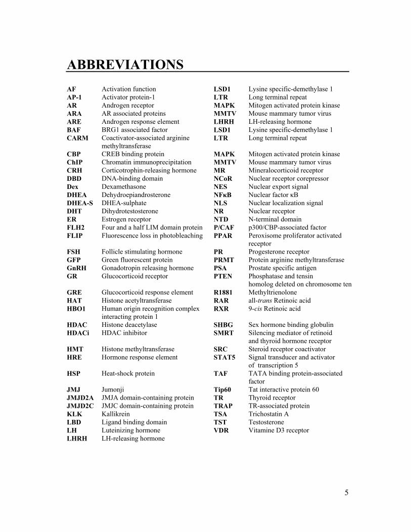
AF Activation function LSD1 Lysine specific-demethylase 1 AP-1 Activator protein-1 LTR Long terminal repeat AR Androgen receptor MAPK Mitogen activated protein kinase ARA AR associated proteins MMTV Mouse mammary tumor virus ARE Androgen response element LHRH LH-releasing hormone BAF BRG1 associated factor LSD1 Lysine specific-demethylase 1 CARM Coactivator-associated arginine
methyltransferase LTR Long terminal repeat
CBP CREB binding protein MAPK Mitogen activated protein kinase ChIP Chromatin immunoprecipitation MMTV Mouse mammary tumor virus CRH Corticotrophin-releasing hormone MR Mineralocorticoid receptor DBD DNA-binding domain NCoR Nuclear receptor corepressor Dex Dexamethasone NES Nuclear export signal DHEA Dehydroepiandrosterone NFκB Nuclear factor κB DHEA-S DHEA-sulphate NLS Nuclear localization signal DHT Dihydrotestosterone NR Nuclear receptor ER Estrogen receptor NTD N-terminal domain FLH2 Four and a half LIM domain protein P/CAF p300/CBP-associated factor FLIP Fluorescence loss in photobleaching PPAR Peroxisome proliferator activated
receptor FSH Follicle stimulating hormone PR Progesterone receptor GFP Green fluorescent protein PRMT Protein arginine methyltransferase GnRH Gonadotropin releasing hormone PSA Prostate specific antigen GR Glucocorticoid receptor PTEN
Phosphatase and tensin homolog deleted on chromosome ten
GRE Glucocorticoid response element R1881 Methyltrienolone HAT Histone acetyltransferase RAR all-trans Retinoic acid HBO1 Human origin recognition complex
interacting protein 1 RXR 9-cis Retinoic acid
HDAC Histone deacetylase SHBG Sex hormone binding globulin HDACi HDAC inhibitor SMRT
Silencing mediator of retinoid and thyroid hormone receptor
HMT Histone methyltransferase SRC Steroid receptor coactivator HRE Hormone response element STAT5
Signal transducer and activator of transcription 5
HSP Heat-shock protein TAF TATA binding protein-associated factor
JMJ Jumonji Tip60 Tat interactive protein 60 JMJD2A JMJA domain-containing protein TR Thyroid receptor JMJD2C JMJC domain-containing protein TRAP TR-associated protein KLK Kallikrein TSA Trichostatin A LBD Ligand binding domain TST Testosterone LH Luteinizing hormone VDR Vitamine D3 receptor LHRH LH-releasing hormone

GENERAL INTRODUCTION
6
1. Androgens Androgens belong to a group of chemically related male sex hormones that are
derived from cholesterol. They are required for the normal development of the penis,
scrotum, testicles and the secondary characteristics of the male body as well as for the
growth and development of prostate. In addition, androgens are implicated in the
initiation and progression of prostate cancer.
Androgens are produced by the Leydig cells in the testis (90%) and by the adrenal
cortex, a small gland located above the kidney. Testosterone (TST), of which more
than 95% is secreted from the testis, is the major circulating androgen in men. The
adrenal cortex and the testis also secrete other androgens, mainly dehydro-
epiandrosterone (DHEA), DHEA sulphate (DHEA-S) and androstenedione. These
hormones have only weak androgenic activity but they are important substrates for
extragonadal synthesis of sex steroids (Labrie et al., 2001; Riggs et al., 2002).
Secretion of testosterone is regulated by the hypothalamic-pituitary-testicular axis.
The hypothalamus secretes locally acting luteinizing hormone-releasing hormone
(LHRH), also known as gonadotropin-releasing hormone (GnRH), and
corticotrophin-releasing hormone (CRH), that act on the pituitary gland. In response
to these hormones, pituitary secretes luteinizing hormone (LH), follicle stimulating
hormone (FSH) and adrenocorticotrophin (ACTH) that enter the circulation and
affect the testis and adrenal glands. While LH acts on the Leydig cells to stimulate
production of testosterone, ACTH stimulates production of adrenal androgens that are
converted into testosterone. When testosterone levels in the bloodstream rise, the
hypothalamus reduces the secretion of LHRH, which inhibits the secretion of LH
from the pituitary gland and further reduces testosterone secretion. Thus, testosterone
controls its own release through a negative feedback on the hypothalamic-pituitary-
testicular axis.

GENERAL INTRODUCTION
7
In the bloodstream, testosterone circulates bound to one of two proteins, either sex
hormone binding globulin (SHBG) or albumin. A small percentage of testosterone,
approximately 2%, remains in a free, unbound form. While free testosterone and
testosterone dissociated from albumin can enter the cell passively by diffusion,
SHGB bound testosterone is transported into the cell actively through the membrane
receptor (Rosner et al., 1999) (see Figure 3). In certain tissues, including the prostate,
testosterone functions as a prohormone, where it is irreversibly converted to
dihydrotestosterone (DHT) by the enzyme 5α-reductase. The biological functions of
androgens are mediated through the androgen receptor (AR). This protein binds both
testosterone and DHT, although it has a much higher affinity for the latter. In contrast
to testosterone, DHT dissociates more slowly from AR and its binding induces a
change in receptor conformation that is more resistant to degradation (Heinlein and
Chang, 2004).
2. The Androgen Receptor AR belongs to a superfamily of proteins that are referred to as nuclear receptors
(NRs). This superfamily of structurally conserved, ligand-dependent transcription
factors comprises more than 150 members that most likely evolved from a common
ancestor (Escriva et al., 2000). Phylogenetic analysis has identified three major
subfamilies within this superfamily, based on their ligand-binding and DNA-binding
properties. AR, together with the estrogen receptor (ER), progesterone receptor (PR),
glucocorticoid receptor (GR), and mineralocorticoid receptor (MR) belong to the
classical steroid receptor subfamily. These NRs undergo nuclear translocation upon
ligand activation and usually bind as homodimers to inverted repeat DNA half sites.
A second subfamily of NRs includes receptors for thyroid hormone (TR), vitamin D3
(VDR), 9-cis retinoic acid (RXR), all-trans retinoic acid (RAR), and peroxisome
proliferators (PPAR). This group of NRs is retained in the nucleus and usually binds
to direct DNA repeats regardless of the presence of ligand. In addition, these

GENERAL INTRODUCTION
8
receptors exhibit promiscuous dimerization patterns, many involving heterodime-
rization with RXR. The majority of NRs identified to date form a third subfamily, so-
called orphan receptors, which share a close structural relationship with receptors for
known hormones but have no known ligands. Although most of them bind DNA as
homodimers on direct repeats, some interact with RXRs while others bind as
monomers to half-site sequences (Wilson et al., 1993; Perlmann and Jansson, 1995).
2.1 Structural features of AR The AR gene is a single-copy gene located on the long (q) arm of the X chromosome
between positions 11.2 and 12 (see Figure 1). It spans approximately 90 kilobases of
genomic DNA. The coding region of the AR gene consist of eight exons separated by
seven introns and encodes a polypeptide product of around 910-919 amino acids that
corresponds to a 110 kDa protein.
2.1.1 Domain structure Like other members of the NR superfamily, the AR contains distinct structural and
functional domains that are conserved among the nuclear receptor family members:
an N-terminal transactivation domain (NTD or A/B), a DNA-binding domain (DBD),
a hinge region, and a C-terminal ligand-binding domain (LBD).
The NTD or A/B domain The N-terminal domain is the most variable both in size and sequence between NRs.
In the case of AR, this domain is encoded by exon 1 that comprises more than half of
the molecule (ca. 559 amino acids). It is involved in the transcriptional activation of
target genes and contains a ligand-independent transactivation region, known as
activation function 1 (AF-1). AF-1 is important for functioning of the full-length AR
as its amino acid substitutions have been identified in patients suffering from
androgen insensitivity and oligospermia (Gottlieb et al., 1999; Hiort et al., 2000). The
NTD also contains three polymorphic direct repeats of glutamine (Glu), proline (Pro),

GENERAL INTRODUCTION
9
and glycine (Gly). Several studies have suggested that the change in the size of
glutamine/glycine repeats alters the function of AR. The expansion of the size of the
polyglutamine segment results in decreased AR transcriptional activity and is related
to impaired spermatogenesis, infertility (Tut et al., 1997) and the spinal and bulbar
muscular atrophy (Kennedy’s disease) (La Spada et al., 1991), whilst shorter
glutamine and/or glycine repeats may be related to prostate cancer incidence
(Edwards et al., 1999; Wang et al., 2004). In addition, the androgen receptor NTD
domain contains two motifs that contribute to intramolecular interactions with the
LBD (He et al., 2000).
Figure 1. Schematic presentation of the AR gene and protein structure AR is coded by a 180-kb gene located on the long arm of the X chromosome (11q11.2). The gene has eight exons (boxes) and seven introns (lines). After transcriptional processing, mRNA is translated into a 919-amino acid-long protein. A number of functional domains are recognized in AR protein: The N-terminal transactivation domain with indicated positions of glutamine (Gln), proline (Pro), glycine (Gly) repeats (arrows) and transactivation function AF1 (line); the central DNA binding domain (DBD); the hinge region (HR) and the nuclear localization signal (NLS); and the C-terminal ligand binding domain with the ligand dependent transactivation function AF-2. Figure modified with permission from The Journal of Clinical Endocrinology & Metabolism (Litvinov et al., 2003); Courtesy of John T. Isaacs, Ph.D.
AF2AF1 AF2AF1
GENERAL INTRODUCTION
10
The DBD domain The DBD domain, encoded by exons 2 and 3, is the best conserved domain among
the members of the nuclear receptor superfamily. It is characterized by a high content
of basic amino acids and by nine invariant cysteine residues, of which eight are
implicated in the formation of two zinc finger motifs. The N-terminal located zinc
finger interacts directly with hormone response elements of target genes in the major
groove of the DNA. The ability to determine the specificity of AR interaction with
DNA resides in three amino acids [Gly; Ser; Val], located in the proximal box (P-
box) at the base of the first zinc finger (Freedman, 1992). The second zinc finger
helps to stabilize DNA receptor interaction and contains a five amino acid-long distal
box (D-box), which participates in forming a dimerization interface for receptor
monomers (Wong et al., 1993). Moreover, the AR DBD contains a non-classical
nuclear export signal (NES) that mediates translocation from the nucleus (Black et
al., 2001).
The hinge region Located between the DBD and the LBD is a non-conserved hinge region, which can
be considered as a flexible linker between the LBD and the rest of the receptor
molecule. The hinge region is important for nuclear localization, containing a ligand-
dependent bipartite nuclear localization signal (NLS) that also spans the C-terminus
of the DBD. The nuclear targeting signal contains the consensus motif KxKK which
is subject to acetylation, thus modulating AR function (Fu et al., 2000). In addition,
the hinge region of all mammalian AR contains a PEST [Pro; Glu; Ser; Thr] rich
sequence, which may function in proteasome mediated androgen receptor turnover
(Sheflin et al., 2000).
The LBD domain The second best conserved region of NRs is the C-terminal hormone binding domain.
This domain is encoded by a portion of exon 4 and exons 5-8, and is responsible for
the specific high-affinity ligand binding. The LBD is formed by 12 conserved α-

GENERAL INTRODUCTION
11
helixes and one β-sheet, together folded into a three-layered, antiparallel helical
sandwich, creating a ligand-binding pocket for accommodation of ligand. Studies
indicate that androgens interact with the LBD mainly through hydrophobic and
hydrogen bonds (Matias et al., 2000). In addition to binding ligand, the LBD is also
involved in dimerization, ligand-dependent coregulator recruitment and interaction of
unliganded receptor with heat-shock protein (HSP) complexes. The LBD contains a
ligand-dependent transactivation domain, known as activation function 2 (AF-2),
which is also involved in interactions with coregulators (Slagsvold et al., 2000) and
intramolecular interaction with the NTD (He et al., 2000).
2.1.2 Post-translational modifications AR undergoes post-translational modifications, such as acetylation, ubiquitylation,
sumoylation and phosphorylation (see Figure 2). These covalent changes may affect
receptor stability, subcellular localisation and interactions with other proteins. Adding
to the complexity, regulatory cross-talk between some of these modifications have
been demonstrated (Fu et al., 2004; Rees et al., 2006).
Figure 2. Schematic representation of the AR post-translational modification sites P: phosphorylation, A: acetylation, U: ubiquitylation, S: sumoylation. U? indicates that the exact ubiquitilation site has not been identified.
AR is a phosphoprotein with multiple phosphorylation sites, mainly in the NTD.
Most of these sites show increased phosphorylation in the presence of androgen, with
the exception of Ser-94, which is constitutively phosphorylated. The kinases involved
1 919
NTD DBD hinge LBD
S16 S81
P
S213S94
PP P P P P PS S PP
S308S256 K386 S424K520
S515 K630K632
S791S650
K633
U?AAA1 919
NTD DBD hinge LBD
S16 S81
P
S213S94
PP P P P P PS S PP
S308S256 K386 S424K520
S515 K630K632
S791S650
K633
U?AAA

GENERAL INTRODUCTION
12
in the phosphorylation of AR and the role of these modifications are still being
debated. However, some studies suggest that MAPKs (Mitogen Activated Protein
Kinases) and Akt play a role (Wen et al., 2000; Gioeli et al., 2006). Concerning
acetylation, three lysines residues located in the hinge region at positions 630, 632
and 633 have been identified as acetylation sites. They play a role in the modulation
of transcriptional activity by favouring nuclear translocation and by balancing
coactivator and corepressor binding (Fu et al., 2002). Furthermore, cross-talk with
phosphorylation has been suggested based on the fact that AR acetylation mutants
change the pattern of AR phosphorylation (Fu et al., 2004). AR, similar to other
steroid receptors, is subject to ubiquitylation. Several of the enzymes involved in this
modification have been identified; however, the exact sites and functional relevance
of this modification are still missing. AR is also postranslationally modified by
sumoylation, namely at K386 and K520 in the NTD (Poukka et al., 2000). AR
sumoylation is hormone dependent and results in mainly repressive effects.
Sumoylation involves SUMO-1-conjugating enzyme Ubc9 that binds AR within the
hinge region (Poukka et al., 1999), raising the possibility of cross-talk between
acetylation and sumoylation.
2.2 AR transcriptional activation and regulation In its unliganded state, AR exists in the cytoplasm in a complex with heat shock
proteins (HSPs) such as Hsp90, Hsp70, and Hsp56. The complex is essential for the
generation of a high-affinity, ligand-binding conformation of AR. Upon ligand
binding, AR undergoes a conformational change, dissociates from HSPs and becomes
hyperphosphorylated. HSP release unmasks the dimerization motifs and NLS site of
the receptor that allows dimerization with another ligand-bound AR and nuclear
translocation. The homodimer translocates to the nucleus where it binds androgen
response elements (AREs) located in the promoters and/or enhancers of various
androgen regulated genes (see Figure 3).

GENERAL INTRODUCTION
13
Figure 3. Schematic presentation of AR transcriptional activation Testosterone (TST) diffuses into the cell or enters the cell through a sex hormone binding globulin (SHBG) receptor. TST is converted to 5α-dihydrotestosterone (DHT) by the enzyme 5α-reductase, and binds to the androgen receptor (AR). AR dissociates from the heat shock protein (HSP) complex, becomes phosphorylated and dimerize with another ligand-bound AR. The homodimer translocates to the nucleus where it binds to androgen response elements (AREs) of target genes, recruits coregulators and factors of the general transcriptional machinery. Transcription of AR target genes, mainly responsible for proliferation and differentiation, is induced. Figure reproduced with permission from (Klokk, 2007); Courtesy of Tove I. Klokk, Ph.D.
Although AR normally functions as a homodimer, it has been found to
heterodimerize with other NRs including ER (Panet-Raymond et al., 2000), GR
(Chen et al., 1997) and TR4 (Lee et al., 1999). The consensus response elements
identified for AR are composed of two palindromic hexanucleotide half sites
separated by a three-nucleotide spacer (AGAACAnnnTGTTCT). Despite the very
different physiological effects of steroids, these AREs are also recognized by GR, PR

GENERAL INTRODUCTION
14
and MR. This paradox may be related to differential recruitment of coregulators upon
the ligand-receptor interaction, although it remains to be further investigated. In
addition, AREs composed of direct repeats and elements with modified site sequence
have also been identified (Zhou et al., 1997; Geserick et al., 2005). After binding to
ARE, AR initiates transcription of target genes through the recruitment of coregulator
proteins, other transcription factors and factors of the general transcription apparatus.
A schematic presentation of AR transcriptional activation is given in Figure 3.
2.2.1 AR coregulators The transcriptional activity of AR, as well as other members of the NR superfamily,
is influenced by coregulatory proteins. AR coregulators are generally defined as
proteins that are recruited by AR and either enhance (coactivators) or reduce
(corepressors) transactivation of target genes, without having significant effect on the
basal transcription rate. In addition, coregulators do not typically possess DNA
binding ability but contribute to AR mediated transcription through multiple
mechanisms. Coregulators can influence AR transcription by acting with AR at the
target gene promoter region to promote DNA occupancy, chromatin remodeling,
histone modification, recruitment of general transcription factors associated with
RNA polymerase II or by enabling the competency of the AR to direct target gene
expression. The last mentioned can be achieved by modulating the appropriate
folding of AR, ensuring its correct subcellular localization, facilitating ligand binding
or intramolecular N/C interaction, thereby contributing to AR stability. In the last
decade, an increasing number of proteins have been proposed to possess AR
coactivating or corepressing characteristics (reviewed in Heinlein and Chang, 2002).
A vast diversity of functions has been ascribed to these proteins, indicating that
multiple cellular functions and signals regulate AR function. An overview of some
well-known AR coregulators with their primary function is given in Table 1.
GENERAL INTRODUCTION
15
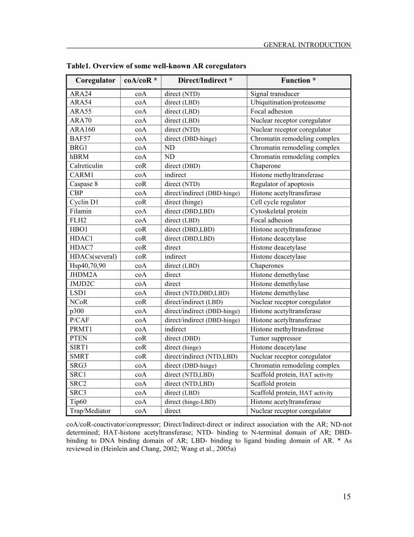
Table1. Overview of some well-known AR coregulators
Coregulator coA/coR * Direct/Indirect * Function *
ARA24 coA direct (NTD) Signal transducer ARA54 coA direct (LBD) Ubiquitination/proteasome ARA55 coA direct (LBD) Focal adhesion ARA70 coA direct (LBD) Nuclear receptor coregulator ARA160 coA direct (NTD) Nuclear receptor coregulator BAF57 coA direct (DBD-hinge) Chromatin remodeling complex BRG1 coA ND Chromatin remodeling complex hBRM coA ND Chromatin remodeling complex Calreticulin coR direct (DBD) Chaperone CARM1 coA indirect Histone methyltransferase Caspase 8 coR direct (NTD) Regulator of apoptosis CBP coA direct/indirect (DBD-hinge) Histone acetyltransferase Cyclin D1 coR direct (hinge) Cell cycle regulator Filamin coA direct (DBD,LBD) Cytoskeletal protein FLH2 coA direct (LBD) Focal adhesion HBO1 coR direct (DBD,LBD) Histone acetyltransferase HDAC1 coR direct (DBD,LBD) Histone deacetylase HDAC7 coR direct Histone deacetylase HDACs(several) coR indirect Histone deacetylase Hsp40,70,90 coA direct (LBD) Chaperones JHDM2A coA direct Histone demethylase JMJD2C coA direct Histone demethylase LSD1 coA direct (NTD,DBD,LBD) Histone demethylase NCoR coR direct/indirect (LBD) Nuclear receptor coregulator p300 coA direct/indirect (DBD-hinge) Histone acetyltransferase P/CAF coA direct/indirect (DBD-hinge) Histone acetyltransferase PRMT1 coA indirect Histone methyltransferase PTEN coR direct (DBD) Tumor suppressor SIRT1 coR direct (hinge) Histone deacetylase SMRT coR direct/indirect (NTD,LBD) Nuclear receptor coregulator SRG3 coA direct (DBD-hinge) Chromatin remodeling complex SRC1 coA direct (NTD,LBD) Scaffold protein, HAT activity SRC2 coA direct (NTD,LBD) Scaffold protein SRC3 coA direct (LBD) Scaffold protein, HAT activity Tip60 coA direct (hinge-LBD) Histone acetyltransferase Trap/Mediator coA direct Nuclear receptor coregulator
coA/coR-coactivator/corepressor; Direct/Indirect-direct or indirect association with the AR; ND-not determined; HAT-histone acetyltransferase; NTD- binding to N-terminal domain of AR; DBD- binding to DNA binding domain of AR; LBD- binding to ligand binding domain of AR. * As reviewed in (Heinlein and Chang, 2002; Wang et al., 2005a)

GENERAL INTRODUCTION
16
Coactivators On a growing list of coactivators that regulate AR are the well studied coactivators of
members of the p160 family of steroid receptor coactivator (SRC) family [SRC-1,
SRC-2 and SRC-3], p300, the p300 homologue CREB binding protein (CBP),
p300/CBP-associated factor (P/CAF), as well as Tat interactive protein 60 kDa
(Tip60). In addition, protein arginine methyltransferases (PRMTs) such as
coactivator-associated arginine methyltransferase 1 (CARM1) and protein arginine
methyltransferase 1 (PRMT1) have also been shown to play a role in AR
transactivation. The majority of these coactivators possesses histone acetyl
transferase (HAT) or methyltransferase (HMT) activity and is believed to act mainly
through histone acetylation or methylation, thus modifying chromatin structure. In
addition to their effects on histones, some can act through functional modification of
proteins such as transcription factors and coregulators. Noteworthy in this regard is
the fact that AR itself is acetylated by p300, P/CAF and Tip60 at three lysine residues
in its hinge region (Fu et al., 2000; Gaughan et al., 2002). The concept that
demethylation of histones could be also involved in transcriptional activation by the
AR has emerged only recently. Since then, lysine specific-demethylase 1 (LSD1), the
Jumonji A (JMJA) domain-containing protein JMJD2A, and the Jumonji C (JMJC)
domain-containing protein JMJD2C, that demethylate lysine 9 on histone 3, have
been shown to interact with and function as coactivators for AR (Metzger et al.,
2005; Yamane et al., 2006; Wissmann et al., 2007). Several AR coactivators have
also been identified as components of the ATP-dependent chromatin remodeling
complex SWI/SNF, including the ATPases BRG1 and hBRM (Marshall et al., 2003),
BAF57 (Link et al., 2005) and SRG3 (Hong et al., 2005). The recruitment of these
proteins to the AR transcriptional complex is consistent with altered DNA topology
following exposure to androgens. Another type of coactivator complex, that enhances
ligand-dependent AR activity, is the multimeric thyroid hormone receptor (TR)-
associated protein (TRAP)-mediator complex (Wang et al., 2002a), which appears to
influence the basal transcription machinery, possibly through the direct recruitment of
GENERAL INTRODUCTION
17
RNA polymerase II. Transcriptional activity of the AR also relies on coactivators that
bind to the AR-LBD or AR-NTD, facilitating AR stability or nuclear transport.
Among these are AR associated proteins (ARAs) [ARA24, ARA54, ARA55, ARA70
and ARA160], the four and a half LIM domain protein (FHL2), filamin and Hsp40
(reviewed in Heinlein and Chang, 2002). In addition to the above mentioned
coactivators, several other AR coactivators have been identified. However, the
precise mechanisms by which many of these modulate AR activity are yet to be
determined.
Corepressors Most of the AR coregulators identified to date have been shown to enhance
transcription of AR. Nonetheless, AR corepressors also play critical roles in
regulating AR activity. Corepressor complexes often contain histone deacetylase
(HDAC) activity that alters the acetylation state of histones, thereby regulating AR-
mediated transcription. Cyclin D1 is an example of an AR interacting corepressor that
functions through its ability to recruit HDACs and inhibit AR N/C interactions
(reviewed in Burd et al., 2005). The two best characterized corepressors, the nuclear
receptor corepressor (NCoR) and silencing mediator of retinoid and thyroid hormone
receptor (SMRT), can directly associate with AR in the absence or in the presence of
an agonist/antagonist and repress AR transactivation (Cheng et al., 2002; Liao et al.,
2003). Even though both NCoR and SMRT recruit HDACs to target genes, evidence
for a direct functional linkage between a specific HDAC and corepressor for AR is
still missing. Thus, NCoR and SMRT may exert their repressive effects through other
mechanisms, such as inhibition of AR N/C interaction or preventing coactivator
binding. In contrast to the indirect recruitment of HDACs to the AR transcriptional
complex, HDAC7, Sir2 and HDAC1 can interact directly with AR and repress its
ligand-induced signaling (Gaughan et al., 2002; Fu et al., 2006; Karvonen et al.,
2006). Similarly, calreticulin (Dedhar et al., 1994), the pro-apoptotic caspase-8 (Qi et
al., 2007) and the phosphatase and tensin homolog deleted on chromosome 10
(PTEN) tumor suppressor (Lin et al., 2004) interact directly with AR and repress its

GENERAL INTRODUCTION
18
transcriptional activity. However, these corepressors limit AR function by inhibiting
AR nuclear translocation and/or DNA binding and not by direct repression of AR
transcriptional activity. Another coregulator that associates with the AR and inhibits
its activation is human origin recognition complex interacting protein (HBO1), a
member of the MYST family (Sharma et al., 2000). The identification of HBO1 as an
AR corepressor was surprising, as this protein contains HAT activity possessed by
many coactivators. Nonetheless, the direct involvement of HBO1 enzymatic activity
in its role as a corepressor has not yet been assessed. A number of other coregulators
have been identified as AR corepressors. However, the mechanisms by which these
corepressors inhibit AR transcativation remain to be elucidated.
2.2.2 AR and specific transcription factors While there has been progress in describing the role of AR coregulators in AR
dependent gene regulation, little is known about the roles of other DNA-binding
transcription factors that may cooperate with AR in mediating androgen response.
Over the last decade, numerous transcription factors have been shown to interact
physically and functionally with the AR and regulate its transcription by different
mechanisms. Some of these proteins interact directly with the AR and affect its
ability to bind to AREs. One such example is dosage-sensitive sex reversal, adrenal
hypoplasia critical region, on chromosome X gene 1 (DAX-1) transcription factor
that binds to the AR LBD and potently inhibits androgen-dependent transcriptional
activation as well as the N/C terminal interaction (Holter et al., 2002). Other proteins
can compete with the AR for coregulators that are present in limiting amounts in cells
(Aarnisalo et al., 1998; Fronsdal et al., 1998). Moreover, some transcription factors,
including FoxA1, Oct1 and GATA2 can bind DNA sequences in close proximity to
AREs and cooperate to regulate AR target gene expression (Wang et al., 2007). The
presence of these collaborating transcription factors may assist AR in binding to sites
other than canonical AREs. Furthermore, some of these factors may function as
pioneer factors that alter chromatin to permit AR binding. Overall, these studies

GENERAL INTRODUCTION
19
indicate that a hierarchical network of transcription factors with distinct functional
roles can regulate distinct steps in AR dependent gene transcription.
3. Glucocorticoid Receptor Glucocorticoids, a major subclass of steroid hormones, were originally named for
their ability to influence glucose metabolism. During fasting, glucocorticoids help to
maintain blood glucose levels by increasing for example gluconeogenesis, glycogen
release, lipolysis and protein catabolism. In addition, glucocorticoids have effects on
mood, cognitive functions and are important for inflammation and immune responses
(reviewed in McMaster and Ray, 2007). Glucocorticoids are produced by the adrenal
cortex and their biological effects are mediated via a 94-kDa intracellular protein, the
glucocorticoid receptor (GR). GR is a member of the nuclear receptor superfamily
and is one of the close relatives of AR. Like other steroid receptors, the GR consists
of a variable N-terminal domain that also contains transactivation domain 1 (τ1), a
central DNA binding domain with two zinc finger motifs, a hinge region, and a C-
terminal hormone binding domain that harbors a second transactivation domain (τ2).
GR exists in a number of splice variants that are expressed at different levels in
different cell types and have differential activity on gene regulation (reviewed in Lu
and Cidlowski, 2004). These include, for example, the conventional ligand binding
GR termed GRα and a C-terminal variant that does not bind ligand, GRβ, which may
have a dominant negative effect on GRα. In addition, it was reported that multiple
proteins are translated from the GRα transcript, further increasing the diversity of GR
protein expression (Lu and Cidlowski, 2005).
3.1 GR transcriptional activation and regulation In its inactive, unliganded state GR is found predominantly in the cytoplasm
complexed with HSPs, although a small fraction of GR/HSP complex may reside in
the nucleus (Wikstrom et al., 1987), or recirculate to the nuclear compartment (Hache

GENERAL INTRODUCTION
20
et al., 1999). Independent of its intracellular localization, the main function of the
GR/HSP complex is to keep the receptor protein in an inactive, ligand-activable state.
Similar to AR, GR undergoes a conformational change upon ligand binding,
dissociates from the HSP complex, becomes hyperphosphorylated, homodimerizes
with another activated GR molecule and if cytoplasmic, translocates to the nucleus. In
the nucleus, GR binds to glucocorticoid response elements (GREs) in promoters or
enhancers of target genes, thereby inducing or repressing gene transcription
(reviewed in Schoneveld et al., 2004). However, GR can also act as a monomer and
modulate the transcriptional rates of non-GRE-containing genes by interacting with
nuclear transcription factors, including activator protein-1 (AP-1), nuclear factor κB
(NFκB) and signal transducer and activator of transcription 5 (STAT5) (reviewed in
Bamberger et al., 1996). When associated with its response elements, GR initiates
gene transcription through the recruitment of coregulatory complexes that modify and
remodel chromatin, promoting a more open structure and further assembly of the
basal transcriptional machinery (Baumann et al., 2001; McKenna and O'Malley,
2002; Kinyamu and Archer, 2004; O'Malley, 2004; Stavreva et al., 2004). Like other
members of the steroid receptor subfamily, GR is subject to post-translational
modifications, including phosphorylation, acetylation, ubiquitination and
sumoylation (reviewed in Faus and Haendler, 2006). These may affect its
transcriptional activity, stability and interactions with other receptors.
4. Nuclear receptor dynamics
4.1 Transcriptional action of NRs AR and nuclear receptors in general mediate the action of their specific ligands
through interaction with chromatin and protein-protein interactions with a variety of
coregulators and basal transcription factors. The dynamic process by which the
receptors recruit these factors to activate transcription was until recently poorly

GENERAL INTRODUCTION
21
understood. Currently, two opposing views exist for the development of
transcriptional complexes on nuclear receptor regulated promoters: the classical view
and the dynamic view, which are reviewed below.
4.1.1 The classic model The classic model of nuclear receptor function proposes stable binding of the
liganded receptor to the promoter. According to this view, nuclear receptors are
stably associated with their recognition sites in promoters of target genes for as long
as the ligand is present in the cellular milieu, serving as a platform for the sequential
assembly of large transcriptional complexes (Shang et al., 2002). These complexes
would have long residence times on the template, measured in minutes or hours.
Indeed, AR activity was shown to involve sustained chromatin association with
regulatory regions (Wang et al., 2005b). The occupancy of the AR-coactivator
complex on regulatory regions increases gradually after androgen exposure, peaking
at 16 hours and then gradually declining following longer stimulation. In contrast to
AR, the ER transcription complex appears to cycle onto and off target promoters
under continuous stimulation by estrogen, leading to a cyclical induction pattern with
a periodicity of 40-60 minutes, at least on the well characterized ER target gene pS2
promoter (Shang et al., 2000). Even though the cyclic assembly of ER transcription
complexes is a dynamic event, the central concept of a slow evolution of factor
complexes (i.e. long term residency measured in 10s of minutes) remains. Evidence
supporting this model has been obtained mainly from experiments based on
chromatin immunoprecipitation (ChIP) studies. In the case of AR, ChIP studies have
focused mainly on two AR target genes, prostate-specific antigen (PSA) and
Kallikrein 2 (KLK2) (Kang et al., 2004; Wang et al., 2005b). Although the ChIP
assay is a powerful tool to assess promoter occupancy and complex formation, it
remains limited by the biochemical nature of the technique. Due to the difficulty in
sample preparation and the need of fixation, ChIP cannot detect rapid protein
movements. Furthermore, ChIP can assess the promoter occupancy only indirectly,

GENERAL INTRODUCTION
22
and thus it cannot confirm whether proteins are truly in a complex on a promoter. It
can only show that they are somehow associated with the promoter sometime during
the course of fixation. In addition, the results represent the promoter occupancy of an
averaged cell population and cannot account for heterogenous cell responses. These
features need to be considered in interpreting ChIP data.
4.1.2 The dynamic model Recent studies making use of technological advances in live cell microscopy and
genetically engineered cell lines challenged the classical view of stable template
bound receptor complexes. This led to the proposal of an alternative, dynamic model
for nuclear receptor action, called the “hit-and-run” model. According to this model,
the receptor interacts transiently with the promoter, recruits other factors, and is itself
dynamically displaced from its target sites (Hager et al., 2002). In contrast to the
static view of receptor action, the residence time of NRs and interacting coregulators
on the promoter would be measured in seconds, rather than minutes or hours.
Evidence for this model was first provided by demonstration of the rapid exchange of
green fluorescent protein (GFP)-tagged GR between chromatin and the
nucleoplasmic compartment on a tandem array of mouse mammary tumor virus
(MMTV) promoters, using fluorescence recovery after photobleaching (FRAP) and
fluorescence loss in photobleaching techniques (FLIP) (McNally et al., 2000).
Tagging the protein of interest with GFP and use of photobleaching techniques, such
as FRAP and FLIP, allows a real time view of protein interactions with the chromatin
template in live cells. In order to visualize and measure real-time mobility of NRs on
their specific regulatory elements, the regulatory sites must be amplified in the
chromosome. This was accomplished by constructing an artificial array with 200
copies of the steroid hormone receptor inducible MMTV promoter that contains
HREs to which steroid receptors can bind directly (McNally et al., 2000) (see Figure
4).
GENERAL INTRODUCTION
23
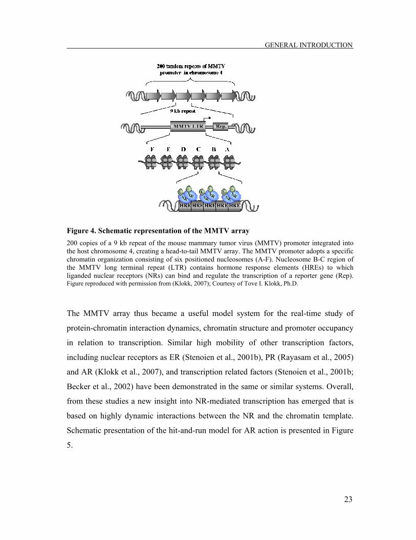
Figure 4. Schematic representation of the MMTV array 200 copies of a 9 kb repeat of the mouse mammary tumor virus (MMTV) promoter integrated into the host chromosome 4, creating a head-to-tail MMTV array. The MMTV promoter adopts a specific chromatin organization consisting of six positioned nucleosomes (A-F). Nucleosome B-C region of the MMTV long terminal repeat (LTR) contains hormone response elements (HREs) to which liganded nuclear receptors (NRs) can bind and regulate the transcription of a reporter gene (Rep). Figure reproduced with permission from (Klokk, 2007); Courtesy of Tove I. Klokk, Ph.D.
The MMTV array thus became a useful model system for the real-time study of
protein-chromatin interaction dynamics, chromatin structure and promoter occupancy
in relation to transcription. Similar high mobility of other transcription factors,
including nuclear receptors as ER (Stenoien et al., 2001b), PR (Rayasam et al., 2005)
and AR (Klokk et al., 2007), and transcription related factors (Stenoien et al., 2001b;
Becker et al., 2002) have been demonstrated in the same or similar systems. Overall,
from these studies a new insight into NR-mediated transcription has emerged that is
based on highly dynamic interactions between the NR and the chromatin template.
Schematic presentation of the hit-and-run model for AR action is presented in Figure
5.

GENERAL INTRODUCTION
24
Figure 5. Hit-and-run model for AR action AR bound to the agonist 5α-dihydrotestosterone (DHT) dissociates from the heat shock protein (Hsp) complex, translocates to the nucleus where it binds to androgen response elements (AREs) of target genes. Coregulator proteins are recruited, including chromatin remodeling complexes (SWI/SNF), coactivators (SRC), coactivators with histone acetyl transferase activity (CBP) and RNA polymerase II (PolII). The chromatin is remodeled, followed by association of a transcription initiation complex. AR is dynamically displaced (symbolized by the arrows) and shuttles between the chromatin-bound and free nucleoplasmic state with a half-maximal recovery time, t1/2 of approximately 5 seconds. AR bound to the antagonist hydroxyflutamide (OHF), similar to agonist-bound AR, dissociates from (Hsps) and translocates to the nucleus. However, because of conformational changes induced by OHF, corepressors as NCoR and SMRT are recruited, leading to further recruitment of molecules, such as histone deacetylases (HDACs), which keep the chromatin in a compact state and inhibit the assembly of the transcription initiation complex. AR is displaced and shuttles between the chromatin-bound and free nucleoplasmic state with a half-maximal recovery time, t1/2 of approximately 0.5 seconds. These rapid, stochastic interactions of AR with chromatin are influenced by the nature and outcome of its bound ligand. Figure adapted from (Kaarbo et al., 2007) with permission from authors.
Transcription

GENERAL INTRODUCTION
25
4.1.3 The “static” versus “dynamic” view The differences in nuclear receptor dynamics seen with live-cell approaches (time
scale of seconds) and ChIP assays (time scale of minutes to hours) arise in part from
the different time scales studied by these techniques. However, it has been proposed
that these two apparently opposing views can be integrated into one consistent model
(Hager et al., 2006; Metivier et al., 2006). The “return to template” model suggests
that NRs exist in the nucleoplasmic space in various coregulator complexes that
rapidly interact with their target regulatory site. Most of these rapid, transient binding
events are stochastic and nonproductive, with only a small fraction resulting in
template modification or the recruitment of alternate complexes. Thus, the promoter
evolves through multiple specific events that modulate the receptor complex stability
and initiation of transcription. In addition, both receptors and their coregulators are
subject to modifications, which may alter the activity of the complex. Whereas FRAP
experiments detect rapid and nonproductive binding of factors, ChIP assays would
determine associations of productive complexes at a specific stage of promoter
development, averaged over large number of cells, giving the impression of a
statically bound complex. The rapid exchange of factors on promoters favors rapid
activation and efficient promoter function and thus is very important for the cell to
respond to changes in the environment. Alternatively, some genes that do not require
a rapid response may still contain stable complexes. This suggests that individual
promoters achieve transcription in different ways, depending on their architecture.
4.2 Mechanisms contributing to NR mobility Despite the increasing number of transcription factors and their complexes that have
been found to be highly mobile within the nucleus, the mechanisms involved in this
behavior are yet not well understood. Current studies propose that several parameters
affect and/or influence observed dynamic protein-chromatin interactions, including
chromatin remodeling complexes, molecular chaperones, the proteasome degradation

GENERAL INTRODUCTION
26
apparatus, specific ligands, and modification of histones (reviewed in Hager et al.,
2004) which are briefly reviewed below.
4.2.1 Ligand-specific dynamics of NRs Live cell imaging experiments revealed that dynamics of steroid/nuclear receptors,
such as GR (McNally et al., 2000), ER (Stenoien et al., 2001b), PR (Rayasam et al.,
2005) and AR (Klokk et al., 2007), are strongly influenced by the nature of their
ligands. For example, it has recently been shown in our laboratory that there is a
significant difference between the dynamics of the AR-chromatin interactions in the
presence of an agonist and that of an antagonist (anti-androgen) at an HRE in living
cells (Klokk et al., 2007). Agonist-bound AR exhibits an approximately 10-fold
slower FRAP recovery kinetics compared to antagonist–bound AR, suggesting that
recovery kinetics is connected to transcriptional activity (see Figure 5). Furthermore,
PR and GR have faster exchange rates compared to AR, indicating that receptors may
use different mechanisms for interaction with their promoters. In general, the slower
turnover rates of agonist/antagonist bound receptors might reflect the time needed for
coregulator recruitment and assembly of the transcription machinery. In addition,
mobilities of steroid receptors might also be affected by their ligand-induced
conformational changes.
4.2.2 Chromatin remodeling and NR dynamics The basic repeating unit of chromatin, the nucleosome, consists of 146 bp of DNA
wrapped around an octamer of core histones, which is made of two copies each of the
histone proteins H2A, H2B, H3, and H4. In addition to the four core histones, the
linker histone H1, associates with DNA between nucleosomes and may facilitate the
formation of larger chromatin fibers (higher order chromatin), leading to a fully
condensed chromosome. The organization of DNA into chromatin restricts the access
of promoters to regulatory proteins and the transcriptional machinery. This structural
restriction of chromatin is overcome by two distinct classes of chromatin-remodeling

GENERAL INTRODUCTION
27
complexes: those that covalently modify histones (reviewed in Kouzarides, 2007) and
those that rearrange the organization of the nucleosomes in the chromatin fiber in an
ATP-dependent manner (Vignali et al., 2000; Chen et al., 2006). Recent data suggest
that these complexes affect NR mobility and disassembly, in addition to their central
role in remodeling (Fletcher et al., 2000; Nagaich et al., 2004; Metivier et al., 2006).
ATP-dependent chromatin remodeling complexes ATP-dependent complexes use the energy derived from ATP hydrolysis to directly
alter the position and/or stability of nucleosomes. They contain a core ATPase
catalytic subunit that belongs to the Swi2/Snf2 superfamily of DNA helicases. Based
on the identity of this subunit, five major families of ATP-dependent remodeling
complexes have been described: SWI/SNF, ISWI, Mi-2/NuRD, INO80, and SWR1.
Among them, the SWI/SNF family was the first identified and thus has been the best
characterized. The human SWI/SNF complex contains one of two catalytic ATPase
subunits, BRG1 or BRM, and several accessory BRG1-associated factors (BAFs).
Although the BRG1 subunit of the SWI/SNF complex has been shown to be the
primary component with regard to GR (Fryer and Archer, 1998) and PR (Mymryk
and Archer, 1995) action, AR activity shows a strong dependence on the BRM
subunit (Marshall et al., 2003). Results obtained over the last years demonstrate that
the SWI/SNF remodeling complexes are also implicated in nuclear receptor
dynamics. Using the template pull-down assays, both GR (Fletcher et al., 2002) and
PR (Rayasam et al., 2005) were shown to be actively displaced from the nucleosome
array during the process of chromatin remodeling, as a direct consequence of
SWI/SNF action. In addition, recruitment of chromatin remodeling complexes by PR
and GR were found to be ligand-dependent and the type of ligand associated with the
PR, affected its displacement from chromatin during the process of remodeling.
These results were more recently also extended to the AR (Klokk et al., 2007). The
findings described above suggest that steroid receptors, in their liganded form, can
recruit the ATPase subunit of the SWI/SNF complex, which is involved in their
dynamic association with the chromatin template.

GENERAL INTRODUCTION
28
Histone modifications The amino-terminal tails of histones are subject to a variety of reversible
posttranslational modifications. At least eight distinct types of histone modifications
have been identified to date, including acetylation, methylation, phosphorylation, and
ubiquitination (Table 2). These alternations are directed by chromatin remodeling
complexes that consist of the specific histone modifying enzymes (for example,
kinases, histone acetyltransferases, methyltransferases, and ubiquitin ligases)
(reviewed in Kouzarides, 2007).
Table 2. Modifications identified on histones
Modifications Residues modified Modifying enzymes Acetylation K-ac Acetyltransferases (HATs)
Methylation (lysines) K-me1 K-me2 K-me3 Lysine methyltransferases (HKMTs)
Methylation (arginines) R-me1 R-me2a R-me3s Arginine methyltransferases (PRMTs)
Phosphorylation S-ph T-ph Serine/Threonine kinases
Ubiquitylation K-ub Ubiquitilases
Sumoylation K-su Sumoylases
ADP ribosylation E-ar ADP-ribosyltransferases
Deimination R → Cit Peptidyl arginine deiminases
Proline isomerization P-cis > P-trans Proline isomerases
Overview of different modifications identified on histones. Modified residues: Lys (K), Arg (R), Ser (S), Thr (T), Glu (E), Pro (P) and Arg (R) to citrulline (C), and enzymes that direct each modifi- cation are shown. Modified from (Kouzarides, 2007).
Histone modifications may alter chromatin structure by influencing contacts between
different histones in adjacent nucleosomes or the interaction of histones with DNA,
or by recruitment of nonhistone proteins. However, current experimental evidence
favors the view that histone modifications are epigenetic markers that facilitate the
recruitment of chromatin binding proteins to dictate a distinct chromatin structure
(histone code hypothesis) (Jenuwein and Allis, 2001). A number of proteins have
been identified that are recruited to specific modifications and bind via specific
domains. For example, acetylated histones are recognized by bromodomains (Yang,

GENERAL INTRODUCTION
29
2004), whereas chromodomains and PHD domains associate with methylated
histones (Brehm et al., 2004), and 14-3-3 proteins bind phosphorylated histone H3
(Macdonald et al., 2005). The presence of such histone specific protein domains in
some of ATP-dependent chromatin remodeling complexes indicates that there is a
functional relationship between ATP-dependent chromatin remodeling complexes
and histone modification. The most studied histone modifications are the acetylation
and deacetylation of histone lysine residues which are reviewed below.
Histone acetylation Histone acetylation is catalyzed by the enzymatic activities of histone
acetyltransferases (HATs) that are divided into three main families: GNAT, MYST,
and CBP/p300 (reviewed in Lee and Workman, 2007). Acetylation of histones
neutralizes positively charged lysine side chains, which could weaken histone-DNA
or nucleosome-nucleosome interactions, thereby creating a more open chromatin
structure and enhance its accessibility to multiple transcription factors, such as the
transcription complex. Indeed, acetylated chromatin has long been associated with
transcriptionally active genes, with the rate of transcription correlating positively with
the degree of histone H3 and H4 acetylation (Berger, 2002). In agreement with this
many transcription coactivators that are recruited to target promoters by transcription
activators, such as NRs, contain intrinsic HAT activity (Kuo and Allis, 1998). These
include CBP/p300, P/CAF, TATA binding protein-associated factor (TAF) II250, and
the p160 family of coactivators. ChIP assays analyzing the timing of recruitment of
different coregulators after ligand treatment have revealed that HAT containing
complexes, similar to other coregulators, are recruited to target promoters in a
dynamic manner and in a specific order (Metivier et al., 2006). The ordered
recruitment of coactivators, changes in histone modifications and the recruitment of
the transcription machinery, which leads to gene expression, were shown to correlate
with cyclical recruitment of ER to the pS2 promoter. Additional transcription
responses mediated by other NRs, such as AR and PR, have shown similar dynamic
GENERAL INTRODUCTION
30
temporal pattern of coactivator recruitment and histone modifications (Kang et al.,
2004; Aoyagi and Archer, 2007). These findings indicate that association of
HATs/HDACs complexes with target promoters and following changes in histone
acetylation contribute to NR dynamics and promoter clearance.
Histone deacetylation and HDAC inhibitors Acetylation of histones can be reversed by deacetylation that is catalysed by histone
deacetylases (HDACs). Mammalian HDACs have been classified into four classes
based on sequence homology to the yeast HDACs: class I (HDACs1-3 and HDAC8),
class II (HDACs 4-7, HDAC9 and HDAC 10), class III (Sirt1-Sirt7), and class IV
(HDAC11) which has properties of both class I and classII. Class III HDACs, so-
called sirtuins, are homologs of yeast Sir2 and form a structurally distinct class of
nicotinamine adenine dinucleotide (NAD)-dependent enzymes. HDACs remove the
acetyl groups from histone lysine side chains, thus re-establishing the positive charge
of histones and the less accessible form of chromatin that is commonly associated
with transcriptional repression. In contrast to HATs, HDACs are often found as
components of transcriptional repressor complexes such as NCoR and SMRT (Tsai
and Fondell, 2004). Although HDACs have been generally correlated with gene
repression, there are several examples where HDACs appear to be required for gene
activation, thus functioning as coactivators (Berghagen et al., 2002; Wang et al.,
2002b; Ferguson et al., 2003; Mulholland et al., 2003; Qiu et al., 2006).
The correct balance between HAT and HDAC activity plays an important regulatory
role in gene expression. In addition to transcriptional regulation, HAT–HDAC
interplay is also linked to other chromatin-associated processes such as replication,
site-specific recombination and DNA repair, thereby playing a major role in
modulating overall cellular fate (reviewed in Kouzarides, 2007). Increasing evidence
indicates that alternations in HAT/ HDAC genes (such as translocation, amplification,
over-expression or mutation) are connected to tumor growth and cancer (Cress and
Seto, 2000). For example, histone deacetylation by HDACs may be a mechanism for

GENERAL INTRODUCTION
31
silencing some tumor suppressor genes responsible for cell progression, cell
proliferation, differentiation and apoptosis. Inhibition of HDACs, and thereby
activation of silenced genes, is therefore of interest in cancer therapy. To date, several
natural and synthetic compounds with HDAC inhibitor (HDACi) activity have been
identified. With a few exceptions, they can be divided into five main classes:
hydroxamic acids, short-chain fatty acids, cyclic peptides, benzamides, and
electrophilic ketones (reviewed in Minucci and Pelicci, 2006). Even though the action
of HDACis in tumorigenesis has been explored and some of them are in clinical
trials, several basic aspects are not yet fully understood and need further
investigation.
5. Aim of the study The main aim of this work was to study the molecular mechanisms by which AR
regulates transcription. Previous studies suggested that the transcriptional activity of
AR, as well as other steroid receptors, correlates with receptor mobility in the nucleus
(Klokk et al., 2007). Furthermore, the dynamic behavior of AR was shown to be
influenced by the nature of ligand (Klokk et al., 2007) and by changes in histone
acetylation at the target promoter (results from Saatcioglu laboratory, unpublished
data). However, other steroid receptors, such as GR, have been shown to have
differential responses to changes in histone acetylation at the same promoter,
compared to AR. To elucidate what links histone acetylation to the changes in AR
dynamics, it is necessary to know the details of AR-chromatin interactions and the
associated proteins at AR response elements under these conditions. The aim of this
study was thus to examine in more detail local acetylation status of the MMTV
promoter during AR-mediated transcriptional activation; this was compared with that
of GR-mediated transactivation at the same response element.

MANUSCRIPT
32
Androgen and glucocorticoid receptor mediated changes in histone acetylation
at the MMTV promoter
1. Summary Post-translational modifications of histones play an important role in regulation of
gene transcription. The most well studied histone modification is acetylation that is
regulated by the enzymatic activities of histone acetyl transferases (HATs) and
histone deacetylases (HDACs). Histone acetylation has generally been associated
with transcriptional activation and deacetylation with repression. However, there are
a number of genes for which activation is associated with deacetylation. Previous
results from our laboratory show that increased histone acetylation induced by the
HDAC inhibitor Trichostatin A (TSA) reduced androgen receptor (AR) mobility at
the mouse mammary tumor virus (MMTV) promoter, concomitant with an increase in
transcriptional activity. The effect of TSA was specific to AR as the dynamics and
transcriptional activity of the glucocorticod receptor (GR), another ligand-regulated
transcription factor of the steroid receptor family, was not affected by TSA. These
data further demonstrated that histone acetylation does not always induce
transcription, but is dependent on promoter and transcription factor context. In this
study, the impact of TSA on the acetylation level of histones H3 and H4 at the
MMTV promoter during AR- and GR-mediated transcriptional activation was
investigated. Chromatin immunoprecipitation (ChIP) analysis revealed no significant
change in histone acetylation at the MMTV promoter following TSA treatment, even
though global levels of histone acetylation were greatly increased. Furthermore,
global acetylation of histones occurred independently of the presence of androgen or
MANUSCRIPT
33
glucocorticoid. These results demonstrate that although TSA treatment induces a
global increase in histone acetylation, specific locations of the genome, such as the
MMTV promoter may be relatively unaffected. Interestingly, androgen treatment
resulted in a decrease in the basal histone H3 acetylation level at the MMTV
promoter. Preliminary studies suggest a different acetylation profile of histone H3 in
the presence of GR compared to AR. However, additional studies are necessarry to
reveal the details of histone acetylation during AR- and GR-mediated transcriptional
activation.
2. Introduction Androgens play a critical role in the development and maintenance of the male
reproductive system and are involved in important physiological and pathological
processes, such as normal prostate biology and prostate cancer (Jenster, 1999). The
effects of androgens are mediated by the androgen receptor (AR), a ligand-regulated
transcription factor that belongs to the nuclear receptor superfamily. Like other
members of this family, AR is characterized by a structure composed of four distinct
functional domains: an N-terminal transactivation domain (NTD) containing a ligand-
independent activation function 1 (AF-1), a DNA-binding domain (DBD), a hinge
region, and a ligand binding domain (LBD) possessing a ligand-dependent activation
function 2 (AF-2). AR is a steroid hormone receptor which together with the closely
related estrogen receptor (ER), progesterone receptor (PR), glucocorticoid receptor
(GR), and mineralocorticoid receptor (MR), form a subfamily of steroid hormone
receptors. Upon ligand binding, steroid receptors change conformation, bind to their
cognate hormone response elements (HREs) in promoters and/or enhancers of target
genes and modulate transcription through the recruitment of chromatin modifying
and remodelling complexes, coregulators, additional transcription factors as well as
the components of the basal transcription machinery (Dilworth and Chambon, 2001;
Hager, 2001; Marshall et al., 2003; Metivier et al., 2003; Wang et al., 2005a). The

MANUSCRIPT
34
classical view of steroid/nuclear receptor function suggests the static binding of the
liganded receptors to regulatory elements in chromatin, which serves as a platform
for the assembly of large transcriptional complexes (McKenna and O'Malley, 2002;
Shang et al., 2002).
Advances in green fluorescent protein (GFP) technology and live-cell microscopy
have led to the discovery of new principles for transcription factor action and the
proposal of an alternative “hit-and-run” model (reviewed in Hager et al., 2006).
According to this model, receptors interact only transiently with their HREs, recruit
other factors and are dynamically displaced from the promoter. Dynamic movement
on target promoters have been characterized for the steroid receptors GR (McNally et
al., 2000), PR (Rayasam et al., 2005), ER (Stenoien et al., 2001a), and AR (Klokk et
al., 2007), as well as for several other DNA binding proteins (Becker et al., 2002). In
addition, various factors have been demonstrated to influence receptor mobility
(reviewed in Hager et al., 2004). These include, among others, chromatin remodelling
complexes, specific ligands and histone modifications. Indeed, it was revealed in our
laboratory that dynamic interactions of AR are strongly dependent on the nature of
the ligand, as agonist-bound AR had reduced mobility compared to antagonist-bound
AR (Klokk et al., 2007). Moreover, longer residence time in the presence of agonist
coincided with the recruitment of the ATPase BRM, chromatin remodeling and
transcriptional activation. The involvement of specific ligands and the chromatin
remodelling complex SWI/SNF in receptor mobility and transcriptional activation has
also been demonstrated for other steroid receptors such as PR (Rayasam et al., 2005)
and GR (Fletcher et al., 2002).
In addition to chromatin remodelling, the accessibility of promoters and
transcriptional activity are also regulated by histone modifications (Berger, 2002).
These are thought to contribute to the changes in histone-histone and histone-
chromatin interactions that could lead to modulation of chromatin structure. Histone
modifications can also act as signals for recruitment of additional chromatin-
modifying factors, leading to changes in chromatin architecture and gene regulation
MANUSCRIPT
35
(Strahl and Allis, 2000; Jenuwein and Allis, 2001). In particular, acetylation of lysine
residues within the N-terminal tails of histone proteins has been well studied in the
context of gene regulation. Histone acetylation is regulated by the actions of histone
acetyl transferases (HATs) and histone deacetylases (HDACs). The dynamic
interplay between HATs and HDACs is thought to regulate histone acetylation at
cellular and local promoter level (Struhl, 1998). Acetylation of histones has long been
associated with transcriptional activation (Allfrey et al., 1964) and with an “open”
and accessible chromatin conformation (Kuo and Allis, 1998; Verdone et al., 2005).
In contrast, histone deacetylation is commonly correlated with gene repression and a
more “closed”, non-accessible form of chromatin. This view was solidified when
several transcriptional coactivators, recruited to target genes, were identified to
possess HAT activity, whereas many corepressor complexes were found in
association with HDACs (Xu et al., 1999; Hu and Lazar, 2000; Tsai and Fondell,
2004).
However, a number of studies provide evidence that the relationship between histone
acetylation and transcription is more complicated as transactivation of some
promoters is associated with deacetylation. For instance, the treatment with histone
deacetylase inhibitors (HDACis) that results in hyperacetylation of histones, showed
inhibitory effects on steroid-inducible promoters, such as ER regulated ovalbumin
promoter (McKnight et al., 1980) and mouse mammary tumor virus (MMTV)
promoter regulated by GR (Bresnick et al., 1990; Mulholland et al., 2003).
The steroid-regulated MMTV promoter that assumes a well-defined chromatin
structure when stably integrated into the host genome (Richard-Foy and Hager, 1987)
has been a useful model system to study the relationship between chromatin structure,
receptor dynamics and transcriptional activation. Previously, it has been shown that
AR transcriptional activity on the MMTV promoter is induced by HDACi TSA (List
et al., 1999). More recent results in our laboratory revealed that increased histone
acetylation induced by the HDACis TSA and SAHA resulted in increased
transcriptional activity of agonist bound AR, which correlated with reduced mobility

MANUSCRIPT
36
of AR at the MMTV promoter (unpublished data). The effect of HDACis on AR
transcriptional activity and dynamics was receptor specific, as another member of the
steroid hormone receptor family, GR, has been shown to have differential responses
to changes in histone acetylation at the same promoter (unpublished data). It was
previously reported that, in contrast to AR, TSA had inhibitory effect on GR activity
on the MMTV promoter (Bresnick et al., 1990; List et al., 1999; Mulholland et al.,
2003). In addition, results in our laboratory showed that transcriptional activity and
mobility of agonist bound GR were not affected by the HDACis TSA or SAHA on
the MMTV promoter, supporting the notion that reduced mobility of AR in response
to HDACi was directly correlated with transcriptional activity. However, what links
histone acetylation to the changes in AR dynamics and the molecular details
underlying the differential response of AR and GR to HDAC inhibitors in the same
promoter background is currently not clear. It was therefore of interest to examine the
local acetylation status of the MMTV promoter in response to the HDAC inhibitor
TSA during AR- and GR-mediated transcriptional activation that could possibly
contribute to changes in receptor dynamics and transactivation potential.
3. Materials and Methods
3.1 Materials Dulbecco’s Modified Eagle Medium (DMEM), DMEM-without Phenol Red, L-
glutamine, Penicillin/Streptomycin and Trypsin/EDTA were purchased from
BioWhittaker, Cambrex Bio Science and fetal calf serum (FCS) was purchased from
PAA Laboratories GmbH. The following reagents were obtained from Sigma-
Aldrich: Bovine serum albumin (BSA), magnesium chloride (MgCl2), puromycin,
dithiothreitol (DTT), β-glycerophosphate, sodiumortovanadate (Na3VO4), HEPES,
Tween 20, leupeptin, phenylmethylsulphonyl-fluoride (PMSF), octyl phenoxy
polyoxy ethanol (Triton X-100), sodium azide (NaN3), trichostatin A (TSA),

MANUSCRIPT
37
dexamethasone (DEX), tetracycline, formaldehyde, phenol chloroform-isoamyl
alcohol (25:24:1), lithium chloride (LiCl), sodium deoxycholate, sodium butyrate,
yeast transfer ribonucleic acid (tRNA), NP-40/Igepal CA-630, anti-α-Tubulin mouse
monoclonal antibody, horseradish peroxidase-conjugated (HRP) anti-rabbit IgG
antibody and HRP-conjugated anti-mouse IgG antibody. Sodium chloride (NaCl),
sodium hydroxide (NaOH) and ethylenediaminetetraacetic acid (EDTA) were
purchased from BDH Chemicals Ltd. and sodium dodecyl sulphate (SDS) was
obtained from Fluka Chemie GmbH. Methanol, trisaminomethane (Tris),
hydrochloric acid (HCl) were obtained from VWR International, Inc. and glycine was
from Duchefa Biochemie BV. Skim milk powder was from Acumedia Manufacturers,
Inc. and salmon sperm DNA, Protein A Sepharose (liquid beads) and anti-GFP rabbit
fraction antibody were purchased from Invitrogen. Lightcycler® 480 SYBR Green I
Master mix, Lightcycler® Multiwell Plates 96, Protease inhibitor cocktail and
proteinase K were obtained from Roche Diagnostics GmbH. Protein A Sepharose
(powder beads) and ECL Western Blotting Analysis System were purchased from GE
Healthcare Bio-Science. Anti-acetyl-histone H3, anti-acetyl-histone H4 and anti-AR
rabbit polyclonal antibodies were obtained from Upstate Biotechnology, Inc. and
another anti-AR rabbit polyclonal antibody (N-20) was from Santa Cruz
Biotechnology, Inc. Anti-histone H3 rabbit polyclonal antibody was purchased from
Cell Signaling Technology, Inc. and anti-GR mouse monoclonal antibody (BuGR2)
was from Abcam, Ltd. Anti-GR rabbit polyclonal antibody was purchased from
Affinity BioReagents, Inc. and ethanol was obtained from Arcus Kjemi AS. Precision
Plus ProteinTM Standards Dual Color, PVDF membrane and Bio-Rad protein assay
were purchased from Bio-Rad Laboratories, Inc. Sodium acetate was from Merck
Chemicals Ltd. and geneticin sulphate (G418) was from Gibco, Invitrogen
Corporation. DyNazymeTM II DNA Polymerase with its buffer was obtained from
Finnzymes Oy and deoxyribonucleosine-5’-triphosphates (dNTPs) and 2-log DNA
ladder were purchased from New England BioLabs, Inc. MatTek cultureware 35mm
glass bottom microwell dishes were obtained from MatTek Corporation and

MANUSCRIPT
38
MycoAlert® Mycoplasma Detection Kit was from Lonza Biologics, Inc.
Methyltrienolone (R1881) was purchased from DuPont NEN Research Products and
primers were manufactured by Sigma-Genosys.
3.2 Methods
3.2.1 Cell lines and Cell culture The ell lines 3108 and 3617 are stably transfected derivates of the murine mammary
adenocarcinoma cell line 3134 that contains 200 tandem repeats of a 9 kb element
composed of the MMTV promoter followed by ras and BPV genes. These cell lines
stably express GFP-tagged AR (3108) and GFP-tagged GR (3617),respectively,
under the control of a tetracycline-off inducible system as previously described
(McNally et al., 2000; Klokk et al., 2007). The passage number of both cell lines used
in experiments was between 4 and 12. The cells were routinely maintained at 37ºC in
a humidified 5% CO2 and 95% air incubator in DMEM supplemented with 10% fetal
calf serum (FCS), 5mg/ml penicillin-streptomycin, 2mM L-glutamine and 10µg/ml
tetracycline (to suppress GFP-AR and GFP-GR expression). The 3108 cell line was
additionally supplemented with 1mg/ml G418 and 0.55 µg/ml puromycin. The
culture medium was changed every second day. The MycoAlert® Mycoplasma
Detection Kit was used to test cells for mycoplasma contamination. For the
experiments cells were plated in culture medium without G418 and puromycin at a
density of 3×10⁵ (3108) and 2×10⁵ (3617) cells per 10 cm dish and grown in the
absence of tetracycline for induction of GFP-AR and GFP-GR, respectively, if not
indicated differently. After reaching 30% confluence, cells were washed with
phosphate-buffered saline (PBS) and serum starved for 2 days in medium containing
10% charcoal treated (CT)-FCS to deplete the cells from steroids that could activate
AR or GR. Prior to the experiments, cells were either left untreated or treated with
TSA (100 nM) for 18 h, the synthetic androgen R1881 (10-8M) for 1 h or the
synthetic glucocorticoid DEX (10-7M) for 30 min.
MANUSCRIPT
39
3.2.2 Protein extraction and Western analysis Cells grown and starved in the presence or absence of tetracycline were harvested by
scraping in PBS and collected by centrifugation. Whole cell extracts were prepared
by resuspending the cell pellets in 200 µl lysis buffer containing 20 mM HEPES (pH
7.7), 300 mM NaCl, 0.2 mM EDTA, 0.1% Triton X-100, 1.5 mM MgCl2, 0.5 mM
DTT, 20 mM β-glycerophosphate, 0.1 M Na3VO4, 2µg/ml leupeptin and 0.5 M
PMSF. After rotating for 2 h at 4ºC, supernatants were collected by centrifugation at
13,000 rpm for 20 min and the protein concentrations were determined by the Bio-
Rad protein assay. For western analysis, proteins (100µg) were separated on 8%
SDS-PAGE gels. The Precision Plus ProteinTM Standards Dual Color was used as a
molecular weight marker. Proteins were then transferred overnight to PVDF
membranes activated in methanol. Blotted membranes were blocked in 5% nonfat dry
milk in PBS containing 0.1% Tween for 1 h, followed by incubation overnight at 4ºC
with primary antibodies diluted in PBS-Tween containing different amounts of
bovine serum albumin (BSA). Antibodies used were rabbit polyclonal anti-AR
antibody (N-20, Santa Cruz) diluted in 3% BSA (1:500), rabbit IgG fraction anti-GFP
antibody diluted in 5% BSA (1:1000), mouse monoclonal anti-GR antibody (BuGR2,
AbCam) diluted in 3% BSA (1:400), and mouse monoclonal anti-α-tubulin antibody
diluted in 0.5% BSA (1:4000). Membranes were washed with PBS-Tween and
incubated for 1.5 h at room temperature with secondary horseradish peroxidase HRP-
conjugated anti-rabbit (1:10000) or anti-mouse (1:5000) IgG antibodies diluted in
PBS-Tween with 0.5% nonfat dry milk. All blots were visualized with the ECL
Western Blotting Analysis System according to the manufacturer’s instructions.
3.2.3 Live cell microscopy For live cell imaging, cells were plated at a density of 7×10⁴ (3108) and 4×10⁴ (3617)
cells per MatTek dish. The cells were grown in medium without tetracycline and
starved as described above. Prior to the microscopy experiments, cells were washed
with PBS and the regular starving medium was replaced with phenol red-free

MANUSCRIPT
40
medium supplemented with 10% CT-FCS to eliminate autofluorescence. The cells
were then treated with R1881 (10-8M) or DEX (10-7M) for indicated time periods or
left untreated. Microscopy was performed with an Olympus FluoView 1000 confocal
laser scanning microscope, equipped with an incubator maintained at 37ºC, and
images were acquired with a 60x1.3 numerical aperture oil immersion objective and a
488nm argon laser.
3.2.4 Histone extraction and Western analysis Cells were plated, grown and starved under the same conditions as described
previously. The cells were either left untreated or treated with R1881 (10-8M), DEX
(10-7M), or TSA (100nM), alone or in combinations as indicated for the desired time
periods. After washing with PBS, the cells were harvested by scraping in PBS and
centrifugation. Harvested cells were washed twice with ice-cold PBS and
resuspended in 500µl Triton extraction buffer (TEB) containing PBS with 0.5%
Triton X-100, 2mM PMSF and 0.02% NaN3. PBS was supplemented with 5mM
sodium butyrate to retain levels of histone acetylation. The cells were lysed on ice for
10 min with gentle stirring followed by centrifugation at 2000 rpm for 10 min at 4ºC.
After centrifugation, pelleted nuclei were washed in 250µl TEB and centrifuged as
before. Nuclei were then resuspended in 150µl 0.2M HCl and lysed on a rotator at
4ºC overnight. The supernatant containing histones were collected after
centrifugation at 2000 rpm for 10 min at 4ºC and protein concentrations were
determined using the Bio-Rad protein assay, followed by Western analysis. 10 µg
aliquots of each sample together with the Precision Plus ProteinTM Standards Dual
Color marker were resolved on 15% SDS-PAGE gels and transferred to methanol
activated PVDF membranes overnight. Histone H3 and histone H4 acetylation were
determined by blocking membranes in Tris-buffered saline (TBS) containing 0.1%
Tween and 5% nonfat dry milk, and then probed with rabbit polyclonal anti-acetyl-
histone H3 antibody (1:10000 dilution) or anti-acetyl-histone H4 antibody (1:5000
dilution) in TBS-Tween with 3% BSA overnight at 4ºC. Membranes were washed

MANUSCRIPT
41
and incubated for 1.5 h at room temperature with the secondary HRP-conjugated anti-
rabbit IgG antibody diluted in TBS-Tween with 0.5% nonfat milk (1:20000). Protein
bands were visualized using Western Blotting Analysis System. To verify equal
loading of samples, membranes were stripped with 0.5M NaOH followed by
incubation in TBS-Tween with 5% nonfat dry milk and reprobed with rabbit
polyclonal anti-histone H3 antibody diluted in TBS-Tween with 3% BSA (1:2000).
Following secondary antibody application visualization was performed as described
above.
3.2.5 Chromatin Immunoprecipitation assay (ChIP) Cells were plated, starved and pretreated with TSA, R1881or DEX for indicated time
periods as described above. Three independent experiments were carried out for each
cell line. Following treatment with hormone, cells were cross-linked with 1%
formaldehyde in culture medium at room temperature for 10 min. The fixing process
was stopped by the addition of glycine to a final concentration of 0.125M and
incubation at room temperature for 5 min. After washing with PBS, the cells were
harvested by scraping in ice-cold PBS supplemented with 1x protease inhibitor
cocktail and centrifugation. The cell pellet was resuspended in 300µl lysis buffer [1%
SDS, 10mM EDTA, 50mM Tris-HCl pH8, 1x protease inhibitor cocktail] and
sonicated using Biorupter UCD-200 (Diagenode). During sonication, samples were
kept in ice-cold water and sonication was performed on high setting using 21 pulses
of 7 sec each, with a 7 sec rest between each pulse (in total 5 min). Cell lysates were
then centrifuged at 13,000 rpm at 4ºC for 10 min to pellet the insoluble material.
Supernatants were collected and for determination of DNA concentration and
shearing efficiency, 15µl aliquots were removed prior to dilution in dilution buffer
[0.01% SDS, 1.1% Triton X-100, 1.2mM EDTA, 16.7mM Tris-HCl pH8, 1x protease
inhibitor cocktail] to a total volume of 3ml followed by immunoprecipitation.

MANUSCRIPT
42
DNA concentration measurement 4µl of each non-diluted sheared chromatin and 5µl proteinase K were added to 191µl
lysis buffer supplemented with 40mM NaCl. The mixture was incubated at 65ºC for 1
h and DNA concentration was measured with Biophotometer spectrophotometer
(Eppendorf) at 260 and 280nm.
Chromatin shearing 5µl of each non-diluted sheared chromatin was added to a mixture containing 3µl
proteinase K, 184 µl lysis buffer and 8µl 5M NaCl. The mixture was incubated at
65ºC for 4 h or overnight and DNA was purified by extraction with phenol/chloro-
form, followed by addition of 0.1 volume of 3M sodium acetate and 2x volumes of
ethanol and precipitation at -20ºC overnight. After 10 min centrifugation at 4ºC, the
DNA pellet was washed once with 70% ethanol, dried and dissolved in 15µl double
distilled water (ddH2O). The size of the sheared DNA was determined on a 1%
agarose gel. Sheared chromatin runs as a smear in the size range of ~100-1500 base
pairs, where the major fraction is at about 300-800 base pairs.
Immunoprecipitation Equal amounts of diluted sheared chromatin was adjusted to the volume of 3ml with
the dilution buffer and precleared on a rotator for 2 h at 4ºC with 200µl salmon sperm
DNA/protein-A sepharose slurry (50% v/v beads in TE buffer supplemented with
0.2mg salmon sperm DNA and 0.5mg BSA/ml) to avoid non-specific binding of
protein-A sepharose. After preclearing, the beads were briefly centrifuged at 1000
rpm at 4ºC for 1 min, the supernatants were collected and 1/10 of each sample was
stored at 4ºC as an input control. The supernatant fractions were then subjected to
chromatin immunoprecipitation overnight at 4ºC with 8 µg of either anti-acetyl
histone H3 or H4 antibodies, 2µg anti-AR antibody (N-20, Santa Cruz) and 10µg
anti-GR antibody (Affinity BioReagents). A control without any antibody was
included for assessing non-specific binding. Following incubation with antibody,
80µl of salmon sperm DNA/protein-A sepharose was added to each tube for 1 h with

MANUSCRIPT
43
rotation at 4ºC to isolate the immune complexes. The sepharose-antibody-chromatin
complexes were then captured by centrifugation at 1000 rpm for 3 min at 4ºC and
washed sequentially for 15 min (washes on a rotator platform, followed by
centrifugation at 1000 rpm for 3 min), first in TSE I buffer [0.1% SDS, 1% Triton X-
100, 2mM EDTA, 20mM Tris-HCl pH8, 150mM NaCl], TSE II buffer [ TSE I with
500mM NaCl], buffer III [0.25M LiCl, 1% NP-40, 1% sodium deoxycholate, 1mM
EDTA, 10mM Tris-HCl pH8], and then in TE [10mM Tris-HCl pH8, 1mM EDTA].
All steps starting with the harvesting of the cells to the elution step were done in the
presence of 5mM sodium butyrate. Sepharose bound proteins were then eluted twice
with 250µl elution buffer [1% SDS in ddH2O] on a rotator at room temperature for 15
min and eluates were combined. The stored input samples were eluted with 200µl
elution buffer [ddH2O + 1% SDS in final solution]. Eluates were mixed with 20µl 5M
NaCl, 10µl 0.5M EDTA, 20µl 1M Tris-HCl pH6.5 and 15.6 µg of proteinase K, and
heated at 65ºC for at least 4 h to reverse the formaldehyde cross-linking between
DNA and protein. DNA fragments were purified by phenol/chloroform extraction
and precipitated with ethanol and 10µg carrier RNA (yeast tRNA) overnight at -20ºC.
Following centrifugation, the DNA pellet was washed with 70% ethanol, dried and
dissolved in 40µl ddH2O for further analysis by PCR.
PCR analysis Following purification, the DNA was subjected to PCR amplification using primers
specific for nucleosome B region of the MMTV promoter. The corresponding PCR
product was 120 base pairs. Primers used were: forward 5`-TTTCCATACCAAG-
GAGGGGACAGTG-3` and reverse 5`-CTTACTTAAGCCTTGGGAACCGCAA-3`.
With all experiments, negative (no template-ddH2O) and positive (LTR plasmid)
PCR controls were included. The PCR reaction was performed in a mixture (25µl),
containing 2µl DNA, 10x reaction buffer, 0.4mM each of dATP, dTTP, dGTP and
dCTP, 1.6mM MgCl2, 1 unit of DyNazyme II DNA polymerase, and 0.4µM of each
primer. The reaction was carried out in a Peltier Thermal Cycler (PTC-200; MJ
Research) using the following amplification conditions: 5 min denaturation at 95ºC

MANUSCRIPT
44
followed by 30 cycles of denaturation at 95ºC for 30 sec, annealing at 63ºC for 1min
and extension at 72ºC for 1 min. In the end, there was a final extension for 5 min at
72°C. The amplified DNA was analyzed on a 1.5% agarose gel.
3.2.6 Quantitative PCR (qPCR) DNA isolated from precipitated chromatin was analysed by qPCR on a LightCycler®
480 (Roche Diagnostics) using SYBR Green as a DNA-specific binding dye and
continuous fluorescence monitoring. The PCR reaction (10µl) contained 2x SYBR
Green I Master mix, 2µl of DNA template and 0.5µl of each primer. The primers,
positive and negative controls used were same as described in conventional PCR
analysis. The PCR program was comprised of an initial denaturating step at 95ºC for
5 min followed by 45 cycles of denaturation (15sec at 95ºC), annealing (30sec at
63ºC) and extension (20sec at 72ºC). To avoid non-specific PCR by-products and
detect presence of possible primer dimers, melting curve was analysed: 5sec at 95ºC,
1 min at 65ºC followed by continuous fluorescence measurement between 65ºC and
97ºC. The crossing points (Cp) values calculated by the LightCycler computer
software were used for determination of the initial amount of DNA template.
Data analysis The data analysis was performed using Microsoft Office Excel 2003 software. The
qPCR signals derived from the ChIP samples were normalized by the signals derived
from the input samples. The level of acetylation in the absence of ligand and TSA
treatment and the level of receptor enrichment in the absence of ligand were set to 1.
The data presented represent the average of three independent experiments. Statistical
analysis was performed using the Student’s two tailed t-test. Values of p < 0.05 were
considered statistically significant.

MANUSCRIPT
45
4. Results
4.1 Tetracycline-regulated expression of GFP-AR and GFP-GR
In order to examine AR and GR mediated chromatin modifications of the MMTV
promoter, the previously established 3108 and 3617 cell lines were used (McNally et
al., 2000; Klokk et al., 2007). These cell lines contain an integrated tandem repeat of
the MMTV promoter and express GFP-AR (3108) or GFP-GR (3617) in a stable and
tetracycline repressible manner. Regulation of GFP-AR and GFP-GR expression in
these cells by tetracycline was confirmed by Western analysis of total cell lysates,
using antibodies against AR, GR and GFP. As can be seen in Figure 1, there was
clear induction of GFP-AR (A) and GFP-GR (B) expression upon 72 h withdrawal of
tetracycline from the medium, resulting in a band of expected size (~130 kDa).
Figure 1. Tetracycline-regulated expression of GFP-AR and GFP-GR 3108 (A) and 3617 (B) cells were grown and starved, as described in Materials and Methods, in the presence (+) or absence (-) of tetracycline (Tet). Whole cell extracts were prepared and subjected to Western analysis with antibodies specific to AR (A, upper-left panel), GR (B, upper-left panel) and GFP (A-B, upper-right panels). Tubulin was used as a loading control (A-B, lower panels). Molecular weight markers are given to the right.
GFP-AR
50 kDa100 kDa
Tet
Tubulin
Anti-AR Anti-GFP
150 kDa-+ + -
A)
Tet
50 kDa
100 kDa
150 kDa
Anti-GR Anti-GFP
Tubulin
GFP-GR
+ - + -B)
GFP-AR
50 kDa100 kDa
Tet
Tubulin
Anti-AR Anti-GFP
150 kDa-+ + -
A)
Tet
50 kDa
100 kDa
150 kDa
Anti-GR Anti-GFP
Tubulin
GFP-GR
+ - + -B)
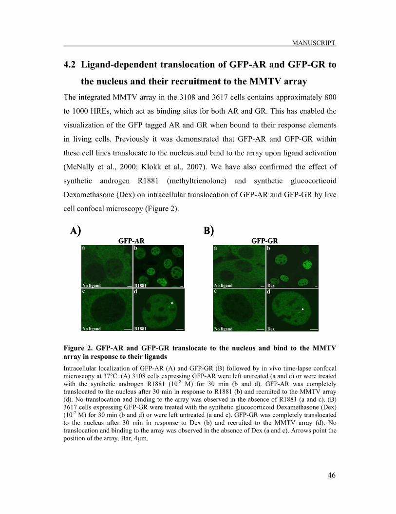
MANUSCRIPT
46
4.2 Ligand-dependent translocation of GFP-AR and GFP-GR to
the nucleus and their recruitment to the MMTV array The integrated MMTV array in the 3108 and 3617 cells contains approximately 800
to 1000 HREs, which act as binding sites for both AR and GR. This has enabled the
visualization of the GFP tagged AR and GR when bound to their response elements
in living cells. Previously it was demonstrated that GFP-AR and GFP-GR within
these cell lines translocate to the nucleus and bind to the array upon ligand activation
(McNally et al., 2000; Klokk et al., 2007). We have also confirmed the effect of
synthetic androgen R1881 (methyltrienolone) and synthetic glucocorticoid
Dexamethasone (Dex) on intracellular translocation of GFP-AR and GFP-GR by live
cell confocal microscopy (Figure 2).
Figure 2. GFP-AR and GFP-GR translocate to the nucleus and bind to the MMTV array in response to their ligands Intracellular localization of GFP-AR (A) and GFP-GR (B) followed by in vivo time-lapse confocal microscopy at 37°C. (A) 3108 cells expressing GFP-AR were left untreated (a and c) or were treated with the synthetic androgen R1881 (10-8 M) for 30 min (b and d). GFP-AR was completely translocated to the nucleus after 30 min in response to R1881 (b) and recruited to the MMTV array (d). No translocation and binding to the array was observed in the absence of R1881 (a and c). (B) 3617 cells expressing GFP-GR were treated with the synthetic glucocorticoid Dexamethasone (Dex) (10-7 M) for 30 min (b and d) or were left untreated (a and c). GFP-GR was completely translocated to the nucleus after 30 min in response to Dex (b) and recruited to the MMTV array (d). No translocation and binding to the array was observed in the absence of Dex (a and c). Arrows point the position of the array. Bar, 4µm.
No ligand
No ligand
R1881
R1881
a
c d
b
B)
cNo ligand Dex
No ligand Dex
a b
dc
A)GFP-AR GFP-GR
No ligand
No ligand
R1881
R1881
a
c d
b
B)
cNo ligand Dex
No ligand Dex
a b
dc
A)GFP-AR GFP-GR
MANUSCRIPT
47
Because the array in these cells exists as a unique, single amplified element on
chromosome 4, a single bright GFP fluorescence signal was expected to be observed
within the nucleus. In the absence of ligand, GFP-AR and GFP-GR were distributed
predominantly in the cytoplasm and no significant binding to the MMTV array was
observed (Figure 2Aa, 2Ac, 2Ba and 2Bc). The presence of agonists R1881 or Dex
for 30 min caused complete translocation of AR or GR, respectively, to the nucleus
(Figure 2Ab and 2Bb). Furthermore, a single bright fluorescent signal (Figure 2Ad
and 2Bd), in addition to the diffuse nucleoplasmic GFP-AR and GFP-GR, was
detected within the nucleus. These data confirm the intact nature of these cell lines as
described in previous studies (McNally et al., 2000; Klokk et al., 2007).
4.3 HDAC inhibitor TSA induces global histone acetylation
independently of the presence of hormone
HDAC inhibitors (HDACis) are generally used to manipulate the acetylation status of
histones and to induce global histone hyperacetylation. Trichostatin A (TSA) is an
efficient HDAC inhibitor (HDACi) and was shown to induce an increase in acetyl-
histone H3 levels in 3108 and 3617 cells in a time-dependent manner (Saatcioglu
laboratory, unpublished data). To assess whether exposure of 3108 and 3617 cells to
TSA treatment give rise to expected increase in histone H3 and H4 acetylation, cells
were left untreated or were treated with 100 nM TSA for 18 h. Moreover, in order to
examine if the TSA-induced histone acetylation was dependent on the presence of
hormone, 3108 and 3617 cells were additionally treated with R1881 or Dex,
respectively, alone or were pretreated with TSA prior to ligand addition. The histone
extracts were obtained and subjected to Western analysis using antisera specific for
acetyl-histone H3 and acetyl-histone H4 (Figure 3).

MANUSCRIPT
48
.
Figure 3. Acetylation of histone H3 and H4 in response to TSA and ligand activation 3108 and 3617 cells were treated with TSA (100nM) for 18 h, or R1881 (10-8 M) for 1h or Dex (10-7 M) for 30 min, alone or in combinations, or were left untreated. Histone extracts were obtained and subjected to Western analysis with antibodies specific to acetyl-histone H3 (A) or acetyl-histone H4 (B). Anti-histone H3 antibody was used as a loading control (lower panels A and B). Molecular weight markers are indicated to the right of the figure.
In the absence of TSA, both histones H3 (A) and H4 (B) exhibited low levels of
acetylation that were strongly increased in response to TSA; total H3 levels were not
affected. This confirmed the efficient inhibition of HDAC activity by TSA in 3108
and 3617 cells. Furthermore, addition of R1881 or Dex did not alter the histone
acetylation profiles generated by treatment with TSA alone. Thus, exposure to TSA
results in global increase of histone acetylation patterns, independently of the
presence of hormone. The acetyl-histone H4 antiserum also detected additional non-
specific band in 3108 cells at around 13 kDa (Figure 3B, left panel). This band shows
similar response to TSA treatment as observed for acetylated histone H4 and might
represent cross reaction with acetylated histone H2A (~14kDa). Alternatively, this
+++
+-
---
TSA R1881
Acetyl-H4
H3
B)
+++
+-
---
TSA R1881
Acetyl-H3
H3
A) GFP-AR
GFP-AR
++--TSA + +--Dex
15 kDa
15 kDa
GFP-GR
TSA ++--+ +--Dex
10 kDa
15 kDa
GFP-GR
+++
+-
---
TSA R1881
Acetyl-H4
H3
B)
+++
+-
---
TSA R1881
Acetyl-H3
H3
A) GFP-AR
GFP-AR
++--TSA + +--Dex
15 kDa
15 kDa
GFP-GR
TSA ++--+ +--Dex
10 kDa
15 kDa
GFP-GR
MANUSCRIPT
49
could be a proteolytic fragment of histone H4 produced during extract preparation.
However, the nature of this band was not investigated further.
4.4 ChIP optimization
Having established that the cell lines we wished to use in this study behaved as
expected, we set out to use chromatin immunoprecipitation (ChIP) in these cells to
probe dynamic changes of protein interactions at the MMTV array. ChIP is a
powerful method used to identify regions of the genome associated with specific
proteins, histone modifications, chromatin remodeling and other chromatin-related
processes. In this study, the ChIP assay was used to study histone acetylation profiles
at the Nuc-B region of the MMTV promoter in 3108 and 3617 cells. In addition,
association of AR and GR with HREs of the MMTV promoter were also assessed by
this method. The ChIP assays were carried out according to the standard protocol that
was previously established in our laboratory, as described in Materials and Methods.
However, after performing some preliminary ChIP assays to optimize it for use in
these experiments, we lost all ChIP signal which was weak to start with; therefore, we
needed to do several rounds of troubleshooting.
To that end, the 3108 cells were left untreated or were treated with androgen R1881
and subjected to the ChIP assay according to our standard protocol (Figure 4).
Because the resolution obtained by the ChIP procedure depends on the size of
chromatin fragments, it was first important to check if the shearing conditions used in
the protocol resulted in the expected DNA smear in the desired size range
(~200~1000 bp). As seen in Figure 4A, the major fraction of sheared DNA had
optimal length of ~300~800 bp, similar to that detected by previous ChIP
experiments performed in our laboratory (data not shown). Having established this,
ChIP assay was processed to the end with the AR antibody which resulted in no ChIP
based signal (Figure 4B).

MANUSCRIPT
50
Figure 4. ChIP assay to assess AR binding to its HREs in the MMTV promoter using standard protocol conditions 3108 cells were left untreated (-) or treated (+) with R1881 (10-8M) for 1 h and subjected to the ChIP assay using an antibody specific to AR, as described in Materials and Methods. The DNA molecular weight markers (M; in kb) are shown at the left of the figures. (A) Sheared DNA, (B) ChIP assay performed with AR antibody (2µg). No antibody (N.A.) was used as negative background control. Inputs represent the starting material before immunoprecipitation. Negative (N.c.) and positive (P.c.) are PCR performed in the absence of DNA containing the MMTV promoter, respectively.
Since the PCR controls and inputs showed expected results, we reasoned that it may
be the immunoprecipitation or the following steps that failed during the experiment.
This could have been due to inefficient crosslinking, unsuccessful binding to the
beads, incomplete/lack of elution or problems with the antibody. Thus, we first
checked crosslinking conditions to assess if it was functional. Crosslinking is a time
critical procedure, depending on the cell type and protein of interest. Excessive
crosslinking can lead to decrease in the amount of protein bound to DNA and to
reduction in the availability of epitopes for antibody binding, while too little
crosslinking may not fix protein to its chromatin binding site. In the experiment
presented above, crosslinking was carried out for 10 min at RT, but no signal was
detected. Thus it became of interest to increase crosslinking time to see if this can
R1881M
+ - +kb
A)1 2 3
-
-
----
-
-
0.5
1.0
10.0
2.0
6.0
3.0
1.5
4.0
B)kb M
R18811+ -
2 3 4 5 6P. c.N. c.+ + +-
ARN. A. Inputs
--0.2
0.1
R1881M
+ - +kb
A)1 2 3
-
-
----
-
-
0.5
1.0
10.0
2.0
6.0
3.0
1.5
4.0
B)kb M
R18811+ -
2 3 4 5 6P. c.N. c.+ + +-
ARN. A. Inputs
--0.2
0.1

MANUSCRIPT
51
help. As can be seen in Figure 5A, even longer crosslinking time did not improve the
signal obtained in ChIP. In addition, crosslinking was also performed on ice for 20
and 30 min (Figure 5B) which either did not help either.
Figure 5. ChIP assay, optimization of crosslinking 3108 cells were untreated (-) or treated (+) with androgen R1881 (10-8M) for 1 h and subjected to the ChIP assay using an antibody specific to AR, as described in Materials and Methods. The DNA molecular weight markers (M; in kb) are shown at the left of the figures. No antibody (N.A.) was used as negative background control. Inputs represent the starting material before immunoprecipitation. Negative (N.c.) and positive (P.c.) PCR controls are indicated. (A) ChIP assay with crosslinking time (CT) of 15 and 20 min at room temperature (RT). (B) ChIP assay with crosslinking time (CT) of 20 and 30 min on ice.
Having shown that crosslinking was not a problem, the next step was to check if use
of different antibodies specific for GFP-AR would improve the signal of precipitated
samples. Antibodies are used to capture the DNA/protein complex and this step is
typically the most critical one for a successful ChIP experiment. Thus, the antibody
specific to AR (N-20, Santa Cruz) that was used in the experiments presented above
for immunoprecipitation was changed with another AR-specific antibody (Upstate)
A)
kb MR1881
1+ -
2 3 4 5 6
N. c.
P. c.+ + +-
ARN.A.
--0.2
0.1
ARN.A.CT (min): 15 20
RT
B)
kb MR1881
1+ -
2 3 4 5 6P. c.+ + +-
ARN.A.
--0.2
0.1
ARN.A.
Elutes
CT (min): 20 30
ICE
N. c.
Inputs0.10.2---0.3
A)
kb MR1881
1+ -
2 3 4 5 6
N. c.
P. c.+ + +-
ARN.A.
--0.2
0.1
ARN.A.CT (min): 15 20
RT
B)
kb MR1881
1+ -
2 3 4 5 6P. c.+ + +-
ARN.A.
--0.2
0.1
ARN.A.
Elutes
CT (min): 20 30
ICE
N. c.
Inputs0.10.2---0.3

MANUSCRIPT
52
and an antibody specific to GFP (Invitrogen). However, neither of these antibodies
resulted in a signal similar to experiments above (Figure 6A).
Since varying the antibody did not help to solve the problem, we then turned to the
Protein A Sepharose beads that are necessary for successful immunoprecipitation. In
previous experiments we used Protein A Sepharose beads obtained from Invitrogen.
To examine if there was something wrong with these beads, the next ChIP was
performed with beads from a different supplier (GE-Healthcare) side by side with
those from Invitrogen (Figure 6B).
A)
kb MR1881
1+ -
2 3 4 5 6P. c.+ + +-
ARN.A.
--0.2
0.1
GFPN.A.
Elutes
N. c.
Inputs0.1-
B)
kb MR1881
1+ -
2 3 4 5 6
N. c.
P. c.+ + +-
Ac-H3N.A.
--0.3
0.1
Ac-H3N.A.
Elutes
Beads: Invitrogen GE-Healthcare
--
0.2
0.4
Inputs0.1---0.3
0.2
A)
kb MR1881
1+ -
2 3 4 5 6P. c.+ + +-
ARN.A.
--0.2
0.1
GFPN.A.
Elutes
N. c.
Inputs0.1-
B)
kb MR1881
1+ -
2 3 4 5 6
N. c.
P. c.+ + +-
Ac-H3N.A.
--0.3
0.1
Ac-H3N.A.
Elutes
Beads: Invitrogen GE-Healthcare
--
0.2
0.4
Inputs0.1---0.3
0.2 Inputs0.1---0.3
0.2

MANUSCRIPT
53
Figure 6. Optimization of the ChIP assay with different antibodies and beads 3108 cells were untreated (-) or treated (+) with androgen R1881 (10-8M) for 1 h and subjected to ChIP assay, as described in Materials and Methods. The DNA molecular weight markers (M; in kb) are shown at the left of the figures. No antibody (N.A.) was used as negative background control. Inputs represent the starting material before immunoprecipitation. Negative (N.c.) and positive (P.c.) PCR controls are indicated. (A) ChIP assay performed with antibodies specific to AR (Upstate) and GFP (Invitrogen). (B) ChIP assay performed with Protein A Sepharose (Invitrogen) and (GE-Healthcare), and acetyl-histone H3 antibody. (C) ChIP assay performed with different amounts of AR (Santa Cruz) antibody, respectively 2, 4 and 6 µg.
We used an antibody for acetyl-histone H3 which was previously shown to function
well in ChIP (data not shown); since histones are far more abundant than chromatin
bound AR, we expected to see a better signal if the ChIP failure was not due to a bead
problem. As can be seen in Figure 6B, there is an expected signal only in samples
that were processed with Protein A Sepharose obtained from GE-Healthcare. The
Sepharose beads that were used in previous experiments resulted in a very weak band
in the sample without hormone treatment (Figure 6B, lane 2). These results indicate
that the failure of the ChIP assays presented previously was due to a problem with the
Protein A Sepharose beads.
As Protein A Sepharose beads were shown to be the cause of previous ChIP failures,
we performed ChIP using new Protein A Sepharose beads and the AR antibody (used
in Figure 4 and 5), (Figure 6C). In addition, a titration of the AR antibody was carried
out to determine the optimal amount to use. Figure 6C shows that 2µg of AR
C)
kb MR1881
1+ -
2 3 4 5 6 N. c.+ + +-
AR (2µg)N.A.
--0.3
0.1 Elutes-0.2
P. c.
Inputs0.1---
0.30.2
7-
AR (4µg) AR (6µg)
C)
kb MR1881
1+ -
2 3 4 5 6 N. c.+ + +-
AR (2µg)N.A.
--0.3
0.1 Elutes-0.2
P. c.
Inputs0.1---
0.30.2
7-
AR (4µg) AR (6µg)

MANUSCRIPT
54
antibody gave the best result, considering the ligand effect (lanes 2 and 3), compared
to other concentrations that were used. With increasing amount of the antibody, there
was a parallel increase in the background signal which diminished the ligand effect
that was observed.
Taken together, these results document that new Protein A Sepharose beads and the
same amount of antibody as used in the standard protocol result in a proper ChIP
signal. However, the signal is still weak and need further optimization, possibly by
trying different antibodies and/or changing protocol conditions.
4.5 Effect of TSA on histone acetylation at the MMTV promoter
in the presence of AR
Although TSA treatment induces an increase in global histone acetylation and
modulates the activity of various promoters, it is often not clear whether there are
changes in histone acetylation at specific target promoters. To investigate the effect
of TSA on acetylation of histones at the MMTV promoter in response to AR
activation, 3108 cells were either left untreated or treated with R1881, or TSA, alone
or in combination, and ChIP assays were carried out with antibodies specific to the
acetylated form of histone H3 and H4 (Figure 7). The MMTV promoter has six well
defined positioned nucleosomes (Nuc) termed A-F (Richard-Foy and Hager, 1987).
Nuc-B contains most of the HREs or and was subject of primary interest in this study
(Figure 7A). To that end, the analysis of ChIP experiments was performed by PCR
with primers specific to the Nuc-B region of the MMTV promoter. A representative
analysis is shown in Figure 7B.
There is a relatively high basal acetylation level of both histone H3 and H4 prior to
treatment with R1881 and TSA (lanes 4 and 8). When the cells were treated with
TSA alone, no change in acetylation of histone H3 (lane 6) and a moderate increase
in acetylation of histone H4 (lane 10) was observed, compared to untreated cells.
Surprisingly, androgen treatment reduced the basal acetylation level of histone H3

MANUSCRIPT
55
(lane 5), which further decreased in combination with TSA (lane 7). However,
acetylation of histone H4 remained relatively unchanged upon hormone treatment,
compared to that observed for untreated cells (lanes 9 and 11).
The analysis of the ChIP experiments was also performed by quantitative PCR
(qPCR) and a graphic summary of three independent experiments is shown in Figure
7C.
ARR1881 Basal
Complex
Basal ComplexAR
R1881
ARR1881AR
R1881
ARR1881
ARR1881
F E D C B A
A)
ARR1881
ARR1881 Basal
Complex
Basal ComplexAR
R1881
ARR1881
ARR1881
ARR1881AR
R1881
ARR1881
ARR1881
ARR1881
ARR1881
ARR1881
F E D C B A
A)
Elutes
-- --+ + +
+
P. c.
N. c.
0. 10. 20. 3
M M
Inputs0. 1
0. 30. 2
kbR1881
TSA - -- - - --+ ++
+ + +-
1 2 3 4 5 6 7 9 10 118
Acetyl-H4Acetyl-H3ARN.A.B)
Elutes
-- --+ + +
+
P. c.
N. c.
0. 10. 20. 3
M M
Inputs0. 1
0. 30. 2 Inputs0. 1
0. 30. 2
kbR1881
TSA - -- - - --+ ++
+ + +-
1 2 3 4 5 6 7 9 10 118
Acetyl-H4Acetyl-H3ARN.A.B)

MANUSCRIPT
56
Figure 7. Effect of TSA on acetylation of histone H3 and H4 at MMTV promoter nucleosome B in the presence and absence of R1881 3108 cells were left untreated or were treated with R1881 (10-8M) for 1 h, treated with TSA (100nM) for 18 h, or pretreated with TSA (100nM) for 17 h followed by R1881 (10-8M) for 1 h. ChIP assays were performed as described in Materials and Methods, using antibodies against AR or acetylated histone H3 or H4. (A) A schematic presentation of the MMTV promoter. The approximate positions of the nucleosomes (A-F) as well as the binding sites for AR and basal transcription factors are indicated. The arrow shows the transcription start site. (B) A representative ChIP assay. Inputs represent the starting material before immunoprecipitation. No antibody (N.A.) was used as negative background control. Negative (N.c.) and positive (P.c.) PCR controls are indicated. The DNA molecular weight markers (M; in kb) are shown at the left of the figure. (C) The graph represents quantification of the real-time PCR results. ChIP signals determined by real-time PCR were normalized with the corresponding inputs and normalized-untreated samples were set to 1. Values mark the mean of three independent experiments with the error bars representing standard deviation. * means p < 0.05 as determined by Student’s t-test.
The qPCR results showed a decrease in acetylation of histone H3 in the presence of
androgen R1881, which is consistent with observations in Figure 7B. In fact,
combination of hormone and TSA treatment resulted in a significant loss of histone
H3 acetylation to about 50% of that seen in untreated cells. Furthermore, the presence
of hormone appeared to decrease acetylation of histone H4, which increased when
combined with TSA treatment. This does not correspond with the results observed in
Figure 7B. Additionally, TSA treatment in the absence of R1881 reduced histone H3
0
0.2
0.4
0.6
0.8
1
1.2
1.4
1.6
acH4acH3AR
R1881
TSA++
- - -- + ++
Fold
cha
nge
- - -
C)
*
0
0.2
0.4
0.6
0.8
1
1.2
1.4
1.6
acH4acH3AR
acH4acH3AR
R1881
TSA++
- - -- + ++
Fold
cha
nge
- - -
C)
*

MANUSCRIPT
57
and increased histone H4 acetylation which also differs from previous observations.
Overall, these results indicate that acetylation of histone H3 in the Nuc-B region of
the MMTV promoter is not increased by TSA under any treatment condition and is
reduced upon R1881 treatment whereas there are no significant changes in
acetylation of histone H4 under these conditions.
In order to assess R1881-induced binding of AR with its binding sites at Nuc-B
region of the MMTV promoter, ChIP experiments were also performed with antibody
specific to AR. However, there was only a small increase in AR binding to HREs
upon androgen activation observed, compared to untreated cells (Figure 7B, lanes 2
and 3). In fact, qPCR analysis revealed that AR enrichment level in the presence of
androgen is somewhat lower than that observed in untreated cells (Figure 7C).
Furthermore, ChIP experiments showed a weak background signal in no antibody
control, as expected (Figure 7B, lane 1). These results again indicate that the ChIP
experiments for AR need to be optimized further.
4.6 Effect of TSA on histone acetylation at the MMTV promoter
in the presence of GR
The glucocorticoid receptor (GR), a member of steroid hormone receptor family, is
closely related to the AR. Previously, it has been demonstrated that GR and AR bind
in a similar dynamic manner to the same HREs of the MMTV promoter (McNally et
al., 2000; Klokk et al., 2007). However, in response to HDACis, GR has been
reported to possess a different behavior at the MMTV promoter compared with the
AR. It was shown that, in contrast to AR, TSA inhibits GR transcriptional activity
and does not affect the mobility of the agonist bound GR on the MMTV promoter
(Mulholland et al., 2003; Saatcioglu lab, unpublished data). Therefore it became of
interest to investigate the effect of TSA on acetylation of histones at the MMTV
promoter in response to GR activation. ChIP assays were performed with antibodies
specific to the acetylated form of histone H3 and H4 in 3617 cells in the presence or

MANUSCRIPT
58
absence of Dex/TSA (Figure 8). As GR binds to the same HREs of the MMTV
promoter as AR (see Figure 7A), Nuc-B contains most of the GR-binding sites. The
analysis of ChIP experiments were carried out by PCR with primers specific to the
Nuc-B region of the MMTV promoter similar to AR. A representative analysis is
shown in Figure 8A. There was an expected increase in GR binding to HREs upon
Dex activation compared to untreated cells (lanes 2 and 3). This was consistent with
the results obtained by qPCR analysis. There was high level of basal acetylation of
both histone H3 and H4 prior to treatment with Dex and TSA (lanes 4 and 8), similar
to that observed in 3108 cells for AR. Furthermore, histone H3 showed relatively
similar acetylation levels under all treatment conditions that were lower than basal
acetylation (lanes 5-7). However, acetylation of histone H4 remained relatively
unchanged upon hormone and TSA treatment alone, compared to untreated cells
(lanes 9 and 10), with a small increase observed upon combination of these two
treatments (lane 11).
The analysis of the ChIP experiments was also performed by qPCR and a graphic
summary is shown in Figure 8B. The graph presented in Figure 8B represents results
obtained from one single ChIP experiment; additional experiments are in progress.
The qPCR results showed a small decrease in acetylation of histone H3 in the
presence of Dex alone and in combination with TSA, which is consistent with
observations in Figure 8B. Additionally, acetylation of histone H4 was decreased in
the presence of TSA alone and in combination with Dex, and increased upon
hormone treatment. These results do not correspond with results observed in Figure
8B that show relatively unchanged levels of histone H4 acetylation, which indicates
the need for additional experiments.
Taken together, TSA treatment does not appear to increase acetylation levels of
histone H3 and H4 in the Nuc-B region of the MMTV promoter of 3617 cells and it
does not seem to be reduction in H3 acetylation upon Dex treatment. Thus, there may
be differences between AR- and GR-mediated changes in histone acetylation at the
MMTV promoter.

MANUSCRIPT
59
Figure 8. Effect of TSA on acetylation of histone H3 and H4 at MMTV promoter nucleosome B in the presence and absence of Dex
3617 cells were treated with Dex (10-7M) for 30 min or TSA (100nM) for 18 h, alone or in combination or were left untreated. ChIP assays were performed as described in Materials and Methods, using antibodies against GR or acetylated histone H3 or H4. (A) A representative analysis of ChIP assay. Inputs represent the starting material before immunoprecipitation. No antibody (N.A.) was used as negative background control. Negative (N.c.) and positive (P.c.) PCR controls are indicated. The DNA molecular weight markers (M; in kb) are shown at the left of the figure. (B) The graph represents quantification of the real-time PCR results. ChIP signals determined by real-time PCR were normalized with the corresponding inputs and normalized-untreated samples were set to 1. Values represent results of one single experimen
Elutes
-- --+ + +
+
P. c.
N. c.
0. 10. 20. 3
M MkbDexTSA - -- - - --
+ +++ + +
-1 2 3 4 5 6 7 9 10 118
Acetyl-H4Acetyl-H3GRN.A.
A)
Inputs0. 1
0. 30. 2
Elutes
-- --+ + +
+
P. c.
N. c.
0. 10. 20. 3
M MkbDexTSA - -- - - --
+ +++ + +
-1 2 3 4 5 6 7 9 10 118
Acetyl-H4Acetyl-H3GRN.A.
A)
Inputs0. 1
0. 30. 2
00.20.40.60.81.01.21.41.61.82.0
acH4acH3GR
Dex
TSA
++- - -- + +
+
Fold
cha
nge
- - -
B)
00.20.40.60.81.01.21.41.61.82.0
acH4acH3GR
acH4acH3GR
Dex
TSA
++- - -- + +
+
Fold
cha
nge
- - -
B)

MANUSCRIPT
60
5. Discussion and Future Perspectives Histone acetylation represents an important mechanism in the regulation of gene
activity. Acetylation is generally associated with increased gene transcription whereas
deacetylation is linked to transcriptional repression (Ng and Bird, 2000; Berger,
2002; Verdone et al., 2005). However, a number of studies suggest a more complex
scenario with respect to individual genes and/or promoters (Deckert and Struhl, 2001;
Mulholland et al., 2003) and the underlying mechanisms are not fully understood.
Previous work in our laboratory demonstrated the consequences of changes in histone
acetylation induced by the HDAC inhibitor TSA on dynamics and transcriptional
activity mediated by AR and GR at the MMTV promoter (unpublished data). While
TSA reduced AR mobility that correlated with an increase in transcriptional activity,
the mobility and transcriptional activity of GR was not affected by TSA. These
observations were consistent with previous investigations that reported differential
response of AR and GR to TSA at the MMTV promoter (List et al., 1999;
Mulholland et al., 2003).
In this study, the acetylation status of the MMTV promoter in response to the HDAC
inhibitor TSA in the presence of AR and GR was examined with the aim to get clues
as to why these two receptors respond differently to TSA. The data presented indicate
that there are some differences in acetylation levels of histone H3 at the MMTV
promoter in response to TSA in the presence of AR, but not GR (Figure 7B and 8A).
The treatment with AR-agonist R1881, independent of the presence of TSA, resulted
in a decrease in histone H3 acetylation at Nuc-B within the MMTV promoter (Figure
7B, lane 5), which was further decreased after TSA treatment (Figure 7B, lane 7).
These observations indicate an unexpected negative correlation between MMTV
transcriptional activation by AR reported previously (List et al., 1999; Klokk et al.,
2007), and histone acetylation at the MMTV promoter. These data suggest that the
increased transcriptional activity of AR at the MMTV promoter in response to TSA is
associated with another mechanism than increased histone acetylation. Results

MANUSCRIPT
61
obtained from preliminary studies of the MMTV promoter in the presence of GR
suggest a different pattern of histone H3 acetylation compared to AR (Figure 8B).
The acetylation level of histone H3 is relatively unchanged after treatment with the
synthetic glucocorticoid Dex alone or in combination with TSA; this is correlated
with no effect of TSA on GR transcriptional activity at the MMTV promoter.
However, it should be pointed out that additional studies, including replication of the
experiments presented here, are needed in order to draw final conclusions regarding
the details of histone acetylation during AR- and GR-mediated transcriptional
activation at the MMTV promoter in response to TSA. The variations that we
experienced in the different ChIP experiments could be due to variations between
batches of antibody, the chromatin preparation, or differences in the efficiency of the
actual immunoprecipitation process for a given antibody; further work is in progress
to check these possibilities.
We found that in any of the ChIP analyses of histone H3 or H4 at Nuc-B of MMTV
promoter, under any treatment conditions, there was no increased acetylation (Figure
7B and 8A, lanes 6, 7, 10 and 11). Thus, even though TSA treatment induced global
histone hyperacetylation (Figure 3A and 3B), the MMTV promoter, at least the Nuc-
B region, was unaffected. Together, these results suggest that the primary target of
HDAC inhibition by TSA at the MMTV promoter may be a non-histone coregulatory
protein that is involved in an essential step of AR- or GR-mediated transcription
rather than core histones. Alternatively, dynamic acetylation and deacetylation of
specific residues of histone H3 and H4 may be occurring that cannot be detected in a
global search for changes.
Potential non-histone targets of TSA could be HDACs, which have shown to possess
both activation and repression properties as coregulators of nuclear receptors (for a
review, see Gallinari et al., 2007). For instance, if TSA inhibited the repressive
function of an AR-associated HDAC and activation action of a GR-associated
HDAC, it could potentially explain opposite effect of the same compound. HDAC1 is
indeed sensitive to inhibition by TSA (Marks et al., 2001) and have a distinct role in

MANUSCRIPT
62
transactivation by AR and GR (Shang et al., 2002; Qiu et al., 2006). While HDAC1
had an inhibitory effect on AR activity on the PSA promoter, suggesting that it acts
as a corepressor for AR (Shang et al., 2002), it was shown to serve as a coactivator
for GR-mediated activation of the MMTV promoter (Qiu et al., 2006). Interestingly,
the coactivator activity of HDAC1 was shown to be regulated by acetylation that
suppresses its deacetylase activity, allowing GR-induced transcription (Qiu et al.,
2006). It would therefore be of interest to study the acetylation status of HDAC1 and
its association with both receptors under the conditions used in this study.
AR and GR are also subject to posttranslational modifications that play a role in
modulation of their transcriptional activities (reviewed in Faus and Haendler, 2006).
Among these, both AR and GR have acetylation motifs (KxKK) within their DBD-
hinge domains (Fu et al., 2000; Ito et al., 2006). Hormone dependent activation of
AR was shown to be dependent on acetylation of three lysine residues in this region
by coactivators that harbour HAT activity and modulate AR transcriptional activity
by favouring nuclear translocation and altering its association with coactivators and
corepressors (Fu et al., 2000; Fu et al., 2002; Gaughan et al., 2002). GR, similar to
AR, is also acetylated at this motif upon hormone activation, although this does not
seem to affect its transcriptional activity (Ito et al., 2006). Thus, TSA treatment may
inhibit HDACs that deacetylate both AR and GR, resulting in increased acetylation
that would possibly increase AR activity without effecting function of GR. Indeed,
earlier studies have demonstrated that TSA treatment results in increased acetylation,
and thereby increased activity, of AR (Fu et al., 2000; Fu et al., 2003). Even though
AR acetylation may explain its increased activity in response to HDAC inhibition,
there is also a possibility of other modifications or cross-talk between the
modifications being involved. For example, AR is also known to be a phosphoprotein
(Gioeli et al., 2002); there could potentially be interactions between acetylation and
phosphorylation events that determine AR activity. Exploring the acetylation status of
AR/GR and use of acetylation mutants may further reveal if the different responses of

MANUSCRIPT
63
AR and GR to TSA are connected to their posttranslational modification by
acetylation.
An alternative explanation of the current results is that inhibition of HDAC activity
could lead to increased acetylation of a member of the transcription initiation
complex, such as general transcription factors or coactivators that could account for
increased/decreased transcriptional activity. In fact several coactivators that interact
with the basal transcription machinery, including PCAF, p300, and SRC1 are known
to be acetylated (Chen et al., 1999; Sterner and Berger, 2000) and these act as
cofactors for AR and GR (for reviews, see Jenkins et al., 2001; Heinlein and Chang,
2002). However, the effect of acetylation on the function of these proteins is currently
unknown. Alternatively, it is possible that an HDAC involved in GR-mediated
transcription would be insensitive to TSA treatment, resulting in a different response
than with AR.
It is important to note that in the studies described in this thesis, the cells were
exposed to TSA for relatively long periods of time (18 hours). During this time
period, some secondary effects of HDAC inhibition could occur, such as changes in
expression levels of AR or GR, or their coregulatory proteins. Recent data from our
laboratory indicate that there is an increase in expression of both AR and GR in
response to TSA (unpublished data). However, it is not clear whether these changes
alone can account for the transcriptional effects observed. In addition, expression
levels of HDAC1 in response to TSA and corresponding ligands were also examined
by Western analysis and immunostaining. The results showed no change in
expression levels of HDAC1 in the presence of TSA and androgen or glucocorticoid
(Saatcioglu laboratory, unpublished results). Thus, different effect of TSA on AR-
and GR-mediated transcription is most probably not connected to differences in
expression levels of the receptors or HDAC1.
Overall, although ChIP analyses indicate differences in the acetylation profile of the
MMTV promoter in the presence of AR compared to GR in response to TSA, these
do not seem to directly correlate with AR and GR transcriptional activity, suggesting

MANUSCRIPT
64
a more complex mechanism of transcriptional regulation. It is possible that increased
acetylation of non-histone proteins may have equal or greater importance in
mediating effects of TSA. However, as pointed out above, more direct and significant
changes in the acetylation status of selected sites on histones may occur which could
be critical for the observed differences between AR and GR. Further work is required
to examine these possibilities.
Finally, it should be emphasized that although ChIP analysis is a powerful method
used to examine chromatin-related processes and was the primary method used in this
study, additional optimization is required for some of the experiments. For example,
although ChIP conditions were extensively optimized prior to the experiments, only a
small difference could be observed in the enrichment of AR on the MMTV promoter
in response to androgen. We now know that this is due to the limitation of the AR
antibody that we used, as most recent studies using GFP antibody reproducibly give
significant enrichment of MMTV-bound AR in response to hormone (Saatcioglu
laboratory, unpublished results). Thus, some of the experiments presented in this
thesis need to be repeated for conclusive results.
In order to determine what accounts for differential responses of AR and GR to TSA
on the MMTV promoter, it is important to identify the exact factors that are used in
transcriptional activation by the two receptors in this context. For example, it is of
interest to determine which HDACs are involved with each receptor and how they
affect their transactivation potential on the MMTV promoter. As briefly discussed
above, one obvious candidate in this regard is HDAC1 whose possible contributions
could be checked through overexpression and/or knock-down experiments.
Furthermore, it may be essential to examine the acetylation status of other
nucleosomes at the MMTV promoter upon hormone and TSA treatment, and also
investigate the acetylation status of specific residues within each histone. In addition,
the investigation of AR acetylation mutants would conclusively assess whether AR
acetylation is involved in its response to TSA. These and other studies will shed light
on the details of AR/GR-chromatin interplay at the MMTV promoter.

REFERENCES
65
Allfrey, V.G., Faulkner, R., and Mirsky, A.E. (1964). Acetylation and Methylation of Histones and Their Possible Role in the Regulation of Rna Synthesis. Proceedings of the National Academy of Sciences of the United States of America 51, 786-794.
Aoyagi, S., and Archer, T.K. (2007). Dynamic histone acetylation/deacetylation with progesterone receptor-mediated transcription. Molecular endocrinology (Baltimore, Md 21, 843-856.
Bamberger, C.M., Schulte, H.M., and Chrousos, G.P. (1996). Molecular determinants of glucocorticoid receptor function and tissue sensitivity to glucocorticoids. Endocrine reviews 17, 245-261.
Baumann, C.T., Ma, H., Wolford, R., Reyes, J.C., Maruvada, P., Lim, C., Yen, P.M., Stallcup, M.R., and Hager, G.L. (2001). The glucocorticoid receptor interacting protein 1 (GRIP1) localizes in discrete nuclear foci that associate with ND10 bodies and are enriched in components of the 26S proteasome. Molecular endocrinology (Baltimore, Md 15, 485-500.
Becker, M., Baumann, C., John, S., Walker, D.A., Vigneron, M., McNally, J.G., and Hager, G.L. (2002). Dynamic behavior of transcription factors on a natural promoter in living cells. EMBO reports 3, 1188-1194.
Berger, S.L. (2002). Histone modifications in transcriptional regulation. Current opinion in genetics & development 12, 142-148.
Berghagen, H., Ragnhildstveit, E., Krogsrud, K., Thuestad, G., Apriletti, J., and Saatcioglu, F. (2002). Corepressor SMRT functions as a coactivator for thyroid hormone receptor T3Ralpha from a negative hormone response element. The Journal of biological chemistry 277, 49517-49522.
Black, B.E., Holaska, J.M., Rastinejad, F., and Paschal, B.M. (2001). DNA binding domains in diverse nuclear receptors function as nuclear export signals. Curr Biol 11, 1749-1758.
Brehm, A., Tufteland, K.R., Aasland, R., and Becker, P.B. (2004). The many colours of chromodomains. Bioessays 26, 133-140.
Bresnick, E.H., John, S., Berard, D.S., LeFebvre, P., and Hager, G.L. (1990). Glucocorticoid receptor-dependent disruption of a specific nucleosome on the mouse mammary tumor virus promoter is prevented by sodium butyrate. Proceedings of the National Academy of Sciences of the United States of America 87, 3977-3981.
Burd, C.J., Petre, C.E., Moghadam, H., Wilson, E.M., and Knudsen, K.E. (2005). Cyclin D1 binding to the androgen receptor (AR) NH2-terminal domain inhibits activation function 2 association and reveals dual roles for AR corepression. Molecular endocrinology (Baltimore, Md 19, 607-620

REFERENCES
66
Chen, H., Lin, R.J., Xie, W., Wilpitz, D., and Evans, R.M. (1999). Regulation of hormone- induced histone hyperacetylation and gene activation via acetylation of an acetylase. Cell 98, 675-686.
Chen, J., Kinyamu, H.K., and Archer, T.K. (2006). Changes in attitude, changes in latitude: nuclear receptors remodeling chromatin to regulate transcription. Molecular endocrinology (Baltimore, Md 20, 1-13.
Chen, S., Wang, J., Yu, G., Liu, W., and Pearce, D. (1997). Androgen and glucocorticoid receptor heterodimer formation. A possible mechanism for mutual inhibition of transcriptional activity. The Journal of biological chemistry 272, 14087-14092.
Cheng, S., Brzostek, S., Lee, S.R., Hollenberg, A.N., and Balk, S.P. (2002). Inhibition of the dihydrotestosterone-activated androgen receptor by nuclear receptor corepressor. Molecular endocrinology (Baltimore, Md 16, 1492-1501.
Cress, W.D., and Seto, E. (2000). Histone deacetylases, transcriptional control, and cancer. Journal of cellular physiology 184, 1-16.
Deckert, J., and Struhl, K. (2001). Histone acetylation at promoters is differentially affected by specific activators and repressors. Molecular and cellular biology 21, 2726-2735.
Dedhar, S., Rennie, P.S., Shago, M., Hagesteijn, C.Y., Yang, H., Filmus, J., Hawley, R.G., Bruchovsky, N., Cheng, H., Matusik, R.J., et al. (1994). Inhibition of nuclear hormone receptor activity by calreticulin. Nature 367, 480-483.
Dilworth, F.J., and Chambon, P. (2001). Nuclear receptors coordinate the activities of chromatin remodeling complexes and coactivators to facilitate initiation of transcription. Oncogene 20, 3047-3054.
Edwards, S.M., Badzioch, M.D., Minter, R., Hamoudi, R., Collins, N., Ardern-Jones, A., Dowe, A., Osborne, S., Kelly, J., Shearer, R., et al. (1999). Androgen receptor polymorphisms: association with prostate cancer risk, relapse and overall survival. International journal of cancer 84, 458-465.
Escriva, H., Delaunay, F., and Laudet, V. (2000). Ligand binding and nuclear receptor evolution. Bioessays 22, 717-727.
Faus, H., and Haendler, B. (2006). Post-translational modifications of steroid receptors. Biomedicine & pharmacotherapy = Biomedecine & pharmacotherapie 60, 520-528.
Ferguson, M., Henry, P.A., and Currie, R.A. (2003). Histone deacetylase inhibition is associated with transcriptional repression of the Hmga2 gene. Nucleic acids research 31, 3123-3133.
Fletcher, T.M., Ryu, B.W., Baumann, C.T., Warren, B.S., Fragoso, G., John, S., and Hager, G.L. (2000). Structure and dynamic properties of a glucocorticoid receptor-induced chromatin transition. Molecular and cellular biology 20, 6466-6475.
Fletcher, T.M., Xiao, N., Mautino, G., Baumann, C.T., Wolford, R., Warren, B.S., and Hager, G.L. (2002). ATP-dependent mobilization of the glucocorticoid receptor during chromatin remodeling. Molecular and cellular biology 22, 3255-3263.
REFERENCES
67
Freedman, L.P. (1992). Anatomy of the steroid receptor zinc finger region. Endocrine reviews 13, 129-145.
Fronsdal, K., Engedal, N., Slagsvold, T., and Saatcioglu, F. (1998). CREB binding protein is a coactivator for the androgen receptor and mediates cross-talk with AP-1. The Journal of biological chemistry 273, 31853-31859.
Fryer, C.J., and Archer, T.K. (1998). Chromatin remodelling by the glucocorticoid receptor requires the BRG1 complex. Nature 393, 88-91.
Fu, M., Liu, M., Sauve, A.A., Jiao, X., Zhang, X., Wu, X., Powell, M.J., Yang, T., Gu, W., Avantaggiati, M.L., et al. (2006). Hormonal control of androgen receptor function through SIRT1. Molecular and cellular biology 26, 8122-8135.
Fu, M., Rao, M., Wang, C., Sakamaki, T., Wang, J., Di Vizio, D., Zhang, X., Albanese, C., Balk, S., Chang, C., et al. (2003). Acetylation of androgen receptor enhances coactivator binding and promotes prostate cancer cell growth. Molecular and cellular biology 23, 8563-8575.
Fu, M., Rao, M., Wu, K., Wang, C., Zhang, X., Hessien, M., Yeung, Y.G., Gioeli, D., Weber, M.J., and Pestell, R.G. (2004). The androgen receptor acetylation site regulates cAMP and AKT but not ERK-induced activity. The Journal of biological chemistry 279, 29436-29449.
Fu, M., Wang, C., Reutens, A.T., Wang, J., Angeletti, R.H., Siconolfi-Baez, L., Ogryzko, V., Avantaggiati, M.L., and Pestell, R.G. (2000). p300 and p300/cAMP-response element-binding protein-associated factor acetylate the androgen receptor at sites governing hormone-dependent transactivation. The Journal of biological chemistry 275, 20853-20860.
Fu, M., Wang, C., Wang, J., Zhang, X., Sakamaki, T., Yeung, Y.G., Chang, C., Hopp, T., Fuqua, S.A., Jaffray, E., et al. (2002). Androgen receptor acetylation governs trans activation and MEKK1-induced apoptosis without affecting in vitro sumoylation and trans-repression function. Molecular and cellular biology 22, 3373-3388.
Gallinari, P., Di Marco, S., Jones, P., Pallaoro, M., and Steinkuhler, C. (2007). HDACs, histone deacetylation and gene transcription: from molecular biology to cancer therapeutics. Cell research 17, 195-211.
Gaughan, L., Logan, I.R., Cook, S., Neal, D.E., and Robson, C.N. (2002). Tip60 and histone deacetylase 1 regulate androgen receptor activity through changes to the acetylation status of the receptor. The Journal of biological chemistry 277, 25904-25913.
Geserick, C., Meyer, H.A., and Haendler, B. (2005). The role of DNA response elements as allosteric modulators of steroid receptor function. Molecular and cellular endocrinology 236, 1-7.
Gioeli, D., Black, B.E., Gordon, V., Spencer, A., Kesler, C.T., Eblen, S.T., Paschal, B.M., and Weber, M.J. (2006). Stress kinase signaling regulates androgen receptor phosphorylation, transcription, and localization. Molecular endocrinology (Baltimore, Md 20, 503-515.
REFERENCES
68
Gioeli, D., Ficarro, S.B., Kwiek, J.J., Aaronson, D., Hancock, M., Catling, A.D., White, F.M., Christian, R.E., Settlage, R.E., Shabanowitz, J., et al. (2002). Androgen receptor phosphorylation. Regulation and identification of the phosphorylation sites. The Journal of biological chemistry 277, 29304-29314.
Gottlieb, B., Vasiliou, D.M., Lumbroso, R., Beitel, L.K., Pinsky, L., and Trifiro, M.A. (1999). Analysis of exon 1 mutations in the androgen receptor gene. Human mutation 14, 527-539.
Hache, R.J., Tse, R., Reich, T., Savory, J.G., and Lefebvre, Y.A. (1999). Nucleocytoplasmic trafficking of steroid-free glucocorticoid receptor. The Journal of biological chemistry 274, 1432-1439.
Hager, G.L. (2001). Understanding nuclear receptor function: from DNA to chromatin to the interphase nucleus. Progress in nucleic acid research and molecular biology 66, 279-305.
Hager, G.L., Elbi, C., and Becker, M. (2002). Protein dynamics in the nuclear compartment. Current opinion in genetics & development 12, 137-141.
Hager, G.L., Elbi, C., Johnson, T.A., Voss, T., Nagaich, A.K., Schiltz, R.L., Qiu, Y., and John, S. (2006). Chromatin dynamics and the evolution of alternate promoter states. Chromosome Res 14, 107-116.
Hager, G.L., Nagaich, A.K., Johnson, T.A., Walker, D.A., and John, S. (2004). Dynamics of nuclear receptor movement and transcription. Biochimica et biophysica acta 1677, 46-51.
He, B., Kemppainen, J.A., and Wilson, E.M. (2000). FXXLF and WXXLF sequences mediate the NH2-terminal interaction with the ligand binding domain of the androgen receptor. The Journal of biological chemistry 275, 22986-22994.
Heinlein, C.A., and Chang, C. (2002). Androgen receptor (AR) coregulators: an overview. Endocrine reviews 23, 175-200.
Heinlein, C.A., and Chang, C. (2004). Androgen receptor in prostate cancer. Endocrine reviews 25, 276-308.
Hiort, O., Holterhus, P.M., Horter, T., Schulze, W., Kremke, B., Bals-Pratsch, M., Sinnecker, G.H., and Kruse, K. (2000). Significance of mutations in the androgen receptor gene in males with idiopathic infertility. The Journal of clinical endocrinology and metabolism 85, 2810-2815.
Holter, E., Kotaja, N., Makela, S., Strauss, L., Kietz, S., Janne, O.A., Gustafsson, J.A., Palvimo, J.J., and Treuter, E. (2002). Inhibition of androgen receptor (AR) function by the reproductive orphan nuclear receptor DAX-1. Molecular endocrinology (Baltimore, Md 16, 515-528.
Hong, C.Y., Suh, J.H., Kim, K., Gong, E.Y., Jeon, S.H., Ko, M., Seong, R.H., Kwon, H.B., and Lee, K. (2005). Modulation of androgen receptor transactivation by the SWI3-related gene product (SRG3) in multiple ways. Molecular and cellular biology 25, 4841-4852.

REFERENCES
69
Hu, X., and Lazar, M.A. (2000). Transcriptional repression by nuclear hormone receptors. Trends in endocrinology and metabolism: TEM 11, 6-10.
Ito, K., Yamamura, S., Essilfie-Quaye, S., Cosio, B., Ito, M., Barnes, P.J., and Adcock, I.M. (2006). Histone deacetylase 2-mediated deacetylation of the glucocorticoid receptor enables NF-kappaB suppression. The Journal of experimental medicine 203, 7-13.
Jenkins, B.D., Pullen, C.B., and Darimont, B.D. (2001). Novel glucocorticoid receptor coactivator effector mechanisms. Trends in endocrinology and metabolism: TEM 12, 122-126.
Jenster, G. (1999). The role of the androgen receptor in the development and progression of prostate cancer. Seminars in oncology 26, 407-421.
Jenuwein, T., and Allis, C.D. (2001). Translating the histone code. Science (New York, NY 293, 1074-1080.
Kang, Z., Janne, O.A., and Palvimo, J.J. (2004). Coregulator recruitment and histone modifications in transcriptional regulation by the androgen receptor. Molecular endocrinology (Baltimore, Md 18, 2633-2648.
Karvonen, U., Janne, O.A., and Palvimo, J.J. (2006). Androgen receptor regulates nuclear trafficking and nuclear domain residency of corepressor HDAC7 in a ligand-dependent fashion. Experimental cell research 312, 3165-3183.
Kinyamu, H.K., and Archer, T.K. (2004). Modifying chromatin to permit steroid hormone receptor-dependent transcription. Biochimica et biophysica acta 1677, 30-45.
Klokk, T.I. (2007). Molecular mechanisms of androgen action: Characterization of the androgen regulated gene KLK4 and the role of nuclear dynamics in androgen receptor-mediated transcription. In Department of Molecular Biosciences (Oslo, University of Oslo).
Klokk, T.I., Kurys, P., Elbi, C., Nagaich, A.K., Hendarwanto, A., Slagsvold, T., Chang, C.Y., Hager, G.L., and Saatcioglu, F. (2007). Ligand-specific dynamics of the androgen receptor at its response element in living cells. Molecular and cellular biology 27, 1823-1843.
Kouzarides, T. (2007). Chromatin modifications and their function. Cell 128, 693-705.
Kuo, M.H., and Allis, C.D. (1998). Roles of histone acetyltransferases and deacetylases in gene regulation. Bioessays 20, 615-626.
Kaarbo, M., Klokk, T.I., and Saatcioglu, F. (2007). Androgen signaling and its interactions with other signaling pathways in prostate cancer. Bioessays 29, 1227-1238.
La Spada, A.R., Wilson, E.M., Lubahn, D.B., Harding, A.E., and Fischbeck, K.H. (1991). Androgen receptor gene mutations in X-linked spinal and bulbar muscular atrophy. Nature 352, 77-79.
Labrie, F., Luu-The, V., Labrie, C., and Simard, J. (2001). DHEA and its transformation into androgens and estrogens in peripheral target tissues: intracrinology. Frontiers in neuroendocrinology 22, 185-212.
REFERENCES
70
Lee, K.K., and Workman, J.L. (2007). Histone acetyltransferase complexes: one size doesn't fit all. Nat Rev Mol Cell Biol 8, 284-295.
Lee, Y.F., Shyr, C.R., Thin, T.H., Lin, W.J., and Chang, C. (1999). Convergence of two repressors through heterodimer formation of androgen receptor and testicular orphan receptor-4: a unique signaling pathway in the steroid receptor superfamily. Proceedings of the National Academy of Sciences of the United States of America 96, 14724-14729.
Liao, G., Chen, L.Y., Zhang, A., Godavarthy, A., Xia, F., Ghosh, J.C., Li, H., and Chen, J.D. (2003). Regulation of androgen receptor activity by the nuclear receptor corepressor SMRT. The Journal of biological chemistry 278, 5052-5061.
Lin, H.K., Hu, Y.C., Lee, D.K., and Chang, C. (2004). Regulation of androgen receptor signaling by PTEN (phosphatase and tensin homolog deleted on chromosome 10) tumor suppressor through distinct mechanisms in prostate cancer cells. Molecular endocrinology (Baltimore, Md 18, 2409-2423.
Link, K.A., Burd, C.J., Williams, E., Marshall, T., Rosson, G., Henry, E., Weissman, B., and Knudsen, K.E. (2005). BAF57 governs androgen receptor action and androgen-dependent proliferation through SWI/SNF. Molecular and cellular biology 25, 2200-2215.
List, H.J., Smith, C.L., Rodriguez, O., Danielsen, M., and Riegel, A.T. (1999). Inhibition of histone deacetylation augments dihydrotestosterone induction of androgen receptor levels: an explanation for trichostatin A effects on androgen-induced chromatin remodeling and transcription of the mouse mammary tumor virus promoter. Experimental cell research 252, 471-478.
Litvinov, I.V., De Marzo, A.M., and Isaacs, J.T. (2003). Is the Achilles' heel for prostate cancer therapy a gain of function in androgen receptor signaling? The Journal of clinical endocrinology and metabolism 88, 2972-2982.
Lu, N.Z., and Cidlowski, J.A. (2004). The origin and functions of multiple human glucocorticoid receptor isoforms. Annals of the New York Academy of Sciences 1024, 102-123.
Lu, N.Z., and Cidlowski, J.A. (2005). Translational regulatory mechanisms generate N-terminal glucocorticoid receptor isoforms with unique transcriptional target genes. Molecular cell 18, 331-342.
Macdonald, N., Welburn, J.P., Noble, M.E., Nguyen, A., Yaffe, M.B., Clynes, D., Moggs, J.G., Orphanides, G., Thomson, S., Edmunds, J.W., et al. (2005). Molecular basis for the recognition of phosphorylated and phosphoacetylated histone h3 by 14-3-3. Molecular cell 20, 199-211.
Marks, P., Rifkind, R.A., Richon, V.M., Breslow, R., Miller, T., and Kelly, W.K. (2001). Histone deacetylases and cancer: causes and therapies. Nature reviews 1, 194-202.
Marshall, T.W., Link, K.A., Petre-Draviam, C.E., and Knudsen, K.E. (2003). Differential requirement of SWI/SNF for androgen receptor activity. The Journal of biological chemistry 278, 30605-30613.
REFERENCES
71
Matias, P.M., Donner, P., Coelho, R., Thomaz, M., Peixoto, C., Macedo, S., Otto, N., Joschko, S., Scholz, P., Wegg, A., et al. (2000). Structural evidence for ligand specificity in the binding domain of the human androgen receptor. Implications for pathogenic gene mutations. The Journal of biological chemistry 275, 26164-26171.
McKenna, N.J., and O'Malley, B.W. (2002). Combinatorial control of gene expression by nuclear receptors and coregulators. Cell 108, 465-474.
McKnight, G.S., Hager, L., and Palmiter, R.D. (1980). Butyrate and related inhibitors of histone deacetylation block the induction of egg white genes by steroid hormones. Cell 22, 469-477.
McMaster, A., and Ray, D.W. (2007). Modelling the glucocorticoid receptor and producing therapeutic agents with anti-inflammatory effects but reduced side-effects. Experimental physiology 92, 299-309.
McNally, J.G., Muller, W.G., Walker, D., Wolford, R., and Hager, G.L. (2000). The glucocorticoid receptor: rapid exchange with regulatory sites in living cells. Science (New York, NY 287, 1262-1265.
Metivier, R., Penot, G., Hubner, M.R., Reid, G., Brand, H., Kos, M., and Gannon, F. (2003). Estrogen receptor-alpha directs ordered, cyclical, and combinatorial recruitment of cofactors on a natural target promoter. Cell 115, 751-763.
Metivier, R., Reid, G., and Gannon, F. (2006). Transcription in four dimensions: nuclear receptor-directed initiation of gene expression. EMBO reports 7, 161-167.
Metzger, E., Wissmann, M., Yin, N., Muller, J.M., Schneider, R., Peters, A.H., Gunther, T., Buettner, R., and Schule, R. (2005). LSD1 demethylates repressive histone marks to promote androgen-receptor-dependent transcription. Nature 437, 436-439.
Minucci, S., and Pelicci, P.G. (2006). Histone deacetylase inhibitors and the promise of epigenetic (and more) treatments for cancer. Nature reviews 6, 38-51.
Mulholland, N.M., Soeth, E., and Smith, C.L. (2003). Inhibition of MMTV transcription by HDAC inhibitors occurs independent of changes in chromatin remodeling and increased histone acetylation. Oncogene 22, 4807-4818.
Mymryk, J.S., and Archer, T.K. (1995). Dissection of progesterone receptor-mediated chromatin remodeling and transcriptional activation in vivo. Genes & development 9, 1366-1376.
Nagaich, A.K., Walker, D.A., Wolford, R., and Hager, G.L. (2004). Rapid periodic binding and displacement of the glucocorticoid receptor during chromatin remodeling. Molecular cell 14, 163-174.
Ng, H.H., and Bird, A. (2000). Histone deacetylases: silencers for hire. Trends in biochemical sciences 25, 121-126.
O'Malley, B.W. (2004). Results of a search for the mechanisms of steroid receptor regulation of gene expression. Annals of the New York Academy of Sciences 1038, 80-87.

REFERENCES
72
Panet-Raymond, V., Gottlieb, B., Beitel, L.K., Pinsky, L., and Trifiro, M.A. (2000). Interactions between androgen and estrogen receptors and the effects on their transactivational properties. Molecular and cellular endocrinology 167, 139-150.
Perlmann, T., and Jansson, L. (1995). A novel pathway for vitamin A signaling mediated by RXR heterodimerization with NGFI-B and NURR1. Genes & development 9, 769-782.
Poukka, H., Karvonen, U., Janne, O.A., and Palvimo, J.J. (2000). Covalent modification of the androgen receptor by small ubiquitin-like modifier 1 (SUMO-1). Proceedings of the National Academy of Sciences of the United States of America 97, 14145-14150.
Poukka, H., Aarnisalo, P., Karvonen, U., Palvimo, J.J., and Janne, O.A. (1999). Ubc9 interacts with the androgen receptor and activates receptor-dependent transcription. The Journal of biological chemistry 274, 19441-19446.
Qi, W., Wu, H., Yang, L., Boyd, D.D., and Wang, Z. (2007). A novel function of caspase-8 in the regulation of androgen-receptor-driven gene expression. The EMBO journal 26, 65-75.
Qiu, Y., Zhao, Y., Becker, M., John, S., Parekh, B.S., Huang, S., Hendarwanto, A., Martinez, E.D., Chen, Y., Lu, H., et al. (2006). HDAC1 acetylation is linked to progressive modulation of steroid receptor-induced gene transcription. Molecular cell 22, 669-679.
Rayasam, G.V., Elbi, C., Walker, D.A., Wolford, R., Fletcher, T.M., Edwards, D.P., and Hager, G.L. (2005). Ligand-specific dynamics of the progesterone receptor in living cells and during chromatin remodeling in vitro. Molecular and cellular biology 25, 2406-2418.
Rees, I., Lee, S., Kim, H., and Tsai, F.T. (2006). The E3 ubiquitin ligase CHIP binds the androgen receptor in a phosphorylation-dependent manner. Biochimica et biophysica acta 1764, 1073-1079.
Richard-Foy, H., and Hager, G.L. (1987). Sequence-specific positioning of nucleosomes over the steroid-inducible MMTV promoter. The EMBO journal 6, 2321-2328.
Riggs, B.L., Khosla, S., and Melton, L.J., 3rd (2002). Sex steroids and the construction and conservation of the adult skeleton. Endocrine reviews 23, 279-302.
Rosner, W., Hryb, D.J., Khan, M.S., Nakhla, A.M., and Romas, N.A. (1999). Sex hormone-binding globulin mediates steroid hormone signal transduction at the plasma membrane. The Journal of steroid biochemistry and molecular biology 69, 481-485.
Schoneveld, O.J., Gaemers, I.C., and Lamers, W.H. (2004). Mechanisms of glucocorticoid signalling. Biochimica et biophysica acta 1680, 114-128.
Shang, Y., Hu, X., DiRenzo, J., Lazar, M.A., and Brown, M. (2000). Cofactor dynamics and sufficiency in estrogen receptor-regulated transcription. Cell 103, 843-852.
Shang, Y., Myers, M., and Brown, M. (2002). Formation of the androgen receptor transcription complex. Molecular cell 9, 601-610.

REFERENCES
73
Sharma, M., Zarnegar, M., Li, X., Lim, B., and Sun, Z. (2000). Androgen receptor interacts with a novel MYST protein, HBO1. The Journal of biological chemistry 275, 35200-35208.
Sheflin, L., Keegan, B., Zhang, W., and Spaulding, S.W. (2000). Inhibiting proteasomes in human HepG2 and LNCaP cells increases endogenous androgen receptor levels. Biochemical and biophysical research communications 276, 144-150.
Slagsvold, T., Kraus, I., Bentzen, T., Palvimo, J., and Saatcioglu, F. (2000). Mutational analysis of the androgen receptor AF-2 (activation function 2) core domain reveals functional and mechanistic differences of conserved residues compared with other nuclear receptors. Molecular endocrinology (Baltimore, Md 14, 1603-1617.
Stavreva, D.A., Muller, W.G., Hager, G.L., Smith, C.L., and McNally, J.G. (2004). Rapid glucocorticoid receptor exchange at a promoter is coupled to transcription and regulated by chaperones and proteasomes. Molecular and cellular biology 24, 2682-2697.
Stenoien, D.L., Nye, A.C., Mancini, M.G., Patel, K., Dutertre, M., O'Malley, B.W., Smith, C.L., Belmont, A.S., and Mancini, M.A. (2001a). Ligand-mediated assembly and real-time cellular dynamics of estrogen receptor alpha-coactivator complexes in living cells. Molecular and cellular biology 21, 4404-4412.
Stenoien, D.L., Patel, K., Mancini, M.G., Dutertre, M., Smith, C.L., O'Malley, B.W., and Mancini, M.A. (2001b). FRAP reveals that mobility of oestrogen receptor-alpha is ligand- and proteasome-dependent. Nature cell biology 3, 15-23.
Sterner, D.E., and Berger, S.L. (2000). Acetylation of histones and transcription-related factors. Microbiol Mol Biol Rev 64, 435-459.
Strahl, B.D., and Allis, C.D. (2000). The language of covalent histone modifications. Nature 403, 41-45.
Struhl, K. (1998). Histone acetylation and transcriptional regulatory mechanisms. Genes & development 12, 599-606.
Tsai, C.C., and Fondell, J.D. (2004). Nuclear receptor recruitment of histone-modifying enzymes to target gene promoters. Vitamins and hormones 68, 93-122.
Tut, T.G., Ghadessy, F.J., Trifiro, M.A., Pinsky, L., and Yong, E.L. (1997). Long polyglutamine tracts in the androgen receptor are associated with reduced trans-activation, impaired sperm production, and male infertility. The Journal of clinical endocrinology and metabolism 82, 3777-3782.
Verdone, L., Caserta, M., and Di Mauro, E. (2005). Role of histone acetylation in the control of gene expression. Biochemistry and cell biology = Biochimie et biologie cellulaire 83, 344-353.
Vignali, M., Hassan, A.H., Neely, K.E., and Workman, J.L. (2000). ATP-dependent chromatin-remodeling complexes. Molecular and cellular biology 20, 1899-1910.
Wang, L., Hsu, C.L., and Chang, C. (2005a). Androgen receptor corepressors: an overview. The Prostate 63, 117-130.

REFERENCES
74
Wang, Q., Carroll, J.S., and Brown, M. (2005b). Spatial and temporal recruitment of androgen receptor and its coactivators involves chromosomal looping and polymerase tracking. Molecular cell 19, 631-642.
Wang, Q., Li, W., Liu, X.S., Carroll, J.S., Janne, O.A., Keeton, E.K., Chinnaiyan, A.M., Pienta, K.J., and Brown, M. (2007). A hierarchical network of transcription factors governs androgen receptor-dependent prostate cancer growth. Molecular cell 27, 380-392.
Wang, Q., Sharma, D., Ren, Y., and Fondell, J.D. (2002a). A coregulatory role for the TRAP-mediator complex in androgen receptor-mediated gene expression. The Journal of biological chemistry 277, 42852-42858.
Wang, Q., Udayakumar, T.S., Vasaitis, T.S., Brodie, A.M., and Fondell, J.D. (2004). Mechanistic relationship between androgen receptor polyglutamine tract truncation and androgen-dependent transcriptional hyperactivity in prostate cancer cells. The Journal of biological chemistry 279, 17319-17328.
Wang, X.Q., Alfaro, M.L., Evans, G.F., and Zuckerman, S.H. (2002b). Histone deacetylase inhibition results in decreased macrophage CD9 expression. Biochemical and biophysical research communications 294, 660-666.
Wen, Y., Hu, M.C., Makino, K., Spohn, B., Bartholomeusz, G., Yan, D.H., and Hung, M.C. (2000). HER-2/neu promotes androgen-independent survival and growth of prostate cancer cells through the Akt pathway. Cancer research 60, 6841-6845.
Wikstrom, A.C., Bakke, O., Okret, S., Bronnegard, M., and Gustafsson, J.A. (1987). Intracellular localization of the glucocorticoid receptor: evidence for cytoplasmic and nuclear localization. Endocrinology 120, 1232-1242.
Wilson, T.E., Fahrner, T.J., and Milbrandt, J. (1993). The orphan receptors NGFI-B and steroidogenic factor 1 establish monomer binding as a third paradigm of nuclear receptor-DNA interaction. Molecular and cellular biology 13, 5794-5804.
Wissmann, M., Yin, N., Muller, J.M., Greschik, H., Fodor, B.D., Jenuwein, T., Vogler, C., Schneider, R., Gunther, T., Buettner, R., et al. (2007). Cooperative demethylation by JMJD2C and LSD1 promotes androgen receptor-dependent gene expression. Nature cell biology 9, 347-353.
Wong, C.I., Zhou, Z.X., Sar, M., and Wilson, E.M. (1993). Steroid requirement for androgen receptor dimerization and DNA binding. Modulation by intramolecular interactions between the NH2-terminal and steroid-binding domains. The Journal of biological chemistry 268, 19004-19012.
Xu, L., Glass, C.K., and Rosenfeld, M.G. (1999). Coactivator and corepressor complexes in nuclear receptor function. Current opinion in genetics & development 9, 140-147.
Yamane, K., Toumazou, C., Tsukada, Y., Erdjument-Bromage, H., Tempst, P., Wong, J., and Zhang, Y. (2006). JHDM2A, a JmjC-containing H3K9 demethylase, facilitates transcription activation by androgen receptor. Cell 125, 483-495.
Yang, X.J. (2004). Lysine acetylation and the bromodomain: a new partnership for signaling. Bioessays 26, 1076-1087.

REFERENCES
75
Zhou, Z., Corden, J.L., and Brown, T.R. (1997). Identification and characterization of a novel androgen response element composed of a direct repeat. The Journal of biological chemistry 272, 8227-8235.
Aarnisalo, P., Palvimo, J.J., and Janne, O.A. (1998). CREB-binding protein in androgen receptor-mediated signaling. Proceedings of the National Academy of Sciences of the United States of America 95, 2122-2127.