Embed Size (px)
Citation preview
Spring 2016 Dr. Maher Hadidi, University of Jordan 1
ThoraxLecture 2
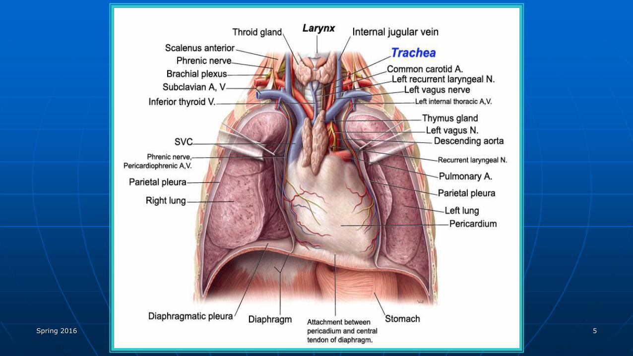
• Thoracic cavity.
Spring 2016 Dr. Maher Hadidi, University of Jordan 2
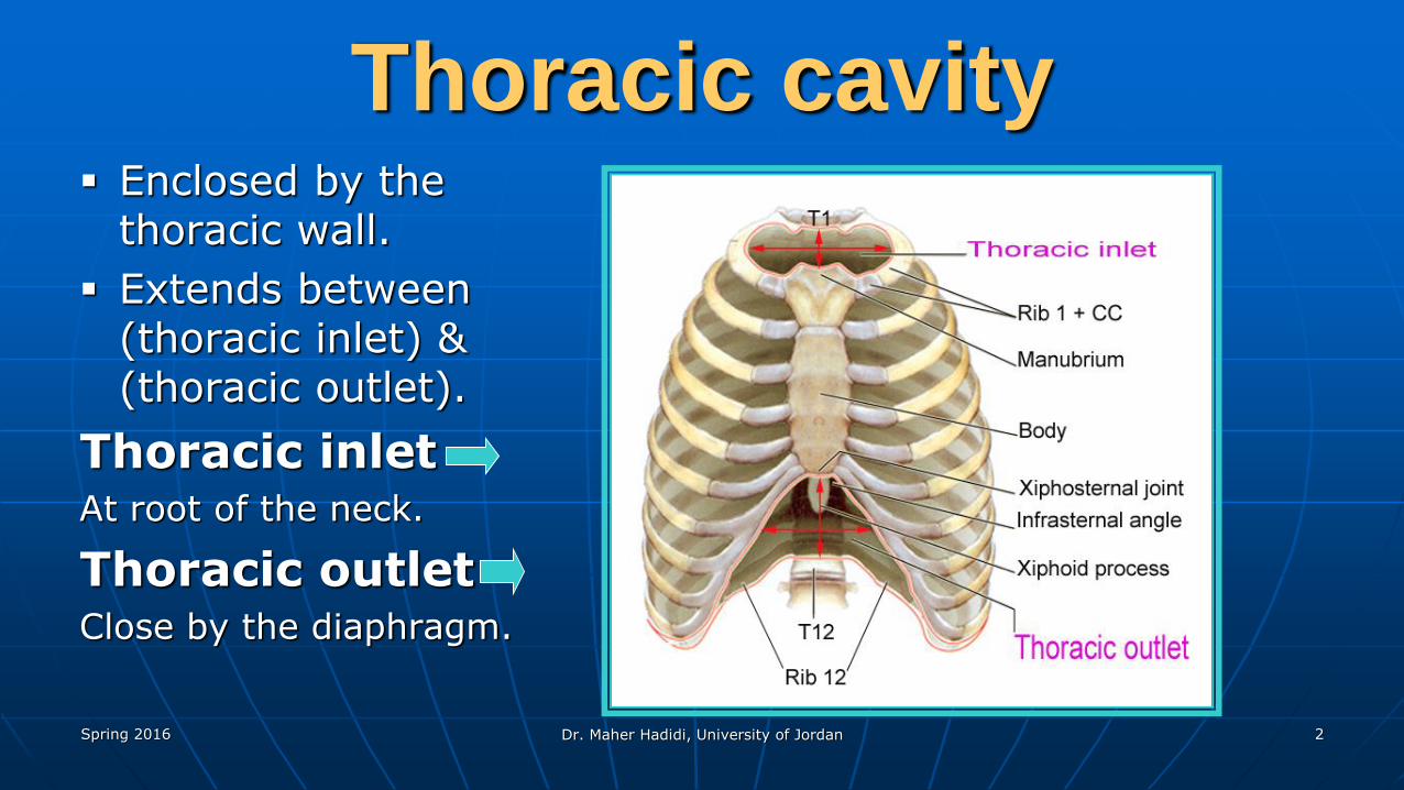
Thoracic cavity Enclosed by the
thoracic wall.
Extends between (thoracic inlet) & (thoracic outlet).
Thoracic inletAt root of the neck.
Thoracic outletClose by the diaphragm.
Spring 2016 Dr. Maher Hadidi, University of Jordan 3
Thoracic cavity
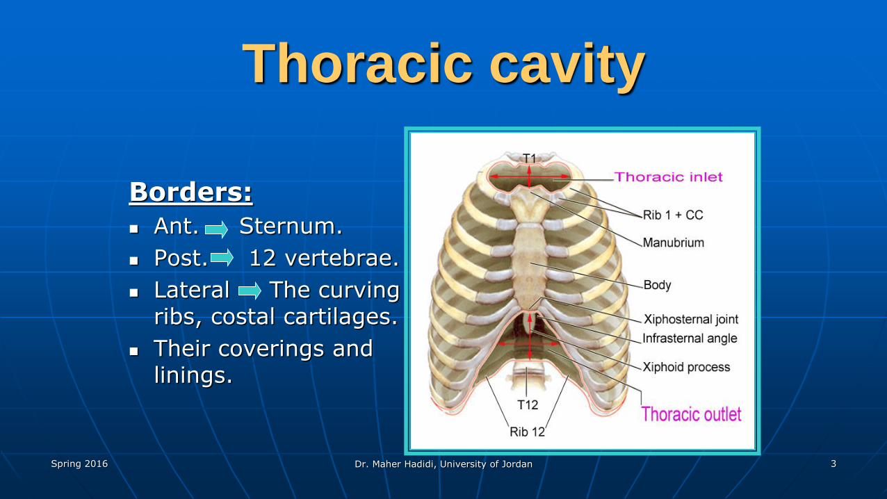
Borders:
Ant. Sternum.
Post. 12 vertebrae.
Lateral The curving ribs, costal cartilages.
Their coverings and linings.
Spring 2016 Dr. Maher Hadidi, University of Jordan 4
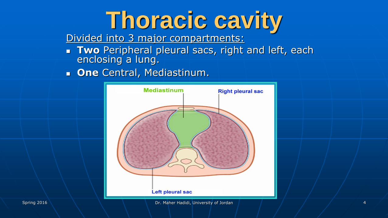
Thoracic cavityDivided into 3 major compartments:
Two Peripheral pleural sacs, right and left, each enclosing a lung.
One Central, Mediastinum.
Spring 2016 Dr. Maher Hadidi, University of Jordan 5
Spring 2016 Dr. Maher Hadidi, University of Jordan 6
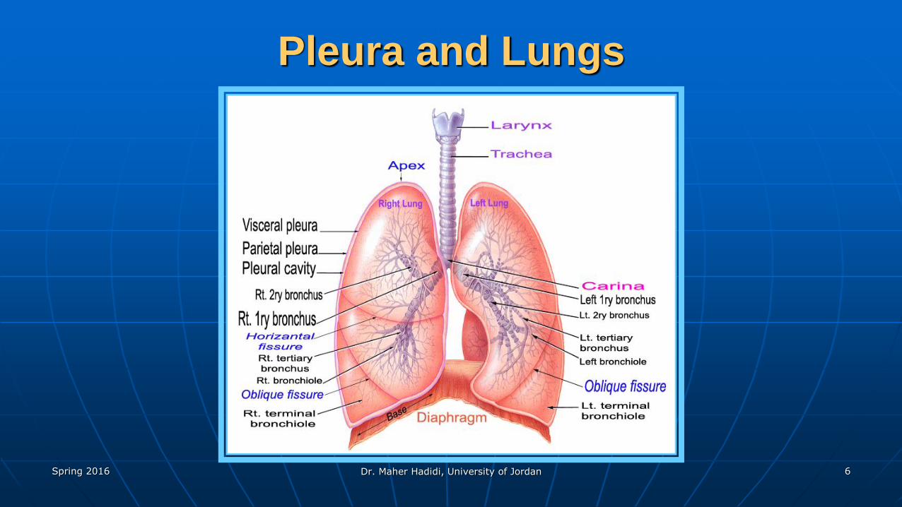
Pleura and Lungs
Spring 2016 Dr. Maher Hadidi, University of Jordan 7
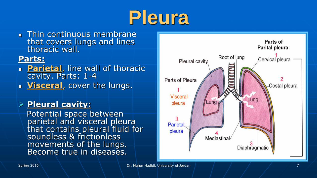
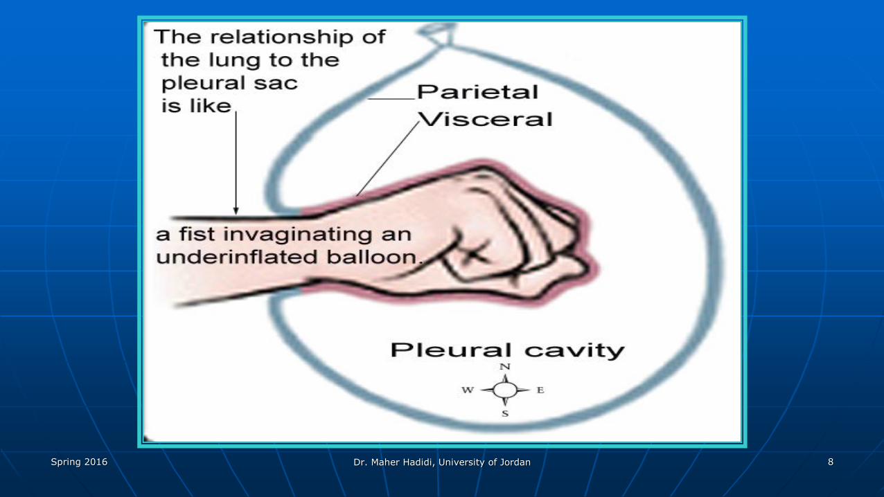
Pleura Thin continuous membrane
that covers lungs and lines thoracic wall.
Parts: Parietal, line wall of thoracic
cavity. Parts: 1-4 Visceral, cover the lungs.
Pleural cavity:Potential space between parietal and visceral pleura that contains pleural fluid for soundless & frictionless movements of the lungs. Become true in diseases.
Spring 2016 Dr. Maher Hadidi, University of Jordan 8
Spring 2016 Dr. Maher Hadidi, University of Jordan 9
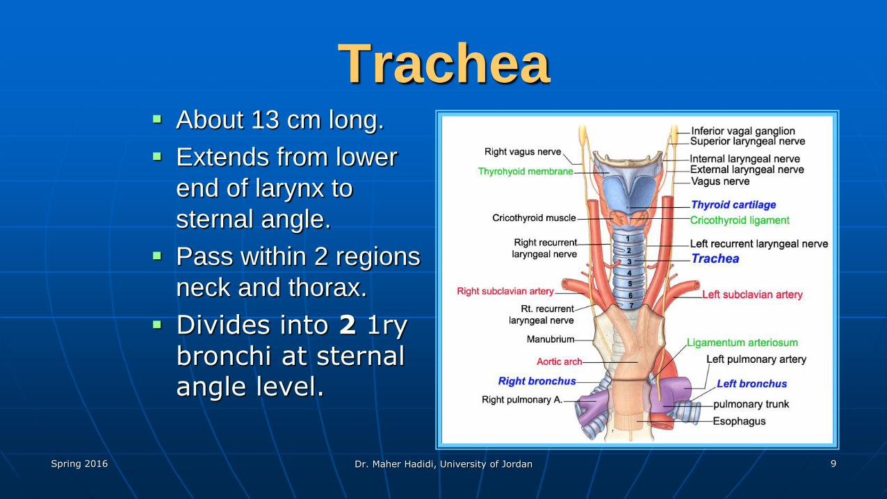
Trachea About 13 cm long.
Extends from lower
end of larynx to
sternal angle.
Pass within 2 regions
neck and thorax.
Divides into 2 1ry bronchi at sternal angle level.
Spring 2016 Dr. Maher Hadidi, University of Jordan 10
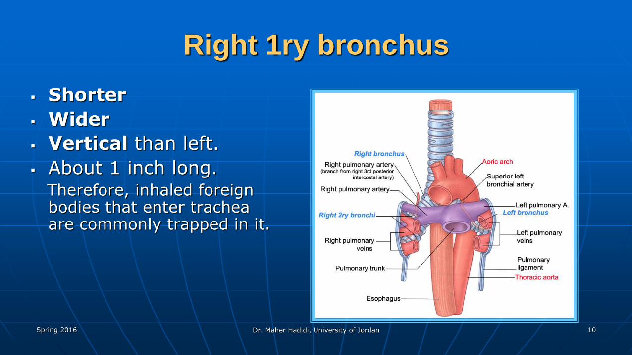
Right 1ry bronchus
Shorter
Wider
Vertical than left.
About 1 inch long.Therefore, inhaled foreign bodies that enter trachea are commonly trapped in it.
Spring 2016 Dr. Maher Hadidi, University of Jordan 11
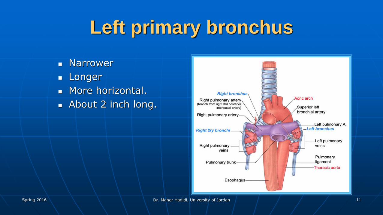
Left primary bronchus
Narrower
Longer
More horizontal.
About 2 inch long.
Spring 2016 Dr. Maher Hadidi, University of Jordan 12
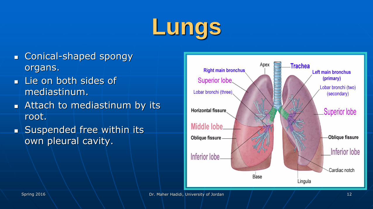
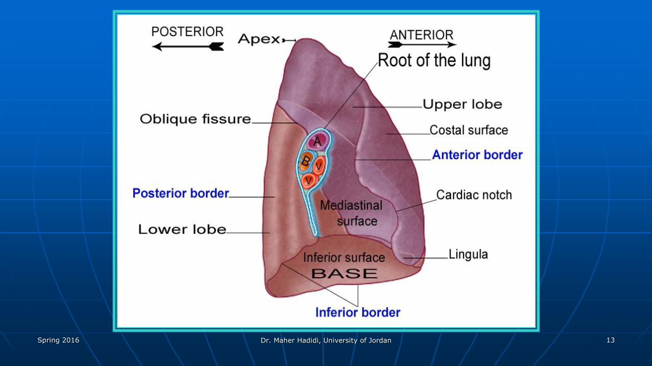
Lungs Conical-shaped spongy
organs.
Lie on both sides of mediastinum.
Attach to mediastinum by its root.
Suspended free within its own pleural cavity.
Spring 2016 Dr. Maher Hadidi, University of Jordan 13
Spring 2016 Dr. Maher Hadidi, University of Jordan 14
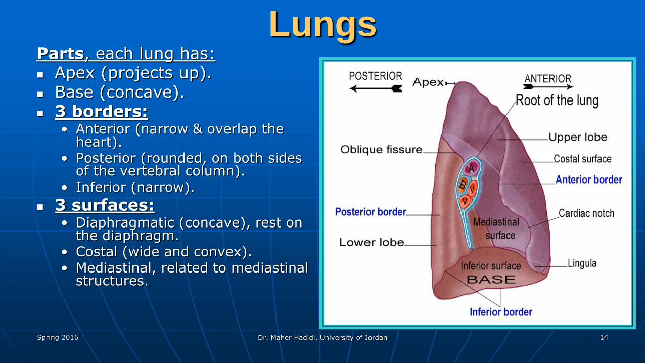
LungsParts, each lung has: Apex (projects up). Base (concave). 3 borders:
• Anterior (narrow & overlap the heart).
• Posterior (rounded, on both sides of the vertebral column).
• Inferior (narrow).
3 surfaces:• Diaphragmatic (concave), rest on
the diaphragm.• Costal (wide and convex). • Mediastinal, related to mediastinal
structures.
Spring 2016 Dr. Maher Hadidi, University of Jordan 15
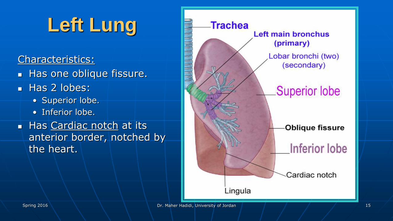
Left Lung
Characteristics:
Has one oblique fissure.
Has 2 lobes:
• Superior lobe.
• Inferior lobe.
Has Cardiac notch at its anterior border, notched by the heart.
Spring 2016 Dr. Maher Hadidi, University of Jordan 16
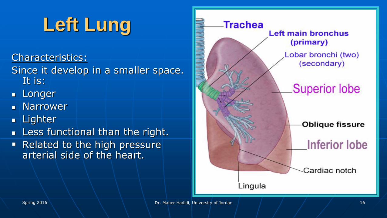
Left Lung
Characteristics:
Since it develop in a smaller space. It is:
Longer
Narrower
Lighter
Less functional than the right.
Related to the high pressure arterial side of the heart.
Spring 2016 Dr. Maher Hadidi, University of Jordan 17
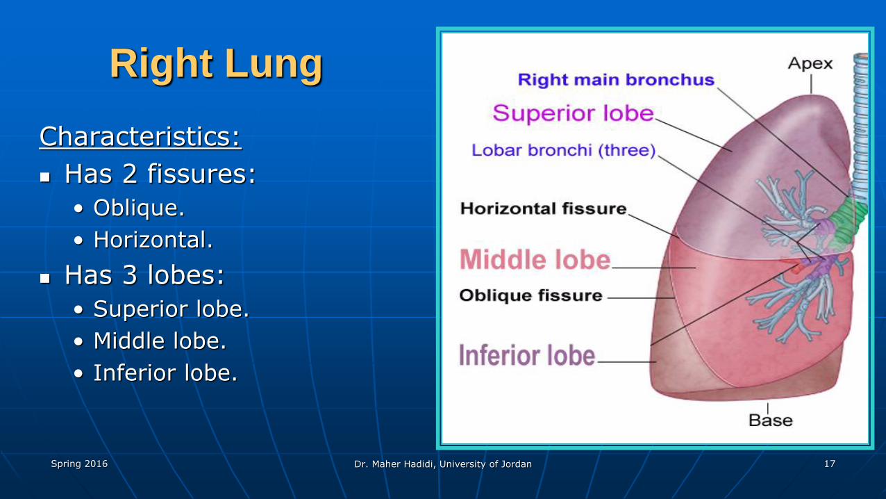
Right Lung
Characteristics:
Has 2 fissures:
• Oblique.
• Horizontal.
Has 3 lobes:
• Superior lobe.
• Middle lobe.
• Inferior lobe.
Spring 2016 Dr. Maher Hadidi, University of Jordan 18
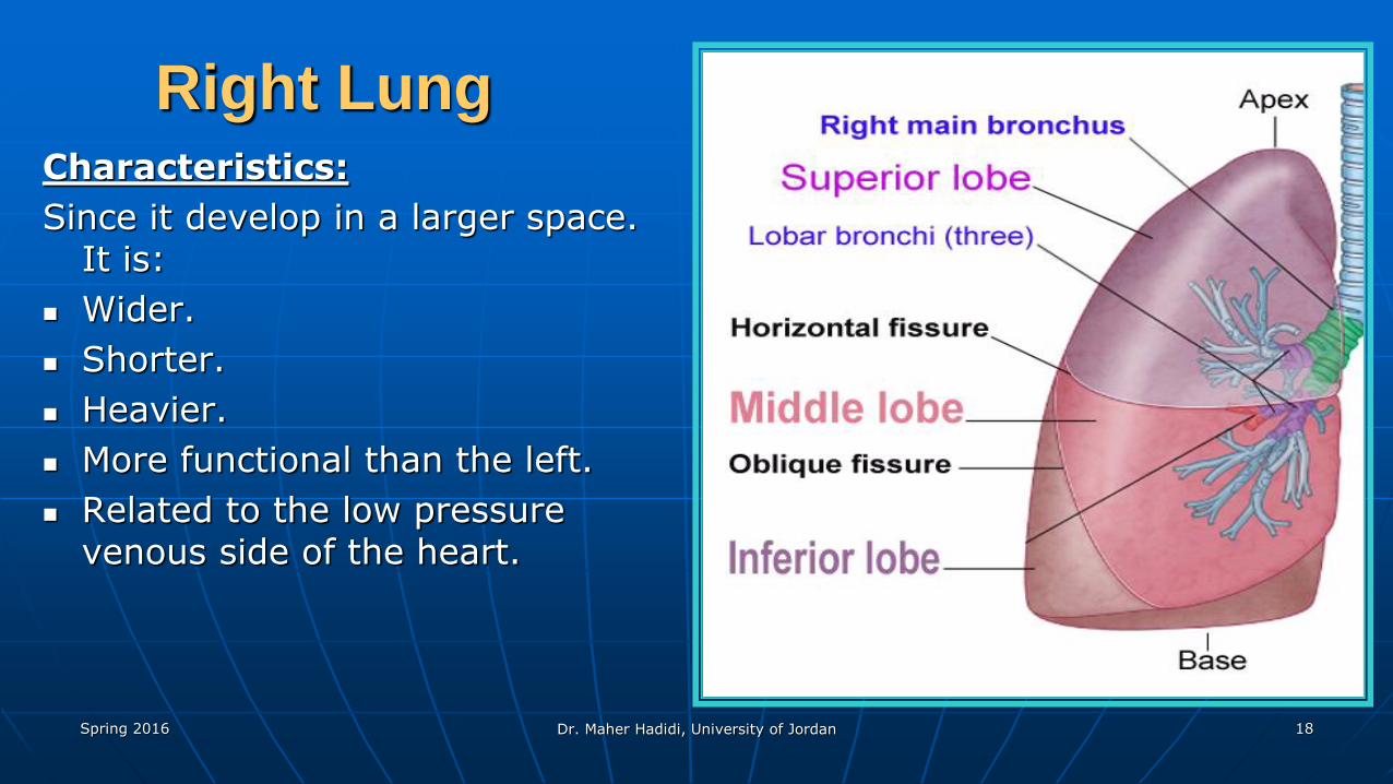
Right LungCharacteristics:
Since it develop in a larger space. It is:
Wider.
Shorter.
Heavier.
More functional than the left.
Related to the low pressure venous side of the heart.
Spring 2016 Dr. Maher Hadidi, University of Jordan 19
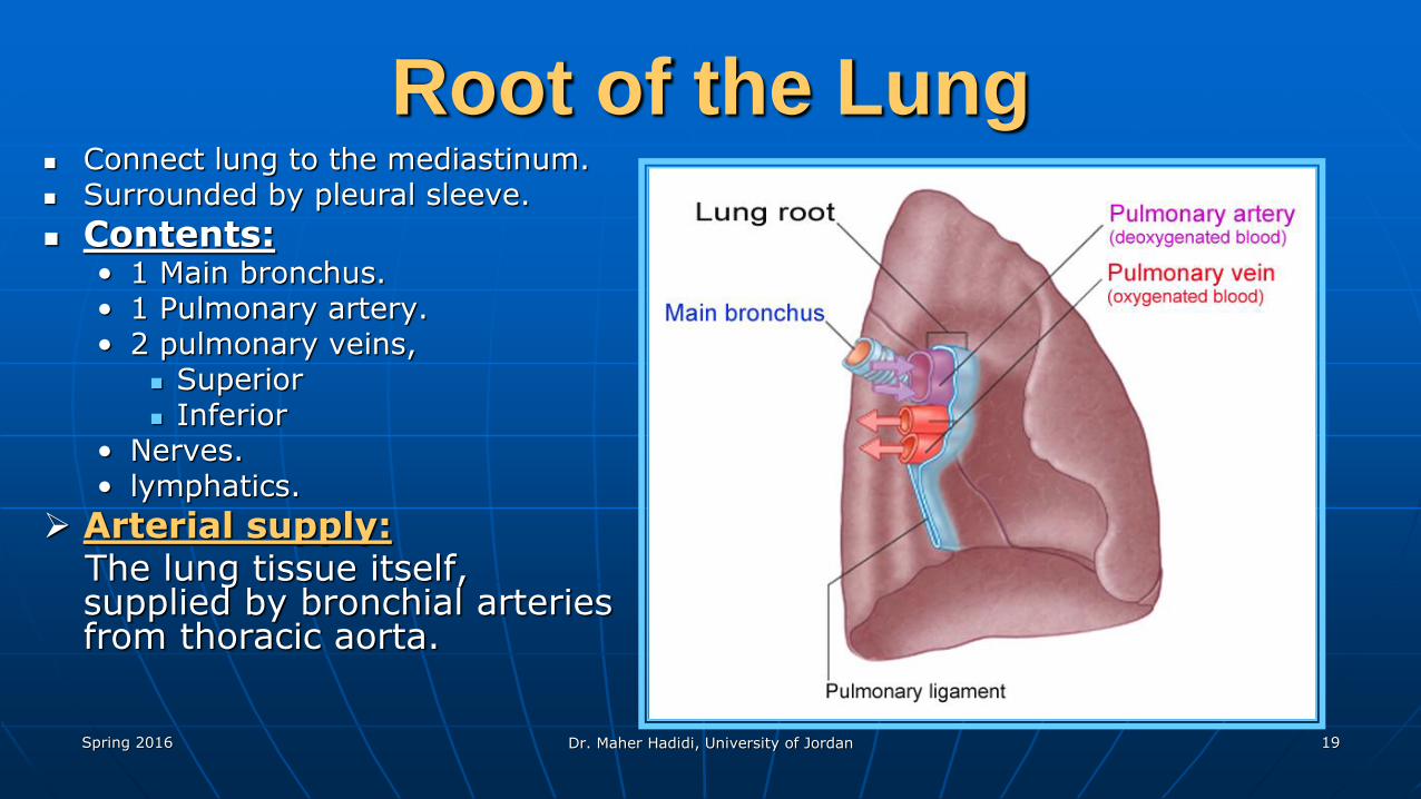
Root of the Lung Connect lung to the mediastinum. Surrounded by pleural sleeve.
Contents:• 1 Main bronchus.• 1 Pulmonary artery.• 2 pulmonary veins,
Superior Inferior
• Nerves. • lymphatics.
Arterial supply:The lung tissue itself, supplied by bronchial arteries from thoracic aorta.
Spring 2016 Dr. Maher Hadidi, University of Jordan 20
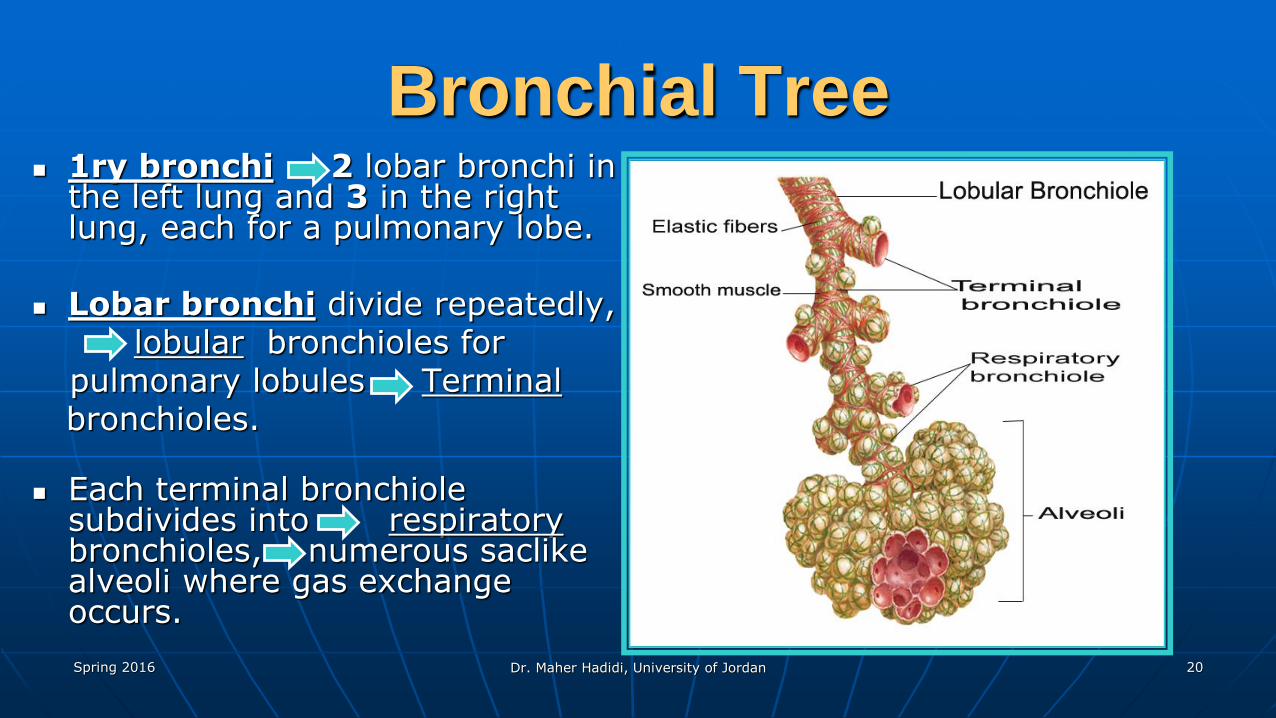
Bronchial Tree 1ry bronchi 2 lobar bronchi in
the left lung and 3 in the right lung, each for a pulmonary lobe.
Lobar bronchi divide repeatedly,lobular bronchioles for
pulmonary lobules Terminalbronchioles.
Each terminal bronchiole subdivides into respiratorybronchioles, numerous saclike alveoli where gas exchange occurs.