Embed Size (px)
Citation preview

Thuesday:MACS® Technology for magnetic isolation of cells and molecules
• Introduction • Features of paramagnetic MicroBeads • General procedure of magnetic cell isolation • Overview of applications in molecular biology
µMACS epitope tagged protein isolation • Expression of tagged/fusion proteins, e.g. GFP fusion proteins • Magnetic protein isolation
Wednesday: • Detection of proteins, results and trouble-shooting • Optimizing protein expression analysis by transfected cell selection (MACSelect)
• organism for protein expression (bacteria, eukaryotic)
• expression vector (promoter etc.)
• transient or stable transfection
• choice of tag
Expression of tagged/fusion proteins, e.g. GFP fusion proteins
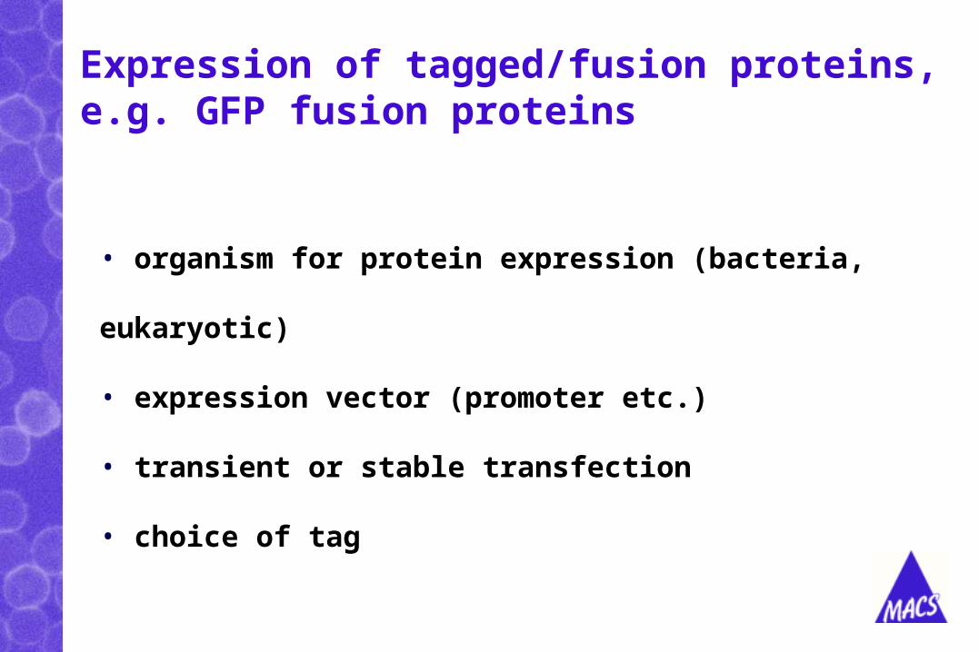
General features of the MicroBeads
• Super-paramagnetic
• Colloidal non-sedimenting Extremly fast reactionkinetics & efficient
• Extremely small magnetic labeling
• Biodegradable & non-toxic

Magnetic protein isolation
1. Cell lysis
2. Magnetic labeling with MicroBeads
• µMACS Protein A and Protein G MicroBeads
• µMACS Anti Epitope Tag MicroBeads, e.g. Anti-GFP
MicroBeads
• µMACS Streptavidin MicroBeads
3. Specific protein isolation with µ or M Columns and MACS® Separator
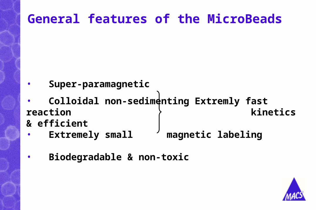
Immunoprecipitation with µMACSTM Protein A / Protein G MicroBeads
Cell lysis andimmunolabeling
Magneticseparation
SDS-PAGE orenzymatic assay
Pure protein in less than 2 hours
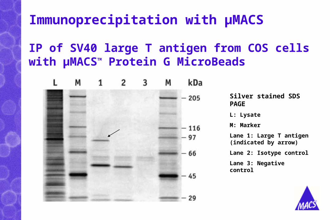
Immunoprecipitation with µMACS
Silver stained SDS PAGE
L: Lysate
M: Marker
Lane 1: Large T antigen (indicated by arrow)
Lane 2: Isotype control
Lane 3: Negative control
IP of SV40 large T antigen from COS cells with µMACSTM Protein G MicroBeads
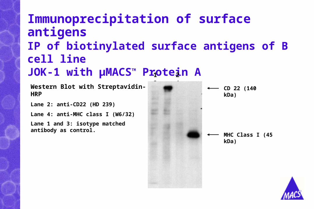
Immunoprecipitation of surface antigens
IP of biotinylated surface antigens of B cell line JOK-1 with µMACSTM Protein A
Western Blot with Streptavidin-HRP
Lane 2: anti-CD22 (HD 239)
Lane 4: anti-MHC class I (W6/32)
Lane 1 and 3: isotype matched antibody as control.
1 2 3 4
CD 22 (140 kDa)
MHC Class I (45 kDa)
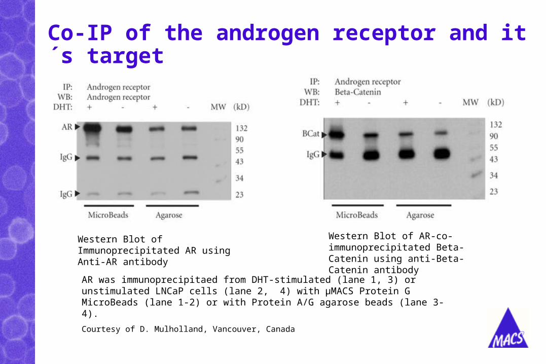
Co-IP of the androgen receptor and it´s target
Western Blot of Immunoprecipitated AR using Anti-AR antibody
Western Blot of AR-co-immunoprecipitated Beta-Catenin using anti-Beta-Catenin antibody
AR was immunoprecipitaed from DHT-stimulated (lane 1, 3) or unstimulated LNCaP cells (lane 2, 4) with µMACS Protein G MicroBeads (lane 1-2) or with Protein A/G agarose beads (lane 3-4).
Courtesy of D. Mulholland, Vancouver, Canada
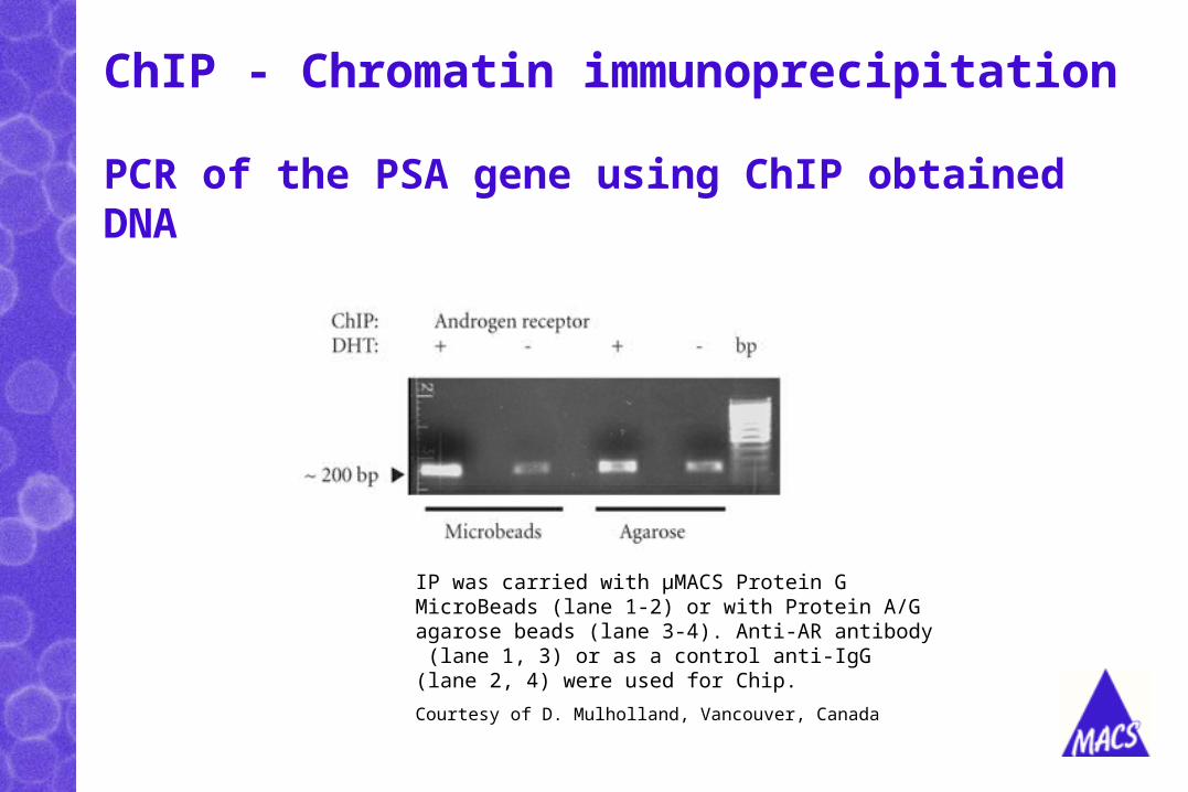
ChIP - Chromatin immunoprecipitation
PCR of the PSA gene using ChIP obtained DNA
IP was carried with µMACS Protein G MicroBeads (lane 1-2) or with Protein A/G agarose beads (lane 3-4). Anti-AR antibody (lane 1, 3) or as a control anti-IgG (lane 2, 4) were used for Chip.
Courtesy of D. Mulholland, Vancouver, Canada

µMACSTM Epitope Tag Protein Isolation Kits
Application
isolation of recombinant proteins with epitope tags
and their interaction partners from eukaryotic cells
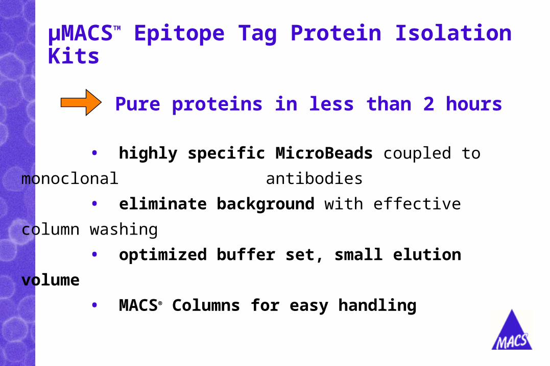
µMACSTM Epitope Tag Protein Isolation Kits
Pure proteins in less than 2 hours
• highly specific MicroBeads coupled to monoclonal
antibodies
• eliminate background with effective column washing
• optimized buffer set, small elution volume
• MACS® Columns for easy handling

Epitope-tags
• His : artificial tag of 6 Histidines
• HA= Hemaglutinin (or Hemagglutinin) : viral gene
• c-myc : human c-myc is proto-oncogene
• GST = Glutathion S-transferase
• GFP = Green Fluorescent Protein
BM 10/2002
µMACS Epitope Tagged Protein Isolation Kits
GFP = Green Fluorescent Protein from Aequoria victoria
BM 10/2002
Zur Anzeige wird der QuickTime™ Dekompressor “GIF”
benötigt.
MACS in Molecular Biology
MACS in Molecular Biology
Epitope tagged proteins - Advantages:
• study of proteins possible without a specific antibody (production of specific Ab difficult & time-consuming)
• simplified isolation: known epitope + known interaction
• standardized isolation: using one established system (expression vector/tag) for study of different proteins
BM 10/2002
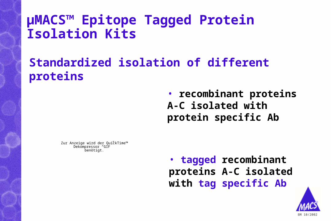
Standardized isolation of different proteins
• recombinant proteins A-C isolated with protein specific Ab
BM 10/2002
Zur Anzeige wird der QuickTime™ Dekompressor “GIF”
benötigt.
• tagged recombinant proteins A-C isolated with tag specific Ab
µMACS™ Epitope Tagged Protein Isolation Kits
MACS in Molecular Biology
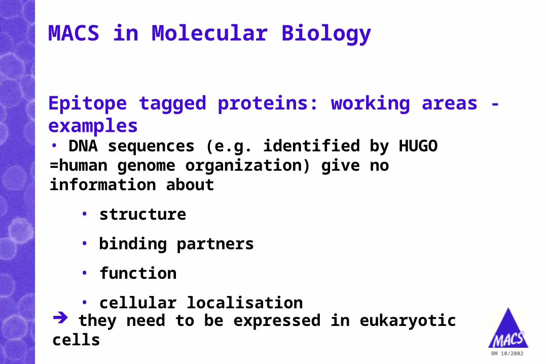
Epitope tagged proteins: working areas - examples
• DNA sequences (e.g. identified by HUGO =human genome organization) give no information about
• structure
• binding partners
• function
• cellular localisation
BM 10/2002
they need to be expressed in eukaryotic cells
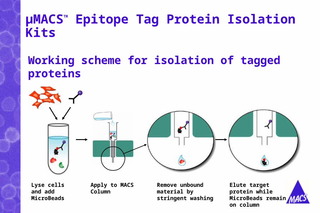
Working scheme for isolation of tagged proteins
Lyse cells and add MicroBeads
Apply to MACS Column
Remove unbound material by stringent washing
Elute target protein while MicroBeads remain on column
µMACSTM Epitope Tag Protein Isolation Kits
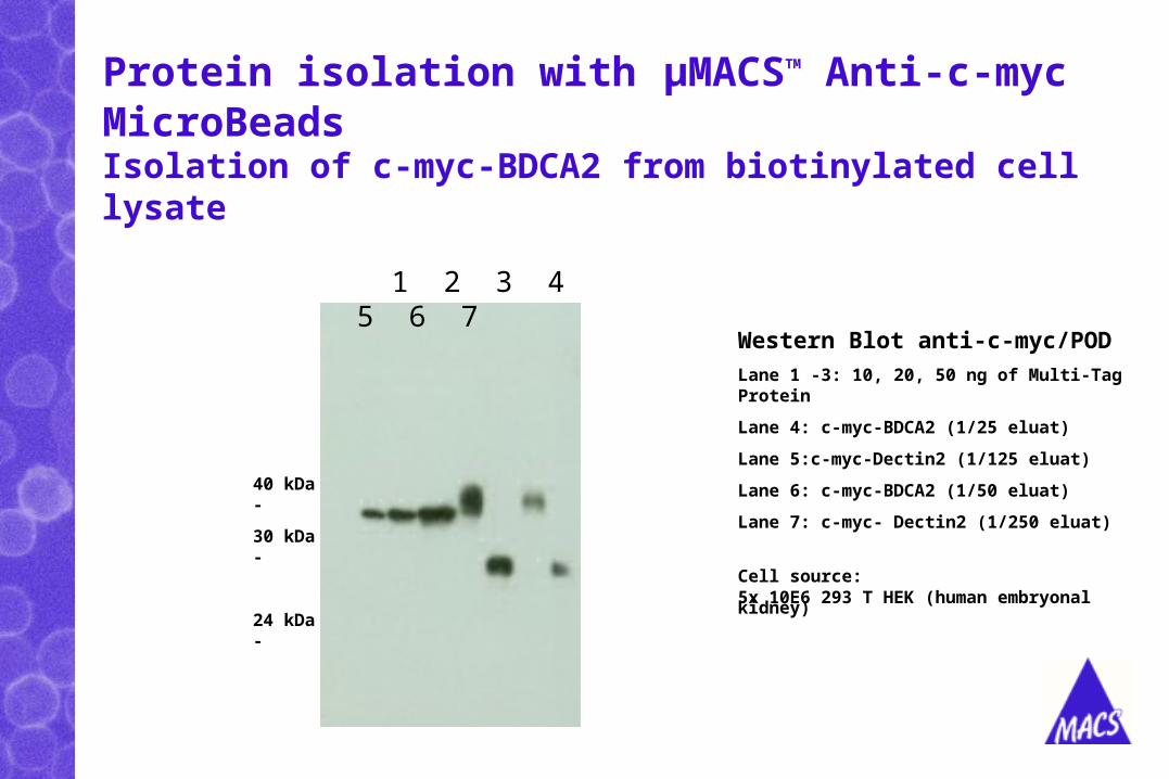
Protein isolation with µMACSTM Anti-c-myc MicroBeads
Western Blot anti-c-myc/POD
Lane 1 -3: 10, 20, 50 ng of Multi-Tag Protein
Lane 4: c-myc-BDCA2 (1/25 eluat)
Lane 5:c-myc-Dectin2 (1/125 eluat)
Lane 6: c-myc-BDCA2 (1/50 eluat)
Lane 7: c-myc- Dectin2 (1/250 eluat)
Cell source: 5x 10E6 293 T HEK (human embryonal kidney)
Isolation of c-myc-BDCA2 from biotinylated cell lysate
1 2 3 4 5 6 7
40 kDa -
30 kDa -
24 kDa -
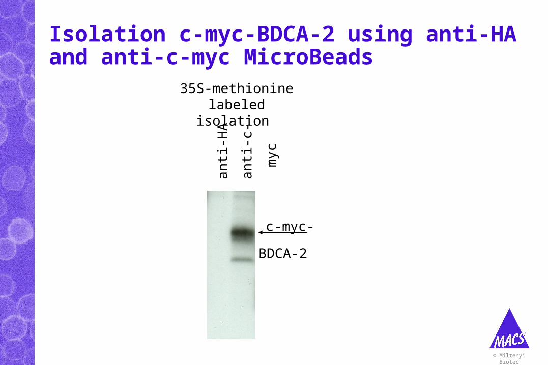
Isolation c-myc-BDCA-2 using anti-HA and anti-c-myc MicroBeads
© Miltenyi Biotec
c-myc-
BDCA-2 a
nti-
HA
ant
i-c-
myc
35S-methionine labeled isolation
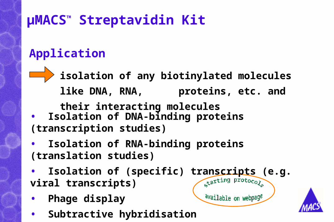
Application
isolation of any biotinylated molecules like DNA,
RNA, proteins, etc. and their interacting molecules
µMACSTM Streptavidin Kit
• Isolation of DNA-binding proteins (transcription studies)
• Isolation of RNA-binding proteins (translation studies)
• Isolation of (specific) transcripts (e.g. viral transcripts)
• Phage display
• Subtractive hybridisation
• SAGE
• Isolation of tRNA molecules
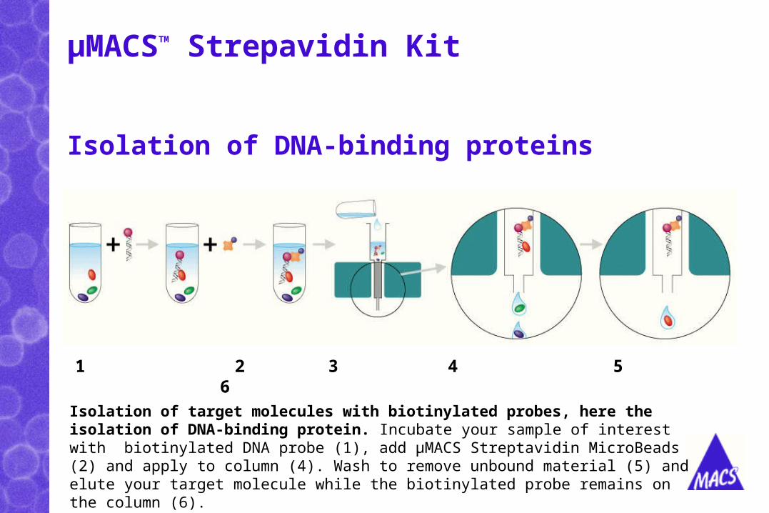
µMACSTM Strepavidin Kit
Isolation of target molecules with biotinylated probes, here the isolation of DNA-binding protein. Incubate your sample of interest with biotinylated DNA probe (1), add µMACS Streptavidin MicroBeads (2) and apply to column (4). Wash to remove unbound material (5) and elute your target molecule while the biotinylated probe remains on the column (6).
Isolation of DNA-binding proteins
1 2 3 4 5 6
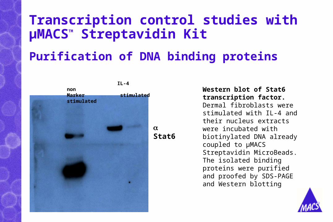
Transcription control studies with µMACSTM Streptavidin Kit
Purification of DNA binding proteins
IL-4 nonMarker stimulated stimulated
Stat6
Western blot of Stat6 transcription factor. Dermal fibroblasts were stimulated with IL-4 and their nucleus extracts were incubated with biotinylated DNA already coupled to µMACS Streptavidin MicroBeads. The isolated binding proteins were purified and proofed by SDS-PAGE and Western blotting
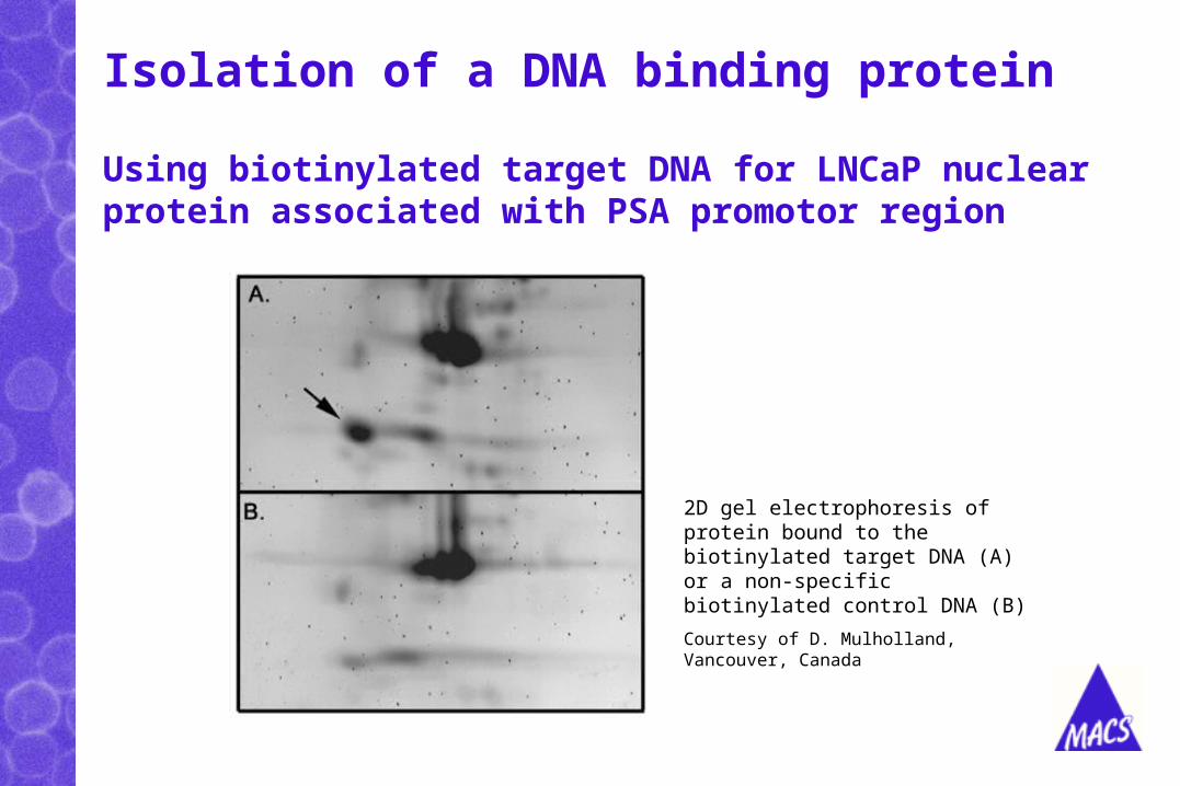
Isolation of a DNA binding protein
Using biotinylated target DNA for LNCaP nuclear protein associated with PSA promotor region
2D gel electrophoresis of protein bound to the biotinylated target DNA (A) or a non-specific biotinylated control DNA (B)
Courtesy of D. Mulholland, Vancouver, Canada
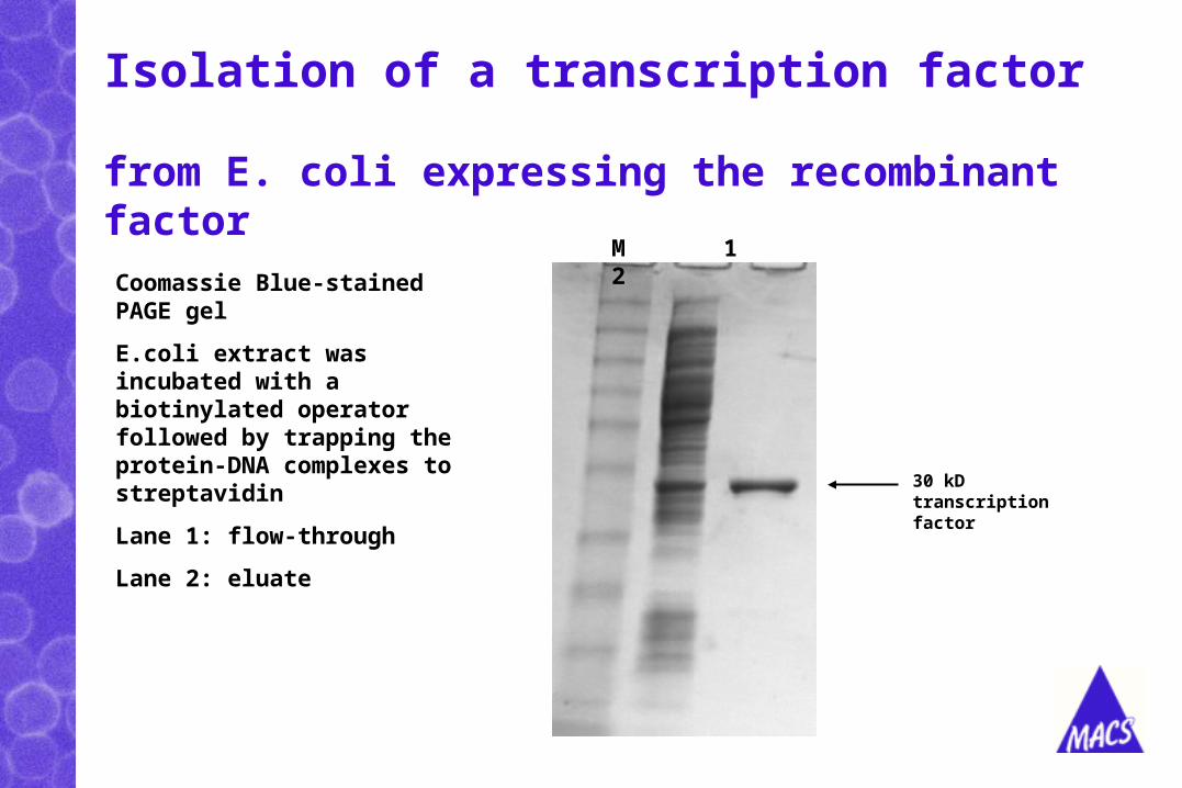
Isolation of a transcription factor
Coomassie Blue-stained PAGE gel
E.coli extract was incubated with a biotinylated operator followed by trapping the protein-DNA complexes to streptavidin
Lane 1: flow-through
Lane 2: eluate
from E. coli expressing the recombinant factor
M 1 2
30 kDtranscription factor
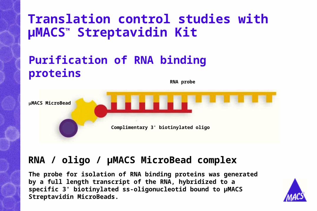
Translation control studies with µMACSTM Streptavidin Kit
The probe for isolation of RNA binding proteins was generated by a full length transcript of the RNA, hybridized to a specific 3' biotinylated ss-oligonucleotid bound to µMACS Streptavidin MicroBeads.
Purification of RNA binding proteins
RNA / oligo / µMACS MicroBead complex
Complimentary 3' biotinylated oligo
RNA probe
MACS MicroBead
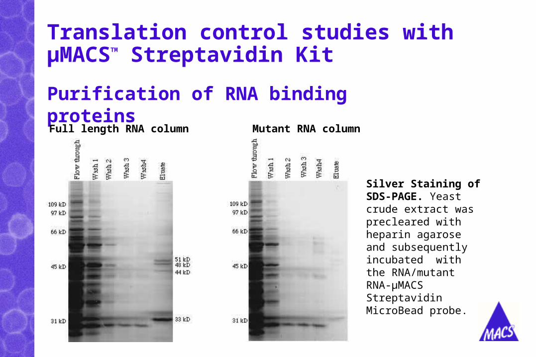
Translation control studies with µMACSTM Streptavidin Kit
Silver Staining of SDS-PAGE. Yeast crude extract was precleared with heparin agarose and subsequently incubated with the RNA/mutant RNA-µMACS Streptavidin MicroBead probe.
Purification of RNA binding proteins
Full length RNA column Mutant RNA column

µMACS in proteomics research
• Protein isolation
• Interaction partner isolation
• Protein complex isolation
• Enzymatic, on-column reactions with the temperature-controlled thermoMACS Separator