Embed Size (px)
Citation preview

American Journal of Pathology, Vol. 147, No. 6, December 1995
Copyright X American Societyfor Investigative Pathology
Time Course of Increased Cellular Proliferation inCollateral Arteries after Administration ofVascular Endothelial Growth Factor in a RabbitModel of Lower Limb Vascular Insufficiency
Satoshi Takeshita,* Susan T. Rossow,*Marianne Kearney,* Lu P. Zheng,*Christophe Bauters,* Stuart Bunting,tNapoleone Ferrara,t James F. Symes,* andJeffrey M. Isner*From the Departments ofMedicine (Cardiology), Surgery(Cardiovascular), and Biomedical Research,* St. Elizabeth'sMedical Center, Tufts University School ofMedicine, Boston,Massachusetts, and the Department of CardiovascularResearch,t Genentech Inc., South San Francisco, California
Proliferation of vascular ceUs has been previ-ously shown to contribute to spontaneous devel-opment of coronary coUaterals. Recent studiesfrom several laboratories have established thatcoUateral artery growth in both the heart andlimb can be enhanced by administration ofangio-genic growth factors, or therapeutic angiogene-sis. In this study, we sought (1) to define theextent and time course of endothelial cell (EC)and smooth muscle ceU (SMC) proliferation ac-companying spontaneous collateral developmentduring limb ischemia and (2) to determine theextent to which proliferative activity ofECs andSMCs is augmented during therapeutic angiogen-esis with vascular endothelial growth factor(VEGF), a heparin-binding EC-specific mitogen.Ten days after induction oflimb ischemia by sur-gicaly excising the femoral artery of rabbits,either VEGF (500 to 1000 pg) or saline was ad-ministered as a bolus into the iliac artery of theischemic limb. Celular proliferation was evalu-ated by bromodeoxyuridine labeling for 24hours at day 0 (immediately before VEGF admin-istration) and at days 3, 5, and 7 after VEGF. ECproliferation in the midzone coUaterals of VEGF-treated animals increased 2.8-fold at day 5 (P <
0.05 versus control), and returned to baselinelevels by day 7. SMC proliferation in midzone
coUaterals also increased 2. 7-fold in response to
VEGF (P < 0.05). No significant increase in EC orSMC proliferation was observed in either thestem or re-entry coUaterals of VEGF-treated ani-mals compared with untreated ischemic controlanimals. Reduction ofhemodynamic deficit in theischemic limb measured by lower limb bloodpressure was documented at day 7 after VEGF(P < 0.01 versus untreated, ischemic control).These data thus (1) establish the contribution ofcelularproliferation to coUateral vessel develop-ment in limb ischemia and (2) support the con-cept that augmented celular proliferation con-tributes to the enhancedformation of coUateralvessels after therapeutic angiogenesis withVEGF. (AmJPathol 1995, 1477:1649-1660)
The paradigm for new blood vessel growth, elegantlyoutlined by D'Amore and Thompson,1 suggests thatthe initiating feature of angiogenesis is the activationof endothelial cells (ECs) within a parent vessel,followed by disruption of the basement membraneand subsequent migration of the ECs into the inter-stitial space, possibly in the direction of an ischemicstimulus. Concomitant and/or subsequent EC prolif-eration, intracellular-vacuolar lumen formation, peri-cyte capping, and production of a basement mem-brane complete the developmental sequence. Thisparadigm is typically applied to new capillary forma-tion, or angiogenesis, a process that some have
Supported in part by grants HL-40518 and HL-02824 (J. M. I.) fromthe National Heart, Lung, and Blood Institute, National Institutes ofHealth, Bethesda, MD. Work performed during Dr. Bauter's tenureas the recipient of the French Federation of Cardiology ResearchFellowship 1993.
Accepted for publication August 2, 1995.
Address reprint requests to Dr. Jeffrey M. Isner, St. Elizabeth'sMedical Center, 736 Cambridge Street, Boston, MA 02135.
1649

1650 Takeshita et alAJP December 1995, Vol. 147, No. 6
suggested may be distinct from the formation oflarger blood vessels, or vasculogenesis.2The proliferative component of this paradigm has
been investigated in several animal models of natu-rally occurring collateral vessel development.i8Studies carried out in the canine and swine coronarycirculations, as well as the canine and rodent renalarterial circulations, have established that cellularproliferation is indeed a feature of collaterals thatdevelop consequent to arterial occlusion, therebyproviding evidence of new blood vessel formation orangiogenesis.
Recent investigations have documented the fea-sibility of using recombinant formulations of angio-genic growth factors to further augment collateralartery development in similar animal models. Thisnovel strategy for the treatment of vascular insuffi-ciency has been termed therapeutic angiogenesis.9The angiogenic growth factors first employed for thispurpose comprised members of the fibroblastgrowth factor (FGF) family.10-13 More recently,we9'14'15 and others16 have shown that the EC-spe-cific mitogen, vascular endothelial growth factor(VEGF),17'18 is a potent agent for collateral vesselaugmentation in the lower extremity and coronarycirculations, respectively. Studies performed previ-ously in our laboratory, for example, showed that 500to 1000 ,ug of VEGF administered as a single intra-arterial bolus to the internal iliac artery of rabbits inwhich the ipsilateral femoral artery was excised toinduce severe, unilateral hind limb ischemia pro-duced angiographic and histological evidence ofenhanced collateral vessel formation within 10 days;consequent amelioration of blood pressure9 andflowl were significantly greater in the ischemic limbof animals treated with VEGF compared with con-trols.
The extent to which cellular proliferation contrib-utes to collateral vessel development in the setting oflower extremity ischemia has not been previouslydefined. Accordingly, in the current study we soughtto document the contribution to and sequence ofcellular proliferation during spontaneous collateralvessel development in the rabbit hind limb model oflower extremity ischemia and, furthermore, deter-mine the extent to which this template is modified bytherapeutic angiogenesis.
Materials and Methods
Animal ModelCellular proliferation of collateral arteries during ther-apeutic angiogenesis by VEGF was investigated with
a rabbit model of hind limb ischemia as previouslydescribed.9'1'14 15 All protocols were approved bySt. Elizabeth's Institutional Animal Care and UseCommittee. Male New Zealand White rabbits weigh-ing 3.5 to 4.0 kg (Pine Acre Rabbitry, Norton, MA)were anesthetized with a mixture of ketamine (50mg/kg) and acepromazine (0.8 mg/kg) after premed-ication with xylazine (2.5 mg/kg). A vertical incisionwas performed in one limb, extending inferiorly fromthe inguinal ligament. The entire femoral artery, in-cluding its branches (the inferior epigastric, deepfemoral, lateral circumflex, and superficial epigastricarteries), was then dissected, ligated, and com-pletely excised from its proximal origin to the pointdistally where it bifurcates into the saphenous andpopliteal arteries. Once the femoral artery is excised,thrombotic occlusion of the external iliac artery ex-tends retrograde to its origin from the common iliacartery. The internal iliac artery, which usually suppliesperfusion to the pelvic organs of the rabbit, but not thehind limb, now becomes the sole source of perfusionfor the distal hind limb; as such, the blood supply to thedistal limb of this animal model becomes almost en-tirely dependent upon collateral arteries that may orig-inate from the internal iliac artery (Figure 1). Prophylac-tic antibiotics (enrofloxacin, 2.5 mg/kg; Miles, ShawneeMission, KS) were administered subcutaneously for atotal of 5 days post-operatively. Analgesia (levorphanoltartrate, 60 mg/kg; Roche Laboratories, Nutley, NJ)was administered subcutaneously as required for evi-dence of discomfort throughout the duration of theexperiment. All animals used in this investigation wereprepared specifically for this study and were not usedfor any other separately reported studies.
Intra-Arterial VEGFAdministrationThe soluble 165-amino-acid isoform of VEGF waspurified from transfected Chinese hamster ovarycells as previously described.19 The purity of thematerial was assessed by silver-stained sodium do-decyl sulfate polyacrylamide gel electrophoresis geland by the presence of a single NH2-terminal aminoacid sequence.An interval of 10 days between the time of surgery
and the administration of VEGF was incorporated toallow for the recovery of rabbits and the stabilizationof endogenous development of collateral arteries. At10 days post-surgery (day 0), a 3 Fr. infusion cath-eter (Tracker-1 8, Target Therapeutics, San Jose, CA)was introduced from the carotid artery and ad-vanced into the internal iliac artery of the ischemiclimb under fluoroscopic guidance. After the catheterwas washed with 3 ml of saline containing 0.1%

Cellular Proliferation after VEGF 1651AJP December 1995, Vol. 14 7, No. 6
- _ Common iliac arlery
A< Thrombotic occlusion ot external iliac artery
A-" Surgically excised femoral artery
Stem -r-1 , Re-entry
(Internal iliacasrery) (Saphenous and popilteal arteries)
Mldzone
Figure 1. Classification ofcollateral vessel anatomy. A: Aortic angiog-raphy ofan iscbemic rabbit limb. Arrows indicate the stem, midzone,and re-entry collateral arteries. Curved arrow indicates the site oftbrombotic occlusion of the external iliac artery. B: Schematic repre-sentation of angiographic findings illustrated in A.
rabbit serum albumin (Sigma Chemical Co., St.Louis, MO), 500 to 1000 ,ug of VEGF in 5 ml of salinecontaining 0.1% albumin was selectively injectedinto the internal iliac artery as a bolus over a periodof 1 minute. The dose of VEGF employed in thisstudy (500 to 1000 ,ug, or 125 to 285 ,tg/kg) was
previously confirmed to be sufficient to augment col-lateral artery development in this animal model.9 14
These same previous studies indicated a lesser ther-apeutic effect at smaller doses of VEGF (eg, 100 ,ug),reaching a plateau at 500 ,ug. Sham-treated animalsreceiving vehicle alone (an identical volume of salinecontaining 0.1% albumin) were used as controls todelineate cellular proliferation during natural (ie,spontaneous) collateral artery development.
Lower Limb Calf Blood Pressure Ratio
To evaluate collateral development, the time course
of calf blood pressure ratio was assessed in bothhind limbs with a Doppler Flowmeter (Model 1050,Parks Medical Electronics, Aloha, OR) immediatelybefore administration of VEGF or saline (day 0) andagain at the time of sacrifice (days 3, 5, and 7). Oneach occasion, the hind limbs were shaved andcleaned, the pulse of the posterior tibial artery was
identified with a Doppler probe, and the systolicblood pressure of both limbs was determined bystandard techniques.9 14 All measurements were
performed by a single observer blinded to the treat-
ment regimen. The calf blood pressure ratio wasdefined for each rabbit as the ratio of systolic pres-sure of the ischemic limb to systolic pressure of thenormal limb.
Bromodeoxyuridine LabelingProliferation of ECs and smooth muscle cells (SMCs)was documented with the thymidine analogue 5-bro-mo-2'-deoxyuridine (BrdU; Sigma). At 24 hours be-fore sacrifice, animals received an intravenous bolusinjection of BrdU (25 mg/kg) followed by continuousdelivery (25 mg/kg) over a 24-hour period from asubcutaneously implanted osmotic pump (Alzet,Alza, Palo Alto, CA). The animals were euthanizedwith an overdose of sodium pentobarbital at each ofthe following time points: day 0 (immediately beforeVEGF), and days 3, 5, and 7 after VEGF. After ani-mals had been euthanized, a saline flush was imme-diately performed from a catheter placed in the ab-dominal aorta to clear the blood. The aorta and thedistal vasculature were then perfusion-fixed in situwith 100% methanol. Tissue harvesting was per-formed as follows based in part upon anatomic cri-teria proposed by Longland20 (Figure 1). (1) Sec-tions were taken from the principal stem artery, ie,the feeder from which collateral arteries were de-rived, in this case the distal internal iliac artery. (2)The midzone arteries were removed from the plexusof branching arteries that interconnect the stem andre-entry arteries. Because the midzone collateral ar-teries were too diminutive to identify macroscopi-cally, tissue harvesting was in this case standardizedby removing in a systematic fashion those connec-tive tissues along the mid-portion of the sciatic nervein the thigh. After tissue processing and immunohis-tochemical staining, all the arteries in each histolog-ical section were identified by light microscopy. Col-laterals with a luminal diameter smaller than 20 lumcannot be consistently quantified on standard lightmicroscopic sections;8 these vessels were thereforeexcluded from analysis (see below). (3) Sectionswere taken from the proximal portions of the principalre-entry arteries, ie, those arteries that rejoin themidzone plexus with the distal circulation; in thiscase the principal re-entry arteries are the saphe-nous and popliteal arteries. All harvested tissueswere further immersion-fixed with 100% methanol forat least 12 hours at 40C, processed, and embeddedin paraffin.
In addition, tissue specimens from the small intes-tine were harvested from each animal at necropsy.Tissues were fixed in methanol and then embeddedin paraffin as described above. These tissues were

1652 Takeshita et alAJP December 1995, Vol. 14 7, No. 6
used as a positive control for BrdU staining. Thebrown nuclear staining of epithelial crypt cells wasconfirmed in all such tissue specimens (Figure 2).
Tissue StainingProliferating cells were identified with a mousemonoclonal antibody against BrdU (Dako, Carpinte-ria, CA). Paraffin-embedded sections (6 ,um) werecut on to poly-L-lysine-coated slides and air driedovernight. Sections were incubated with 2 N HCI for10 minutes at 370C to denature DNA. Endogenousperoxidase activity was blocked with 3.0% hydrogenperoxide in phosphate-buffered saline. Sectionswere then incubated with anti-BrdU antibody (1:50dilution) for 45 minutes. Bound primary antibody wasdetected by an avidin-biotin-immunoperoxidasemethod according to the supplier's guidelines (EliteAvidin-Biotin Detection System, Signet Laboratories,Dedham, MA). Sections were lightly counterstainedwith hematoxylin. Additional staining for Bandeiraeasimplicifolia lectin B4 (BSLB4)21 and smooth musclea-actin was performed on adjacent sections to iden-tify ECs and SMCs. For lectin staining, sections pre-treated with 0.3% hydrogen peroxide were incu-bated with 5 ,ug/ml biotinylated lectin (VectorLaboratories, Burlingame, CA) overnight at 40C. Af-ter washing, slides were incubated with peroxidase-conjugated streptavidin (BioGenex Laboratories,San Ramon, CA) for 1 hour; 3-amino-9-ethyl-carba-zole (Signet Laboratories) was then applied as asubstrate for the enzyme, resulting in a brown reac-tion product. Sections were lightly counterstainedwith hematoxylin. Monoclonal antibody againstsmooth muscle a-actin (HHF-35, Enzo Diagnostics,New York, NY) was used for SMC staining in con-
-q-ga -s s * t
*# vFigure 2. Immunostaining for BrdU. Tissue specimensfrom the smallintestine were harvested from each animal at necropsy. These tissueswere used as a positive controlfor BrdU staining. The brown nuclearstaining of epithelial crypt cells was confirmed in all such tissuespecimens.
junction with the avidin-biotin peroxidase complextechnique as described previously.22 Sections werelightly counterstained with hematoxylin.
Cell CountingProliferative activity of limb arteries was quantified bycounting labeled versus unlabeled cells to determinethe BrdU labeling index. ECs and SMCs were iden-tified by their position within the vessel wall as well astheir morphology in conjunction with lectin and/orsmooth muscle a-actin staining. Each animal repre-sents an n of one, so that the labeling index for eachrabbit represents the combined average of 20 sec-tions. These 20 sections, 3 to 5 mm2 in size, werequantitatively analyzed in their entirety under a 40xobjective. Approximately 9,000 to 10,000 cells, in-cluding all ECs and SMCs in all vessels of all sizes,in the intima and the media of these 20 sections wereindividually counted, following which the BrdU label-ing index, ie, the fraction (percentage) of labelednuclei, was determined for each rabbit by averagingthe indices of all sections examined. BrdU-positivecells were identified principally in vessels 20 to 100,Ag in diameter. The EC labeling index (percentage)was defined as the number of BrdU-positive ECsdivided by the total number of ECs in the intima. TheSMC labeling index (percentage) was defined as thenumber of BrdU-positive SMCs divided by the totalnumber of SMCs in the media. Previous work fromour laboratory has indicated that >95% of the cells inthe intima of this model are ECs, and >95% of thecells in the media are SMCs (S. Takeshita, unpub-lished data). Nevertheless, we cannot exclude withcertainty the possibility that some small fraction ofthe 9,000 to 10,000 denominator cells counted indi-vidually for each of the 41 rabbits could be non-ECsor non-SMCs, including pericytes or fibroblasts. Allmeasurements were performed by a single observerblinded to the treatment regimen.
StatisticsResults were expressed as mean ± SE. Statisticalsignificance was evaluated by unpaired Student'st-test for comparisons between two means and anal-ysis of variance followed by Scheffe's procedure formore than two means. A value of P < 0.05 wasinterpreted to denote statistical significance.
ResultsProliferative activity of collateral artery ECs andSMCs was studied as a function of anatomic collat-
Cellular Proliferation after VEGF 1653AJP December 1995, Vol. 14 7, No. 6
eral zone and time course in 41 rabbits with surgi-cally induced lower limb ischemia; 5 rabbits weresacrificed at day 0 (immediately before administra-tion of VEGF or saline); 19 rabbits at days 3 (n = 5),5 (n = 8), and 7 (n = 6) after VEGF administration;and 17 rabbits at days 3 (n = 5), 5 (n = 8), and 7(n = 4) after saline administration. All 41 rabbits werelabeled with BrdU for 24 hours before sacrifice asdescribed above.
Time Course of Lower Limb Blood PressureRatio (Figure 3)The time course of lower limb blood pressure ratiowas measured to document physiological evidenceof collateral artery development.9'14 The blood pres-sure ratio at day 0 was 0.25 ± 0.04 in control groupand 0.29 ± 0.04 in VEGF-treated group (P valuenot significant). In the ischemic, untreated controlanimals, no significant increase in blood pressureratio was recorded between days 0 and 7; specif-ically, blood pressure ratio in the control groupmeasured 0.23 + 0.13 at day 3; 0.33 + 0.09 at day5; and 0.23 ± 0.11 at day 7. In contrast, among theVEGF-treated group, a statistically significant in-crease in the blood pressure ratio was observedby day 7 (P < 0.01 versus day 0); specifically,blood pressure ratio in the VEGF group measured0.40 + 0.06 at day 3; 0.32 ± 0.09 at day 5; and0.57 + 0.04 at day 7. The day 7 blood pressureratio for VEGF-treated animals was also signifi-cantly increased when compared with that re-
c0
coa ---
-
'D0.2-i0
0
co0.1~
0.0
0 3 5 7
Days
Figure 3. Time coirse of loner limb blood pressure ratio measuire-ments. The time course of lower limb blood pressure ratio (ischemiclnormal limb) was measured to document physiological evidence ofcollateral artery development. In iscbemic, untreated control animals(-----) no statistically significant increase in bloodpressure ratio uasobserved from day 0 to day 7. In contrast, among the VEGF-treatedgroup (-), a statistically significant increase in the blood pressureratio was observed by day 7( *P < 0.01, versus day 0). The day 7 bloodpressure ratio of VEGF-treated animals uas also significantly higherthan that of the controls at the same time point (*P < 0.05, versus
control). Interruption of both dashed and solid lines indicates thatblood pressure measurement was not continuously performed on adaily basis but was limited to days 0. 3. 5 and 7.
corded for the control group at the same time point(P < 0.05).
Time Course of Cellular ProliferationAssociated with Spontaneous CollateralDevelopment (Tables 1 and 2; Figure 4)
Stem Artery
In the stem artery of ischemic, untreated controlanimals, no significant change in proliferative activityof either ECs or SMCs was observed during the7-day period after administration of saline (Figure 4,A and B). Although the percentage of labeled ECsappeared to increase progressively from day 0 (0.21± 0.13%) to day 3 (0.46 ± 0.29%), day 5 (0.89 ±0.24%), and day 7 (1.35 ± 0.99%), these incrementsin the labeling index did not achieve statistical sig-nificance. Likewise, a similar frequency of labeledSMCs was observed at day 0 (0.40 + 0.21 %), day 3(0.52 ± 0.33%), day 5 (0.38 ± 0.08%), and day 7(0.47 ± 0.38%).
Midzone Artery
Proliferative activity of both ECs and SMCs wasalso stable in the midzone collateral arteries overthe 7-day study period (Figure 4, C and D). ForECs, proliferative activity was 1.78 ± 1.53% at day0, 0.47 ± 0.38% at day 3, 1.51 + 0.39% at day 5,and 1.71 ± 0.95% at day 7. For SMCs, the labelingindex was 1.87 ± 1.05% at day 0, 0.59 + 0.34% atday 3, 2.08 ± 0.48% at day 5, and 1.88 ± 1.11%at day 7.
Re-Entry Artery
In re-entry arteries, proliferative activity of bothECs and SMCs of control arteries was again sta-tistically unchanged from 0 (0.49 ± 0.35% and0.23 + 0.10%, respectively) through 7 days (0.85± 0.85% and 0.03 ± 0.03%, respectively) aftersaline administration (Figure 4, E and F).
Time Course of Cellular Proliferation afterVEGF (Tables 1 and 2; Figure 4)Stem Artery
For the VEGF group, proliferative activity of both ECsand SMCs in the stem artery failed to show a statisti-cally significant deviation (peak or nadir) at any singletime point over the 7-day period. Moreover, prolifera-tive activity of stem ECs in the VEGF-treated animals at
1654 Takeshita et alAJP December 1995, Vol. 14 7, No. 6
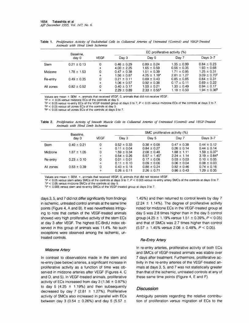
Table 1. Proliferative Activity of Endothelial Cells in Collateral Arteries of Untreated (Control) and VEGF-TreatedAnimals with Hind Limb Ischemia
Baseline, EC proliferative activity (%)day 0 VEGF Day 3 Day 5 Day 7 Days 3-7
Stem 0.21 ± 0.13 0 0.46 ± 0.29 0.89 ± 0.24 1.35 ± 0.99 0.84 ± 0.23+ 4.00 ± 2.25 1.65 + 0.59 0.56 ± 0.35 1.93 ± 0.68
Midzone 1.78 ± 1.53 0 0.47 ± 0.38 1.51 ± 0.39 1.71 ± 0.95 1.25 ± 0.31+ 1.56 ± 0.87 4.25 ± 1.19* 2.81 ± 1.27 3.09 ± 0.70t
Re-entry 0.49 ± 0.35 0 0.21 + 0.11 0.69 ± 0.43 0.85 ± 0.85 0.64 ± 0.31+ 1.06 + 0.57 0.92 ± 0.38 0.17 ± 0.11 0.69 ± 0.22
All zones 0.82 ± 0.52 0 0.40 ± 0.17 1.03 ± 0.21 1.30 ± 0.49 0.94 ± 0.17+ 2.29 ± 0.89 2.33 ± 0.55t 1.18 ± 0.50 1.94 ± 0.36§
Values are mean ± SEM. +, animals that received VEGF; 0, animals that did not receive VEGF.*P = < 0.05 versus midzone ECs of the controls at day 5.tp < 0.05 versus re-entry ECs of the VEGF-treated group at days 3 to 7; P < 0.05 versus midzone ECs of the controls at days 3 to 7.tP < 0.05 versus all zones ECs of the controls at day 5.§P < 0.05 versus all zones ECs of the controls at days 3 to 7.
Table 2. Proliferative Activity of Smooth Muscle Cells in Collateral Arteries of Untreated (Control) and VEGF-TreatedAnimals with Hind Limb Ischemia
Baseline, SMC proliferative activity (%)day 0 VEGF Day 3 Day 5 Day 7 Days 3-7
Stem 0.40 ± 0.21 0 0.52 ± 0.33 0.38 ± 0.08 0.47 ± 0.38 0.44 ± 0.12+ 0.11 ± 0.04 0.84 ± 0.27 0.26 ± 0.14 0.44 ± 0.14
Midzone 1.87 ± 1.05 0 1.59 ± 0.34 2.08 ± 0.48 1.88 ± 1.11 1.59 ± 0.37*+ 0.54 ± 0.26 5.57 + 1.45t 2.24 ± 1.14 3.19 ± 0.84t
Re-entry 0.23 ± 0.10 0 0.01 ± 0.01 0.17 ± 0.08 0.03 ± 0.03 0.10 ± 0.05+ 0.11 ± 0.10 0.09 ± 0.06 0.06 ± 0.04 0.08 ± 0.03
All zones 0.83 ± 0.39 0 0.43 ± 0.18 0.88 + 0.24 0.82 + 0.46 0.74 ± 0.16+ 0.26 ± 0.11 2.26 ± 0.71 0.86 + 0.43 1.29 ± 0.35
Values are mean + SEM. +, animals that received VEGF; 0, animals that did not receive VEGF.*P < 0.05 versus stem artery SMCs of the controls at days 3 to 7; P < 0.005 versus re-entry artery SMCs of the controls at days 3 to 7.tp < 0.05 versus midzone SMCs of the controls at day 5.tP < 0.005 versus stem and re-entry SMCs of the VEGF-treated group at days 3 to 7.
days 3, 5, and 7 did not differ significantly from findingsin ischemic, untreated control animals at the same timepoints (Figure 4, A and B). It was nevertheless intrigu-ing to note that certain of the VEGF-treated animalsshowed very high proliferative activity of the stem ECsat day 3 after VEGF. The highest EC-BrdU index ob-served in this group of animals was 11.4%. No suchexceptions were observed among the ischemic, un-treated controls.
Midzone Artery
In contrast to observations made in the stem andre-entry (see below) arteries, a significant increase inproliferative activity as a function of time was ob-served in midzone arteries after VEGF (Figures 4, Cand D, and 5). In VEGF-treated animals, proliferativeactivity of ECs increased from day 3 (1.56 ± 0.87%)to day 5 (4.25 ± 1.19%) and then subsequentlydecreased by day 7 (2.81 ± 1.27%). Proliferativeactivity of SMCs also increased in parallel with ECsbetween day 3 (0.54 ± 0.26%) and day 5 (5.57 ±
1.45%) and then returned to control levels by day 7(2.24 ± 1.14%). The degree of proliferative activitynoted for midzone ECs in the VEGF-treated group atday 5 was 2.8 times higher than in the day 5 controlgroup (4.25 ± 1.19% versus 1.51 ± 0.39%, P < 0.05)and that of SMCs was 2.7 times higher than control(5.57 ± 1.45% versus 2.08 ± 0.48%, P < 0.05).
Re-Entry Artery
In re-entry arteries, proliferative activity of both ECsand SMCs of VEGF-treated animals was stable over7 days after treatment. Furthermore, proliferative ac-tivity in the re-entry arteries of the VEGF-treated an-imals at days 3, 5, and 7 was not statistically greaterthan that of the ischemic, untreated controls at any ofthese same time points (Figure 4, E and F).
DiscussionAmbiguity persists regarding the relative contribu-tion of proliferation versus migration of ECs to the

Cellular Proliferation after VEGF 1655AJP December 1995, Vol. 14 7, No. 6
--o.-- Control* - VEGF
3 5 7Days
D 8.7.
- 6.x 5a)V 4.- 3.2 2.m 1
0 3 5 7Days
F--v--- Control. - VEGF 1-0
xa)
"-
m
3 5 7Days
--o.-- Control. VEGF
s__S,_ 4-234
VI.0 3 5 7
Days
8 __o--- Control
67 VEGF543210O.I 9 I I10 3 5
Days7
Figure 4. Time course ofproliferative activity ofcollateral arteries. BrdUlabeling index( %) ofstem ECs (A) and SMCs (B); midzone ECs (C) and SMCs(D); and re-entry ECs (E) and SMCs (F). Midzone EC and SMCproliferation of VEGF-treated animals ( ) was significantly higher than that ofischemic, untreated controls (- - ) at day 5 after treatment (*P < 0.05). Interruption ofsolid and dashed lines indicates that tissue retrieval wasnot continuously performed on a daily basis but was limited to days 0, 3, 5, and 7.
development of new blood vessels, or angiogenesis.Sholley et al,23 using a model of inflammation-in-duced angiogenesis of the rat cornea, demonstratedthat initial vascular sprouting does not require ECproliferation. EC proliferation in this model was sup-
pressed by X-irradiation with 2000 or 8000 rads be-fore application of the inflammatory stimulus. In irra-diated corneas displaying no cellular proliferation,vascular sprouting at 2 days was similar to that seenin contralateral shielded corneas. Although neovas-
cular growth was subsequently blunted and ulti-mately ceased by 4 to 7 days, these experimentsdocumented the critical if not exclusive roles of mi-gration and redistribution of pre-existing ECs in thecommencement of neovascularization. Similar impli-cations resulted from work by Nicosia et a124; fi-bronectin was shown to promote, in a dose-depen-
dent fashion, the elongation of microvessels thatsprout from explants of rat aorta placed in serum-
free collagen gel, despite the fact that neither DNAsynthesis nor mitotic activity was increased in com-parison with fibronectin-negative gels. Fibronectinwas therefore inferred to promote angiogenesis invitro by migratory recruitment of pre-existing ECs.
In contrast to these in vivo inflammatory and in vitroorgan culture models, angiogenesis that develops inresponse to experimental vascular obstruction, ie,collateral vessel development, has been shown byseveral previous investigators to involve proliferationof not only ECs but SMCs as well (Table 3). Severalimportant principles were elucidated by these stud-ies.
First, evidence of EC proliferation is nearly absentin normal arteries,4'5 a finding that is consistent with
A0
xm)
m
B 8-7.
-6-x 5.-' 4.= 3.l 2
1'
Days
T(1+ "I ZD
0
C 87-
._6
x 5-V 4.c3a 2m 1-
0 4
E 8-7-
o- 6-
x 5.CD-0 4 -c
3-D" 2 -m 1-
00
1- uI I.1- - - .& -
*

1656 Takeshita et alAJP December 1995, Vol. 14 7, No. 6
Figure 5. Representative histological section ofmidzone collateral arteries at day5 after VEGF. A: ImmunostainingforBrdU. Brown nuclearstainingindicates BrdUpositivity. B: Higher magnification ofBrdU-positive EC indicated by arrow in A. Identification of the cell type as EC was confirmedby positive staining for lectin (C) and negative staining for smooth muscle a-actin (D) in adjacent sections. Hematoxylin counterstain.
an estimated EC turnover time of "thousands ofdays" in quiescent microvasculature.25 Even a rela-tively low percentage of EC proliferation observed inresponse to arterial occlusion or exogenous growthfactors may therefore represent considerable en-
hancement of EC proliferative activity and, whenconsidered in relation to a denominator of thousandsof ECs, is clearly sufficient to provide the basis fornew blood vessel formation. Second, peak EC pro-liferation, which contributes to naturally occurringcollateral development in the setting of vascular oc-
clusion, varies from 2.6 to 3.5% in the canine coro-nary circulation,47 from 5 to 6% in the rodent renalvasculature,56 and <1% in swine coronaries.8 Thecontrasting rates of EC proliferation between the ca-nine and swine coronary circulations are indeedrepresentative of the contrasting propensity fornatural collateral artery development in these twospecies. Third, proliferation of SMCs, the addi-
tional requisite cell type for the formation of largerblood vessels (vasculogenesis), is an implicit com-ponent of angiogenesis, regardless of animal spe-cies or circulatory site. Schaper et al3 in fact spec-ulated nearly 25 years ago that "it is tempting toassume that EC proliferation not only serves thepurpose of forming the endothelium of a finallylarger artery but rather actively participates in thedevelopment of the tunica media." Fourth, prolifer-ative activity, for SMCs as well as ECs, is highest atthe level of the smallest diameter collateral ves-
sels, the so-called midzone collateral seg-ments.3478 Fifth, although evidence of EC andSMC proliferation alone does not necessarily dis-tinguish new vessel development from an increasein the size of pre-existing vessels, adjunctive dataregarding increased capillary density9'26 supportthe notion that proliferative activity does in factreflect true angiogenesis.
Table 3. Evidence for Vascular Cell Proliferation during Spontaneous Angiogenesis in Vivo
Proliferative activityFirst author (year) Species (n) Circulation Index of proliferation Time course ECs SMCs
Schaper (1969) Dog (6) Coronary Mitoses Peak = 2-3 weeks* 60 cells 53 cellsSchaper (1971) Dog (11) Coronary [3H]Thymidine Peak = 3 weeks* 3.5% of all ECs and
SMCsCowan (1978) Dog (3) Renal [3H]Thymidine 18-39 days 5.5% IncreasedIlich (1979) Rat (38) Renal [3H]Thymidine Peak = 1-4 days 5-6% NDPasyk (1982) Dog (10) Coronary [3H]Thymidine Peak = 4 days 2.6% 1.6%White (1992) Pig (17) Coronary [3H]Thymidine Peak = 2-3 weeks* 0.7% 0.5%
ND, not done.*In these studies, peak refers to time after placement of an ameroid constrictor; occlusion in this case typically develops at
approximately 17 days post-operatively.

Cellular Proliferation after VEGF 1657AJP December 1995, Vol. 147, No. 6
In contrast to those studies shown in Table 1, all ofwhich concern spontaneous collateral development,limited investigation has been performed regardingthe extent to which EC and/or SMC proliferation are
altered by interventions designed to augment collat-eral vessel development, ie, therapeutic angiogene-sis.9 Graham et a126 established arteriovenous (AV)fistulae at the popliteal level in a canine model ofhind limb ischemia, following which the vein proximalto the AV anastomosis was ligated; this so-called"arteriovenous reversal (AVR)" was designed to aug-ment perfusion to the distal limb in a retrogrademanner. A consistent feature of this procedure (inhumans27 as well as animals) was the developmentof an extensive network of new vessels in the prox-
imity of the AV anastomosis. A significant increase inuptake of tritiated thymidine, administered 3 hoursbefore death, was observed in hind limbs treated byAVR versus controls and, when coupled with histo-logical evidence of increased capillary density, was
interpreted as evidence for therapeutically aug-
mented EC proliferation. More recently, Unger et al13administered BrdU to dogs in which collateral arterydevelopment had been provoked by application ofan ameroid constrictor around the left circumflexcoronary artery; after administration of basic FGF(bFGF) directly into the circumflex distal to the con-
strictor, cellular proliferation was found to be in-creased in collateral-dependent viable myocardiumas well as sites of myocardial infarction. Based on
morphology and location, cells immunopositive forBrdU were judged to consist predominantly of ECs.The current study aimed to document the contri-
bution to and sequence of cellular proliferation dur-ing spontaneous collateral vessel development in ananimal model of lower extremity ischemia and deter-mine the extent to which this is modified by thera-peutic angiogenesis. EC proliferative activity (BrdUindex) among collateral arteries developing in theuntreated, ischemic limbs of control animals variedfrom 0 to 3.3% in stem segments, from 0 to 4.1% inmidzone segments, and from 0 to 3.6% in the re-
entry segments. Thus, regardless of anatomic site,EC proliferation varied between 0 and 4.1% per day,similar to the range reported previously by Pasyk eta17 and White et al8 for canine and swine coronary
artery collaterals, respectively. EC proliferationamong the ischemic untreated controls was not dis-proportionately greater for any individual collateralzone or individual time point. For SMCs, proliferativeactivity among the control animals was lower thanthat of ECs in the stem and re-entry zones; the de-gree of SMC proliferative activity in the midzone was
similar to that of ECs and thereby significantly
greater than that of the stem and re-entry zones. Withregard to time course, however, the proliferative ac-tivity of SMCs during spontaneous collateral arterydevelopment did not vary significantly as a functionof time.
The apparently modest extent of EC proliferationdocumented during spontaneous collateral develop-ment in this model is in reality quite substantial con-sidering that (1) EC proliferation is nearly absent innormal arteries4'5; (2) the estimated EC turnover timefor ECs in quiescent microvasculature is "thousandsof days"25; and (3) the denominator in the currentanalyses represented thousands of cells.A single intra-arterial bolus of recombinant human
VEGF165 was used to accomplish therapeutic angio-genesis in this study. The 165-amino-acid isoform ofVEGF employed is the most prevalent of the fourVEGF isoforms in humans and is intermediate be-tween the 121 and 189 VEGF isoforms with regard toheparin affinity. Previous studies performed in ourlaboratory have demonstrated that the dose androute of administration employed in the current studyproduce statistically significant augmentation of col-lateral vessel development9; moreover, ameliorationof hemodynamic deficit in the ischemic limb, as-sessed both by measurements of blood pressureand more recently blood flow,14 have been shown tobe significantly greater in animals receiving VEGFthan in untreated controls. It has been previouslyproposed12 that the heparin-binding feature of an-giogenic growth factors such as VEGF may explainin part their protracted efficacy as a result of bindingof the growth factor by heparan sulfate proteogly-cans present on the luminal surface of the vascularendothelium and/or within the extracellular matrix.Such heparan sulfate binding of VEGF165 may in-deed contribute to the observation made in thepresent study that peak EC proliferative activity wasdelayed to day 5 after VEGF administration.
Administration of VEGF in the current study wasshown to augment EC proliferation in the midzonecollateral vessels by roughly threefold comparedwith control animals; this difference was most pro-found, and statistically significant, at day 5 afterVEGF. The increase in EC proliferation observed atday 5 after VEGF was followed by reduction of thehemodynamic deficit in the ischemic limb by day 7.The improved hemodynamic measurements docu-mented in the treatment group in the current studyare unlikely to be the result of vasodilatory effects ofVEGF; in our experience such effects are quite tran-sient and do not persist beyond 2 hours after admin-istration of VEGF.28 Previous studies from our grouphave clearly documented that the increase in lower

1658 Takeshita et alAJP December 1995, Vol. 147, No. 6
limb blood pressure ratio correlates with an increasein the number of angiographically visible collateralarteries as well as an increase in the limb blood flowmeasured by Doppler flow guidewire,914 ie, im-provement of collateral artery formation in the is-chemic limb.
In the stem arteries, EC proliferation among VEGF-treated animals was quite high (up to 11.4%) inselected cases but did not reach statistical signifi-cance for the treated group as a whole when com-pared with controls. At the level of the re-entry arter-ies, EC proliferation among VEGF-treated animalswas in no case >2.6%. Thus, a statistically signifi-cant increase in EC proliferation in response to VEGFversus controls was limited to the midzone collater-als. These findings suggest that the locus of aug-mented EC proliferation consequent to therapeuticangiogenesis is similar to that previously noted dur-ing naturally occurring collateral development,4namely, the smallest diameter, or midzone, collateralvessels.
Despite the fact that the mitogenic effects of VEGFhave been previously shown to be limited to ECs,17the proliferative activity of SMCs in the midzone col-laterals also increased by approximately threefold.The augmentation in midzone SMC proliferation atday 5 was statistically significant, compared with thatobserved in naturally occurring collateral artery de-velopment in the present study and exceeds thatreported in previous studies of natural collateral de-velopment4'7'8 as well. Although VEGF has beenshown to interact with lower affinity binding sites toinduce mononuclear phagocyte chemotaxiS,29,30higher affinity binding sites presumed to mediate themitogenic effects of VEGF are limited to ECs.17'28Increased SMC proliferation observed in the presentstudy is therefore not likely to represent a directeffect of VEGF. Two indirect effects are possible. Theability to induce vascular permeability is a wellknown feature of VEGF, responsible in fact for itsalternate designation as vascular permeability fac-tor.31-33 It is possible that extravasation of certainangiogenic growth factors from circulating bloodmight result in activation of SMC proliferation.
Alternatively, ECs stimulated by VEGF may se-crete factor(s) that promote SMC proliferation. It hasbeen previously shown, for example, that VEGF in-duces expression of tissue-type plasminogen activa-tors, potent mitogens for cultured human SMCs,34 incultured bovine microvascular ECs.35 Platelet-de-rived growth factor, a mitogen and chemoattractantfor vascular SMCs,36 which is expressed byECs37-39 in a polarized manner40 sufficient to pro-vide an organizing gradient for SMCs, has been
previously shown to be induced in human umbilicalvein ECs by the addition of another angiogenicgrowth factor, aFGF;41 the possibility that VEGF maysimilarly up-regulate EC production of growth factorsmitogenic for SMCs,42 has to our knowledge not yetbeen studied.
In summary, spontaneous collateral artery devel-opment observed in response to limb ischemia, aswell as that seen after administration of VEGF, ischaracterized by active cellular proliferation of ECsand SMCs in the midzone arteries, maximal at day 5after VEGF and followed by hemodynamic evidenceof improvement in limb perfusion by day 7. Thesedata thus support the concept that augmented cel-lular proliferation plays an important role in the pro-cess of enhanced collateral artery development aftertherapeutic angiogenesis and, furthermore, providea basis for rational selection and/or timing of adjunc-tive therapies designed to optimize collateral devel-opment via effects on cellular proliferation, migration,and/or associated lysis of the extracellular matrix.
AcknowledgmentsWe are grateful to Dr. John Ogez for purification ofVEGF165 and to Mrs. Mickey Neely for her secretarialassistance.
References
1. D'Amore PA, Thompson RW: Mechanisms of angiogen-esis. Annu Rev Physiol 1987, 49:453-464
2. Schaper, W: Coronary collateral development: con-cepts and hypothesis. Collateral Circulation: Heart,Brain, Kidney, Limbs. Edited byW Schaper, J Schaper.Boston, Kluwer Academic Publishers, 1993, pp 41-64
3. Schaper W, Schaper J, Xhonneux R, Vandesteene R:The morphology of intercoronary anastomoses inchronic coronary artery occlusion. Cardiovasc Res1969, 3:315-323
4. Schaper W, de Brabander M, Lewi P: DNA synthesisand mitoses in coronary collateral vessels of the dog.Circ Res 1971, 28:671-679
5. Cowan DF, Hollenberg NK, Connelly CM, Williams DH,Abrams HL: Increased collateral arterial and venousendothelial cell turnover after renal artery stenosis inthe dog. Invest Radiol 1978, 13:143-149
6. Ilich N, Hollenberg NK, Williams DH, Abrams H: Timecourse of increased collateral arterial and venous en-dothelial cell turnover after renal artery stenosis in rat.Circ Res 1979, 45:579-582
7. Pasyk S, Schaper W, Schaper J, Pasyk K, MiskiewiczG, Steinseifer B: DNA synthesis in coronary collateralsafter coronary artery occlusion in conscious dog. Am JPhysiol 1982, 242:H1031-H1037

Cellular Proliferation after VEGF 1659AJP December 1995, Vol. 14 7, No. 6
8. White FC, Carroll SM, Magnet A, Bloor CM: Coronarycollateral development in swine after coronary arteryocclusion. Circ Res 1992, 71:1490-1500
9. Takeshita S, Zheng LP, Brogi E, Kearney M, Pu LQ,Bunting S, Ferrara N, Symes JF, Isner JM: Therapeuticangiogenesis: a single intraarterial bolus of vascularendothelial growth factor augments revascularization ina rabbit ischemic hind limb model. J Clin Invest 1994,93:662-670
10. Baffour R, Berman J, Garb JL, Rhee SW, Kaufman J,Friedmann P: Enhanced angiogenesis and growth ofcollaterals by in vivo administration of recombinant ba-sic fibroblast growth factor in a rabbit model of acutelower limb ischemia: dose-response effect of basicfibroblast growth factor. J Vasc Surg 1992,16:181-191
11. Pu LQ, Sniderman AD, Brassard R, Lachapelle KJ,Graham AM, Lisbona R, Symes JF: Enhanced revas-cularization of the ischemic limb by means of angio-genic therapy. Circulation 1993, 88:208-215
12. Yanagisawa-Miwa A, Uchida Y, Nakamura F, TomaruT, Kido H, Kamijo T, Sugimoto T, Kaji K, Utsuyama M,Kurashima C, Ito H: Salvage of infarcted myocardiumby angiogenic action of basic fibroblast growth factor.Science 1992, 257:1401-1403
13. Unger EF, Banai S, Shou M, Lazarous DF, Jakiltsch MT,Scheinowitz M, Correa R, Klingbeil C, Epstein SE: Ba-sic fibroblast growth factor enhances myocardial col-lateral flow in a canine model. Am J Physiol 1994,266:H 1588-H 1595
14. Bauters C, Asahara T, Zheng LP, Takeshita S, BuntingS, Ferrara N, Symes JF, Isner JM: Physiologic assess-ment of augmented vascularity induced by VEGF inrabbit ischemic hindlimb. Am J Physiol 1994, 36:H1263-H1271
15. Takeshita S, Pu LQ, Stein LA, Sniderman AD, BuntingS, Ferrara N, Isner JM, Symes JF: Intramuscular ad-ministration of vascular endothelial growth factor in-duces dose-dependent collateral artery augmentationin a rabbit model of chronic limb ischemia. Circulation1994, 90(Part 2):11-228-11-234
16. Banai S, Jaklitsch MT, Shou M, Lazarous DF, Schei-nowitz M, Biro S, Epstein SE, Unger EF: Angiogenic-induced enhancement of collateral blood flow to isch-emic myocardium by vascular endothelial growthfactor in dogs. Circulation 1994, 89:2183-2189
17. Ferrara N, Henzel WJ: Pituitary follicular cells secrete anovel heparin-binding growth factor specific for vascu-lar endothelial cells. Biochem Biophys Res Commun1989, 161:851-855
18. Leung DW, Cachianes G, Kuang WJ, Goeddel DV,Ferrara N: Vascular endothelial growth factor is a se-creted angiogenic mitogen. Science 1989, 246:1306-1309
19. Ferrara N, Leung DW, Cachianes G, Winer J, HenzelWJ: Purification and cloning of vascular endothelialgrowth factor secreted by pituitary follicolostellatecells. Methods Enzymol 1991, 198:391-404
20. Longland CJ: The collateral circulation of the limb. AnnR Coll Surg Engl 1953, 13:161-181
21. Coffin JD, Harrison J, Schwartz S, Heimark R: Angio-blast differentiation and morphogenesis of the vascularendothelium in the mouse embryo. Dev Biol 1991, 148:51-62
22. Takeshita S, Gal D, Leclerc G, Pickering JG, Riessen R,Weir L, Isner JM: Increased gene expression after li-posome-mediated arterial gene transfer associatedwith intimal smooth muscle cell proliferation: in vitro andin vivo findings in a rabbit model of vascular injury. JClin Invest 1994, 93:652-661
23. Sholley MM, Ferguson GP, Seibel HR, Montour JL,Wilson JD: Mechanisms of neovascularization: vascu-lar sprouting can occur without proliferation of endo-thelial cells. Lab Invest 1984, 51:624-634
24. Nicosia RF, Bonanno E, Smith M: Fibronectin promotesthe elongation of microvessels during angiogenesis invitro. J Cell Physiol 1993, 154:654-661
25. Folkman J, Shing Y: Angiogenesis. J Biol Chem 1992,267:10931-10934
26. Graham AM, Baffour R, Burdon T, DeVarennes B, RicciMA, Common A, Lisbona R, Sniderman AD, Symes JF:A demonstration of vascular proliferation in response toarteriovenous reversal in the ischemic canine hindlimb. J Surg Res 1989, 47:341-347
27. Symes JF, Graham AM, Stein L, Sniderman AD: Sal-vage of a severely ischemic limb by arteriovenousrevascularization: a case report. Can J Surg 1984,27:274-276
28. Horowitz J, Hariawala M, Sheriff D, Keyt B, Symes J: Invivo administration of vascular endothelial growth factoris associated with EDRF-dependent systemic hypoten-sion in porcine and rabbit animal models. Circulation1995;92:1630-1631.
29. Clauss M, Gerlach M, Gerlach H, Brett J, Wang F,Familletti PC, Pan Y-CE, Olander JV, Connolly DT, SternD: Vascular permeability factor: a tumor-derivedpolypeptide that induces endothelial cell and mono-cyte procoagulant activity and promotes monocyte mi-gration. J Exp Med 1990, 172:1535-1545
30. Shen H, Clauss M, Ryan J, Schmidt AM, Tijburg P,Borden L, Connolly D, Stern D, Kao J: Characterizationof vascular permeability factor/vascular endothelialgrowth factor receptors on mononuclear phagocytes.Blood 1993, 81:2767-2773
31. Connolly DT, Hewelman DM, Nelson R, Olander JV,Eppley BL, Delfino JJ, Siegel RN, Leimgruber RS,Feder J: Tumor vascular permeability factor stimulatesendothelial cell growth and angiogenesis. J Clin Invest1989, 84:1470-1478
32. Keck PJ, Hauser SD, Krivi G, Sanzo K, Warren T, FederJ, Connolly DT: Vascular permeability factor, an endo-thelial cell mitogen related to PDGF. Science 1989,246:1309-1342
33. Dvorak HF: Tumors: wounds that do not heal: similari-ties between tumor stroma generation and wound heal-ing. N Engl J Med 1986, 315:1650-1659

1660 Takeshita et alAJP December 1995, Vol. 14 7, No. 6
34. Herbert JM, Lamarche I, Prabonnaud V, Dol F, Gau-thier T: Tissue-type plasminogen activator is a potentmitogen for human aortic smooth muscle cells. J BiolChem 1994, 269:3076-3080
35. Pepper MS, Ferrara N, Orci L, Montesano R: Vascularendothelial growth factor (VEGF) induces plasminogenactivators and plasminogen activator inhibitor type 1 inmicrovascular endothelial cells. Biochem Biophys ResCommun 1994, 189:824-831
36. Ross R, Glomset B, Kariya B, Harker L: A platelet-dependent serum factor that stimulates the prolifera-tion of arterial smooth muscle cells in vitro. Proc NatlAcad Sci USA 1974, 71:1207-1210
37. Collins T, Ginsburg D, Boss JM, Orkin SH, Pober JS:Cultured human endothelial cells express platelet-de-rived growth factor B chain: cDNA cloning and struc-tual analysis. Nature 1985, 316:748-750
38. Collins T, Pober JS, Gimbrone MA Jr, Betsholtz C,
Westermark B, Heldin C-H: Cultured human endothelialcells express platelet-derived factor A chain. Am JPathol 1987, 126:7-12
39. Bowen-Pope DF, Hart CE, Seifert RA: Sera and condi-tioned media contain different isoforms of platelet-derivedgrowth factor (PDGF) which bind to different classes ofPDGF receptor. J Biol Chem 1989, 264:2502-2508
40. Zerwes H-G, Risau W: Polarized secretion of aplatelet-derived growth factor-like chemotactic factorby endothelial cells in vitro. J Cell Biol 1987, 105:2037-2041
41. Gay CG, Winkles JA: Heparin-binding growth factor-1stimulation of human endothelial cells induces platelet-derived growth factor A-chain gene expression. J BiolChem 1990, 265:3284-3292
42. Lindner V, Lappi DA, Baird A, Majack RA, Reidy MA:Role of basic fibroblast growth factor in vascular lesionformation. Circ Res 1991, 68:106-113


![· Web viewBy targeting cellular or viral genes, these miRNAs are involved in the regulation of multiple cellular responses such as host cell proliferation, apoptosis [12-15], and](https://img.pdfslide.net/doc/110x75/5f0b48857e708231d42fc04c/web-view-by-targeting-cellular-or-viral-genes-these-mirnas-are-involved-in-the.jpg)