Embed Size (px)
Citation preview
Lecture Presentation by
Lori Garrett
4Tissue Level of
Organization
© 2018 Pearson Education, Inc.

Section 1: Epithelial Tissue
Learning Outcomes
4.1 Identify the four types of tissues in the body,
and describe their roles.
4.2 Describe three microscopy techniques.
4.3 Describe epithelial tissues, including cell shape,
layers, and functions.
4.4 Discuss the types and functions of intercellular
connections between epithelial cells.
4.5 Describe the structure and function of squamous
epithelium.
© 2018 Pearson Education, Inc.

Section 1: Epithelial Tissue
Learning Outcomes (continued)
4.6 Describe the structure, function, and locations of
cuboidal and transitional epithelia.
4.7 Describe the structure, function, and locations of
columnar epithelia.
4.8 Compare the three different methods of exocrine
secretion by glandular epithelia.
4.9 Explain how multicellular exocrine glands are
classified by their structure.
© 2018 Pearson Education, Inc.

Module 4.1: Four types of tissue make up the body
Body organization review
Atoms → Molecules → Cells → Tissues
• Chemical level can only be seen using special
imaging techniques
• Cellular level details seen best with electron
microscope
© 2018 Pearson Education, Inc.

Module 4.1: Types of tissue
Body organization review (continued)
Trillions of cells in the body
Only about 200 different types of cells
Cells working together form tissues
Study of tissues called histology
Four basic types of tissues
1. Epithelial
2. Connective
3. Muscle
4. Neural
© 2018 Pearson Education, Inc.

Levels of organization up to the tissue level
© 2018 Pearson Education, Inc.

Module 4.1: Review
A. Give the term for “the study of tissues.”
B. What is a tissue?
C. List the four basic tissue types, and describe
the functions of each.
Learning Outcomes: Identify the four types of
tissue in the body, and describe their roles.
© 2018 Pearson Education, Inc.

Module 4.2: Microscopes are used to study cells and tissues
Anatomy studied at different scales
Microscopy (the use of microscopes)
• Began about 400 years ago
– Early magnification levels 10–20 times actual size
• Simple microscope—uses only one lens
• Compound microscope—uses >1 lens
• Electron microscope
– Can magnify over 1 million times
• Amount of fine detail (resolution) of an image varies
with magnification and type of microscope used
© 2018 Pearson Education, Inc.

Magnification and resolution of different types of microscopes
© 2018 Pearson Education, Inc.

Module 4.2: Microscopy techniques
Types of microscopes
1. Compound light microscope
• Detects visible light through thin section of tissue
• Two lenses magnify specimen
1. Objective lens located on
revolving nosepiece
2. Ocular lens located in the
eye piece
© 2018 Pearson Education, Inc.

Module 4.2: Microscopy techniques
Types of microscopes (continued)
1. Compound light microscope (continued)
• Total magnification calculated by multiplying the two
lens powers (objective × ocular)
• Resolution
– Ability to distinguish
between two separate points
– Wavelength of light limits
resolution on light
microscope to about
200 nm (0.2 μm)
© 2018 Pearson Education, Inc.
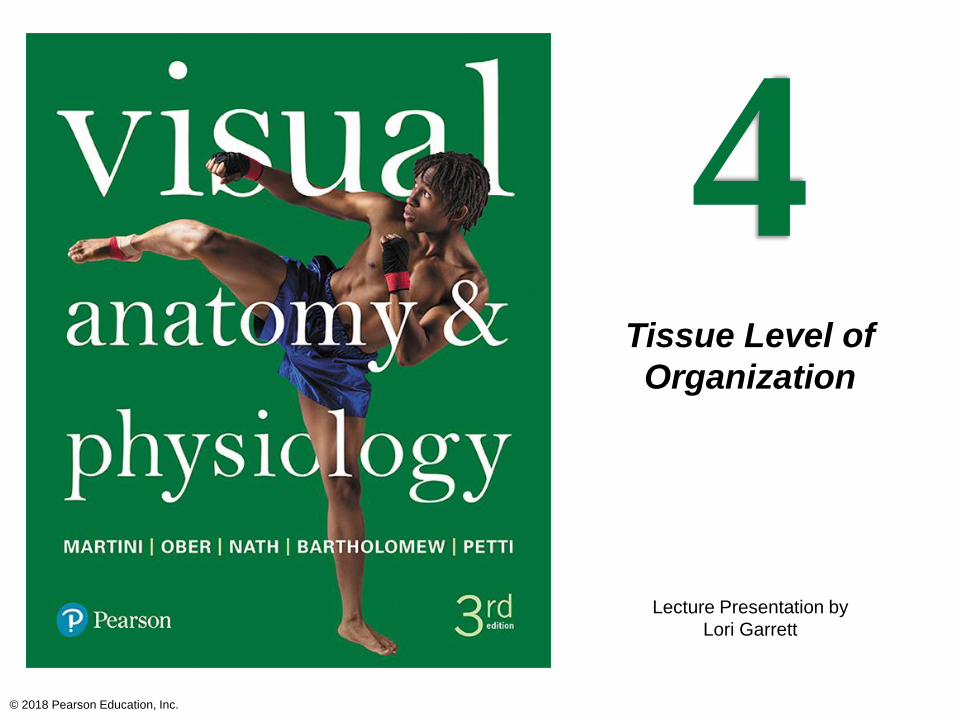
Module 4.2: Microscopy techniques
Types of microscopes (continued)
2. Transmission electron
microscope
• “Transmits” electrons through
specimen
• Uses magnets to direct beam of
electrons through the surface of
a very thin object onto a
photographic plate
• Wavelength of electron beam
0.00001 of white light
• Maximum resolution 0.2 nm (0.0002 μm)
© 2018 Pearson Education, Inc.

Module 4.2: Microscopy techniques
Types of microscopes (continued)
3. Scanning electron microscope
• Uses electrons, but not by
sending them through a
specimen
• Specimen coated with
electron dense material
• Electron beams are focused
on the specimen
• Reflection of electrons bouncing off object produce
three dimensional image of the surface
• Can view surface features only
• Maximum resolution of about 10 nm (0.01 μm)
© 2018 Pearson Education, Inc.
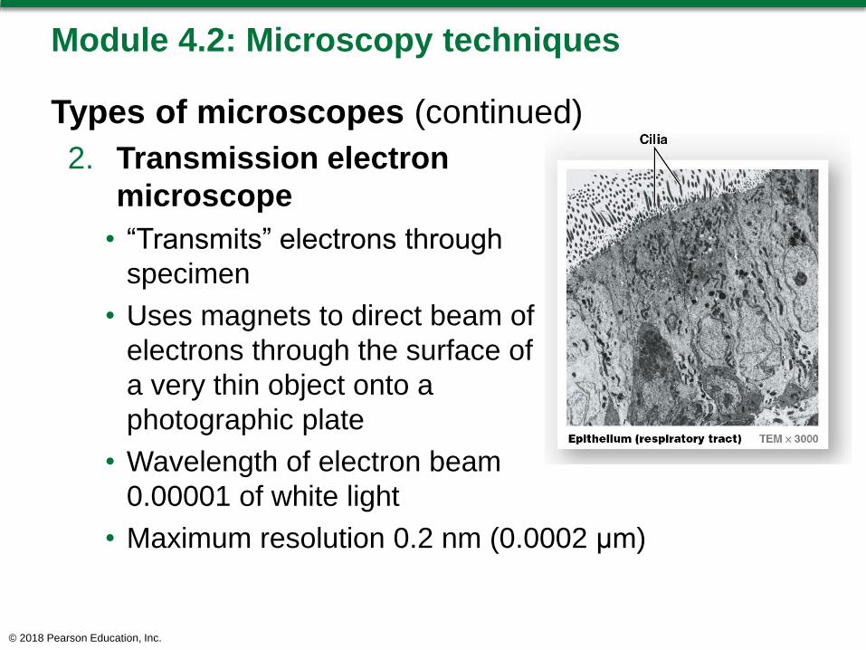
Module 4.2: Microscopy techniques
Magnification notation examples
Type of microscope used (abbreviation) and
magnification
1. LM × 400
– Light micrograph magnified 400 times
2. TEM × 3000
– Transmission electron micrograph magnified 3000
times
3. SEM × 15,846
– Scanning electron micrograph magnified 15,846 times
© 2018 Pearson Education, Inc.

Module 4.2: Microscopy techniques
A&P lab microscopy tips
Light microscopes most often
used
Begin with lowest magnification
objective lens over specimen
When in focus on that power,
carefully rotate objective lenses with greater
magnification into place
Compare images from textbook to microscope
image at same magnification
© 2018 Pearson Education, Inc.

Module 4.2: Review
A. How do early microscopes compare with
modern microscopes?
B. Differentiate among LM, TEM, and SEM.
C. The LM at the top of this page is magnified 400
times (400×). If the ocular lens used to make
this image has a magnification of 10×, what is
the magnification of the objective lens?
Learning Outcome: Describe three microscopy
techniques.
© 2018 Pearson Education, Inc.

Module 4.3: Epithelial tissue covers surfaces, lines cavities, and forms secretory glands
Divisions of epithelial tissue
1. Epithelia
• Avascular layers
• Cover exposed surfaces
• Line internal cavities and passageways
• Often contain secretory or gland cells scattered
among other cell types
© 2018 Pearson Education, Inc.

Module 4.3: Epithelial tissues
Divisions of epithelial tissue (continued)
2. Glands
• Derived from epithelia
• Predominantly secretory cells
• Two types
1. Exocrine glands
o Secrete onto external surfaces or into ducts
2. Endocrine glands
o Secrete hormones into interstitial fluid
o Hormones then distributed by bloodstream
© 2018 Pearson Education, Inc.

Epithelial tissues
© 2018 Pearson Education, Inc.

Module 4.3: Epithelial tissues
Functions of epithelial tissue
Provide physical protection
• Protect surfaces from abrasion, dehydration, or
destruction by chemical or biological agents
Control permeability
• Most epithelia are capable of selective absorption or
secretion
• Epithelial barrier can be modified in response to
stimuli (Example: calluses)
© 2018 Pearson Education, Inc.

Module 4.3: Epithelial tissues
Functions of epithelial tissue (continued)
Provide sensation
• Specialized epithelial cells detect changes in
environment (for example, touch receptors)
• Neuroepithelium
– Sensory epithelium found in special sense organs
Produce specialized secretions
• Glandular epithelial cells produce secretions
© 2018 Pearson Education, Inc.

Module 4.3: Epithelial tissues
Features of epithelial tissue (continued)
Surfaces
• Apical surface
– Faces exterior of body
or internal space
• Base
– Attached to underlying
tissues
– Basolateral surface
o Includes base and sides (lateral surfaces) attached
to neighboring cells
Polarity
• Refers to structural differences between exposed and
attached surfaces© 2018 Pearson Education, Inc.

Module 4.3: Epithelial tissues
Features of epithelial tissue (continued)
Apical surface features
• If lining a tube, apical surface is exposed to space
inside the tube, called lumen
• Microvilli found on this surface in digestive, urinary,
and reproductive tracts
• Cilia found on this surface in parts of the respiratory
and reproductive tracts
Epithelial cells also contain membranous organelles
comparable to other cell types
© 2018 Pearson Education, Inc.

Module 4.3: Epithelial tissues
Epithelial cells have 3 basic shapes (viewed
perpendicular to exposed surface)
1. Squamous
• Thin and flat
2. Cuboidal
• Cube-shaped
• Like little boxes
3. Columnar
• Taller than they are wide
• Slender rectangles
© 2018 Pearson Education, Inc.

Module 4.3: Epithelial tissues
Epithelial cell layers
Single layer
• Simple epithelium
Several layers of cells
• Stratified epithelium
• Found in areas that need protection from abrasion or
chemical stress
– Examples: surface of skin, lining of the mouth
© 2018 Pearson Education, Inc.

Module 4.3: Review
A. List four essential functions of epithelial tissue.
B. Summarize the classification of an epithelium
based on cell shape and number of cell layers.
C. What function is served by motile cilia on
epithelial cell surfaces?
Learning Outcome: Describe epithelial tissues,
including cell shape, layers, and functions.
© 2018 Pearson Education, Inc.

Module 4.4: Epithelial cells are extensively interconnected, both structurally and functionally
Epithelial attachments
Extensive attachments
between adjacent cells
and adjacent tissues
• To function as a barrier,
must have intact,
complete lining
• Must be able to replace
damaged or lost cells
• Epithelia lack blood vessels (avascular)
– Requires attachment to underlying connective tissue for
nourishment from blood vessels there
© 2018 Pearson Education, Inc.

Module 4.4: Intercellular connections
Types of intercellular
connections
1. Hemidesmosomes
• Attach deepest epithelial
cells to basement
membrane
1. Basal lamina
o Contains
glycoproteins and fine
protein filaments
o Produced by basal
surface of epithelium
© 2018 Pearson Education, Inc.

Module 4.4: Intercellular connections
Types of intercellular connections (continued)
1. Hemidesmosomes (continued)
2. Reticular lamina
o Contains bundles of coarse protein fibers
oGives strength and restricts diffusion
© 2018 Pearson Education, Inc.

Module 4.4: Intercellular connections
Types of intercellular connections (continued)
2. Tight (occluding) junctions
• Interlocking membrane proteins bind adjacent plasma
membranes together
• Prevent passage of water and solutes between cells
• Isolate basolateral surfaces and deeper tissues from
contents in lumen
• Found in intestinal tract
© 2018 Pearson Education, Inc.

Module 4.4: Intercellular connections
Types of intercellular connections (continued)
3. Adhesion belts
• Continuous band of
membrane proteins
• Strengthens apical region
of cells
– Reinforces tight junctions
• Dense proteins attached to microfilaments of the
terminal web (part of cytoskeleton)
• Belts encircle cells and bind to adjacent cells
© 2018 Pearson Education, Inc.
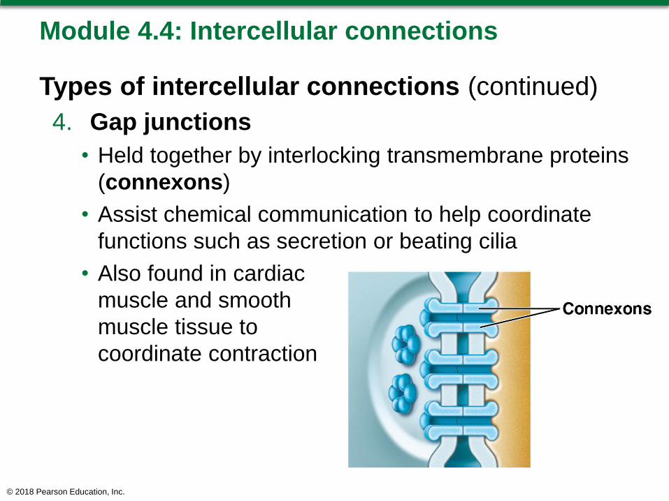
Module 4.4: Intercellular connections
Types of intercellular connections (continued)
4. Gap junctions
• Held together by interlocking transmembrane proteins
(connexons)
• Assist chemical communication to help coordinate
functions such as secretion or beating cilia
• Also found in cardiac
muscle and smooth
muscle tissue to
coordinate contraction
© 2018 Pearson Education, Inc.

Module 4.4: Intercellular connections
Types of intercellular connections (continued)
5. Desmosomes
• Provide firm attachments
by interlocking adjacent
cells’ cytoskeletons
• Opposing plasma
membranes locked
together by cell
adhesion molecules (CAMs)
– Thin layer of proteoglycans may also bond
o Contain polysaccharide, notably hyaluronic acid
• Very strong; resist stretching and twisting
• Found in superficial layers of skin
© 2018 Pearson Education, Inc.

Module 4.4: Review
A. Identify the various types of epithelial intercellular
connections.
B. What is the functional significance of gap
junctions?
C. How do epithelial tissues obtain needed
nutrients?
D. What two types of tissues contribute to the
formation and maintenance of the basement
membrane?
Learning Outcome: Discuss the types and functions
of intercellular connections between epithelial cells.
© 2018 Pearson Education, Inc.

Module 4.5: The cells in a squamous epithelium are flat and irregularly shaped
Squamous epithelium
(squama, plate or scale)
Thin, flat, irregularly shaped
cells (like jigsaw puzzle
pieces)
• Viewed from above, cells look
like fried eggs
• In sectional view, disc-shaped
nucleus found in thickest part
of cell
May be single layer (simple)
or multiple layers (stratified)
© 2018 Pearson Education, Inc.

Module 4.5: Squamous epithelium
Simple squamous epithelium
Most delicate epithelium (one layer thick)
Functions include absorption, diffusion, reduction of
friction
Found in protected
regions such as
peritoneum, capillary
walls, inside eye,
lung alveoli
© 2018 Pearson Education, Inc.
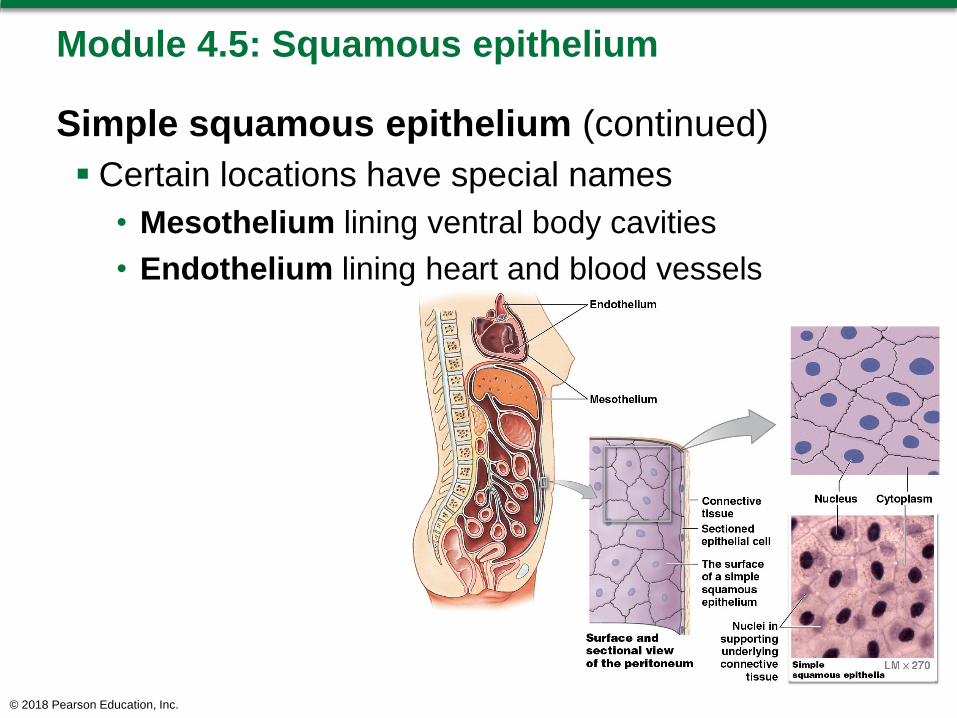
Module 4.5: Squamous epithelium
Simple squamous epithelium (continued)
Certain locations have special names
• Mesothelium lining ventral body cavities
• Endothelium lining heart and blood vessels
© 2018 Pearson Education, Inc.

Module 4.5: Squamous epithelium
Stratified squamous epithelium
Located where severe mechanical or chemical
stresses exist
Many layers of cells
Superficial layer flattened
Forms surface of skin and lines mouth, throat,
esophagus, rectum, anus, vagina
© 2018 Pearson Education, Inc.

Module 4.5: Squamous epithelium
Stratified squamous epithelium (continued)
Two types
1. Keratinized
– Superficial layers packed with keratin
– Tough and water resistant
– Resists both mechanical stress and dehydration
– Found on surface of skin and in hair and nails
2. Nonkeratinized
– Resists abrasion but can dry out
– Found lining oral cavity, pharynx, esophagus, anus,
vagina
© 2018 Pearson Education, Inc.

Keratinized skin layers
© 2018 Pearson Education, Inc.

Module 4.5: Review
A. What do a mesothelium and an endothelium have
in common?
B. Why do the pharynx, esophagus, anus, and vagina
have a similar epithelial organization?
C. What properties are common to keratinized
epithelia?
D. Under a light microscope, a tissue appears as a
simple squamous epithelium. Can this be a sample
of the skin surface? Why or why not?
Learning Outcome: Describe the structure and function
of squamous epithelium.
© 2018 Pearson Education, Inc.

Module 4.6: Cuboidal and transitional epithelia line several passageways and chambers connected to the exterior
Cuboidal epithelium
Cells resemble hexagonal boxes
• In sectional view, cells appear square
Spherical nucleus near center of each cell
© 2018 Pearson Education, Inc.

Module 4.6: Cuboidal and transitional epithelium
Two types of epithelium
1. Simple cuboidal epithelium
• Functions in secretion and absorption
• Lines exocrine glands and ducts
• Lines parts of kidney tubules and thyroid gland
© 2018 Pearson Education, Inc.

Module 4.6: Cuboidal and transitional epithelium
Two types of epithelium (continued)
2. Stratified cuboidal epithelium
• Rare tissue
• Found in ducts of sweat glands and mammary glands
© 2018 Pearson Education, Inc.
Module 4.6: Cuboidal and transitional epithelium
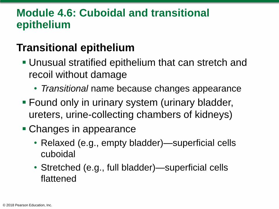
Transitional epithelium
Unusual stratified epithelium that can stretch and
recoil without damage
• Transitional name because changes appearance
Found only in urinary system (urinary bladder,
ureters, urine-collecting chambers of kidneys)
Changes in appearance
• Relaxed (e.g., empty bladder)—superficial cells
cuboidal
• Stretched (e.g., full bladder)—superficial cells
flattened
© 2018 Pearson Education, Inc.

Transitional epithelium
© 2018 Pearson Education, Inc.

Module 4.6: Review
A. Describe the appearance of simple cuboidal
epithelial cells in sectional view.
B. Identify the epithelium that lines the urinary bladder,
and describe its unusual functional characteristic.
C. Describe the changes in appearance of the
transitional epithelium lining the urinary bladder as
stretching occurs.
D. What functions are associated with a simple
cuboidal epithelium and a transitional epithelium?
Learning Outcome: Describe the structure, function,
and locations of cuboidal and transitional epithelia.
© 2018 Pearson Education, Inc.

Module 4.7: Columnar epithelia absorb substances and protect the body from digestive chemicals
Columnar epithelium
In sectional view, cells appear rectangular
Cells taller and more slender than cuboidal
Elongated nuclei in band close to basement
membrane
Types
1. Simple columnar epithelium
2. Pseudostratified columnar epithelium
3. Stratified columnar epithelium
© 2018 Pearson Education, Inc.

Module 4.7: Columnar epithelium
Simple columnar epithelium
Found where absorption or secretion
takes place
• Line stomach, intestine, gallbladder,
uterine tubes, kidney ducts
May have microvilli (for absorption)
or cilia (for movement) on apical
surface
© 2018 Pearson Education, Inc.

Module 4.7: Columnar epithelium
Pseudostratified columnar epithelium
Cells of varying shapes and functions
Distance between nuclei varies, giving appearance
of layering or being stratified
• Each cell contacts basement membrane
Cells usually have cilia
Lines nasal cavities, trachea, larger airways in
lungs, portions of male reproductive tract
© 2018 Pearson Education, Inc.

Module 4.7: Columnar epithelium
Stratified columnar epithelium
Rare tissue type
Two or more layers of cells
• Superficial layer of columnar cells
Found lining large ducts such as those of salivary glands or pancreas
© 2018 Pearson Education, Inc.

Module 4.7: Review
A. Describe the appearance of simple columnar
epithelial cells in a sectional view.
B. Explain why a pseudostratified columnar
epithelium is not truly stratified.
C. Describe the structures found on the surfaces of
simple columnar and pseudostratified columnar
epithelia.
Learning Outcome: Describe the structure,
function, and locations of columnar epithelia.
© 2018 Pearson Education, Inc.

Module 4.8: Glandular epithelia are specialized for secretion
Glands
Collections of epithelial cells (or derived structures)
that produce secretions
Can be scattered cells or complex organs
Categorized into two types
1. Endocrine glands
– Release secretions into interstitial fluid
2. Exocrine glands
– Release secretions into ducts onto epithelial surface
© 2018 Pearson Education, Inc.

Module 4.8: Glandular epithelium
Three types of exocrine gland secretion
1. Merocrine (meros, part) secretion
• Product released from secretory vesicles by
exocytosis
• Most common mode of secretion
• Example: salivary gland secretion
• Mucin
– Merocrine secretion
that mixes with
water to form mucus
© 2018 Pearson Education, Inc.

Module 4.8: Glandular epithelium
Three types of exocrine gland secretion
(continued)
2. Apocrine (apo, off) secretion
• Apical cytoplasm packed with secretory vesicles
• Cell releases cytoplasm as well as secretory product
• Example: mammary gland secretion (involves
combination of merocrine and apocrine secretions)
© 2018 Pearson Education, Inc.
Module 4.8: Glandular epithelium
Three types of exocrine gland secretion
(continued)
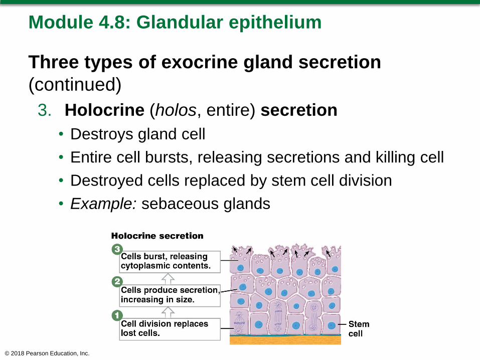
3. Holocrine (holos, entire) secretion
• Destroys gland cell
• Entire cell bursts, releasing secretions and killing cell
• Destroyed cells replaced by stem cell division
• Example: sebaceous glands
© 2018 Pearson Education, Inc.

Module 4.8: Review
A. Describe the two primary types of glands.
B. By what three methods do various secretory
cells of exocrine glands release their
secretions?
Learning Outcome: Compare the three different
methods of exocrine secretion by glandular
epithelia.
© 2018 Pearson Education, Inc.
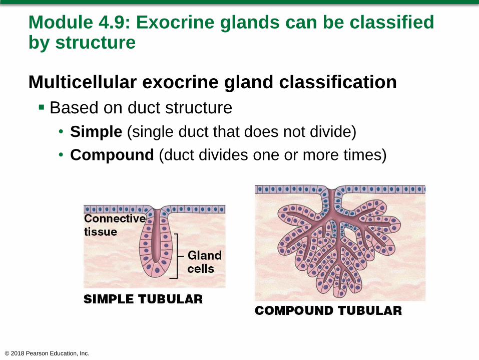
Module 4.9: Exocrine glands can be classified by structure
Multicellular exocrine gland classification
Based on duct structure
• Simple (single duct that does not divide)
• Compound (duct divides one or more times)
© 2018 Pearson Education, Inc.

Module 4.9: Glandular epithelium classification
Multicellular exocrine gland classification
(continued)
Based on the shape of the secretory area
• Tubular (glandular cells form tubes)
• Alveolar or acinar (glandular cells form sacs)
• Tubuloalveolar (glandular cells form both tubes
and sacs)
© 2018 Pearson Education, Inc.

Types of compound glands
© 2018 Pearson Education, Inc.
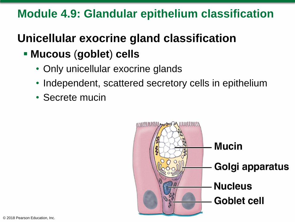
Module 4.9: Glandular epithelium classification
Unicellular exocrine gland classification
Mucous (goblet) cells
• Only unicellular exocrine glands
• Independent, scattered secretory cells in epithelium
• Secrete mucin
© 2018 Pearson Education, Inc.

Module 4.9: Review
A. What three characteristics are used to describe
multicellular exocrine glands?
B. Describe the simplest type of multicellular
exocrine gland.
Learning Outcome: Explain how multicellular
exocrine glands are classified by their structure.
© 2018 Pearson Education, Inc.

Section 2: Connective Tissue
Learning Outcomes
4.10 Describe the general structure of connective
tissue.
4.11 Describe the structure, function, and locations of
areolar tissue, adipose tissue, and reticular
tissue.
4.12 Describe the structure, function, and locations of
dense connective tissues and fluid connective
tissues.
4.13 Describe the structure, function, and locations of
cartilage.
© 2018 Pearson Education, Inc.

Section 2: Connective Tissue
Learning Outcomes (continued)
4.14 Describe the structure and function of bone.
4.15 Describe the arrangements of epithelial and
connective tissues in the four types of tissue
membranes, and describe the structures and
locations of the three types of fasciae.
© 2018 Pearson Education, Inc.

Module 4.10: A matrix surrounds connective tissue cells
Connective tissue overview
Varies widely in
appearance and
function
• Found throughout
the body, but never
exposed to surface
Ranges from highly
vascular to avascular
Many contain sensory receptors that detect pain,
pressure, temperature, and other stimuli
© 2018 Pearson Education, Inc.

Module 4.10: Connective tissue
Three basic components shared by connective
tissues
1. Specialized cells
2. Extracellular protein fibers
3. Fluid called ground substance
• Matrix
– Extracellular fibers and ground substance
– Surrounds the cells
– Accounts for majority of connective tissue volume
– Fewer cells and more extracellular material compared
to epithelial tissue
© 2018 Pearson Education, Inc.

Module 4.10: Connective tissue
Three subdivisions of connective tissue
1. Connective tissue proper
• Contains many types of cells
• Extracellular fibers in
syrupy ground substance
– Loose (fibers create loose,
open framework)
– Dense (fibers densely packed)
© 2018 Pearson Education, Inc.

Module 4.10: Connective tissue
Three subdivisions of connective tissue
(continued)
2. Fluid connective tissue
• Distinctive group of cells
• Watery matrix
– Blood (within cardiovascular
system)
– Lymph (within lymphatic
system)
© 2018 Pearson Education, Inc.

Module 4.10: Connective tissue
Three subdivisions of connective tissue
(continued)
3. Supporting connective
tissue
• Less diverse cell population
• More densely packed matrix
– Cartilage (solid, rubbery
matrix)
– Bone (solid, crystalline
matrix)
© 2018 Pearson Education, Inc.

Module 4.10: Review
A. Identify the three basic components of
connective tissue.
B. Summarize the functions of connective tissue.
C. Distinguish among connective tissue proper,
fluid connective tissues, and supporting
connective tissues.
Learning Outcome: Describe the general structure
of connective tissue.
© 2018 Pearson Education, Inc.

Module 4.11: Loose connective tissues support other tissue types
Connective tissue proper components
Extracellular protein fibers
• Reticular fibers (strong and form branching network)
• Collagen fibers (thick, very strong)
• Elastic fibers (slender, very stretchy)
Ground substance
• Clear and colorless
• Viscous (syrupy) due to presence of proteoglycans
and glycoproteins
© 2018 Pearson Education, Inc.

Module 4.11: Loose connective tissue
Connective tissue proper components
(continued)
Two classes of cells
1. Fixed (stationary; involved with maintenance, repair,
energy storage)
– Melanocytes (synthesize melanin pigment)
– Fixed macrophage (engulfs cell debris and
pathogens)
– Mast cells (stimulate inflammation and mobilize
defenses)
© 2018 Pearson Education, Inc.

Module 4.11: Loose connective tissue
Connective tissue proper components
(continued)
Two classes of cells
1. Fixed
– Fibroblasts (synthesize extracellular fibers)
– Adipocytes (store lipid reserves)
– Fibrocytes (differentiate from fibroblasts and maintain
extracellular fibers)
© 2018 Pearson Education, Inc.

Module 4.11: Loose connective tissue
Connective tissue proper components
(continued)
Two classes of cells
2. Wandering (move throughout tissue; function in
defense and repair)
– Plasma cells (immune cells producing antibodies)
– Free macrophages (engulf debris and pathogens)
– Mesenchymal cells (stem cells that aid
tissue repair)
– Neutrophils and eosinophils (phagocytic
blood cells)
– Lymphocytes (immune system cells)
© 2018 Pearson Education, Inc.

Connective tissue components
© 2018 Pearson Education, Inc.

Module 4.11: Loose connective tissue
Three types of loose connective tissue
1. Areolar tissue
• Most common connective tissue proper
• Packing material of the body
• Has all connective tissue proper cell types
© 2018 Pearson Education, Inc.

Module 4.11: Loose connective tissue
Three types of loose connective tissue
(continued)
2. Adipose tissue
• Found deep to skin in various areas of body
• Forms layer of padding around eyes and kidneys
• Cells (adipocytes) account for most of tissue volume
© 2018 Pearson Education, Inc.

Module 4.11: Loose connective tissue
Three types of loose connective tissue
(continued)
3. Reticular tissue
• Found in liver, kidney, spleen, lymph nodes, and bone
marrow
• Provides support and resists distortion
• Many reticular fibers
forming network (stroma)
© 2018 Pearson Education, Inc.
Module 4.11: Review
A. Identify the types of cells found in connective tissue
proper.
B. Describe the role of fibroblasts in connective tissue.
C. Which type of loose connective tissue contains
primarily lipids?
D. What term means the fibrous supporting network
formed of reticular fibers?
E. What types of phagocytic cells are present in
connective tissue proper?
Learning Outcome: Describe the structure, function,
and locations of areolar tissue, adipose tissue, and
reticular tissue.
© 2018 Pearson Education, Inc.

Module 4.12: Dense connective tissues are dominated by extracellular fibers …
Three types of dense connective tissues
Most volume occupied by extracellular fibers
1. Dense regular connective tissue
– Found in cords (tendons, ligaments) or sheets
– Collagen arranged in parallel bundles
© 2018 Pearson Education, Inc.

Module 4.12: Dense connective tissue
Three types of dense connective tissue
(continued)
2. Dense irregular connective tissue
• Fibers arranged in meshwork (no consistent pattern)
to resist tension in many directions
• Found covering visceral organs; in superficial layers
of bones, cartilages, and peripheral nerves; in dermis
of skin
© 2018 Pearson Education, Inc.

Module 4.12: Dense connective tissue
Three types of dense connective tissue
(continued)
3. Elastic tissue
• More elastic fibers than collagen
• Is springy and resilient
• Found between vertebrae, in walls of large blood
vessels, erectile tissues of penis
© 2018 Pearson Education, Inc.

Module 4.12: … whereas fluid connective tissues have an aqueous matrix
Fluid connective tissue
Fluid matrix with many suspended proteins
Usually contains no insoluble fibers
Two types of liquid connective tissue
1. Blood
2. Lymph
© 2018 Pearson Education, Inc.

Module 4.12: Fluid connective tissue
Fluid connective tissue (continued)
Blood components
• Watery matrix called plasma
• Formed elements suspended in plasma
– Red blood cells (transport oxygen)
– White blood cells (bodily defense)
o Monocytes (large phagocytes)
o Lymphocytes (uncommon in blood)
o Eosinophils/neutrophils (small phagocytes)
o Basophils (promote inflammation)
– Platelets (involved in clotting response)
© 2018 Pearson Education, Inc.

Blood as fluid connective tissue
© 2018 Pearson Education, Inc.

Module 4.12: Fluid connective tissue
Fluid connective tissue (continued)
Lymph
• Watery matrix called lymph located in lymphatic
vessels
• Collected from interstitial fluid
• Majority of cells are lymphocytes
• Returned to blood at large veins near heart
• Functions to maintain solute levels, blood volume,
and alert immune system of infection
© 2018 Pearson Education, Inc.

Module 4.12: Fluid connective tissue
Fluid connective tissue (continued)
Extracellular fluid circulation
• Contractions of the heart move blood through blood
vessels
– Arteries (away from heart)
– Capillaries (smallest vessels; sites of exchange)
– Veins (toward heart)
• Water and solutes exchanged between plasma and
interstitial fluid
• Lymphatic vessels collect excess interstitial fluid
• Lymphatic vessels return lymph to large veins near
heart
© 2018 Pearson Education, Inc.

Lymph as fluid connective tissue
© 2018 Pearson Education, Inc.

Module 4.12: Review
A. What makes a connective tissue “loose” or
“dense”?
B. Summarize the role of extracellular fluid in
maintaining homeostasis.
C. Lack of vitamin C in the diet interferes with the
ability of fibroblasts to produce collagen. How
might this affect connective tissue function?
Learning Outcome: Describe the structure,
function, and locations of dense connective tissues
and fluid connective tissues.
© 2018 Pearson Education, Inc.

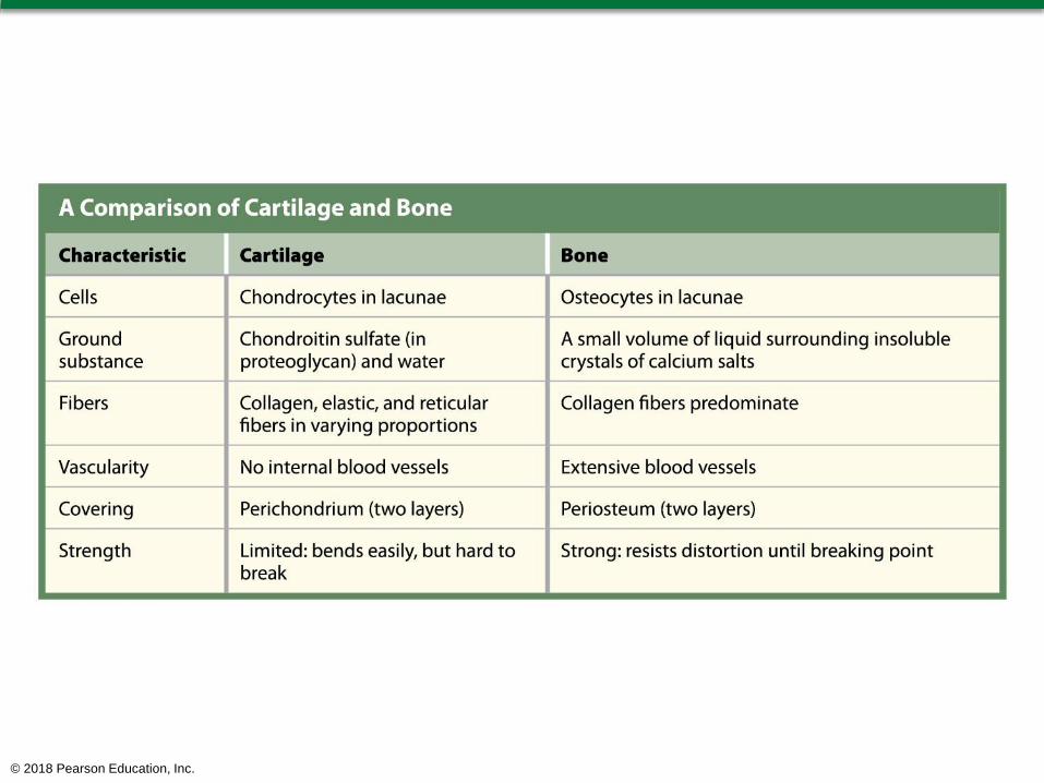
Module 4.13: Cartilage provides a flexible support for body structures
Cartilage overview
Matrix is a firm gel containing chondroitin sulfates
(chondros, cartilage), polysaccharide derivatives
• Form complexes with proteins forming proteoglycans
Only one type of cell (chondrocyte)
• Found in small chambers called lacunae (lacus, lake)
Avascular
© 2018 Pearson Education, Inc.

Module 4.13: Cartilage
Three types of cartilage
1. Hyaline cartilage
• Found between ribs and sternum, covering bones in
mobile joints, part of nasal septum, supporting
respiratory passageways
• Provides stiff but flexible support
• Reduces friction
© 2018 Pearson Education, Inc.

Module 4.13: Cartilage
Three types of cartilage (continued)
2. Elastic cartilage
• Distorts without damage and returns to original shape
• Found in external ear and smaller internal structures
© 2018 Pearson Education, Inc.

Module 4.13: Cartilage
Three types of cartilage (continued)
3. Fibrocartilage
• Found in knee joint, between pubic bones, and in
intervertebral discs
• Durable and tough
• Resists compression, prevents bone-to-bone contact,
and limits relative movement
© 2018 Pearson Education, Inc.

Module 4.13: Cartilage
Cartilage properties
Set apart from surrounding
tissues by perichondrium
(peri-, around)
• Two layers of perichondrium
1. Outer layer of dense
irregular connective tissue
oMechanical support,
protection, attachment
2. Inner cellular layer
oWhere cartilage growth and maintenance occur
• Blood vessels in perichondrium provide oxygen and
nutrients to chondrocytes
© 2018 Pearson Education, Inc.

Module 4.13: Cartilage
Two types of cartilage growth
1. Appositional growth (at cartilage surface)
• Chondroblasts (immature chondrocytes) divide in
cellular layer of perichondrium
• Chondroblasts secrete new matrix
• Once surrounded by matrix, chondroblasts mature
into chondrocytes
© 2018 Pearson Education, Inc.

Module 4.13: Cartilage
Two types of cartilage growth (continued)
2. Interstitial growth (within cartilage)
• Chondrocytes divide within a lacuna
• Daughter cells secrete additional matrix and move apart
• Both types of cartilage growth occur during
development
• Normally no growth and repair in adults
• With slight damage or with hormonal stimulation, some
appositional growth possible
© 2018 Pearson Education, Inc.

Module 4.13: Review
1. Which connective tissue fiber is characteristic of
the cartilage supporting the ear?
2. Describe the two layers making up the
perichondrium.
3. Contrast appositional and interstitial growth of
cartilage.
Learning Outcome: Describe the structure,
function, and locations of cartilage.
© 2018 Pearson Education, Inc.

Module 4.14: Bone provides a strong framework for the body
Osseous (os, bone) tissue (Bone tissue)
Connective tissue with solid, crystalline matrix
• Small volume of ground substance
• 2/3 of matrix is calcium salts (provide strength)
– Mostly calcium phosphate
– Some calcium carbonate
• Many collagen fibers (provide flexibility)
Strong, somewhat flexible, resistant to shattering
© 2018 Pearson Education, Inc.

Module 4.14: Bone
Typical long bone structure
Hollow with two types of bone
1. Compact bone
– Outer layer of bone
2. Spongy bone
– Lines internal cavity
– Finer network
© 2018 Pearson Education, Inc.

Typical long bone structure
© 2018 Pearson Education, Inc.
© 2018 Pearson Education, Inc.

Module 4.14: Bone
Compact bone structure
Matrix organized in concentric layers
• Organized into functional units (osteons)
– Central canal contains blood vessels in center
• Cells (osteocytes) located between layers
• Canaliculi (little canals) connect osteocytes
Superficial layer of solid, calcified bone prevents
interstitial growth
© 2018 Pearson Education, Inc.

Module 4.14: Bone
Compact bone structure (continued)
Surrounded by periosteum
• Two layers
1. Outer fibrous layer
allows attachment
of ligaments
2. Inner cellular layer
allows appositional
growth and repair
© 2018 Pearson Education, Inc.

Module 4.14: Review
A. Describe bone matrix.
B. What are mature bone cells in lacunae called?
C. What is the functional unit of compact bone?
D. Distinguish between the two types of supporting
connective tissues with respect to their
characteristic fibers.
Learning Outcome: Describe the structure and
function of bone.
© 2018 Pearson Education, Inc.

Module 4.15: Tissues membranes are physical barriers, and fasciae support and surround organs
Overview of membranes
Line or cover body surfaces
Typically consist of epithelium supported by
connective tissue
Four types in the body
1. Mucous membranes
2. Serous membranes
3. Cutaneous membrane
4. Synovial membranes
© 2018 Pearson Education, Inc.

Module 4.15: Membranes and fasciae
Four types of membranes
1. Mucous membranes
• Line passageways
open to the exterior of
the body
– Digestive, respiratory,
reproductive, urinary
tracts
• Must be kept moist to facilitate movement, absorption,
or secretion
• Lubricated by mucus or bodily fluids
• Supported by areolar connective tissue (lamina
propria)
© 2018 Pearson Education, Inc.
Module 4.15: Membranes and fasciae
Four types of membranes (continued)
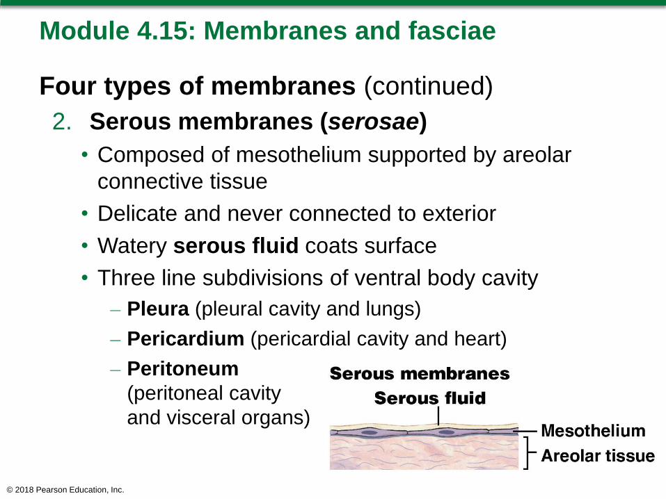
2. Serous membranes (serosae)
• Composed of mesothelium supported by areolar
connective tissue
• Delicate and never connected to exterior
• Watery serous fluid coats surface
• Three line subdivisions of ventral body cavity
– Pleura (pleural cavity and lungs)
– Pericardium (pericardial cavity and heart)
– Peritoneum
(peritoneal cavity
and visceral organs)
© 2018 Pearson Education, Inc.

Module 4.15: Membranes and fasciae
Four types of membranes (continued)
3. Cutaneous membrane
• Covers surface of body (skin)
• Composed of:
– Stratified squamous epithelium
– Layer of areolar tissue
– Underlying dense irregular connective tissue
• Relatively thick, waterproof, and usually dry
© 2018 Pearson Education, Inc.
Module 4.15: Membranes and fasciae
Four types of membranes (continued)
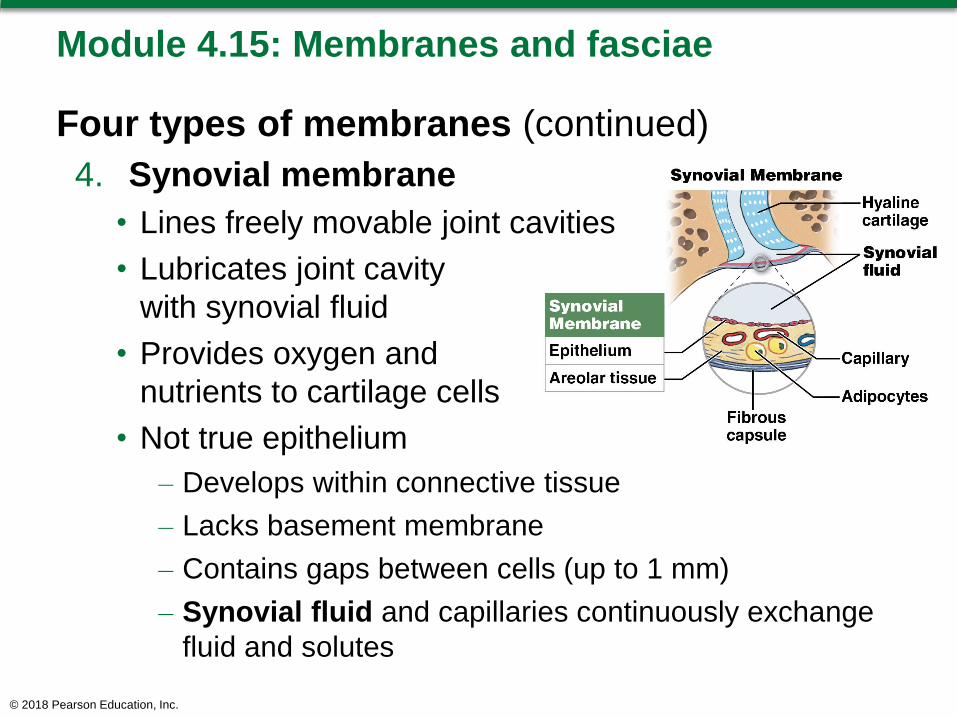
4. Synovial membrane
• Lines freely movable joint cavities
• Lubricates joint cavity
with synovial fluid
• Provides oxygen and
nutrients to cartilage cells
• Not true epithelium
– Develops within connective tissue
– Lacks basement membrane
– Contains gaps between cells (up to 1 mm)
– Synovial fluid and capillaries continuously exchange
fluid and solutes
© 2018 Pearson Education, Inc.

Module 4.15: Membranes and fasciae
Fasciae: Support and surround organs
Three types of layers
1. Superficial fascia
– Under skin
– Consists of areolar and adipose tissue
2. Deep fascia
– Continuous with capsules, ligaments, and other
connective tissue structures
– Consists of dense irregular connective tissue
– Forms strong, fibrous internal framework
3. Subserous fascia
– Between serous membranes and deep fascia
– Consists entirely of areolar tissue
© 2018 Pearson Education, Inc.

Connective tissue framework of the body
© 2018 Pearson Education, Inc.
Module 4.15: Review
A. Which cavities in the body are lined by serous
membranes?
B. Name the four types of membranes found in the
body.
C. Name the three layers of fascia and their types of
connective tissue.
D. Which of the four tissue membranes is relatively
waterproof and usually dry?
Learning Outcome: Describe the arrangements of
epithelial and connective tissues in the four types of
tissue membranes, and describe the structures and
locations of the three types of fasciae.
© 2018 Pearson Education, Inc.

Section 3: Muscle Tissue and Nervous Tissue
Learning Outcomes
4.16 Describe the relative proportions of muscle tissue
and nervous tissue in the body.
4.17 Specify the functions of muscle tissue and
nervous tissue.
4.18 Clinical Module: Describe the roles of
inflammation and regeneration in response to
tissue injury.
© 2018 Pearson Education, Inc.

Module 4.16: Muscle tissue outweighs nervous tissue by 25:1
Tissue types by weight
Muscle tissue is 50 percent total body weight
• The most of any major tissue type
Nervous tissue is 2 percent total body weight
• The least of any major tissue type
Connective tissue (45 percent) and epithelial tissue
(3 percent) provide interwoven framework for body
© 2018 Pearson Education, Inc.

Percentages (by weight) of the four tissue types in the body
© 2018 Pearson Education, Inc.

Module 4.16: Muscle tissue and nervous tissue
Types of muscle tissue
Skeletal muscle tissue
• Moves the body
Cardiac muscle tissue
• Moves blood within the heart and through blood
vessels
Smooth muscle tissue
• Moves fluids and solids along digestive tract
• Regulates diameter of small arteries, among other
functions
© 2018 Pearson Education, Inc.

Module 4.16: Review
A. What is the relative percentage of body weight
from each of the four tissue types?
B. List the three classifications of muscle tissue,
and describe a function for each type.
Learning Outcome: Describe the relative
proportions of muscle tissue and nervous tissue in
the body.
© 2018 Pearson Education, Inc.

Module 4.17: Muscle tissue is specialized for contraction …
Muscle tissue
Specialized for contraction to cause movement
• Movement of the body
• Movement of blood around the cardiovascular system
• Movement of materials along the digestive tract
Three types of muscles
1. Skeletal muscle tissue
2. Cardiac muscle tissue
3. Smooth muscle tissue
© 2018 Pearson Education, Inc.

Module 4.17: Functions of muscle tissue
Three types of muscle tissue
1. Skeletal muscle tissue
• Found in skeletal muscle
• Elongated, cylindrical, banded (striated) cells with
multiple nuclei (multinucleate)
• Functions
– Move and stabilizes skeleton
– Guard entrances and exits to digestive, respiratory,
urinary tracts
– Generate heat
– Protect internal organs
© 2018 Pearson Education, Inc.

Skeletal muscle tissue
© 2018 Pearson Education, Inc.

Module 4.17: Functions of muscle tissue
Three types of muscle tissue (continued)
2. Cardiac muscle tissue
• Found only in heart
• Cells (“cardiocytes”) are short, branched, and usually
have a single nucleus
– Interconnected with special junctions (intercalated
discs) that help synchronize cardiocyte contractions
• Functions to move blood and maintain blood pressure
© 2018 Pearson Education, Inc.

Module 4.17: Functions of muscle tissue
Three types of muscle tissue (continued)
3. Smooth muscle tissue
• Found throughout body (skin, blood vessel walls,
many organs of various systems)
• Cells are short, spindle-shaped, nonstriated, have a
single nucleus
• Functions
– Move food, urine, and reproductive secretions
– Control diameter of respiratory passageways and blood
vessels
© 2018 Pearson Education, Inc.

Module 4.17: … and nervous tissue is specialized for communication
Nervous tissue
Specialized for conduction of electrical impulses
98 percent found in brain and spinal cord
Two basic types of cells
1. Neurons (neuros, nerve)
2. Neuroglia or glial cells (glia, glue)
– Various supporting cells
© 2018 Pearson Education, Inc.

Module 4.17: Functions of nervous tissue
Neurons
Transfer information around body and perform
information processing
Vary in size and shape
• Longest cells in body are neurons (up to 1 meter)
© 2018 Pearson Education, Inc.

Module 4.17: Functions of nervous tissue
Neuron structure
Dendrites (dendron, tree)
• Receive information
Axon
• Conducts information to other cells
• Also called nerve fibers
Cell body
• Contains large nucleus and other organelles
• Cell control center and site of information processing
• Most lack centrioles and cannot divide
© 2018 Pearson Education, Inc.

Neuron structure
© 2018 Pearson Education, Inc.

Module 4.17: Functions of nervous tissue
Neuroglia
Several different structural types with associated
functions
© 2018 Pearson Education, Inc.

Module 4.17: Review
A. Which type of muscle tissue regulates blood
vessel diameter?
B. Distinguish between neurons and neuroglia.
C. Organs are made up of different tissues. What
tissues are found in skeletal muscles?
Learning Outcome: Specify the functions of muscle
tissue and nervous tissue.
© 2018 Pearson Education, Inc.

Module 4.18: CLINICAL MODULE: The response to tissue injury involves inflammation and regeneration
Tissues respond in a coordinated way to restore
homeostasis
Two restoration processes
Inflammation
Regeneration
© 2018 Pearson Education, Inc.

Module 4.18: CLINICAL MODULE: Responses to tissue injury
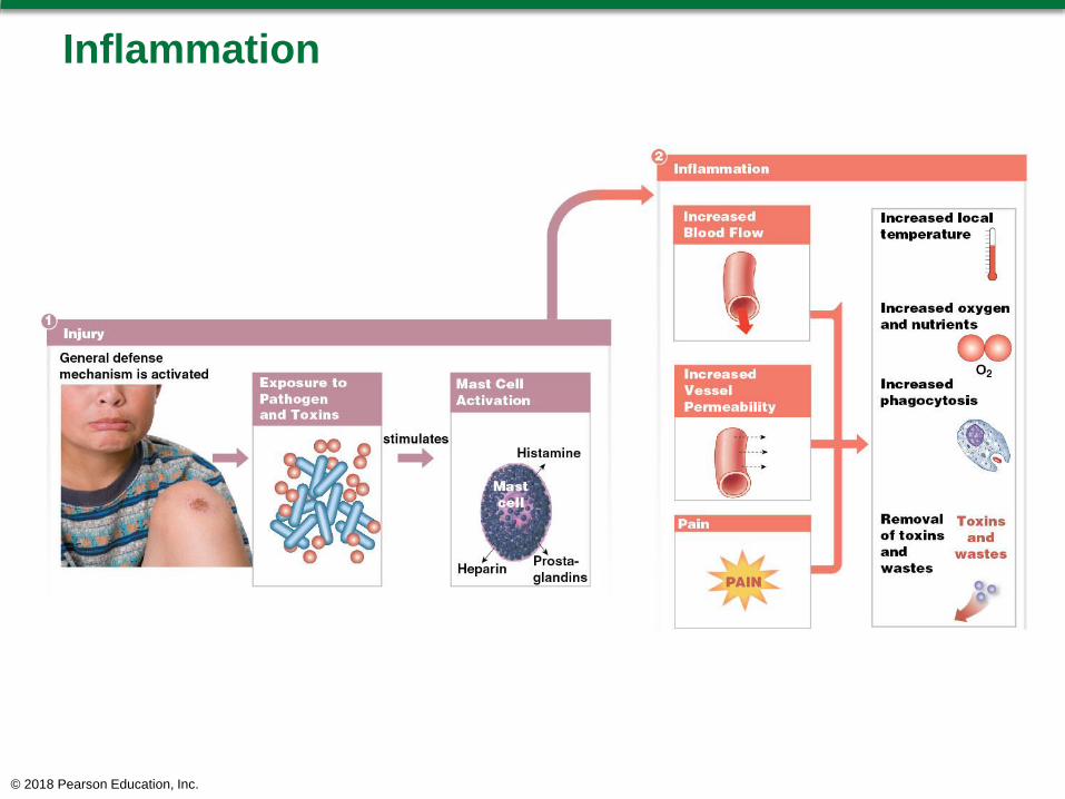
Injury occurs
Body is exposed to pathogens and toxins and
chemicals from injured cells
Body activates defense mechanisms including:
• Mast cell activation
– Mast cells release histamine and other chemicals
– Stimulates inflammation
© 2018 Pearson Education, Inc.

Module 4.18: CLINICAL MODULE: Responses to tissue injury
Inflammation
Produces indications of injury
1. Swelling
2. Redness
3. Warmth
4. Pain
May result from injury or from infection (presence
of pathogens within the tissue)
Occurs in connective tissue, so may occur
anywhere in the body (as all organs contain
connective tissue)
© 2018 Pearson Education, Inc.

Module 4.18: CLINICAL MODULE: Responses to tissue injury
Inflammation (continued)
Dilates (enlarges) blood vessels
Increases blood vessel permeability
Increased blood flow to the area causes:
• Swelling in the area
• Increased local temperature
• Increased delivery of oxygen and nutrients
• Increased removal of toxins and wastes
Stimulates increased phagocytosis in tissues
Sensation of pain from abnormal conditions and
chemicals released by mast cells
© 2018 Pearson Education, Inc.
Inflammation
© 2018 Pearson Education, Inc.

Module 4.18: CLINICAL MODULE: Responses to tissue injury
Cleanup process (removing toxins and wastes)
usually eliminates inflammatory stimuli in hours to
days
Regeneration
Occurs after damaged tissue has stabilized
Fibroblasts produce collagen fibers to stabilize injury
site
Produces dense, collagenous framework called
scar tissue
Scar tissue usually remodeled and normal tissue
conditions restored
© 2018 Pearson Education, Inc.

Module 4.18: CLINICAL MODULE: Responses to tissue injury
Each tissue has a different ability to regenerate
Epithelial, connective (except cartilage), and smooth
muscle regenerate well
Other muscle types and neural tissue regenerate
poorly, if at all
In tissues that regenerate poorly, scar tissue
replaces tissues that do not regenerate
• Fibrosis is the permanent replacement of normal
tissue by scar tissue
© 2018 Pearson Education, Inc.

Module 4.18: CLINICAL MODULE: Responses to tissue injury
© 2018 Pearson Education, Inc.

Module 4.18: Review
A. Identify the two processes in the response to
tissue injury.
B. What are the four indications of inflammation
that occur following an injury?
C. Why can inflammation occur in any organ in the
body?
Learning Outcome: Describe the roles of
inflammation and regeneration in response to
tissue injury.
© 2018 Pearson Education, Inc.