Embed Size (px)
Citation preview
TITLE:
A STUDY TRIAL ON PROTESCAL IN PREVENTING POST CAESAREAN SECTION HYPERTROPHIC SCAR AND KELOID
PRINCIPAL INVESTIGATOR : DR ANIZAH ALI
CO-RESEARCHER : DR. NOR AZILA MOHD NAFIAH
RESEARCH CODE : FF-2017-170
DATE : 26/4/2017
Content Page
1.0 Introduction
2.0 Objectives of the study
2.1 General objective
2.2 Specific objectives
3.0 Outcomes
3.1 Primary outcome
3.2 Secondary outcome
4.0 Methodology
4.1 Study design
4.2 Place of study
4.3 Study population
4.4 Duration of study
4.5 Inclusion Criteria
4.6 Exclusion Criteria
4.7 Randomization
4.8 Data Collection and statistical analysis
4.9 Sample size calculation
4.10 Study flow chart
4.11 Study Protocol
5.0 References
1.0 INTRODUCTION
Every year several million women worldwide acquire an abdominal scar as a result of caesarean delivery. Obstetricians often consider skin closure after a caesarean section as a trivial aspect of the procedure, because the skin scar is deemed the normal and inevitable price we pay foe tissue repair. Moreover, the anatomical location of caesarean scars, which hide easily beneath underwear, and the generally held belief that all transverse suprapubic incision heal about equally well further contribute to the underestimation by practitioners of the importance of scar appearance to patients. Young women place supreme importance on cosmetic outcomes, but scarring can also affect patient in term of symptoms ( pain, tenderness, and itching ) and has the potential to have a negative impact overall quality of life, being a source of considerable distress, loss of self esteem and stigmatization. The final appearance and the function of the healed skin is dependent on patient and wound factors, which are often outside the control of a surgeon, and technical factors, which are completely within the control of the surgeon and include closure material and technique of skin apposition. There were few study trial done with the aims of preventing hyperthropic scar formation following caesarean section. Antonella Cromi et al ( obstet Gynaecol 2010, 203: 36.el-8 ) did a randomized trial on 123 patient to compare scar quality associated with different types of wound closure method after caesarean section. The result showed that the were no difference in scar quality in either staples or 3 different types of subcurticular sutures. Atkinson et al ( 2005 American Society of plastic surgeon) performed a randomized controlled trial involving 70 patient to determine the efficacy of paper tape in preventing hyperthrophic scar formation in surgical incisions that traverse Langer's skin tension lines. Result suggest that tension acting on a scar is the trigger for hyperthrophic scarring, and paper tape is likely to be an effective modality for prevention of hypertrophic scarring through its ability to eliminate scar tension.
The aims of this study is to prevent hypertrophic scar and keloid formation post caesarean section using PROTESCAL adhesion barrier. PROTESCAL, a combination of hyaluronic acid, methylcellulose, and alginate was manufactured by Korean pharmaceutical companies and became available since 2012. PROTESCAL was developed to prevent complications such as ileus, pain and infertility due to postoperative adhesion.
Hyaluronic acid is a natural polymer of disaccharides, one of the components of the extracellular matrix. It is present in the skin, cartilage, bone and brain. Because of its biocompatibility, moisture capacity, and viscoelasticity, hyaluronic acid has been used as
artificial tears in drug delivery systems, and tissue restoration materials, and it plays a role in inflammation, granulation and re-epithelization for wound healing. It has proved valuable in neurosurgery and dermatology because hyaluronic acid and degradation products can modulate wound healing. There is wide scientific evidence on the positive role of hyaluronic acid in tissue regeneration and wound healing.
Carboxymethylcellulose is a high molecular weight polysaccharide that has a concentration and volume that are inversely correlated with its antiadhesive agent. The combination of carboxymethylcellulose and hyaluronic acid has had a preventive effect on the formation of adhesion in various surgical fields.
Alginate has been used as a wound dressing agent, its calcium or sodium form has hemostatic and antimicrobial effects, and it has been shown to prevent adhesion formation in animal studies.
With the combination of hyluronic acid, methylcellulose and alginate, which already proven scientifically benefit in wound healing, we aims to prevent the hypertrophic scar and keloid formation following caesarean section by applying PROTESCAL in subcutaneous layer prior to skin closure.
2.0 OBJECTIVE OF THE STUDY
2.1 General Objective
To evaluate the effectiveness of Protescal in preventing post caesarean section hypertrophic scar and keloid and pelvic adhesion.
2.2 Specific Objectives
To determine the effectiveness of Protescal in preventing hypertrophic scar and keloid compared to control group.
To determine the effectiveness of Protescal in preventing pelvic adhesion
3.0 OUTCOMES
3.1 Primary
To assess the outcome of healing of the external scar
3.2 Secondary
To look for adhesion formed during next caesarean section
4.0 RESEARCH METHODOLOGY
4.1 Study design
Prospective, randomized controlled clinical trial
4.2 Place of study
Obstetric ward and maternity operation theatre of Department of Obstetrics and Gynaecology, Universiti Kebangsaan Malaysia Medical Centre (UKMMC).
4.3 Study Population
All women undergoing elective caesarean section, without any history of previous abdominal surgery; who planned for further pregnancy and consented to participate in this study.
4.4 Duration of study
6 months duration; from April 2017 to October 2017
4.5 Inclusion Criteria
Pregnant women without any history of previous abdominal surgery
Plan for elective caesarean section for this current pregnancy section
Transverse suprapubic scar
4.6 Exclusion Criteria
Patient who are allergic to protescal
Patient with previous abdominal surgery
Patient with surgery complication
4.7 Randomization
The randomization sequence, either to Protescal group or control group, was generated by using a computer randomization program; in the maternity operation theatre or antenatal clinic .
4.8 Data Collection And Statistical Analysis
SPSS (Statistical Package of Social Science) version 20.0 will be employed proportional data
will be compared with chi square. The Fisher exact test will be chosen if the expected size of
any cell of the contingency table is less than 5. Continuous data will be compared using the
Wilcoxon rank-sum test. Multiple logistic regressions will be use to model the relationship
between group assignment, controlling for possible confounders. P<0.05 will be considered
significant.
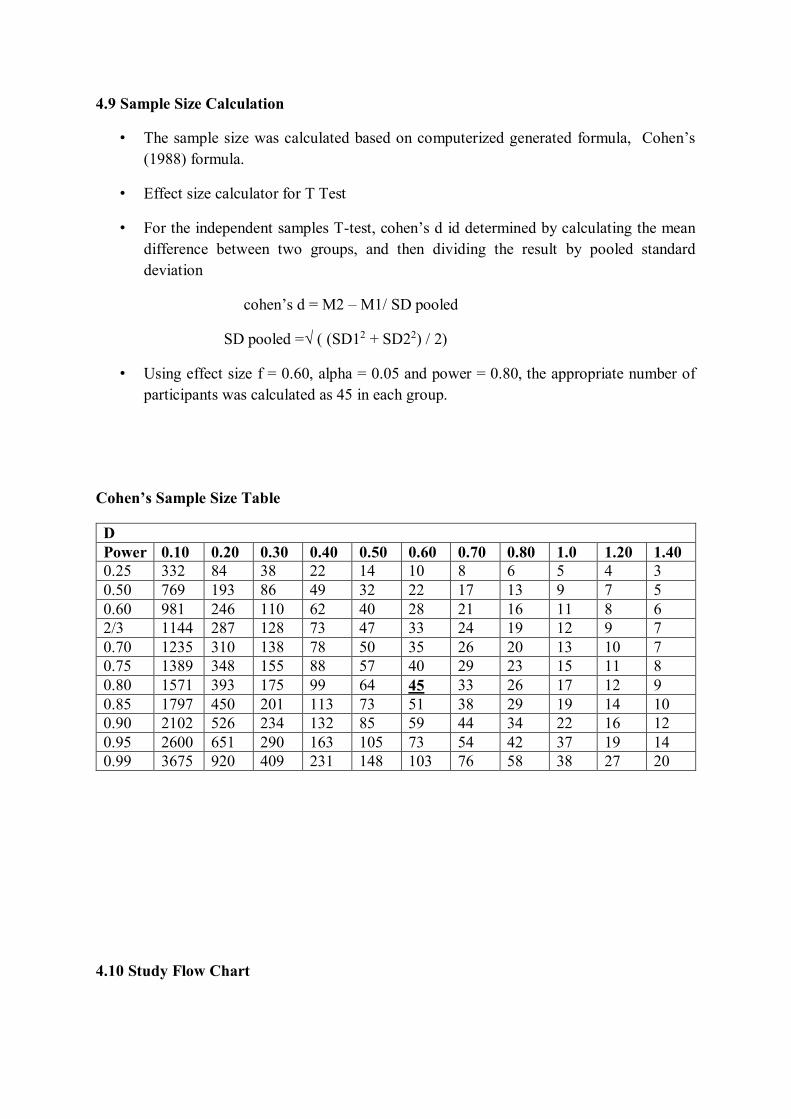
4.9 Sample Size Calculation
• The sample size was calculated based on computerized generated formula, Cohen’s
(1988) formula.
• Effect size calculator for T Test
• For the independent samples T-test, cohen’s d id determined by calculating the mean
difference between two groups, and then dividing the result by pooled standard deviation
cohen’s d = M2 – M1/ SD pooled
SD pooled =√ ( (SD12 + SD22) / 2)
• Using effect size f = 0.60, alpha = 0.05 and power = 0.80, the appropriate number of participants was calculated as 45 in each group.
Cohen’s Sample Size Table
D Power 0.10 0.20 0.30 0.40 0.50 0.60 0.70 0.80 1.0 1.20 1.40 0.25 332 84 38 22 14 10 8 6 5 4 3 0.50 769 193 86 49 32 22 17 13 9 7 5 0.60 981 246 110 62 40 28 21 16 11 8 6 2/3 1144 287 128 73 47 33 24 19 12 9 7 0.70 1235 310 138 78 50 35 26 20 13 10 7 0.75 1389 348 155 88 57 40 29 23 15 11 8 0.80 1571 393 175 99 64 45 33 26 17 12 9 0.85 1797 450 201 113 73 51 38 29 19 14 10 0.90 2102 526 234 132 85 59 44 34 22 16 12 0.95 2600 651 290 163 105 73 54 42 37 19 14 0.99 3675 920 409 231 148 103 76 58 38 27 20
4.10 Study Flow Chart
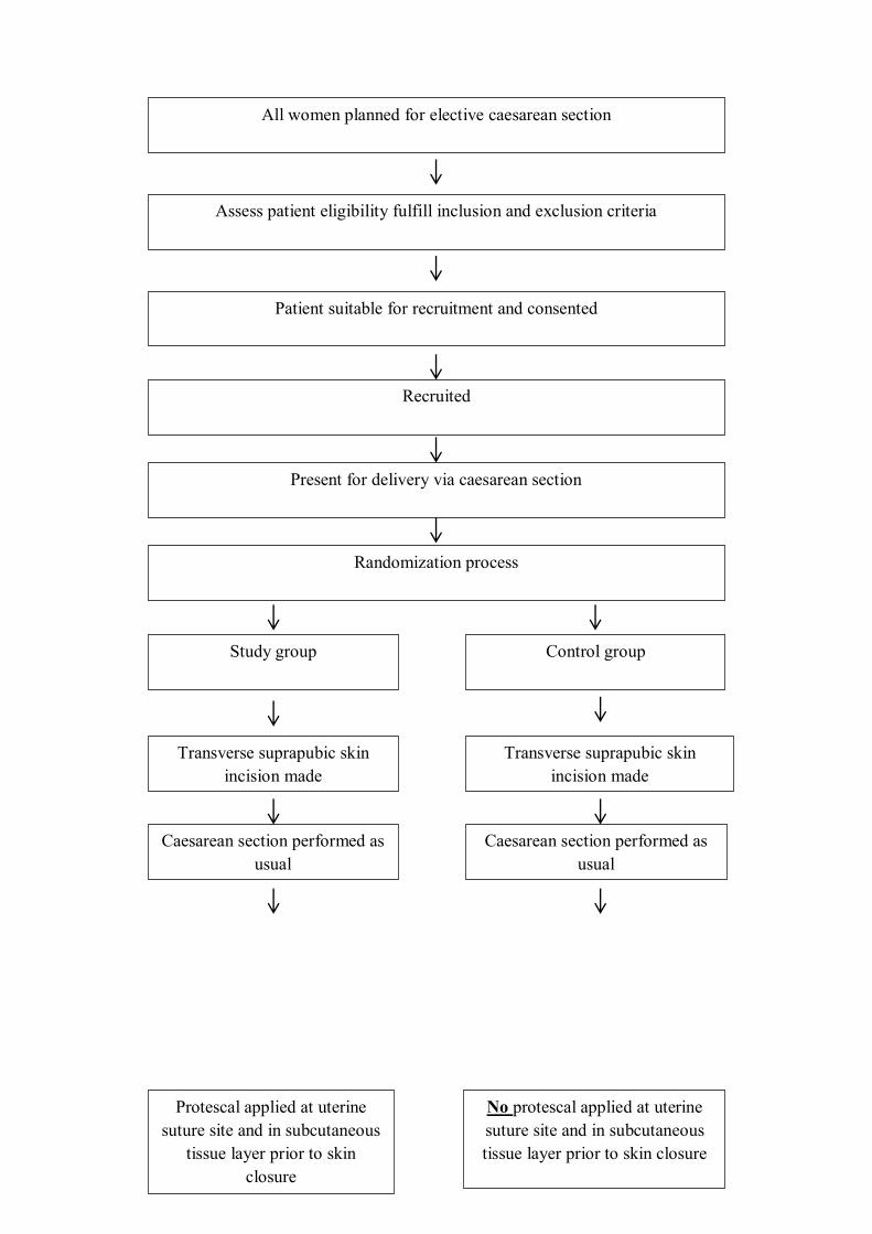
Protescal applied at uterine suture site and in subcutaneous
tissue layer prior to skin closure
No protescal applied at uterine suture site and in subcutaneous tissue layer prior to skin closure
All women planned for elective caesarean section
Assess patient eligibility fulfill inclusion and exclusion criteria
Patient suitable for recruitment and consented
Recruited
Present for delivery via caesarean section
Caesarean section performed as usual
Control group
Transverse suprapubic skin incision made
Randomization process
Study group
Transverse suprapubic skin incision made
Caesarean section performed as
usual
4.11 Study Protocol
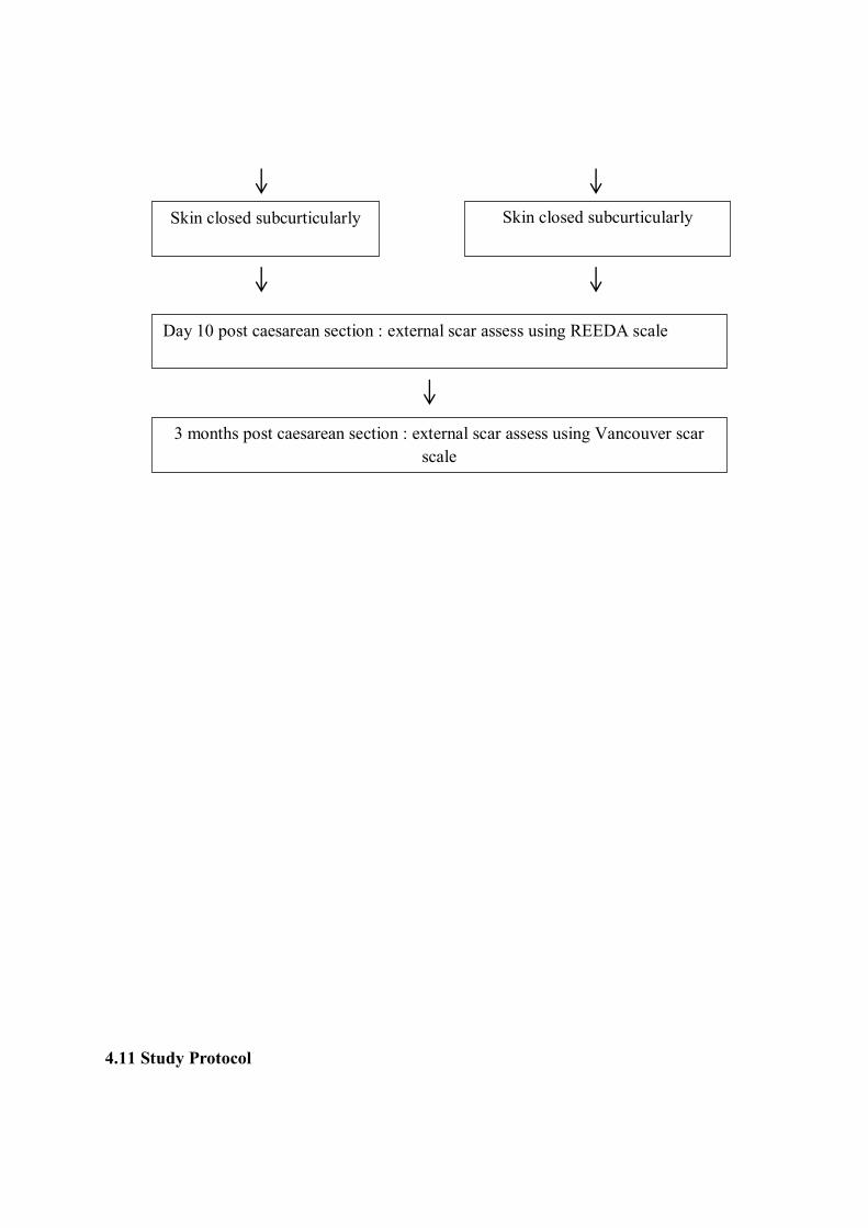
Skin closed subcurticularly Skin closed subcurticularly
Day 10 post caesarean section : external scar assess using REEDA scale
3 months post caesarean section : external scar assess using Vancouver scar scale
After delivering the baby through caesarean section, uterine muscle is closed in 2 layers with braided absorbable suture, polyglactin 910 (vicryl no 1).
After haemostasis secure, 4 ml Protescal gel apply at the uterine suture site.
Peritoneal layer close using braided absorbable suture, polyglactin 910 (vicryl no 1).
Rectus sheath suture using braided absorbable suture, polyglactin 910 (vicryl no 1).
Subcutaneous tissue close interruptedly using braided absorbable suture, polyglactin 910 (vicryl no 1).
1 ml Protescal gel apply in subcutaneous tissue prior to skin closure.
Skin close with subcuticular method using braided absorbable suture, polyglactin 910 (vicryl 3-0).
Wound healing assess on 10th day post caesarean section using REEDA scale, which had criteria including redness, edema, ecchymosis, discharge and approximation.
On 3rd month, the degree of scarring assess using the Vancouver scar scale including pigmentation, height, pliability and vascularity.
5.0 REFERENCES
1. Brown BC, McKenna SP, Siddhi K, Mc-Grouther DA, Bayat A. The hidden cost of skin scars: quality of life after skin scarring. J Plast Reconstr Aesthet Surg 2008;61:1049-58. 2. Brown BC, Moss TP, McGrouther DA, Bayat A. Skin scar pre-conceptions must be challenged: importance of self-perception in skin scarring. J Plast Reconstr Aesthet Surg 2010;63:1022-9.
3. Tully L, Gates S, Brocklehurst P, McKenzie-McHarg K, Ayers S. Surgical techniques used during caesarean section operations: results of a national survey of practice in the UK. Eur J Obstet Gynecol Reprod Biol 2002;102:120-6. 4. Alderdice F, McKenna D, Dornan J. Techniques and materials for skin closure in caesareansection. CochraneDatabaseSystRev2003:CD003577. 5. Berghella V, Baxter JK, Chauhan SP. Evidence-based surgery for cesarean delivery. Am J Obstet Gynecol 2005;193:1607-17. 6. Lindholt JS, Möller-Christensen T, Steele RE. The cosmetic outcome of the scar formation after cesarean section: percutaneous or intracutaneous suture? Acta Obstet Gynecol Scand 1994;73:832-5. 7. Frishman GN, Schwartz T, Hogan JW. Closure of Pfannenstiel skin incisions. Staples vs. subcuticular suture. J Reprod Med 1997;42:627-30. 8. Gaertner I, Burkhardt T, Beinder E. Scar appearance of different skin and subcutaneous tissue closure techniques in caesarean section: a randomized study. Eur J Obstet Gynecol Reprod Biol 2008;138:29-33. 9. Rousseau JA, Girard K, Turcot-Lemay L, Thomas N. A randomized study comparing skin closure in cesarean sections: staples vs subcuticular sutures. Am J Obstet Gynecol 2009; 200:265.e1-4. 10. Cromi A, Ghezzi F, Di Naro E, Siesto G, Loverro G, Bolis P. Blunt expansion of the low transverse uterine incision at cesarean delivery: a randomized comparison of 2 techniques. Am J Obstet Gynecol 2008;199:292.e1-6. 11. Mustoe TA, Cooter RD, Gold MH, et al. International clinical recommendations on scar management. Plast Reconstr Surg 2002;110:560-71. 12. Sullivan T, Smith J, Kermode J, McIver E, Courtemanche DJ. Rating the burn scar. J Burn Care Rehabil 1990;11:256-60. 13. Baryza MJ, Baryza GA. The Vancouver Scar Scale: an administration tool and its interrater reliability. J Burn Care Rehabil 1995;16:535-8. 14. Draaijers LJ, Tempelman FR, Botman YA, et al. The patient and observer scar assessment scale: a reliable and feasible tool for scar evaluation. Plast Reconstr Surg 2004;113:1960-5. 15. van de Kar AL, Corion LU, Smeulders MJ, Draaijers LJ, van der Horst CM, van Zuijlen PP. Reliable and feasible evaluation of linear scars by the Patient and Observer Scar Assessment Scale. Plast Reconstr Surg 2005;116:514-22. 16. Faul F, Erdfelder E, Lang A-G, Buchner A. G*Power 3: a flexible statistical power analysis program for the social, behavioral, and biomedical sciences. Behav Res Methods 2007;39:175-91.
17. Brown NJ, Smyth EA, Cross SS, Reed MW. Angiogenesis induction and regression in human surgical wounds. Wound Repair Regen 2002;10:245-51. 18. Truong PT, Lee JC, Soer B, Gaul CA, Olivotto IA. Reliability and validity testing of the Patient and Observer Scar Assessment Scale in evaluating linear scars after breast cancer surgery. Plast Reconstr Surg 2007;119:487-94. 19. Truong PT, Abnousi F, Yong CM, et al. Standardized assessment of breast cancer surgical scars integrating the Vancouver Scar Scale, Short-Form McGill Pain Questionnaire, and patients’ perspectives. Plast Reconstr Surg 2005;116:1291-9. 20. Ranaboldo CJ, Rowe-Jones DC. Closure of laparotomy wounds: skin staples versus sutures. Br J Surg 1992;79:1172-3.