Embed Size (px)
Citation preview
Title Involvement of auxin distribution in root nodule developmentof Lotus japonicus.
Author(s) Takanashi, Kojiro; Sugiyama, Akifumi; Yazaki, Kazufumi
Citation Planta (2011), 234(1): 73-81
Issue Date 2011-07
URL http://hdl.handle.net/2433/143584
Right
The final publication is available at www.springerlink.com;This is not the published version. Please cite only the publishedversion. この論文は出版社版でありません。引用の際には出版社版をご確認ご利用ください。
Type Journal Article
Textversion author
Kyoto University
1
Involvement of auxin distribution in root nodule development of Lotus japonicus
Kojiro Takanashi, Akifumi Sugiyama, Kazufumi Yazaki
K Takanashi・A Sugiyama・K Yazaki
Research Institute for Sustainable Humanosphere,
Kyoto University, Gokasho, Uji 611-0011, Japan
* Corresponding author
K Yazaki
e-mail: [email protected]
Tel. +81-774-38-3617
Fax. +81-774-38-3623
2
Abstract
The symbiosis between legume plants and rhizobia causes the development of new organs,
nodules, which function as an apparatus for nitrogen fixation. In this study, the roles of auxin
in nodule development in Lotus japonicus have been demonstrated using molecular genetic
tools and auxin inhibitors. The expression of an auxin-reporter GH3 fused to -glucuronidase
(GUS) was analyzed in L. japonicus roots, and showed a strong signal in the central cylinder of
the root, whereas upon rhizobium infection, generation of GUS signal was observed at the
dividing outer cortical cells during the first nodule cell divisions. When nodules were developed
to maturity, strong GUS staining was detected in vascular tissues of nodules, suggesting
distinct auxin involvement in the determinate nodule development. Numbers and the
development of nodules were affected by auxin transport inhibitors (1-naphtylphthalamic acid;
NPA and tri-indobenzoic acid; TIBA), and by a newly synthesized auxin antagonist, α-(phenyl
ethyl-2-one)-indole-3-acetic acid (PEO-IAA). The common phenotypical alteration by these
auxin inhibitors was the inhibition in forming lenticel, which is normally developed on the
nodule surface from the root outer cortex. The inhibition of lenticel formation was correlated
with the inhibition of nodule vascular bundle development. These results indicate that auxin is
required for the normal development of determinate nodules in a multidirectional manner.
Keywords Auxin・Lenticel・Lotus japonicus・Nodule formation・PEO-IAA
Abbreviations
DsRed Discoma sp. red fluorescent protein
dpi days post inoculation
GUS - glucuronidase
NPA 1-naphtylphthalamic acid
PEO-IAA α -(phenyl ethyl-2-one)- indole-3-acetic acid
TIBA tri-indobenzoic acid
3
Introduction
Legume plants can establish symbiosis with rhizobia by forming a nitrogen-fixing apparatus
called nodules, which are mostly developed on roots following legume–rhizobium signal
exchanges that trigger the plant meristem formation that allows bacterial invasion from plant
root hairs to underlying cortical cells (Brewin 2004). The initiation of nodules by rhizobia is
stimulated by lipochitin oligosaccharides, also called Nod factors (Denarie et al. 1996), and the
start of cell division involves the re-initiation of the cell cycle in the cortical and pericycle cells
of the host plant root. In indeterminate legume plants, nodules are initiated from the inner
cortex and form a persistent nodule meristem, which allows continuous growth, and leads to
the formation of elongated nodules, whereas in determinate legumes nodules are mostly
developed from outer cortical cells and form spherical nodules (Hirsch 1992). This nodule
formation process is regulated by several phytohormones, such as auxin, cytokinins,
gibberellins, and brassinosteroids as positive regulators of nodule formation (Ferguson et al.
2005; Frugier et al. 2008; Maekawa et al. 2009; Mathesius 2008), while ethylene, jasmonic acid
and abscic acid as negative regulators (Ding et al. 2008; Nakagawa and Kawaguchi 2006;
Penmetsa et al. 2008).
Auxin regulates plant growth and development in a highly sophisticated manner, in
particular auxin functions by the regulated polar movement along the vertical axis leading to
its concentration gradient through-out the plant. Since the possible involvement of auxin in
nodule formation was first reported by Thimann in 1936, auxin distribution during nodulation
has been studied in particular with indeterminate nodules (Thimann 1936). GH3 and DR5
promoters are often used as auxin reporter genes. In white clover (Trifolium repens),
Mathesius and coworkers reported that rhizobia caused a reduction in the GH3 signal at the
site of infection and at the area beneath. In their study, GH3 was observed to be highly
expressed in early dividing pericycle and cortical cells of nodules, and subsequently, the
4
expression decreased in the forming primordium. Finally, GH3 expression remained only in
the nodule meristem and the vascular bundles (Mathesius et al. 1998). A similar expression
pattern was also observed in Medicago truncatura using a DR5 promoter (Huo et al. 2006).
Compared to these indeterminate nodules, auxin distribution of determinate nodules was
seldom studied. In the determinate nodulating legume Lotus japonicus, GH3 expression did
not disappear below the site of infection, but was found in the first dividing outer cortical cells,
suggesting that there is a different auxin contribution pattern between determinate and
indeterminate nodules (Pacios-Bras et al. 2003).
Inhibitors of auxin transport, i.e., 1-naphtylphthalamic acid (NPA) and
tri-indobenzoic acid (TIBA) have been generally used to observe developmental effects on
changes in auxin transport. NPA is thought to interfere with the intracellular cycling of the
PIN-FORMED (PIN) proteins between the plasma membrane and endosomal vesicles
(Blakeslee et al. 2005; Geldner et al. 2001). NPA also inhibits the auxin transport mediation by
ATP-binding cassette proteins by binding these membrane proteins (Bailly et al. 2008).
Several classes of flavonoids show a similar effect on auxin transport as NPA in white clover
and also in the non-leguminous plant Arabidopsis thaliana (Brown et al. 2001; Jacobs and
Rubery 1988; Mathesius et al. 1998). As another biochemical tool to study auxin functions, tert
-butoxycarbonylaminohexyl-indole-3-acetic acid (BH-IAA) was reported to be an auxin
antagonist (Hayashi et al. 2008). This -alkyl-IAA specifically binds to the auxin binding site
of TIR1/AFBs auxin receptors and blocks their functions in many plant species such as
Arabidopsis, rice (Oryza sativa) and moss (Physcomitrella patens). More recently, α-(phenyl
ethyl-2-one)-indole-3-acetic acid (PEO-IAA) has been provided by Dr. Hayashi as another
α-alkyl-IAA that shows more potent anti-auxin activity, and reveals the antagonistic activities
to auxin-responsive gene expression, cell division, and the elongation pathway mediated by
SCF TIR1/AFBs (Hayashi et al. unpublished data). In this study, we have shown the auxin
distribution in the determinate nodules of L. japonicus using auxin inhibitors of different
mechanisms and have found that the development of lenticels and nodule vascular bundles

5
was strongly affected by auxin during the nodulation process.
Materials and methods
Plant materials, rhizobia strain and growth conditions
Lotus japonicus B129 Gifu and its GH3:GUS transgenic line, Lj3632.5.127, (kindly provided by
Dr. Herman P. Spaink, Leiden State University, Netherlands) were used in this work
(Pacios-Bras et al. 2003). All analyses were conducted in a cultivation chamber under a 16-h
day/8-h night cycle at a temperatures of 23ºC. For inoculation, Mesorhizobium loti
MAFF303099 carrying the DsRed (Discoma sp. red fluorescent protein) reporter gene (kind gift
from Dr. Makoto Hayashi, National Institute of Agrobiological Sciences, Japan) was cultured
overnight in TY medium at 28 ºC (Beringer 1974).
Treatment with auxin inhibitors
To study the effect of auxin transport inhibitors and auxin antagonist on plant growth and
nodulation, NPA (TCI, Tokyo, Japan), TIBA (Sigma–Aldrich, St. Louis, MO), or PEO-IAA
(kindly provided from Dr. Ken-ichiro Hayashi, Okayama University of Science, Japan) were
dissolved in dimethyl sulfoxide (DMSO) and used to treat L. japonicus. Seeds were
surface-sterilized with a 1% sodium hypochloride solution for 10 min, rinsed five times with
sterile distilled water, then germinated on water agar plates (0.8%).
For nodulation experiments, five-day-old seedlings were transferred to plant boxes
filled with sterile fine vermiculite with 100 ml of 1/2 B&D liquid medium (Broughton and
Dilworth 1971), to which 100 l of either TIBA, NPA, or PEO-IAA were added with final
concentration of 10 or 100 M, and 108 cells of M. loti was inoculated after two days. Plants
grown on 1/2 B&D medium containing the same volume of DMSO was used as a control. After
6
19 days plants were sampled and their nodules including primordia were observed under a
digital stereomicroscope system (VB-S20, Keyence, Osaka, Japan). Nodule primodia were
defined as nodules with a diameter of 0.55 mm or less. Total root growth was evaluated as
the sum of main and lateral root lengths.
To evaluate the effect of PEO-IAA on normal growth, five-day-old seedlings were
transferred on 1/2 B&D plate containing PEO-IAA. After 7 days plants were sampled and their
phenotypes were observed under a stereomicroscope VB-S20 (Keyence). For scanning electron
microscopy, roots were put on a dental impression material (Provil novo Light, Heraeus Kulzer,
Wehrheim, Germany). After 2 hrs the negative molds were peeled and then filled with epoxy
resin (GM-6600, Blenny Giken, Isezaki, Japan). The replicas were polymerized at room
temperature for 48 hrs. The positive resin replicas were peeled from the negative molds and
examined with a scanning electron microscope (S3500N, Hitachi, Tokyo, Japan).
For chloral hydrate clearance, nodules were fixed in fixative solution (ethanol : acetic
acid = 3 : 1) for 3 hrs, and then hydrated through an ethanol series. The fixed samples were
immersed in clearing solution containing 4 mg/ml chloral hydrate (Wako, Osaka, Japan) and
0.5 ml/ml glycerol at 4ºC for a week, and then observed under a microscope (Axioscope2, Zeiss,
Oberkochen, Germany).
Histochemical analysis of GH3:GUS transformant of L. japonicus
For GUS staining, tissues were prefixed in ice-cold 90% acetone for 15 min, rinsed once with
cold water, immersed with staining solution (50 mM sodium phosphate buffer, pH 7.0, 0.1%
Triton X-100, 0.5 mM potassium ferrocyanide, 0.5 mM potassium ferricyanide, and 1mM
X-Gluc), and incubated at 37ºC for 40 min. The stained samples were dehydrated through an
ethanol series. The fixed samples were embedded in Technovit 7100 (Heraeus Kulzer)
according to the manufacturer’s protocol. Tissue sections (10 m thick) prepared with a
microtome (RM2155, Leica, Wetzlar, Germany) were mounted on slides and observed with a

7
microscope (Axioscope2).
Results
Distribution of auxin in nodule development
The auxin-regulated soybean promoter GH3 (Hagen et al. 1991) fused to a reporter gene has
provided a molecular tool to monitor auxin distribution in, among others, leguminous plants.
Using GH3:GUS transformants we have investigated the auxin localization in developing
nodules of L. japonicus. In uninoculated L. japonicus root, strong GUS staining was observed
in the central cylinder (Fig. 1a, b), which represents high auxin level in these root tissues. At
two days post inoculation (dpi) of M. loti, the GUS activity became detectable in part of root
cortex, where a young nodule primordium was developing (Fig. 1c). When a section was
prepared from this region, the cell division was seen in cortical cells, where GUS staining was
observed at the basal part connecting the vascular tissues of the main root (closed arrow in Fig.
1d), and at the front of the young developing nodule as well, which is still under root epidermis
(open arrow at Fig. 1d). In mature nodules (14 dpi), strong GUS staining was observed
surrounding the infected zone of the nodule (Fig. 1e). The cross-section of the mature nodule
revealed the strong GUS staining at vascular tissues of the nodule, and the continuous signal
was seen at the root vascular tissues, from which a nodule vascular bundle was developed (Fig.
1f). These data strongly suggest that the possible involvement of auxin in the development of
determinate nodules, which is consistent with the observation by Pacios-Bras (Pacios-Bras et
al. 2003).
Effects of inhibitors of auxin transport on nodule formation in L. japonicus
Auxin is mainly synthesized in the apical region including shoot apical meristem and young
8
leaves, and transported in a basipetal direction in the stem, and in an acropetal direction in
the root (Baluska et al. 2005). This polar transport of auxin maintains the gradient of auxin
concentration in a plant body, which is essential for normal plant growth and development. To
test the effect of this polar auxin transport on the nodulation process, plants were treated with
auxin transport inhibitors, such as NPA and TIBA. Plants grown in the presence of NPA at
concentration of 10 M and 100 M showed an apparent reduction in the total root length (70%
and 73% inhibition, respectively) and nodule numbers (Fig. 2a, b). When plants were treated
with 10 M of TIBA, a weak inhibition in the root growth (19%) was observed while the nodule
number was strongly reduced. Interestingly, a clear increment in the number of nodule
primordia was observed when plants were treated with 100 M of TIBA, while root length was
strongly reduced (68% inhibition) at this concentration. Because NPA and TIBA inhibited the
root growth of L. japonicus in particular at 100 M, the reduction in nodule number might be
due to an indirect effect of auxin transport inhibition, at least in part.
Effects of auxin antagonist on nodule formation
A newly synthesized auxin antagonist, PEO-IAA, binds to TIR1/AFBs receptors and blocks
their functions leading to an auxin defective phenotype. To evaluate the direct auxin effects on
nodulation, we employed this compound in this study. To investigate the effective dose of
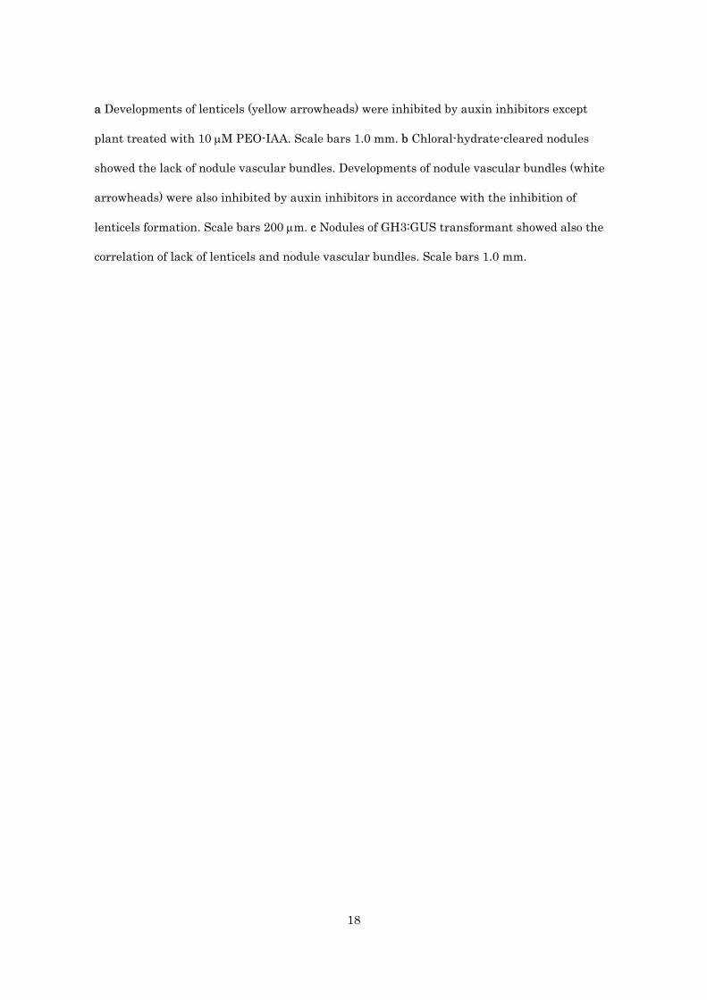
PEO-IAA in L. japonicus, we first observed the growth phenotype in the presence of different
concentrations of PEO-IAA. At 10 M, L. japonicus root elongation was slightly enhanced, but
this auxin antagonist started to inhibit from 100 M on agar plates, and at 300 M the main
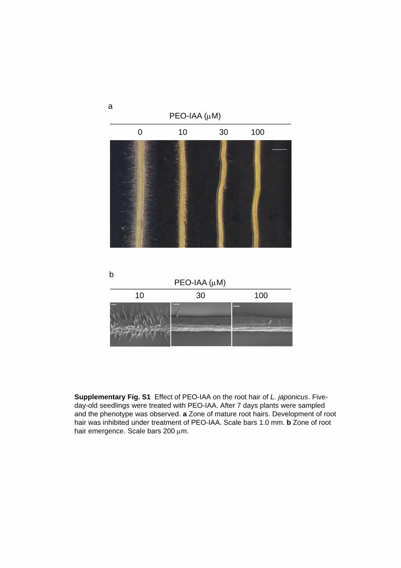
root growth was strongly inhibited (Fig. 3a, b). We also found that PEO-IAA inhibited root hair
emergence and development at low concentrations as 10 to 30 M (supplementary Fig. S1a, b).
In L. japonicus, the rhizobium infection occurs via infection threads that pass through
root hairs in the root cortex, and then the bacteria are distributed to the cells, which become
infected symbiosome-containing nitrogen-fixing cells (Karas et al. 2005; van Spronsen et al.
9
2001). It was also reported that rhizobia could infect a root hairless mutant through cortical
root hairs, which were induced by Nod factors (Karas et al. 2005). In that case, reduction of
nodule number and increase of nodule primordia were observed in the root hairless mutant at
21 dpi. To study the effect of PEO-IAA on nodulation we inoculated M. loti on L. japonicus
grown in plant boxes filled with vermiculite, which had been treated with 10 M or 100 M
PEO-IAA. The results are shown in Fig. 4a and 4b. Surprisingly, when plants were grown on
vermiculites, there was no significant difference in the development of root hairs even at the
high concentration of PEO-IAA, probably due to the apparent difference in the effective
concentration of PEO-IAA between on agar plate, which gives more precise effective
concentration by the tight contact of entire roots with agar medium, and in vermiculite, in
which contact surface of root tissues with inhibitor-containing vermiculite particles is limited.
The number, size and the typical red color of nodules of L. japonicus treated with 10 M or 100
M PEO-IAA were not influenced either, but we found one clear phenotype alteration, i.e., the
lenticels on the surface of nodules disappeared under the treatment of PEO-IAA (100 M) (Fig.
5a). The development of lenticels, which arise from specific phellogen cells, was also strongly
inhibited by the auxin transport inhibitors (Fig. 5a). Lenticels develop above the nodule
vascular bundles which were postulated to provide growth substances to the lenticels
(Pankhurst and Sprent 1975). To investigate the effect of auxin inhibitors on the development
of vascular bundles, nodules were cleared with chloral hydrate after treatment with auxin
inhibitors. Developed vascular bundles were observed in nodules with lenticels (control), while
in nodules treated with auxin inhibitors, neither vascular bundle nor lenticels were observed
accordingly (Fig. 5b). These correlations were also observed in GH3:GUS nodules which were
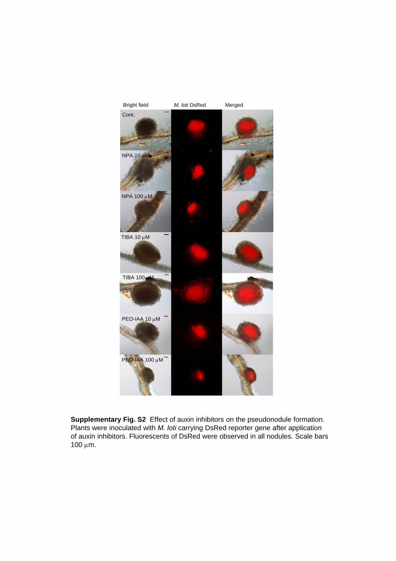
treated with auxin inhibitors (Fig. 5c). Fluorescence of DsRed carried by M. loti was detected
in all nodules observed, indicating that no pseudonodule was developed by the treatment of
these inhibitors (supplementary Fig. S2).
Discussion
10
Among several types of nodules in legume plants, indeterminate nodules and determinate
nodules are well studied their nodulation process. In the former, for which alfalfa and M.
truncatula are representatives, nodules are derived from the inner cortex of the root and
develop a meristem, whereas in the latter (for instance, soybean and L. japonicus) nodules are
derived from the outer cortex cells and grow via non-meristematic cell division. Due to these
morphological and developmental differences, many comparisons have been made between
these two types of nodules. Several previous reports suggested that auxin is involved in
nodulation, especially in indeterminate nodules because, for example, nodules contain elevated
levels of auxin, auxin response genes are differentially expressed during nodule initiation,
auxin transport is inhibited by rhizobia, and auxin transport inhibitors can induce
pseudonodules (Mathesius 2008). Compared to indeterminate nodules, the involvement of
auxin on the development of determinate nodules is still largely unknown. In the present study,
we have shown the distribution of auxin during the formation of determinate nodules, the
effects of inhibitors of auxin polar transport and a new auxin antagonist on nodulation.
The GH3:GUS transformant of L. japonicus showed auxin responses during nodule
development. In uninoculated root, GH3 expression was observed only in central cylinder (Fig.
1a, b), whereas in inoculated roots, GH3 expression started to increase in the outer cortical
cells where cell divisions occurred in nodule primordia. It is also to be noted that the GH3
expression was connected to the main root vascular tissues. In mature nodules, GUS staining
disappeared in both infected and non-infected cells derived from the cortex, whereas strong
expression was observed in vascular tissues. These results suggest that auxin plays an
important role in nodule formation, especially in the development of vascular tissues. Similar
GUS expression was observed in dividing root cortex when M. loti was inoculated into L.
japonicus, which was transformed with the GUS gene under the control of the Arabidopsis
cytokinin-responsive gene, ARR5 (Lohar et al. 2004). Cytokinin signaling is necessary to
induce cortical cell divisions and nodule organogenesis, because the gain-of-function mutant in

11
the cytokinin receptor, LHK1, spontaneously develops nodules in the absence of rhizobia
whereas loss-of-function mutant in LHK1 abolishes nodule primordium formation (Murray et
al. 2007; Tirichine et al. 2007). Together with cytokinin, auxin may participate in the initiation
of cell divisions in a coordinated fashion.
Polar auxin transport is mediated by cell-to-cell movement that is generated by a
chemiosmotic model where the existence of auxin-carrier proteins localized to the plasma
membrane responsible for the auxin uptake and efflux out of the cells has been proposed
(Raven 1975; Rubery and Ar 1974). The model is rationalized by the asymmetric localization of
the efflux carriers at one side of the cells, which determines the direction of the intercellular
auxin movement in plants. In the current model, polar auxin transport is mediated by several
different classes of transporter proteins i.e., AUXIN/LIKE-AUXIN (AUX/LAX) permeases
(Kramer and Bennett 2006), PIN proteins (Teale et al. 2006; Vieten et al. 2007), and ABC
proteins of the ABCB subfamily (Geisler and Murphy 2006). In L. japonicus, rhizobium
inoculation induced GH3 expression and auxin transport was up-regulated after the treatment
of purified Nod factors at a root tip (Pacios-Bras et al. 2003), which suggested that an
increased auxin level is necessary for the nodulation process. We have demonstrated in this
study the effects of auxin transport inhibitors on nodulation, i.e., the treatment of L. japonicus
with NPA or TIBA altered the nodule number at concentrations 10 M and 100 M, in which
the plant growth was not drastically suppressed.
PEO-IAA can block auxin functions since this α-alkyl-IAA competitively binds to the
auxin-binding site of receptor molecules TIR1/AFBs. Some auxin-deficient phenotypes of this
compound are similar to those resulting from treatment with NPA, but most effects are
different and more direct effects can be observed with the auxin antagonist (Hayashi et al.
2008). Thus, we investigated the effects of PEO-IAA on nodulation in L. japonicus. Although
PEO-IAA affected the root growth and root hair formation in L. japonicus at 10 to 300 M in
agar plates, the number of nodules was not strongly altered as compared to that of control at
concentrations 10 M and 100 M in vermiculite. One striking effect of PEO-IAA was that this
12
auxin antagonist strongly suppressed the development of lenticels on nodules in a similar
manner as those with NPA and TIBA (Fig. 5a). We also found the inhibition of nodule vascular
bundle development under the treatment of these inhibitors (Fig. 5b, c). The effective
concentration of PEO-IAA for various auxin responses differs depending on the tissues, e.g.,
the inhibition of lenticel formation was observed at 100 M whereas root hair development was
more sensitive (30 M) (Supplementary Fig. S1). Inhibition of lateral root formation was only
observed at high concentrations of PEO-IAA such as 300 M in L. japonicus (Fig. 3c).
Lenticels develop opposite the nodule vascular bundle and are suggested to arise from
specific phellogen cells in the nodule cortex. These cells become meristematic and by repeated
divisions rupture the epidermis, whereby the lenticels appear on the nodule surface as bands
of loosely packed tissue (Pankhurst and Sprent 1975). Lenticels regulate gas permeability of
nodules and develop more extensively under low oxygen or waterlogged conditions, whereas
during water insufficient condition or high oxygen pressure they collapse, or develop very
little externally (Pankhurst and Sprent 1975; Walsh 1995). Grønlund et al. showed that the
lenticel development was also inhibited as well as the vascular tissues, when the expression of
two homeodomain proteins, Ljndx1 and Ljndx2, was suppressed (Grønlund et al. 2003). The
relation between the lenticels and the nodule vascular system is unknown. However, due to
their relative location, it was suggested that lenticel development is associated with growth
substances supplied from the vascular system (Pankhurst and Sprent 1975). Taken together,
our data suggested that auxin is also one of the candidates of the growth substances
controlling lenticel formation, or that auxin is necessary to form the nodule vascular bundle
and lack of lenticel may be an indirect effect of abnormal development of vascular bundle after
treatment of these auxin inhibitors.
In this study, we have shown the involvement of auxin in the formation of vascular
bundle and lenticel on the nodules. For further understanding of the auxin distribution and
the role of auxin in the nodules of L. japonicus, identification and characterization of auxin
transporter genes that respond to nodulation will be necessary. (Brown et al. 2001)

13
Acknowledgements
We would like to thank National Bioresource Project (Lotus japonicus, Glycine max) for seeds
of L. japonicus and Dr. Ken-ichiro Hayashi (Okayama University of Science, Japan) for the
PEO-IAA. We also wish to thank Dr. Niels Sandal and Dr. Jens Stougaard (University of
Aarhus, Denmark) and Dr. Herman P. Spaink (Leiden State University, Netherlands) for
seeds of GH3:GUS transformed L. japonicus (Lj3632.5.127). We thank Dr. Takashi Aoyama
(Kyoto University, Japan) for the technical assistance in scanning electron microscope and the
sample preparation and Dr. Makoto Hayashi (National Institute of Agrobiological Sciences,
Japan) for the M. loti carrying DsRed. This work was supported in part by Grant-in-Aid for
Scientific Research (No. 21027022 to K.Y.) and by Research Fellowship from the Japan Society
for the Promotion of Science for Young Scientists (No. 09J00170 to K.T.).
References
Bailly A, Sovero V, Vincenzetti V, Santelia D, Bartnik D, Koenig BW, Mancuso S, Martinoia E,
Geisler M (2008) Modulation of p-glycoproteins by auxin transport inhibitors is mediated
by interaction with immunophilins. J Biol Chem 283: 21817-21826
Baluska F, Barlow PW, Baskin TI, Chen RJ, Feldman L, Forde BG, Geisler M, Jernstedt J,
Menzel D, Muday GK, Murphy A, Samaj J, Volkmann D (2005) What is apical and what is
basal in plant root development? Trends Plant Sci 10: 409-411
Beringer JE (1974) R factor transfer in Rhizobium-leguminosarum. J Gen Microbiol 84:
188-198
Blakeslee JJ, Peer WA, Murphy AS (2005) Auxin transport. Curr Opin Plant Biol 8: 494-500
Brewin NJ (2004) Plant cell wall remodelling in the rhizobium-legume symbiosis. Crit Rev
Plant Sci 23: 293-316
Broughton WJ, Dilworth MJ (1971) Control of leghaemoglobin synthesis in snake beans.
14
Biochem J 125: 1075-1080
Brown DE, Rashotte AM, Murphy AS, Normanly J, Tague BW, Peer WA, Taiz L, Muday GK
(2001) Flavonoids act as negative regulators of auxin transport in vivo in Arabidopsis.
Plant Physiol 126: 524-535
Denarie J, Debelle F, Prome JC (1996) Rhizobium lipo-chitooligosaccharide nodulation factors:
Signaling molecules mediating recognition and morphogenesis. Annu Rev Biochem 65:
503-535
Ding YL, Kalo P, Yendrek C, Sun JH, Liang Y, Marsh JF, Harris JM, Oldroyd GED (2008)
Abscisic acid coordinates Nod factor and cytokinin signaling during the regulation of
nodulation in Medicago truncatula. Plant Cell 20: 2681-2695
Ferguson BJ, Ross JJ, Reid JB (2005) Nodulation phenotypes of gibberellin and
brassinosteroid mutants of pea. Plant Physiol 138: 2396-2405
Frugier F, Kosuta S, Murray JD, Crespi M, Szczyglowski K (2008) Cytokinin: secret agent of
symbiosis. Trends Plant Sci 13: 115-120
Geisler M, Murphy AS (2006) The ABC of auxin transport: The role of p-glycoproteins in plant
development. FEBS Lett 580: 1094-1102
Geldner N, Friml J, Stierhof YD, Jurgens G, Palme K (2001) Auxin transport inhibitors block
PIN1 cycling and vesicle traficking. Nature 413: 425-428
Grønlund M, Gustafsen C, Roussis A, Jensen D, Nielsen LP, Marcker KA, Jensen EO (2003)
The Lotus japonicus ndx gene family is involved in nodule function and maintenance.
Plant Mol Biol 52: 303-316
Hagen G, Martin G, Li Y, Guilfoyle TJ (1991) Auxin-induced expression of the soybean GH3
promoter in transgenic tobacco plants. Plant Mol Biol 17: 567-579
Hayashi K, Tan X, Zheng N, Hatate T, Kimura Y, Kepinski S, Nozaki H (2008) Small-molecule
agonists and antagonists of F-box protein-substrate interactions in auxin perception and
signaling. Proc Natl Acad Sci USA 105: 5632-5637
Hirsch AM (1992) Developmental biology of legume nodulation. New Phytol 122: 211-237
15
Huo XY, Schnabel E, Hughes K, Frugoli J (2006) RNAi phenotypes and the localization of a
protein :: GUS fusion imply a role for Medicago truncatula PIN genes in nodulation. J
Plant Growth Regul 25: 156-165
Jacobs M, Rubery PH (1988) Naturally-occurring auxin transport regulators. Science 241:
346-349
Karas B, Murray J, Gorzelak M, Smith A, Sato S, Tabata S, Szczyglowski K (2005) Invasion of
Lotus japonicus root hairless 1 by Mesorhizobium loti involves the nodulation
factor-dependent induction of root hairs. Plant Physiol 137: 1331-1344
Kramer EM, Bennett MJ (2006) Auxin transport: a field in flux. Trends Plant Sci 11: 382-386
Lohar DP, Schaff JE, Laskey JG, Kieber JJ, Bilyeu KD, Bird DM (2004) Cytokinins play
opposite roles in lateral root formation, and nematode and Rhizobial symbioses. Plant J
38: 203-214
Maekawa T, Maekawa-Yoshikawa M, Takeda N, Imaizumi-Anraku H, Murooka Y, Hayashi M
(2009) Gibberellin controls the nodulation signaling pathway in Lotus japonicus. Plant J
58: 183-194
Mathesius U (2008) Auxin: at the root of nodule development? Funct Plant Biol 35: 651-668
Mathesius U, Schlaman HRM, Spaink HP, Sautter C, Rolfe BG, Djordjevic MA (1998) Auxin
transport inhibition precedes root nodule formation in white clover roots and is regulated
by flavonoids and derivatives of chitin oligosaccharides. Plant J 14: 23-34
Murray JD, Karas BJ, Sato S, Tabata S, Amyot L, Szczyglowski K (2007) A cytokinin
perception mutant colonized by Rhizobium in the absence of nodule organogenesis.
Science 315: 101-104
Nakagawa T, Kawaguchi M (2006) Shoot-applied MeJA suppresses root nodulation in Lotus
japonicus. Plant Cell Physiol 47: 176-180
Pacios-Bras C, Schlaman HRM, Boot K, Admiraal P, Langerak JM, Stougaard J, Spaink HP
(2003) Auxin distribution in Lotus japonicus during root nodule development. Plant Mol
Biol 52: 1169-1180

16
Pankhurst CE, Sprent JI (1975) Surface features of soybean root nodules. Protoplasma 85:
85-98
Penmetsa RV, Uribe P, Anderson J, Lichtenzveig J, Gish JC, Nam YW, Engstrom E, Xu K,
Sckisel G, Pereira M, Baek JM, Lopez-Meyer M, Long SR, Harrison MJ, Singh KB, Kiss
GB, Cook DR (2008) The Medicago truncatula ortholog of Arabidopsis EIN2, sickle, is a
negative regulator of symbiotic and pathogenic microbial associations. Plant J 55: 580-595
Raven JA (1975) Transport of indoleacetic-acid in plant-cells in relation to pH and electrical
potential gradients, and Its significance for polar IAA transport. New Phytol 74: 163-172
Rubery PH, Ar S (1974) Carrier-mediated auxin transport. Planta 118: 101-121
Teale WD, Paponov IA, Palme K (2006) Auxin in action: signalling, transport and the control of
plant growth and development. Nature Rev Mol Cell Biol 7: 847-859
Thimann KV (1936) On the physiology of the formation of nodules on legume roots. Proc Natl
Acad Sci USA 22: 511-514
Tirichine L, Sandal N, Madsen LH, Radutoiu S, Albrektsen AS, Sato S, Asamizu E, Tabata S,
Stougaard J (2007) A gain-of-function mutation in a cytokinin receptor triggers
spontaneous root nodule organogenesis. Science 315: 104-107
van Spronsen PC, Grønlund M, Bras CP, Spaink HP, Kijne JW (2001) Cell biological changes
of outer cortical root cells in early determinate nodulation. Mol Plant Microbe In 14:
839-847
Vieten A, Sauer M, Brewer PB, Friml J (2007) Molecular and cellular aspects of
auxin-transport-mediated development. Trends Plant Sci 12: 160-168
Walsh KB (1995) Physiology of the legume nodule and its response to stress. Soil Biol Biochem
27: 637-655

17
Figure Legends
Fig. 1 GH3:GUS expression during the nodulation in L. japonicus. a GH3:GUS expression was
observed in central cylinder of uninoculated root. b Cross section of a. c Enhanced GH3:GUS
expression was observed in outer cortex of the root, where M. loti was inoculated. d Cross
section of c. GH3:GUS expressed in vascular tissues of the main root, and basal (closed arrow)
and apical parts (open arrow) of the nodule primordia. e In mature nodule, strong GH3:GUS
expression was detected only in vascular tissues. f Cross section of e. Scale bars 100 m
Fig. 2 Effect of auxin transport inhibitor on the nodule development. a Growth phenotype and
b nodule number of L. japonicus treated with NPA and TIBA. The data are represented as
averages of 18 individual plants, and values represent mean +SD. *P<0.05 and **P<0.01
compared with control (DMSO) by ANOVA (Bonferroni test). Scale bars 1.0 cm
Fig. 3 Effect of PEO-IAA on the growth of L. japonicus. Five-day-old seedlings were treated
with PEO-IAA. After 7 days plants were sampled and the phenotype was observed. a Seedlings
grown in the presence of PEO-IAA. Scale bars 1.0 cm. b Main root length was measured after
treatment of PEO-IAA. The data are represented as averages of 6 individual plants, and values
represent mean + SD. *P<0.05 compared with control by ANOVA (Bonferroni test).
Fig. 4 Effect of PEO-IAA on the nodule development. a Growth phenotype and b nodule
numbers of L. japonicus. The growth was inhibited by PEO-IAA, but nodule number and
development were not significantly affected. The data are represented as averages of 18
individual plants and values represent mean + SD. Scale bars 1.0 cm
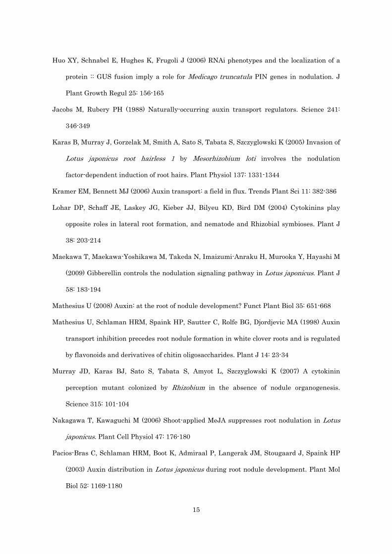
Fig. 5 Nodules of L. japonicus grown with PEO-IAA or auxin transport inhibitors. Five-day-old
seedlings were treated with PEO-IAA, NPA or TIBA, respectively, and then inoculated with M.
loti 2 days later. After 19 days plants were sampled and the phenotype of nodules was observed.
18
a Developments of lenticels (yellow arrowheads) were inhibited by auxin inhibitors except
plant treated with 10 M PEO-IAA. Scale bars 1.0 mm. b Chloral-hydrate-cleared nodules
showed the lack of nodule vascular bundles. Developments of nodule vascular bundles (white
arrowheads) were also inhibited by auxin inhibitors in accordance with the inhibition of
lenticels formation. Scale bars 200 m. c Nodules of GH3:GUS transformant showed also the
correlation of lack of lenticels and nodule vascular bundles. Scale bars 1.0 mm.

Fig. 1. Takanashi, et al.
a
d
b
c
e f

Cont.NPA
10 M 100 M
a
b
Fig. 2. Takanashi, et al.
0
1
2
3
4
5
6
Cont.NPA
10 M 100 M
Nod
ule
num
ber
Nodule primordiaMature nodules
***
*
***
TIBA
10 M 100 M
TIBA
10 M 100 M
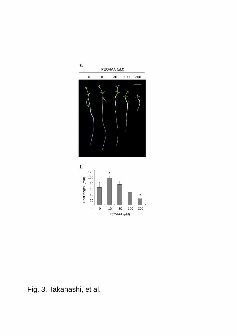
Fig. 3. Takanashi, et al.
a
0 10 30 100 300
PEO-IAA (M)
PEO-IAA (M)
Roo
t le
ngth
(m
m)
0
20
40
60
80
100
120
0 10 30 100 300
b
*
*
a
b
Cont. PEO-IAA
10 M 100 M
Fig. 4. Takanashi, et al.
0
1
2
3
4
5
6
Cont.PEO-IAA
10 M 100 M
Nod
ule
nnum
ber
Nodule primordiaMature nodules
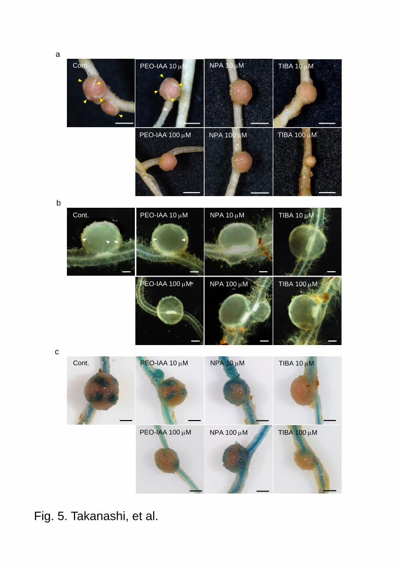
Fig. 5. Takanashi, et al.
aCont. PEO-IAA 10 M NPA 10 M TIBA 10 M
PEO-IAA 100 M NPA 100 M TIBA 100 M
cCont. PEO-IAA 10 M NPA 10 M TIBA 10 M
PEO-IAA 100 M NPA 100 M TIBA 100 M
Cont. PEO-IAA 10 M NPA 10 M TIBA 10 M
PEO-IAA 100 M NPA 100 M TIBA 100 M
b
a
0 10 30 100
PEO-IAA (M)
b
10 30 100
PEO-IAA (M)
Supplementary Fig. S1 Effect of PEO-IAA on the root hair of L. japonicus. Five-day-old seedlings were treated with PEO-IAA. After 7 days plants were sampled and the phenotype was observed. a Zone of mature root hairs. Development of root hair was inhibited under treatment of PEO-IAA. Scale bars 1.0 mm. b Zone of root hair emergence. Scale bars 200 m.
Cont.
NPA 10 M
NPA 100 M
TIBA 10 M
TIBA 100 M
PEO-IAA 10 M
PEO-IAA 100 M
Bright field M. loti DsRed Merged
Supplementary Fig. S2 Effect of auxin inhibitors on the pseudonodule formation. Plants were inoculated with M. loti carrying DsRed reporter gene after application of auxin inhibitors. Fluorescents of DsRed were observed in all nodules. Scale bars 100 m.


![Auxin Influx Activity Is Associated with Frankia Infection ... · Auxin Influx Activity Is Associated withFrankia Infection during Actinorhizal Nodule Formation in Casuarina glauca1[C][W][OA]](https://img.pdfslide.net/doc/110x75/5e06464ee955ea6dd853ad7b/auxin-iniux-activity-is-associated-with-frankia-infection-auxin-iniux-activity.jpg)