Embed Size (px)
Citation preview

Title: New Approaches to Molecular Imaging of Multiple Myeloma
Ravi Vij1, Kathryn J. Fowler2 and Monica Shokeen2*
1Division of Hematology and Oncology, Washington University School of Medicine,
Saint Louis, Missouri; 2Mallinckrodt Institute of Radiology, Washington University School
of Medicine, Saint Louis, Missouri
*Corresponding author: [email protected]
Corresponding Author: Monica Shokeen (Mallinckrodt Institute of Radiology, Department
of Radiology, 4525 Scott Avenue, St. Louis, MO 63110, Phone: 314-362-8979, Fax: 314-
747-5190, Email: [email protected])
First Author: Ravi Vij (Division of Hematology and Oncology, Washington University
Medical School, St.Louis, MO 63110, Phone: 314-454-8204, Email: [email protected])
Running Title: Molecular Imaging of Multiple Myeloma
Journal of Nuclear Medicine, published on November 5, 2015 as doi:10.2967/jnumed.115.163808by on June 28, 2018. For personal use only. jnm.snmjournals.org Downloaded from
Abstract: Molecular imaging plays important role in detection and staging of
hematological malignancies. Multiple myeloma (MM) is an age-related hematological
malignancy of clonal bone marrow plasma cells characterized by destructive bone lesions
and is fatal in the vast majority of patients. Traditional skeletal survey and bone scans
have sensitivity limitations for osteolytic lesions manifested in MM. Progressive
biomedical imaging technologies such as low dose CT, molecularly targeted PET, MRI,
and the functional-anatomical hybrid versions (PET-CT, PET-MRI) provide incremental
advancements in imaging MM. Imaging with PET and MRI using molecularly targeted
probes are promising precision medicine platforms that might successfully address the
clinical ambiguities of myeloma spectrum diseases. The intent of this focus article is to
provide a concise review of the present status and promising developments on horizon
such as the new molecular imaging biomarkers under investigation that can either
complement or potentially supersede existing standards.
Key words: Multiple myeloma (MM), monoclonal gammopathy of undetermined
significance (MGUS), PET, MRI, PET/CT, PET/MRI, receptor very late antigen-4 (VLA-
4), metabolic imaging
by on June 28, 2018. For personal use only. jnm.snmjournals.org Downloaded from
MULTIPLE MYELOMA (MM)
MM is the second most common age-related hematologic malignancy in the United States
and is incurable in the vast majority of patients. MM is a malignancy of clonal bone marrow
plasma cells whose DNA has undergone characteristic class switch recombination and
somatic hypermutation(1). In addition to hallmark genetic mutations, the bone micro-
environmental elements play a critical role in the MM pathogenesis(2). MM is preceded
by a premalignant stage called monoclonal gammopathy of undetermined significance
(MGUS) with a 0.5-1% incidence to MM(3). Smoldering multiple myeloma (sMM) is an
intermediate clinical stage in which the risk of incidence to MM is 10% per year(3). The
International Myeloma Working Group (IMWG) diagnostic criteria for premalignant and
malignant MM has been elegantly summarized by Rajkumar et. al.(3). Due to the aging
population, the incidence of MM is expected to increase along with the associated costs.
Total healthcare costs in the first year after diagnosis of MM is $118,353(4).
Advancements in targeted therapy as well as the success of stem cell transplantation has
contributed to improvements in the 5-year survival rate in MM (26.3% in 1975 versus
46.6% in 2011) (5). Promising new agents are currently under development for relapsed
and refractory MM (6). The treatment regimen for MM is dictated by patient eligibility for
autologous or allogeneic stem cell transplantation. About 80% of MM patients treated at
our institution are transplant eligible. Majority of these patients receive combination
therapy of Bortezomib (proteasome inhibitor), Revlimid (an immunomodulatory drug) and
Dexamethasone (corticosteroid); although treatments are tailored around patient age and
comorbidities. Despite the success in 5-year survival, challenges of relapse and acquired
drug resistance remain in MM. The remissions are transient and vast majority of patients
by on June 28, 2018. For personal use only. jnm.snmjournals.org Downloaded from
eventually relapse and die from progressive disease. The mechanisms by which
premalignant myeloma (MGUS and sMM) progresses to MM are complex and not known.
Malignant myeloma plasma cells accumulate in the bone marrow and cause disruption of
the bone homeostasis leading to bone destruction and marrow failure (Figure 1).
Consequently, the risk of skeletal related events, such as fractures, is very-high in MM
patients and continues to rise even with treatment(7). Malignant plasma cells are
generally avid secretors of immunoglobulins; therefore, MM and its obligate precursor
state, MGUS are readily detected using serum and/or urine markers (either intact
immunoglobulin or free light chains) in majority of cases. However, serum markers are
insufficient to distinguish premalignant MGUS and sMM from fully transformed MM. The
diagnosis of MM requires very high monoclonal tumor burden and/or end organ damage
such as lytic bone lesions. Evaluation of progression and treatment response is also
confounded in 10% of MM patients who display an oligo-secretory phenotype (defined as
serum M-protein < 1g/dL and urine M-protein < 200 mg/24 h) (8). The timely and accurate
diagnosis of MM is important because a delay in the diagnosis of MM can be detrimental
to the patient’s outcome. Imaging might provide critical information such as predicting
high-risk fracture sites, visualization of non- and oligo-secretory MM tumors and
assessing treatment response at various stages of disease (9).
The current clinical imaging practice for MM includes an initial diagnostic full skeleton
radiographic survey for evaluation of lytic bone lesions (recommended by International
Staging System)(10). Skeletal survey involves acquiring a series of radiographs (plain 2D
films) to cover the entire skeleton or common anatomic regions appropriate for the clinical
indications of the whole spine. Despite the advantage of this fast, relatively low cost
by on June 28, 2018. For personal use only. jnm.snmjournals.org Downloaded from
imaging option, a key limitation of radiographic skeletal survey is its low sensitivity to
detect early osteolytic lesions as typically lesions can only be detected after 30-50% of
mineralized bone destruction has occurred(11). Low dose whole-body (WB) computed
tomography (CT) is now frequently used in MM and has higher sensitivity to radiographs
for superimposed skeletal regions such as scapulae, ribs and sternum(12). Additionally,
CT is better for detecting extra-osseous lesions and for radiotherapy planning as
compared to conventional radiography (13). PET and MRI have high sensitivity and
specificity for providing molecular, functional and metabolic information in MM patients.
Recent advances in functional PET and MR imaging for MM are discussed below.
PET IMAGING IN MM
Functional PET imaging is widely used to assess medullary and extra-medullary disease
(EMD), providing diagnostic factors such as maximum standardized uptake values
(SUVmax), quantifying number of focal lesions, and identifying diffuse bone marrow
infiltration. Metabolism in cancer cells is altered as compared to normal cells. PET
imaging of tumor-metabolism using 18F-fluorodeoxyglucose (18F-FDG) has been widely
used in the clinic for staging, treatment planning and monitoring of response(14). Several
reviews on tumor cell metabolism imaging are available including a comprehensive article
by Plathow et. al.(15). Although, the majority of clinical metabolic PET imaging in MM is
performed with 18FDG, 18FDG has significant limitations for MM. MM cells are
hypoproliferative, do not consistently overexpress GLUT-1 transporter, and distinguishing
a benign lesion from a low-metabolic MM lesion can be difficult to achieve. Over a third
of intramedullary myeloma lesions can go undetected by 18FDG-PET(16). There is an
unmet need for myeloma-specific diagnostic imaging agents. New tracers targeting
by on June 28, 2018. For personal use only. jnm.snmjournals.org Downloaded from
different molecular signatures, and therefore biological properties of myeloma, will
enhance knowledge of disease progression and lead to personalized patient
management.
LIPID METABOLISM
11C-ACETATE/PET IN MM.
A variety of cancer cells including myeloma cells can metabolize exogenous acetate for
de-novo membrane biosynthesis through fatty acid synthase (FAS) and enter the
tricarboxylic acid cycle (TCA cycle)(17). FAS is overexpressed in MM cells and has been
shown to sustain the biogenesis of lipids from extracellular acetate(18). Okawa et. al.
have shown the expression of FAS in primary myeloma cells as well as in cell lines, and
demonstrated apoptosis upon pharmacological inhibition of FAS in vitro(19). 11C-Acetate
is a promising clinical PET tracer that has been shown to be sensitive in bone metastases,
primarily prostate cancer and is being evaluated for cancers that have limited avidity for
18FDG(20). Clinically, in a small prospective study Lin et. al. showed significant correlation
between systemic tumor burden as measured by percentage of bone marrow plasma cell
infiltrates and 11C-acetate marrow uptake (r=+0.63, p=0.01), and a higher number of focal
lesions were detected using 11C-acetate compared to 18FDG (13 versus 10)(21). Ho et al.
demonstrated that 11C-acetate had enhanced sensitivity over 18FDG (84.6% versus
57.7%) in detecting diffuse infiltration and focal lesions in MM patients. They also
demonstrated a correlation of 11C-acetate marrow uptake with the clinical serum β-2-
microglobulin levels, and a reduction in 11C-acetate uptake after treatment which was
associated with systemic measures of response(22). These data support additional 11C-
by on June 28, 2018. For personal use only. jnm.snmjournals.org Downloaded from
acetate-PET and 18FDG-PET comparison studies in newly diagnosed and refractory
patients.
11C/18F-CHOLINE/PET IN MM.
Radiolabeled choline (11C or 18F) and its analogues are precursors for biosynthesis of
cellular membrane phospholipids, and are used as metabolic PET markers of membrane
metabolism and turn over. In a small study with 10 patients Nanni et. al. reported 11C-
choline to be better at identifying myeloma lesions in the bone as compared to 18FDG (37
versus 22)(23). There have been reports of incidental findings of MM or solitary
plasmacytoma by choline-PET(24). Additional pre-clinical and clinical evaluations will
help correlate myeloma hallmarks with choline metabolism and uptake mechanisms.
AMINO ACID-PET IMAGING IN MM.
Amino acid transporter targeted probes represent a promising class of imaging agents
that target the increased rates of amino acid transport by cancer cells(25). Tumor uptake
of amino acid tracers primarily reflect the rate and mechanism of transport rather than
other metabolic fates such as protein synthesis. 11C-methionine (11C-MET) is a potential
amino acid PET tracer for MM(26). Luckerath et. al. demonstrated significantly high
uptake of radiolabeled MET in myeloma cells as compared to FDG, and there was
differential MET uptake in myeloma cell lines (with high uptake in cell lines of worse
prognosis)(27). L-type amino-acid transporter 1 (LAT1) mediates sodium-independent
cellular transport of amino acids for protein synthesis and other metabolic pathways, and
high levels of LAT-1 correlate with proliferating cancers. Isoda et. al. have demonstrated
the expression of LAT1 by immunohistochemistry in 100 MM patients and found LAT1 in
56% of patients. 18F-labeled amino acid, 3,4-dihydroxy-6-18F-fluoro-L-phenylananine (18F-
by on June 28, 2018. For personal use only. jnm.snmjournals.org Downloaded from
DOPA), is a tracer for imaging LAT-1, and warrants evaluation as a PET marker of
prognosis and therapeutic planning and response in MM.
RECEPTOR TARGETED PET IMAGING IN MM
MM re-sculpts the bone microenvironment by facilitating neo-angiogenesis, recruitment
of tumor associated macrophages and activating osteoclasts while inhibiting osteoblasts,
thereby causing a vicious cycle of tumor growth and bone destruction. A grim result of
this interplay is that majority of MM patients are diagnosed only after pathologic bone
fracture. Integrins are glycoprotein cell receptors that transmit signals bi-directionally
across the plasma membrane by undergoing conformational changes in response to
stimuli from the intra-cellular products and extra-cellular components(28). Interactions of
the tumor cell surface integrins with the stromal environment play a defining role in the
pathogenesis of MM. Activated form of the receptor very late antigen-4 (VLA-4; also
known as integrin α4β1) is present in high-levels on MM cells. VLA-4 is a critical mediator
of myeloma cell adhesion to the bone marrow stroma, and promotes MM cell trafficking,
proliferation and drug resistance. We have previously demonstrated the sensitive and
specific molecular imaging of activated VLA-4 in MM tumors using the PET
radiopharmaceutical, 64Cu-CB-TE1A1P-LLP2A(29). We are currently developing VLA-4
targeted radiopharmaceuticals for translation into humans to image myeloma spectrum
diseases and compare with 18FDG-PET. The chemokine receptor-4 (CXCR4) is another
key receptor that plays an important role in MM pathogenesis. Philipp-Abbrederis et. al.
recently demonstrated imaging of advanced MM in humans using the CXCR4 targeted
PET probe, [68Ga]Pentixafor(30).
MRI IN MM
by on June 28, 2018. For personal use only. jnm.snmjournals.org Downloaded from
The role of MRI in imaging MM relies on two primary functions: improved sensitivity for
detecting pathologic lesions, and the potential for predictive and prognostic imaging
biomarkers. In regards to sensitivity of disease detection, WB MRI offers high soft tissue
contrast resolution which in turn yields superior sensitivity compared to conventional
radiography for visualization of focal and diffuse tumor infiltration of the bone marrow in
untreated patients(31). The updated criteria for diagnosis of MM by IMWG recommends
MRI as part of initial assessment(3), and MRI is also considered particularly beneficial in
patients with smoldering MM(32). Hillengass and coworkers in a study of 149 patients
with asymptomatic MM demonstrated that patients with greater than one focal lesion had
significantly shorter progression-free survival than those without or with only one focal
lesion (P<0.001)(33). Beyond sensitivity, there has been much interest in developing
prognostic and predictive imaging biomarkers using the functional capabilities of MRI.
One such example is dynamic contrast-enhanced MRI (DCE-MRI) using gadolinium-
based contrast agents. Increased angiogenesis of the bone marrow is associated with
the transition from premalignant states to MM. In a prospective clinical trial, 30 patients
were evaluated for the levels of angiogenesis from MGUS to frank malignancy(34). The
kinetic parameters, Ktrans (transendothelial transport of gadolinium from vascular
compartment to the tumor interstitium (wash in)) and Kep (reverse transport of gadolinium
back into the vascular space (wash out)) derived from DCE-MRI of lumbar vertebrae were
compared with bone marrow microvessel density (MVD) and serum panel of 17
angiogenic markers. The study found moderate-strong correlation between MVD and Kep
in all patients (r=0.59; P=0.001), and weak-moderate correlation between MVD and Ktrans
in all patients (r=0.43; P=0.03). It should be noted that DCE is not a WB application and
by on June 28, 2018. For personal use only. jnm.snmjournals.org Downloaded from
is done on a limited region such as to evaluate a plasmacytoma or a specific anatomic
region. For evaluation of cellularity of the lesion or quantification of the distribution of
plasma cells in bone marrow, apparent diffusion coefficient (ADC) values derived from
diffusion weighted imaging (DWI) sequences are used. DWI can noninvasively quantify
altered diffusion, volume, and flow permeability of new vessels. The relationship between
tumor and background marrow ADC values is complex and depends on the degree of
marrow activation and the status of the tumor. In a pilot study of 11 patients with
metastatic osseous lesions, median global ADC values acquired by semi-automated
segmentation of the DWI data allowed for differentiation of responders from non-
responders(35). In a prospective trial of 26 patients with MM and baseline/follow-up WB-
DWI imaging, there was a significant change in ADC values following therapy which was
reproducible between multiple readers(36). Additional small number of studies have
shown similar results suggesting that DWI/ADC imaging is a potential response biomarker
platform(37-39). While DWI/ADC data provide insight into tumor cellularity and disease
activity, the interpretation of these images can be complicated by physiological factors
such as age and bone marrow activation due to physical activity and infection.
PET/MRI
In recent years simultaneous PET/MRI platforms have become available for clinical use.
These hybrid systems can combine the molecular data of PET with the anatomic and
functional data of MRI. The benefits of simultaneous acquisition are that two previously
separate exams can now be performed in a single imaging session, there is improved
registration between modalities, and dynamic PET and DCE could be done
simultaneously. Drawbacks of hybridizing PET with MRI rest mainly on issues related to
by on June 28, 2018. For personal use only. jnm.snmjournals.org Downloaded from
attenuation correction of the PET data. MR based attenuation correction does not take
into account cortical bone; however, vendors and researchers are actively investigating
the potential impact of this on quantitative evaluation of osseous lesions while working
toward improved technology. In regards to workflow challenges, it is essential to focus
on patient tolerance and comfort when designing whole body PET/MRI protocols (40).
Minimization of MRI sequences to what is essential to answer the clinical or research
question is advised. In the absence of WB PET/MRI, WB-PET imaging and MRI of the
spine and pelvis is recommended. Additionally, any known or concerning areas of disease
involvement may be targeted for imaging. PET/MRI protocols that are being optimized at
our institution for prospective MRI of MGUS, sMM and MM patients are summarized in
Table 1. Figure 2 demonstrates an example of a fused MR-PET image showing an active
site of MM involvement in a lumbar vertebral body. Studies aimed at evaluating PET/MRI
as a diagnostic tool for MM will provide more insights into the benefits of this promising
imaging platform.
ACKNOWLEDGMENTS
We thank Drs. Walter J. Akers, Michael H. Tomasson, Jon McConathy, Farrokh
Dehdashti, Katherine N. Weilbaecher and Francesca Fontana for helpful discussions. We
gratefully acknowledge the grants: 1R01CA176221 (MS) and CTSA UL1 TR000448 (MS).
by on June 28, 2018. For personal use only. jnm.snmjournals.org Downloaded from
REFERENCES
1. Kuehl WM, Bergsagel PL. Molecular pathogenesis of multiple myeloma and its
premalignant precursor. J. Clin. Invest. 2012;122:3456-3463.
2. Roodman GD. Pathogenesis of myeloma bone disease. Journal of cellular
biochemistry. 2010;109:283-291.
3. Rajkumar SV, Dimopoulos MA, Palumbo A, et al. International Myeloma Working
Group updated criteria for the diagnosis of multiple myeloma. The lancet oncology.
2014;15:e538-e548.
4. Teitelbaum A, Ba-Mancini A, Huang H, Henk HJ. Health care costs and resource
utilization, including patient burden, associated with novel-agent-based treatment
versus other therapies for multiple myeloma: findings using real-world claims data.
The oncologist. 2013;18:37-45.
5. Kristinsson SY, Anderson WF, Landgren O. Improved long-term survival in multiple
myeloma up to the age of 80 years. Leukemia. 2014;28:1346-1348.
6. Anderson KC. Therapeutic advances in relapsed or refractory multiple myeloma.
Journal of the National Comprehensive Cancer Network : JNCCN. 2013;11:676-
679.
by on June 28, 2018. For personal use only. jnm.snmjournals.org Downloaded from
7. Kyle RA, Gertz MA, Witzig TE, et al. Review of 1027 Patients With Newly
Diagnosed Multiple Myeloma. Mayo Clinic Proceedings. 2003;78:21-33.
8. Wang TF, Ahluwalia R, Fiala MA, et al. The characteristics and outcomes of
patients with multiple myeloma dual refractory or intolerant to bortezomib and
lenalidomide in the era of carfilzomib and pomalidomide. Leukemia & lymphoma.
2014;55:337-341.
9. Pianko MJ, Terpos E, Roodman GD, et al. Whole-body low-dose computed
tomography and advanced imaging techniques for multiple myeloma bone
disease. Clinical cancer research : an official journal of the American Association
for Cancer Research. 2014;20:5888-5897.
10. Greipp PR, San Miguel J, Durie BG, et al. International staging system for multiple
myeloma. Journal of clinical oncology : official journal of the American Society of
Clinical Oncology. 2005;23:3412-3420.
11. Healy CF, Murray JG, Eustace SJ, et al. Multiple myeloma: a review of imaging
features and radiological techniques. Bone marrow research. 2011;2011:583439.
12. Princewill K, Kyere S, Awan O, et al. Multiple myeloma lesion detection with whole
body CT versus radiographic skeletal survey. Cancer investigation. 2013;31:206-
211.
by on June 28, 2018. For personal use only. jnm.snmjournals.org Downloaded from
13. Kropil P, Fenk R, Fritz LB, et al. Comparison of whole-body 64-slice multidetector
computed tomography and conventional radiography in staging of multiple
myeloma. European radiology. 2008;18:51-58.
14. Zamagni E, Patriarca F, Nanni C, et al. Prognostic relevance of 18-F FDG PET/CT
in newly diagnosed multiple myeloma patients treated with up-front autologous
transplantation. Blood. 2011; 118:5989-5995.
15. Plathow C, Weber WA. Tumor cell metabolism imaging. Journal of nuclear
medicine : official publication, Society of Nuclear Medicine. 2008;49 Suppl 2:43S-
63S.
16. van Lammeren-Venema D, Regelink JC, Riphagen II, et al. 18F-fluoro-
deoxyglucose positron emission tomography in assessment of myeloma-related
bone disease: A systematic review. Cancer. 2012;118:1971-1981.
17. Lyssiotis CA, Cantley LC. Acetate fuels the cancer engine. Cell. 2014;159:1492-
1494.
18. Wang WQ, Zhao XY, Wang HY, et al. Increased fatty acid synthase as a potential
therapeutic target in multiple myeloma. Journal of Zhejiang University Science B.
2008;9:441-447.
by on June 28, 2018. For personal use only. jnm.snmjournals.org Downloaded from
19. Okawa Y, Hideshima T, Ikeda H, et al. Fatty acid synthase is a novel therapeutic
target in multiple myeloma. British journal of haematology. 2008;141:659-671.
20. Grassi I, Nanni C, Allegri V, et al. The clinical use of PET with (11)C-acetate.
American journal of nuclear medicine and molecular imaging. 2012;2:33-47.
21. Lin C, Ho CL, Ng SH, et al. (11)C-acetate as a new biomarker for PET/CT in
patients with multiple myeloma: initial staging and postinduction response
assessment. European journal of nuclear medicine and molecular imaging.
2014;41:41-49.
22. Ho CL, Chen S, Leung YL, et al. 11C-acetate PET/CT for metabolic
characterization of multiple myeloma: a comparative study with 18F-FDG PET/CT.
Journal of nuclear medicine : official publication, Society of Nuclear Medicine.
2014;55:749-752.
23. Nanni C, Zamagni E, Cavo M, et al. 11C-choline vs. 18F-FDG PET/CT in
assessing bone involvement in patients with multiple myeloma. World journal of
surgical oncology. 2007;5:68.
24. Calabria F, Chiaravalloti A, Schillaci O. (18)F-choline PET/CT pitfalls in image
interpretation: an update on 300 examined patients with prostate cancer. Clinical
nuclear medicine. 2014;39:122-130.
by on June 28, 2018. For personal use only. jnm.snmjournals.org Downloaded from
25. Huang C, McConathy J. Fluorine-18 labeled amino acids for oncologic imaging
with positron emission tomography. Current topics in medicinal chemistry.
2013;13:871-891.
26. Nakamoto Y, Kurihara K, Nishizawa M, et al. Clinical value of (1)(1)C-methionine
PET/CT in patients with plasma cell malignancy: comparison with (1)(8)F-FDG
PET/CT. European journal of nuclear medicine and molecular imaging.
2013;40:708-715.
27. Luckerath K, Lapa C, Spahmann A, et al. Targeting paraprotein biosynthesis for
non-invasive characterization of myeloma biology. PloS one. 2013;8:e84840.
28. Shimaoka M, Springer TA. Therapeutic antagonists and conformational regulation
of integrin function. Nature reviews Drug discovery. 2003;2:703-716.
29. Soodgupta D, Hurchla MA, Jiang M, et al. Very late antigen-4 (alpha(4)beta(1)
Integrin) targeted PET imaging of multiple myeloma. PloS one. 2013;8:e55841.
30. Philipp-Abbrederis K, Herrmann K, Knop S, et al. In vivo molecular imaging of
chemokine receptor CXCR4 expression in patients with advanced multiple
myeloma. EMBO molecular medicine. 2015;7:477-487.
31. Dutoit JC, Vanderkerken MA, Verstraete KL. Value of whole body MRI and
dynamic contrast enhanced MRI in the diagnosis, follow-up and evaluation of
by on June 28, 2018. For personal use only. jnm.snmjournals.org Downloaded from

disease activity and extent in multiple myeloma. European journal of radiology.
2013;82:1444-1452.
32. Kristinsson SY, Minter AR, Korde N, Tan E, Landgren O. Bone disease in multiple
myeloma and precursor disease: novel diagnostic approaches and implications on
clinical management. Expert review of molecular diagnostics. 2011;11:593-603.
33. Hillengass J, Fechtner K, Weber MA, et al. Prognostic significance of focal lesions
in whole-body magnetic resonance imaging in patients with asymptomatic multiple
myeloma. Journal of clinical oncology : official journal of the American Society of
Clinical Oncology. 2010;28:1606-1610.
34. Bhutani M, Turkbey B, Tan E, et al. Bone marrow angiogenesis in myeloma and
its precursor disease: a prospective clinical trial. Leukemia. 2014;28:413-416.
35. Blackledge MD, Collins DJ, Tunariu N, et al. Assessment of treatment response
by total tumor volume and global apparent diffusion coefficient using diffusion-
weighted MRI in patients with metastatic bone disease: a feasibility study. PloS
one. 2014;9:e91779.
36. Giles SL, Messiou C, Collins DJ, et al. Whole-body diffusion-weighted MR imaging
for assessment of treatment response in myeloma. Radiology. 2014;271:785-794.
by on June 28, 2018. For personal use only. jnm.snmjournals.org Downloaded from
37. Horger M, Weisel K, Horger W, et al. Whole-body diffusion-weighted MRI with
apparent diffusion coefficient mapping for early response monitoring in multiple
myeloma: preliminary results. AJR American journal of roentgenology.
2011;196:W790-795.
38. Hillengass J, Bauerle T, Bartl R, et al. Diffusion-weighted imaging for non-invasive
and quantitative monitoring of bone marrow infiltration in patients with monoclonal
plasma cell disease: a comparative study with histology. British journal of
haematology. 2011;153:721-728.
39. Messiou C, Giles S, Collins DJ, et al. Assessing response of myeloma bone
disease with diffusion-weighted MRI. The British journal of radiology.
2012;85:e1198-e1203.
40. Martinez-Moller A, Eiber M, Nekolla SG, et al. Workflow and scan protocol
considerations for integrated whole-body PET/MRI in oncology. Journal of nuclear
medicine : official publication, Society of Nuclear Medicine. 2012;53:1415-1426.
by on June 28, 2018. For personal use only. jnm.snmjournals.org Downloaded from
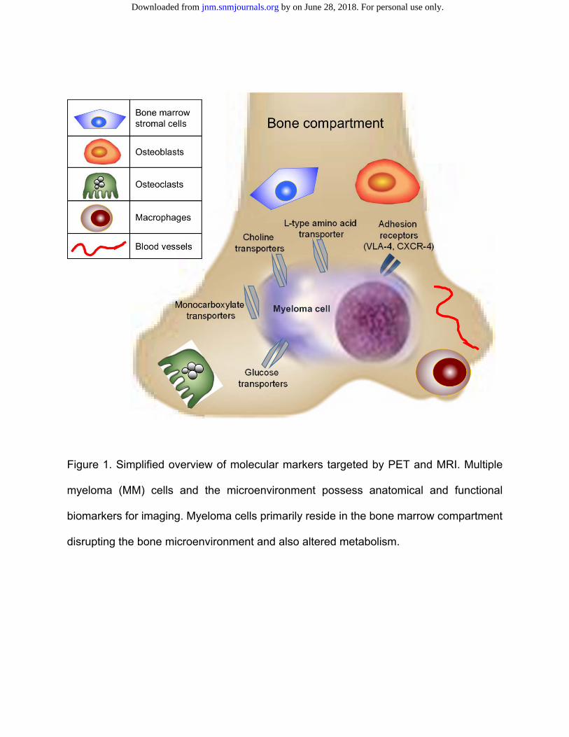
Figure 1. Simplified overview of molecular markers targeted by PET and MRI. Multiple
myeloma (MM) cells and the microenvironment possess anatomical and functional
biomarkers for imaging. Myeloma cells primarily reside in the bone marrow compartment
disrupting the bone microenvironment and also altered metabolism.
by on June 28, 2018. For personal use only. jnm.snmjournals.org Downloaded from
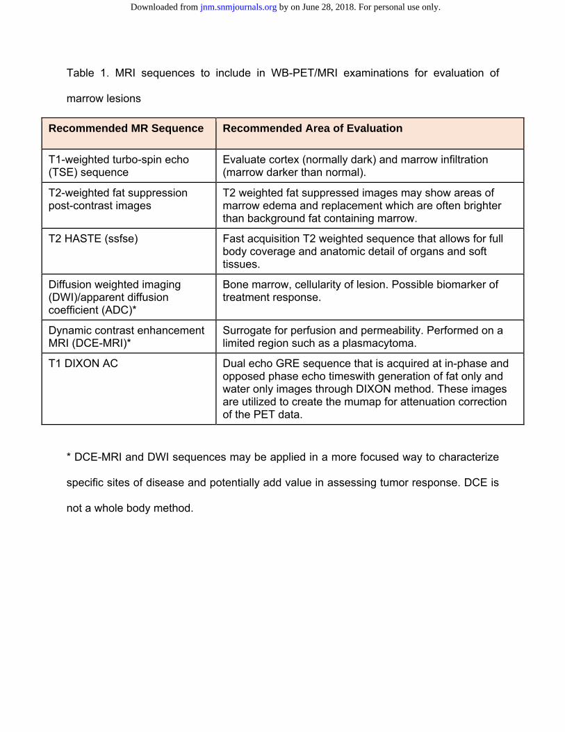
Table 1. MRI sequences to include in WB-PET/MRI examinations for evaluation of
marrow lesions
Recommended MR Sequence Recommended Area of Evaluation
T1-weighted turbo-spin echo (TSE) sequence
Evaluate cortex (normally dark) and marrow infiltration (marrow darker than normal).
T2-weighted fat suppression post-contrast images
T2 weighted fat suppressed images may show areas of marrow edema and replacement which are often brighter than background fat containing marrow.
T2 HASTE (ssfse) Fast acquisition T2 weighted sequence that allows for full body coverage and anatomic detail of organs and soft tissues.
Diffusion weighted imaging (DWI)/apparent diffusion coefficient (ADC)*
Bone marrow, cellularity of lesion. Possible biomarker of treatment response.
Dynamic contrast enhancement MRI (DCE-MRI)*
Surrogate for perfusion and permeability. Performed on a limited region such as a plasmacytoma.
T1 DIXON AC Dual echo GRE sequence that is acquired at in-phase and opposed phase echo timeswith generation of fat only and water only images through DIXON method. These images are utilized to create the mumap for attenuation correction of the PET data.
* DCE-MRI and DWI sequences may be applied in a more focused way to characterize
specific sites of disease and potentially add value in assessing tumor response. DCE is
not a whole body method.
by on June 28, 2018. For personal use only. jnm.snmjournals.org Downloaded from
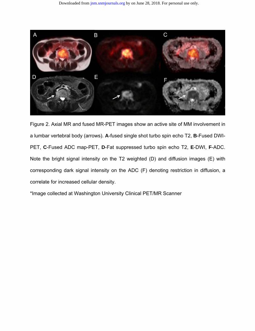
Figure 2. Axial MR and fused MR-PET images show an active site of MM involvement in
a lumbar vertebral body (arrows). A-fused single shot turbo spin echo T2, B-Fused DWI-
PET, C-Fused ADC map-PET, D-Fat suppressed turbo spin echo T2, E-DWI, F-ADC.
Note the bright signal intensity on the T2 weighted (D) and diffusion images (E) with
corresponding dark signal intensity on the ADC (F) denoting restriction in diffusion, a
correlate for increased cellular density.
*Image collected at Washington University Clinical PET/MR Scanner
by on June 28, 2018. For personal use only. jnm.snmjournals.org Downloaded from
Doi: 10.2967/jnumed.115.163808Published online: November 5, 2015.J Nucl Med. Ravi Vij, Kathryn Fowler and Monica Shokeen New Approaches to Molecular Imaging of Multiple Myeloma
http://jnm.snmjournals.org/content/early/2015/11/04/jnumed.115.163808This article and updated information are available at:
http://jnm.snmjournals.org/site/subscriptions/online.xhtml
Information about subscriptions to JNM can be found at:
http://jnm.snmjournals.org/site/misc/permission.xhtmlInformation about reproducing figures, tables, or other portions of this article can be found online at:
and the final, published version.proofreading, and author review. This process may lead to differences between the accepted version of the manuscript
ahead of print area, they will be prepared for print and online publication, which includes copyediting, typesetting,JNMcopyedited, nor have they appeared in a print or online issue of the journal. Once the accepted manuscripts appear in the
. They have not beenJNM ahead of print articles have been peer reviewed and accepted for publication in JNM
(Print ISSN: 0161-5505, Online ISSN: 2159-662X)1850 Samuel Morse Drive, Reston, VA 20190.SNMMI | Society of Nuclear Medicine and Molecular Imaging
is published monthly.The Journal of Nuclear Medicine
© Copyright 2015 SNMMI; all rights reserved.
by on June 28, 2018. For personal use only. jnm.snmjournals.org Downloaded from