Embed Size (px)
Citation preview

1
Title:
Primary repair of symptomatic neonates with tetralogy of Fallot
with or without pulmonary atresia
2
Abstract
Recently, surgical outcomes of tetralogy of Fallot (TOF) have been improving. For
TOF patients more than 3 months of age, primary repair has been advocated
regardless of symptoms. However, a surgical approach of symptomatic TOF
neonates or very young infants remains elusive. Traditionally, there have been two
surgical options for these patients, primary repair versus first aortopulmonary
shunt followed by repair. Early primary repair provides several advantages
including avoidance of shunt related complications, early relief of hypoxia,
promotion of normal lung development, avoidance of ventricular hypertrophy and
fibrosis, and psychological comfort to family. Because of advances in
cardiopulmonary bypass techniques and accumulated experience in neonatal
cardiac surgery, primary repair in neonates with TOF has been performed with
excellent early outcomes (early mortality < 5%), which may be superior to ones of
aortopulmonary shunt. Regarding surgical options, a remaining question to
answer is whether shunt can preserve the pulmonary valve annulus for TOF
3
neonates with pulmonary stenosis. Comparing to older babies, symptomatic
neonates have different anatomy of RVOT obstruction, which is nearly always
caused by a hypoplastic pulmonary valve annulus instead of infundibular
obstruction. Therefore, shunt is less likely to save the pulmonary valve annulus
comparing to primary repair. Primary repair of TOF can be performed safely in
most symptomatic neonates. The patients who had primary repair should be
closely followed up to evaluate the RVOT pathology and right ventricular function.
Keywords: tetralogy of Fallot, neonate, early primary repair, palliation
4
Introduction
Tetralogy of Fallot (TOF) is the most common form of cyanotic congenital heart
disease, and characterized by four distinct anatomic features: (1) pulmonary
outflow tract obstruction (stenosis or atresia), (2) ventricular septal defect (VSD),
(3) overriding aortic root, and (4) right ventricular hypertrophy1-3). Since the
Fallot’s description in 1888, so called “blue baby operation” or Blalock-Taussig
shunt, the first surgical treatment for TOF was performed by Alfred Blalock in
19454), followed by the first successful intracardiac repair using human cross-
circulation by Lillehei in 19545) and using a pump oxygenator by Kirklin in 19556).
For at least 2 decades, initial palliation followed by repair later in childhood had
been the most prevalent strategy. By the early 1980s, primary repair in early
infancy had been advocated by Castaneda7), Barrat-Boyes8), and others. This
approach gained wide acceptance, and many surgeons adopted a therapeutic
strategy based on early primary repair in the absence of specific anatomic
contraindications or comorbidities such as major non-cardiac anomalies. The
5
concept of early primary repair was extended to symptomatic neonates, and low
operative mortality was achieved9-13).
However, the management of symptomatic infants with TOF requiring surgical
intervention in the first month of life remains controversial14). In this article, I
would like to summarize the personal experience and review the current practice
of early primary repair in TOF neonates with pulmonary stenosis or atresia.
Considerations of the anatomy and pathophysiology of TOF in small
babies requiring early intervention
TOF is defined on the basis of anterocephalad deviation of the outlet septum with
associated malformation of the septoparietal trabeculation or results from
underdevelopment of the subpulmonary infundibulum2,15). This one abnormality
or pathologic process during embryogenesis produces RVOT obstruction and also
is responsible for the malalignment type VSD and the aortic override. The right
ventricle hypertrophy is the hemodynamic consequence of the above anatomical
lesions16.
6
TOF has a wide spectrum of disease from mild RVOT obstruction to severe
obstruction and TOF with pulmonary atresia is at one extreme. The level of
obstruction may occur at one or more of the following structures: (1)
infundibulum, (2) pulmonary valve, (3) main pulmonary artery, and/or (4) branch
pulmonary arteries3).
In TOF patients with pulmonary stenosis, the initial manifestation of symptoms
depends on the degree of RVOT obstruction17). Most commonly, cyanosis is mild
at birth and gradually progresses with age as the stenosis increases due to
increasing infundibular hypertrophy. Cyanosis tends to become significant within
the first 6 to 12 months of life. In such situations, the obstruction is entirely or
predominantly at the infundibular level. The pulmonary valve annulus and the
branch pulmonary arteries usually are of good size. However, a smaller
percentage of patients have marked cyanosis at or soon after birth. In this group
the RVOT obstruction is nearly always caused by a hypoplastic pulmonary valve
annulus12,17,18). Cyanosis is constant in these patients because of the fixed nature
of the obstruction of pulmonary blood flow. Castaneda et al7) described the
anatomy of RVOT in young infants with TOF presenting with cyanosis. Although
7
the infundibulum was stenotic, the area of greatest obstruction to pulmonary
blood flow in the majority of infants was at the pulmonary valve annulus, in
which the pulmonary valve was bicuspid or dysplastic with fusion of commissures.
Pulmonary atresia is present in approximately 7% of patients with TOF19). TOF
with pulmonary atresia exhibits considerable morphologic variability, particularly
with respect to the source of pulmonary arterial flow20). When patients with
pulmonary atresia have no major collateral arteries, a PDA is the only source of
pulmonary blood flow. In such cases, there may be varying degrees of hypoplasia
of the pulmonary arteries, but it is most often the case that all or nearly all lung
segments are supplied by branches arborizing from the right and left pulmonary
arteries, that is, pulmonary artery arborization is much more predictable21).
Surgical options in symptomatic TOF neonates
Symptomatic TOF is defined as prostaglandin dependent or having either hypoxic
episodes or severe hypoxemia with resting systemic oxygen saturation of less
than 75%. Neonates born with TOF with pulmonary stenosis are known to
8
uncommonly develop symptoms but a smaller percentage of patients have
marked cyanosis at or soon after birth as previously mentioned. TOF neonates
with pulmonary atresia are symptomatic or asymptomatic but have inevitably
ductal dependent pulmonary circulation. For such patients, surgical options
include early primary repair or palliation with a aortopulmonary shunt.
Recently, Al Habib et al22) reported contemporary patterns of management of TOF
with pulmonary stenosis using The Society of Thoracic Surgeons (STS) Database
(3059 operations in 2002-2007). This study showed that primary repair in the first
year of life is the most prevalent strategy. However, for the neonatal age group,
primary procedures were about equally divided between primary repair and
palliation (154 cases vs. 178 cases among total 332 procedures, respectively). This
demonstrates that an optimal strategy for neonates with symptomatic TOF is still
an unanswered issue even in current years.
Aortopulmonary shunt in neonates with symptomatic TOF
9
For neonates and young infants with symptomatic TOF, palliation with a
aortopulmonary shunt has been performed by many surgeons. Those who prefer
this option are likely to agree with the use of primary repair in TOF at 3 months
of age or older. Recently, Kanter et al14) reported a study comparing two surgical
options such as primary repair and shunt in symptomatic TOF neonates. They
concluded that shunting or primary repair in neonates with symptomatic TOF
provided equivalent mortality and results and also shunted patients had fewer
transannular patch repairs despite having more emergent initial operations.
At this point, two issues should be answered regarding shunting in neonates with
TOF: (1) How safely shunting is performed in these patients and (2) whether
shunting additionally saves the pulmonary annulus?
1. Safety issue of shunting
For infants with TOF, a palliative shunt has been performed safely and with
excellent outcomes23,24). Even in small babies, this option still provides good and
similar outcomes to primary repair14,22). Recently, Kanter et al14) reported excellent
10
early outcomes including shorter intensive care unit and hospital stays for the
first operation and early mortality of 5.9%. STS database said that discharge
mortality of TOF repair in neonates was 11 of 178 (6.2%) for palliation and 12 of
154 (7.8%) for primary repair22). However, outcomes of shunt in neonates have
been reported inconsistently and with fluctuations. In a multicenter study of the
effect of aspirin on shunts, clinical outcomes of palliative shunts in infants were
poor even in the current era25). This study showed that event rates of shunt
thrombosis and death were 5% and 15%, respectively for TOF patients and 16%
and 22%, respectively for patients with pulmonary atresia. Recently, Guzzetta et
al26) reported outcomes of in-hospital shunt occlusion in 207 small infants
undergoing only a modified Blalock-Taussig shunt, in which in-hospital shunt
occlusion occurred in 14 patients (6.8%). Patients who had shunt occlusion had a
harder postoperative course and a higher rate of in-hospital mortality (6.2%
versus 21.4%). They concluded that the risk factors of in-hospital shunt occlusion
were cardiac diagnosis (pulmonary atresia) and the size of the pulmonary arteries.
With regard to the safety of shunt, interim mortality should be issued in infants
undergoing shunting13,27,28). Interim mortality occurs during the palliated state
11
before repair and may account for an additional attrition, which may or may not
be shunt-related29).
2. Issue of preservation of pulmonary valve annulus in neonatal TOF repair
For TOF neonates with pulmonary stenosis, the influence of shunting on the
growth of the pulmonary annulus should be evaluated. Pulmonary regurgitation
after TOF repair results in various adverse long-term outcomes30). Therefore, every
effort should be made to preserve total pulmonary valve function as well as the
pulmonary annulus in TOF repair. Recently, Kanter et al 14) suggested that shunted
patients have a greater likelihood of avoiding a transannular patch at the time of
repair. Sousa Uva et al31) reported that initial palliation promoted the growth of
pulmonary annulus and transannular patching was less prevalent for patients who
underwent initial palliation (13% versus 56%, p = 0.03). One year later, however,
they reported on the same issue, in which the observation of increased size of
the pulmonary annulus after shunt could be due to chance only and initial
palliation did not allow for a reduction in incidence of transannular patching32).
12
The incidence of transannular patching at the time of primary repair in neonates
ranges from 84 to 100%10,12,14,33). Symptomatic neonates have received a
transannular patch because of the morphology, not because of their age. The
need for a transannular patch reflects the severity of the RVOT obstruction at the
annular level7,10,12,18). Parry et al18) reported outcomes of elective primary repair of
acyanotic TOF in early infancy, in which only 28% patients required transannular
patching and follow-up echocardiography suggested a trend towards ‘catch-up’
growth of the annulus. Although in a disease entity different from TOF, Emani et
al34) showed progressive decrease in the pulmonary valve annular size in case of
delayed anatomic repair beyond the neonatal period. They suggested the
importance of antegrade blood flow as a stimulus for growth of ventricular
outflow tract structures and the negative effect of retrograde blood flow from
aortopulmonary shunts.
Primary repair in symptomatic TOF neonates
13
Initial study of Kirklin et al35) suggested that primary repair of TOF at less than
three months of age was associated with a high mortality. Over the past several
years, however, increasing success with primary repair of TOT in younger infants
has been demonstrated by many centers9,18,32,36). These days, early primary repair
in symptomatic neonates with TOF has been performed with excellent early
outcomes (Table 1).
Early primary repair provides multiple advantages including avoidance of shunt
related complications, early relief of hypoxia, promotion of normal lung
development, avoidance of ventricular hypertrophy and fibrosis, and
psychological comfort to the young family37,38).
In the current era, there area essentially no contraindications to early primary
repair38,39). Past considerations for delayed repair include anomalous coronary
artery crossing the right ventricular outflow tract, hypoplastic or discontinuous
pulmonary arteries and multiple ventricular septal defects39).
For symptomatic neonates with TOF, Kanter et al14) suggested that neonates who
were smaller and required emergency operation were shunted and those with
favorable anatomy and good-sized branch pulmonary arteries had primary repair.
14
Shunting in smaller neonates and neonates necessitating emergency operation is
not absolutely safe and most likely associated with considerable mortality and
morbidity. Recently, the accumulation of the experience of neonatal cardiac
surgery and advances in cardiopulmonary bypass techniques may provide more
stable outcomes after primary repair in such situations.
Regarding the size of pulmonary arteries, this issue should be simplified to
identify the presence of major aortopulmonary collateral arteries. If there are no
major collateral arteries present, the pulmonary arteries are adequate for primary
repair of TOF38). Jonas38) emphasized that the pulmonary arteries are underfilled
and underpressurized preoperatively, so whatever imaging technique is used, the
potential size of the pulmonary arteries is unknown. Van Arsdell et al40) pointed
out that problems with true small pulmonary arteries are infrequent (1% to 2%)
and can be managed with a fenestrated VSD.
Surgical techniques of primary repair in neonatal TOF have been progressively
changed over the last several decades. Since the early years, neonatal TOF repair
has been performed using cardiopulmonary bypass with a period of deep
hypothermic circulatory arrest7,10,11,33). Recently, however, many surgeons have
15
tried to avoid deep hypothermic circulatory arrest and perform primary repair
under continuous moderate hypothermic cardiopulmonary bypass and cardiac
arrest instead12-14). The VSD is closed through a transatrial and/or transventricular
approach. RVOT reconstruction is performed using various techniques including
transannular or non-transannular patching in pulmonary stenosis and
transjunctional patching, interposition of a conduit from the right ventricle to the
pulmonary arteries, and other techniques in pulmonary atresia. Farouk et al20)
described the surgical technique in TOF patients with pulmonary atresia and the
feasibility of a transatrial approach for VSD closure in all patients.
Personal experience of primary repair of TOF with or without
pulmonary atresia
My personal strategy for surgical intervention in neonatal TOF is early primary
repair if possible. I have reviewed my clinical experiences of surgical treatment in
symptomatic neonates with TOF.
16
Patient Population
Between May 2004 and Dec 2012, 27 consecutive neonates with TOF underwent a
surgical intervention. Their cardiac diagnoses were TOF with pulmonary stenosis
(n = 6) or atresia (n = 21) and no major aortopulmonary collateral vessels.
All patients had symptoms before surgery. Twenty-six patients were receiving an
infusion of prostaglandin and remaining patient with pulmonary stenosis suffered
from anoxic spell and was treated with propranolol and mechanical ventilator care.
Ten patients needed mechanical ventilator care temporarily or until repair.
Intracardiac anatomy and the RVOT were preoperatively assessed using
transthoracic echocardiography. The morphology and size of pulmonary arteries
and the presence of major collateral arteries were assessed by chest computed
tomography (CT) scan. Preoperative chest CT scan images were available to
measure the size of the branch pulmonary arteries in 24 patients. McGoon ratio41)
and Nakata index42) were median value of 1.1 (range 0.6 to 1.5) and 121 mm2/m2
(range 27 to 189 mm2/m2), respectively (Fig 1). The only one patient who had
17
undergone shunt for small pulmonary arteries had McGoon ratio of 0.6 and
Nakata index of 27 mm2/m2 (Fig. 1, black triangle*).
Twenty-five neonates (93%) underwent primary repair. The age at repair was 16
days (median, range 12-29 days) and body weight was 3.2 kg (median, range
2.2−4.2 kg). Two patients (7%) had aortopulmonary shunts, one patient with
seemingly small LV size and another with diffuse hypoplastic branch pulmonary
arteries despite no major aortopulmonary collateral vessels (Fig 2).
Operative techniques
The early primary repair included VSD closure (a transatrial approach in 20 and a
transvenous approach in 5), resection of the hypertrophied right ventricular
muscle and RVOT reconstruction with various techniques such as a transannular
or transjunctional RVOT widening with an autologous pericardial patch (n = 12),
interposition of a conduit made with an autologous pericardial roll between the
right ventricle and the pulmonary arteries (n = 8, 7 to 10 mm in diameter), RVOT
reconstruction using the left atrial auricle as a flap (n = 3), and REV (reparation a
18
l’etage ventricularie) type reconstruction (native tissue to tissue anastomosis
between the posterior wall of the main PA and right ventricle, n = 2). Any
materials for preventing the pulmonary regurgitation such as artificial monocusp
or bicusp pulmonary valves were not used in any patients. The fenestration of
VSD was not necessary in all patients.
Cardiopulmonary bypass data
All primary repairs were performed after standard median sternotomy using full-
flow cardiopulmonary bypass with moderate systemic hypothermia. Antegrade
crystalloid cardioplegia was used for myocardial protection. Cardiopulmonary
bypass and aortic cross clamp time were 153 min (median, range 95-257 min)
and 75 min (median, range 50-117 min) respectively. Total circulatory arrest was
not necessary in all patients.
19
Early results and postoperative course
There were no hospital deaths. Delayed sternal closure was necessary in 6
patients and postoperative complications requiring an operation occurred in 5
patients including left-side diaphragm palsy (n = 3), postoperative bleeding (n =
1) and wound infection (n = 1). There was no patient showing the development
of junctional ectopic tachycardia or complete atrioventricular heart block.
Late results
Follow-up was performed at a median interval of 56 months (range 2 to 105
months). There were 2 late deaths. Actuarial survival rate at 6-month, 1-year, and
5-year were 96%, 92%, and 92%, respectively. Among 23 late survivors, 15
patients (65%) have not needed any surgical intervention after primary repair.
Eight patients have undergone surgical procedures of RVOT at a median interval
after total repair of 14 months (range 6.8 to 51 months) and 3 patients have
needed second redo operations for RVOT stenosis. The first catheter
20
intervention for RVOT obstruction has been performed in 14 patients (61%) at a
median interval after primary repair of 7.4 months (range 3 to 19 months) and for
left pulmonary artery stenosis in 6 patients, right pulmonary artery stenosis in 2,
stenosis of both branch pulmonary arteries in 5, and pulmonary trunk stenosis in
1. Eight patients have needed further catheter interventions such as re-ballooning
or placement of a stent. Reoperation free survival rate at 6-month, 1-year, and 5
year were 100%, 81%, and 60%, respectively and catheter plus surgery
intervention free survival rate at 6-month, 1-year, and 5 year were 79%, 50%, and
33%, respectively. On most recent follow-up echocardiography right and left
ventricular systolic function was normal in all patients.
For 23 late survivors, a post-repair RV/LV pressure ratio were 0.61 ± 0.15 (range
0.37 − 0.89), which was not related with preoperative size of the pulmonary
arteries (Fig 3). However, in case of the ratio of 0.6 or more, catheter plus surgical
interventions were needed significantly more frequent (10/11, 91% vs. 5/12, 42%;
p = 0.027 by Fisher’s exact test).
21
Conclusions
Surgical strategy of symptomatic TOF neonates such as primary repair or shunt
followed by repair remains elusive. Shunt operation is no longer a safe option in
neonates and is less likely to save the pulmonary valve annulus additionally.
Primary repair of TOF can be performed safely in most symptomatic neonates.
The patients who had primary repair should be closely followed up to evaluate
the RVOT pathology and right ventricular function.
Conflict of interest
No potential conflict of interest relevant to this article was reported.
References
1. Apitz C, Webb GD, Redington AN. Tetralogy of Fallot. Lancet 2009;374:1462
22
–71.
2. Van Praagh R. The first Stella van Praagh memorial lecture: the history and
anatomy of tetralogy of Fallot. Semin Thorac Cardiovasc Surg Pediatr Card
Surg Annu 2009:19–38.
3. Starr JP. Tetralogy of Fallot: yesterday and today. World J Surg. 2010;34:658
–68.
4. Blalock A. The surgical treatment of malformations of the heart in which
there is pulmonary stenosis or pulmonary atresia. JAMA 1945;128:189–202.
5. Lillehei CW, Cohen M, Warden HE, et al. Direct vision intracardiac surgical
correction of the tetralogy of Fallot, pentalogy of Fallot, and pulmonary
atresia defects; report of first ten cases. Annals of surgery 1955;142:418–42.
6. Kirklin JW, Dushane JW, Patrick RT, et al. Intracardiac surgery with the aid of
a mechanical pump-oxygenator system (gibbon type): report of eight cases.
Proc Staff Meet Mayo Clin 1955;30:201–6.
7. Castaneda AR, Freed MD, Williams RG, Norwood WI. Repair of tetralogy of
23
Fallot in infancy. Early and late results. J Thorac Cardiovasc Surg
1977;74:372–81.
8. Barratt-Boyes BG, Neutze JM. Primary repair of tetralogy of Fallot in infancy
using profound hypothermia with circulatory arrest and limited
cardiopulmonary bypass: a comparison with conventional two stage
management. Ann Surg 1973;178:406–11.
9. Reddy VM, Liddicoat JR, McElhinney DB, Brook MM, Stanger P, Hanley FL.
Routine primary repair of tetralogy of Fallot in neonates and infants less
than three months of age. Ann Thorac Surg 1995;60:S592–6.
10. Hirsch JC, Mosca RS, Bove EL. Complete repair of tetralogy of Fallot in the
neonate: results in the modern era. Ann Surg 2000;232:508–14.
11. Kolcz J, Pizarro C. Neonatal repair of tetralogy of Fallot results in improved
pulmonary artery development without increased need for reintervention.
Eur J Cardiothorac Surg 2005;28:394–399.
12. Tamesberger MI, Lechner E, Mair R, Hofer A, Sames-Dolzer E, Tulzer G. Early
24
primary repair of tetralogy of Fallot in neonates and infants less than four
months of age. Ann Thorac Surg 2008;86:1928–35.
13. Kwak JG, Lee C-H, Lee C, Park CS. Surgical management of pulmonary
atresia with ventricular septal defect: early total correction versus shunt. Ann
Thorac Surg 2011;91:1928–1935.
14. Kanter KR, Kogon BE, Kirshbom PM, Carlock PR. Symptomatic neonatal
tetralogy of Fallot: repair or shunt? Ann Thorac Surg 2010;89:858–63.
15. Anderson RH, Jacobs ML. The anatomy of tetralogy of Fallot with
pulmonary stenosis. Cardiol Young 2008;18 Suppl 3:12–21.
16. Bailliard F, Anderson RH. Tetralogy of Fallot. Orphanet J Rare Dis. 2009;4:2.
17. Hirsch JC, Bove EL. Tetralogy of Fallot. In: Mavroudis C, Backer CL, editors.
Pediatric Cardiac Surgery. 3rd ed. Mosby Inc, 2003:383–397.
18. Parry AJ, McElhinney DB, Kung GC, Reddy VM, Brook MM, Hanley FL.
Elective primary repair of acyanotic tetralogy of Fallot in early infancy:
overall outcome and impact on the pulmonary valve. J Am Coll Cardiol
25
2000;36:2279–83.
19. Chiariello L, Meyer J, Wukasch DC, Hallman GL, Cooley DA. Intracardiac
repair of tetralogy of Fallot. Five-year review of 403 patients. J Thorac
Cardiovasc Surg 1975;70:529–35.
20. Farouk A, Zahka K, Siwik E, et al. Individualized approach to the surgical
treatment of tetralogy of Fallot with pulmonary atresia. Cardiol Young
2009;19:76–85.
21. Stewart RD, Mavroudis C, Backer CL. Tetralogy of Fallot. In: Mavroudis C,
Backer CL, editors. Pediatric cardiac surgery. 4 ed. Wiley-Blackwell, 2013:410
–427.
22. Habib Al HF, Jacobs JP, Mavroudis C, et al. Contemporary patterns of
management of tetralogy of Fallot: data from the Society of Thoracic
Surgeons Database. Ann Thorac Surg 2010;90:813–9
23. Karl TR, Sano S, Pornviliwan S, Mee RB. Tetralogy of Fallot: favorable
outcome of nonneonatal transatrial, transpulmonary repair. Ann Thorac
26
Surg 1992;54:903–7.
24. Fraser CDJ, McKenzie ED, Cooley DA. Tetralogy of Fallot: surgical
management individualized to the patient. Ann Thorac Surg 2001;71:1556–
61.
25. Li JS, Yow E, Berezny KY, et al. Clinical outcomes of palliative surgery
including a systemic-to-pulmonary artery shunt in infants with cyanotic
congenital heart disease: does aspirin make a difference? Circulation
2007;116:293–297.
26. Guzzetta NA, Foster GS, Mruthinti N, Kilgore PD, Miller BE, Kanter KR. In-
Hospital Shunt Occlusion in Infants Undergoing a Modified Blalock-Taussig
Shunt. Ann Thorac Surg 2013 (in press)
27. Fermanis G, Ekangaki A, Salmon A, et al. Twelve year experience with the
modified Blalock-Taussig shunt in neonates. Eur J Cardiothorac Surg
1992;6:586–589.
28. Fenton KN, Siewers RD, Rebovich B, Pigula FA. Interim mortality in infants
27
with systemic-to-pulmonary artery shunts. Ann Thorac Surg 2003;76:152–6.
29. Dodge-Khatami A, Tulevski II, Hitchcock JF, de Mol BAJM, Bennink GBWE.
Neonatal complete correction of tetralogy of Fallot versus shunting and
deferred repair: is the future of the right ventriculo-arterial junction at stake,
and what of it? Cardiol Young 2001;11:484–90.
30. Redington AN. Physiopathology of Right Ventricular Failure. Semin Thorac
Cardiovasc Surg Pediatr Card Surg Annu 2006;9:3–10.
31. Sousa-Uva M, Lacour-Gayet F, Komiya T, et al. Surgery for tetralogy of
Fallot at less than six months of age. J Thorac Cardiovasc Surg
1994;107:1291–300.
32. Sousa-Uva M, Chardigny C, Galetti L, et al. Surgery for tetralogy of Fallot at
less than six months of age. Is palliation "old-fashioned"? Eur J Cardiothorac
Surg 1995;9:453–9.
33. Di Donato RM, Jonas RA, Lang P, Rome JJ, Mayer JEJ, Castaneda AR.
Neonatal repair of tetralogy of Fallot with and without pulmonary atresia. J
28
Thorac Cardiovasc Surg 1991;101:126–37.
34. Emani SM, Beroukhim R, Zurakowski D, et al. Outcomes after anatomic
repair for d-transposition of the great arteries with left ventricular outflow
tract obstruction. Circulation 2009;120:S53–8.
35. Kirklin JW, Blackstone EH, Jonas RA, et al. Morphologic and surgical
determinants of outcome events after repair of tetralogy of Fallot and
pulmonary stenosis. A two-institution study. J Thorac Cardiovasc Surg
1992;103:706–23.
36. Pigula FA, Khalil PN, Mayer JE, del Nido PJ, Jonas RA. Repair of tetralogy of
Fallot in neonates and young infants. Circulation 1999;100(19 Suppl):II157–
61.
37. Pozzi M, Trivedi DB, Kitchiner D, Arnold RA. Tetralogy of Fallot: what
operation, at which age. Eur J Cardiothorac Surg 2000;17:631–36.
38. Jonas RA. Early primary repair of tetralogy of Fallot. Semin Thorac
Cardiovasc Surg Pediatr Card Surg Annu 2009:39–47.
29
39. Derby CD, Pizarro C. Routine primary repair of tetralogy of Fallot in the
neonate. Expert Rev Cardiovasc Ther 2005;3:857–63.
40. Van Arsdell G, Yun TJ. An apology for primary repair of tetralogy of Fallot.
Semin Thorac Cardiovasc Surg Pediatr Card Surg Annu 2005:128–31.
41. McGoon DC, Baird DK, Davis GD. Surgical management of large bronchial
collateral arteries with pulmonary stenosis or atresia. Circulation 1975;52:109
–18.
42. Nakata S, Imai Y, Takanashi Y, et al. A new method for the quantitative
standardization of cross-sectional areas of the pulmonary arteries in
congenital heart diseases with decreased pulmonary blood flow. J Thorac
Cardiovasc Surg 1984;88:610–9.
30
Legends
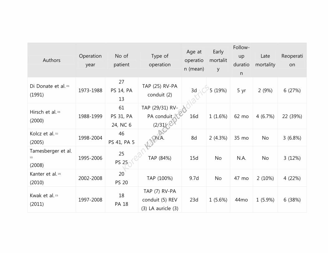
Table 1. Outcomes of early primary repair.
*N.A.; not available, NC; non-confluent, PA; pulmonary atresia, PS; pulmonary
stenosis, REV; reparation a l’etage ventricularie, RV-PA; right ventricle - pulmonary
arteries, TAP; transannular patch
Fig 1. Preoperative size of the pulmonary arteries. Black triangle*; palliative case
due to hypoplastic pulmonary arteries, black triangle**; palliative case due to
seemingly LV hypoplasia
Fig 2. Preoperative chest CT scan in a patient with diffuse hypoplastic pulmonary
arteries who had a palliative shunt (case of black triangle* in Fig 1).
Fig 3. Scatter plots between post-repair RV/LV pressure ratio and preoperative
McGoon ratio (A) and Nakata index (B).

31
Authors Operation
year
No of
patient
Type of
operation
Age at
operatio
n (mean)
Early
mortalit
y
Follow-
up
duratio
n
Late
mortality
Reoperati
on
Di Donate et al. 33)
(1991) 1973-1988
27
PS 14, PA
13
TAP (25) RV-PA
conduit (2) 3d 5 (19%) 5 yr 2 (9%) 6 (27%)
Hirsch et al. 10)
(2000) 1988-1999
61
PS 31, PA
24, NC 6
TAP (29/31) RV-
PA conduit
(2/31)
16d 1 (1.6%) 62 mo 4 (6.7%) 22 (39%)
Kolcz et al. 11)
(2005) 1998-2004
46
PS 41, PA 5 N.A. 8d 2 (4.3%) 35 mo No 3 (6.8%)
Tamesberger et al.
12)
(2008)
1995-2006 25
PS 25 TAP (84%) 15d No N.A. No 3 (12%)
Kanter et al. 14)
(2010) 2002-2008
20
PS 20 TAP (100%) 9.7d No 47 mo 2 (10%) 4 (22%)
Kwak et al. 13)
(2011) 1997-2008
18
PA 18
TAP (7) RV-PA
conduit (5) REV
(3) LA auricle (3)
23d 1 (5.6%) 44mo 1 (5.9%) 6 (38%)


Fig 1
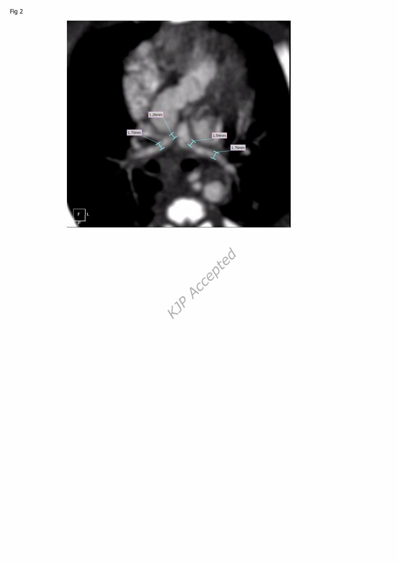
Fig 2

Fig 3