Embed Size (px)
Citation preview

1
Unit 7: Molecular biology and genetics
How many genetic diseases do you think there are? What causes genetic diseases?
On successful completion of this topic you will: • be able to determine patterns of inheritance (LO4).
To achieve a Pass in this unit you will need to show that you can: • relate inherited diseases to patterns of inheritance (4.3).
Inherited diseases7.6

2
Unit 7: Molecular biology and genetics
7.6: Inherited diseases
1 What are inherited diseases?It is important to realise that there are not genes for diseases.
Genes should code for functioning proteins (for example, for a protein used in normal colour vision, a normal blood clotting factor, an enzyme, a membrane channel protein or a muscle protein) or for regulatory molecules, such as RNA. Mutated alleles, however, give rise to the genetic diseases because their products do not function correctly.
There are over 5000 single gene defects and many other chromosomal abnormality defects. As molecular biologists learn more about the complexities and subtleties of the way gene expression is regulated, they are finding out more about inherited disorders and that many chronic conditions such as heart disease and diabetes have large genetic components but do not follow Mendelian inheritance patterns.
In this topic guide we shall consider three autosomal recessive traits and one autosomal dominant trait, all of which exhibit Mendelian inheritance patterns.
Autosomal recessive traitsThere are many genetic diseases with autosomal inheritance patterns and Table 7.6.1 shows information about three of them.
Disease Cause Symptoms Detection Treatment and prognosis
phenylketonuria (PKU)
• Mutation to a gene, on chromosome 12, for the hepatic enzyme phenylalanine hydroxylase. Lack of this enzyme in its functioning form leads to build up of phenylalanine in the blood. This prevents other amino acids reaching brain tissue and forming essential neurotransmitter molecules.
• Lack of neurotransmitters in brain tissue leads to microcephaly, mental retardation, seizures.
• Lack of metabolism of phenylalanine to tyrosine and then to melanin leads to albinism.
• Incidence about 1 in 16 000.
• Excess phenylalanine in blood is converted to phenylketone and phenylacetone and these can be detected in urine.
• Phenylacetone in urine and sweat gives characteristic musty odour.
• Newborns are given Guthrie test (Figure 7.6.1) – see Topic guide 7.5.
• Diet with restricted amounts of phenylalanine and enhanced amounts of tyrosine. If started early enough this prevents mental retardation.
• Medication to reduce phenylalanine levels in blood when patient is ill and has raised phenylalanine levels.
sickle cell anaemia
• Nucleotide base substitution. T replaces A at the 17th nucleotide of the gene for beta haemoglobin. GAG for glutamic acid becomes GTG, which encodes valine.
• At regions of low oxygen tension haemoglobin fails to remain globular. The ‘stringiness’ of haemoglobin makes the red blood cells sickle shaped and these can stick in capillaries, reducing the oxygen supply to tissues and giving a painful crisis. These cells are removed from the circulation and this produces anaemia.
• Incidence about 1 in 500 in black Africans.
• Examination of red blood cells under low oxygen tension under a microscope (see Figure 7.6.3).
• Genetic test. • Electrophoresis of haemoglobin.
• Regular blood transfusions about once every three months.
• Future therapies may focus on gene regulation mechanism that could switch back on the gene for fetal haemoglobin.
Table 7.6.1 continued on next page
Table 7.6.1: Information about three diseases with autosomal recessive inheritance patterns.
Figure 7.6.1: Blood taken from the heel of a newborn baby for the Guthrie test.

3
Unit 7: Molecular biology and genetics
7.6: Inherited diseases
Disease Cause Symptoms Detection Treatment and prognosis
cystic fibrosis (CF)
• Mutation to CFTR (cystic fibrosis transmembrane regulatory) gene on chromosome 7.
• The 1480 amino acid protein is encoded by a gene of over 6000 nucleotides with 27 exons.
• Unlike sickle cell anaemia there are several mutations to the gene, which codes for chloride ion channels on cell membranes of epithelial cells lining airways, intestine and sperm ducts.
• People with CF inherit two mutated alleles of the CFTR gene but each mutated allele does not have to be the same.
• Thick sticky mucus in lungs, intestine and sperm ducts.
• Excess salt in sweat. • Baby fails to thrive as cannot absorb digested food properly, particularly fat.
• Newborn may fail to pass faeces or babies have grey, greasy faeces in nappies.
• Breathlessness. • Chronic chest infections. • Lung fibrosis (see Figure 7.6.2).
• Infertility in males. • Pulmonary hypertension and heart failure.
• Incidence about 1 in 2500 births among Caucasians.
• Sweat test – showing higher than normal salt levels.
• Genetic test can detect many, but not all, of the mutations to the CFTR gene.
• Physiotherapy to expel mucus from lungs.
• Nebuliser to aid breathing. • Encapsulated digestive enzyme taken with meals to aid digestion.
• Prophylactic antibiotics to prevent chest infections.
• Heart-lung transplant later on in life.
• Life expectancy now up to 40–50 years.
Figure 7.6.2: Fibrosis of the lung. Areas of lung tissue have been converted into
macroscopic cystic (pus-filled) air spaces.

4
Unit 7: Molecular biology and genetics
7.6: Inherited diseases
Autosomal dominant traitsHuntington’s disease is classed as a polyglutamine disease. It is caused by an expanding triple nucleotide repeat on chromosome 4.
The gene for Huntingtin protein contains a sequence of between 10 and 30 repeating nucleotide triplets, CAG. This nucleotide triplet codes for glutamine. If this sequence contains more than 36 CAG triplets Huntington’s disease will develop. The greater the number of repeats, the younger the age of onset, with an average age of onset of 37 years.
However, this is not a precise correlation. Huntingtin protein has many functions and the abnormal form may interfere with neurotransmission within brain tissue, or lead to death of brain cells. Symptoms begin with loss of motor control manifested by jerky involuntary movements, and then progress to personality changes and loss of cognitive function. Only one copy of the mutated allele is needed for development of the disorder and children of a sufferer have a 50% chance of inheriting the faulty allele. There is a genetic test available but as there is no cure or effective treatment, about 90% of people with Huntington’s disease in the family are reluctant to have the test. However, this raises issues – should someone with Huntington’s disease among relatives be tested before deciding to have children? If one parent is known to have an allele for Huntington’s disease, they may have fetal cells, obtained from chorionic villus sampling or amniocentesis, tested genetically and, if the test is positive, terminate that pregnancy.
Other polyglutamine diseases include Parkinson’s disease and Lou Gehrig’s disease (also called ALS – amyotrophic lateral sclerosis, a type of motor neurone disease). Both of these involve extra glutamines in a protein called ataxin 2.
For some autosomal dominant conditions the homozygous dominant genotype is a lethal combination.
Figure 7.6.3: Light micrograph of human blood of patient with sickle cell anaemia.
Activity: More genetic disorders Research and give a presentation to others in your class about one of the following disorders. Outline the mutation causing the condition, symptoms, inheritance pattern, diagnosis (including pedigree analysis) and prognosis.
• familial hypercholesterolaemia (FHC)
• achondroplasia • erythromelalgia • Marfan syndrome • haemophilia B • red-green colour blindness • Tay–Sachs disease • Duchenne muscular dystrophy • X-linked hypophosphataemic
(vitamin D-resistant) rickets • testicular feminisation syndrome.

5
Unit 7: Molecular biology and genetics
7.6: Inherited diseases
Taking it further: Another genetic disorder caused by an expanding triple nucleotide repeatFragile X syndrome is a sex-linked disorder involving an expanding triple nucleotide repeat. It is the most common form of mental retardation, affecting 1 in 1250 males and 1 in 2000 females in all ethnic groups. Males with the syndrome have facial abnormalities (narrow face, prominent forehead, jaw and ears) and large testes as well as moderate to severe learning difficulties. However, 20% of males with the mutation are normal. One third of heterozygous females are clinically affected but only have learning difficulties if the affected X chromosome came from their mother; they do not show symptoms even if they inherit an X chromosome from their affected father. However, the grandchildren of an affected transmitting male have a higher risk of inheriting the disorder than do their cousins. The gene is the FMR1 gene and it encodes FMR1, an RNA-binding protein. There is a repeat sequence, normally 6–52 repeats, of CGG triplets in the untranslated first exon. In affected individuals the repeat has expanded to more than 230 CGG triplets, the 5’ end of this gene becomes hypermethylated (methyl groups are added to C bases) and the gene is not transcribed. Having 50–230 repeats is a permutation and, although this gives no symptoms, the gene is unstable and likely to expand more during meiosis and transmit the condition to offspring.
Children with severe learning difficulties can be genetically tested by analysing DNA obtained from their blood using PCR and Southern blotting. The DNA is digested using restriction enzymes that recognise a methylated site. Antibodies to the FMR1 protein may also be used to detect presence or absence of the product of this gene.
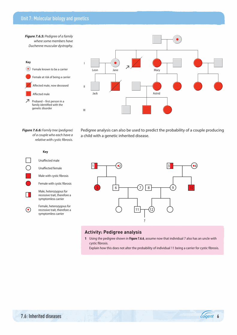
Pedigree analysisPedigree diagrams, using standard symbols such as those shown in Figure 7.6.4, have been used to find the inheritance pattern of newly-discovered genetic conditions, using the following guidance:
• for conditions with a Mendelian recessive pattern, affected individuals may not appear in each generation as offspring of two unaffected parents can inherit the disease (see Figure 7.6.5)
• for conditions having a dominant inheritance pattern, affected male and female individuals appear in every generation and only one parent of an affected individual has to have the condition
• if a condition appears far more in males than in females, it is likely to be a sex-linked trait. Sons of affected males will not inherit the condition if it is X-linked.
Figure 7.6.4: Some standard pedigree symbols.
P
Male, deceased
Female, affected
Mating
Consanguineousmating
One male,three females
Two individuals,sex not specified
Male, heterozygousfor autosomalrecessive trait
Female, heterozygousfor X-linkedrecessive trait
Non-identical twins
Identical twins
Miscarriage
Stillbirth
Pregnancy
Parents (father, age 36, isaffected and is the proband)
Children (ages 13, 12, and 6).Third pregnancy ended inmiscarriage
Male, unaffected
1
2
P
SB
1I
II1
2
36
13
2 3
12
29
4
6
3
P
Male, deceased
Female, affected
Mating
Consanguineousmating
One male,three females
Two individuals,sex not specified
Male, heterozygousfor autosomalrecessive trait
Female, heterozygousfor X-linkedrecessive trait
Non-identical twins
Identical twins
Miscarriage
Stillbirth
Pregnancy
Parents (father, age 36, isaffected and is the proband)
Children (ages 13, 12, and 6).Third pregnancy ended inmiscarriage
Male, unaffected
1
2
P
SB
1I
II1
2
36
13
2 3
12
29
4
6
3
6
Unit 7: Molecular biology and genetics
7.6: Inherited diseases
I
AstridJack
Jane MaryLeon
II
III
Female known to be a carrier
Female at risk of being a carrier
Proband – first person in a family identified with the genetic disorder
Affected male, now deceased
Affected male
Key
Pedigree analysis can also be used to predict the probability of a couple producing a child with a genetic inherited disease.
12
?
11
6
Unaffected male
Key
Unaffected female
Male with cystic fibrosis
Female with cystic fibrosis
Male, heterozygous forrecessive trait, therefore asymptomless carrier
Female, heterozygous forrecessive trait, therefore asymptomless carrier
5 7 8 9
3 421
10
Activity: Pedigree analysis1 Using the pedigree shown in Figure 7.6.6, assume now that individual 7 also has an uncle with
cystic fibrosis.Explain how this does not alter the probability of individual 11 being a carrier for cystic fibrosis.
Figure 7.6.5: Pedigree of a family where some members have
Duchenne muscular dystrophy.
Figure 7.6.6: Family tree (pedigree) of a couple who each have a
relative with cystic fibrosis.
7
Unit 7: Molecular biology and genetics
7.6: Inherited diseases
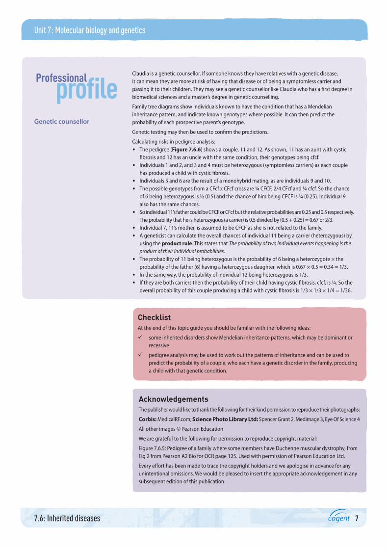
Claudia is a genetic counsellor. If someone knows they have relatives with a genetic disease, it can mean they are more at risk of having that disease or of being a symptomless carrier and passing it to their children. They may see a genetic counsellor like Claudia who has a first degree in biomedical sciences and a master’s degree in genetic counselling.
Family tree diagrams show individuals known to have the condition that has a Mendelian inheritance pattern, and indicate known genotypes where possible. It can then predict the probability of each prospective parent’s genotype.
Genetic testing may then be used to confirm the predictions.
Calculating risks in pedigree analysis: • The pedigree (Figure 7.6.6) shows a couple, 11 and 12. As shown, 11 has an aunt with cystic
fibrosis and 12 has an uncle with the same condition, their genotypes being cfcf. • Individuals 1 and 2, and 3 and 4 must be heterozygous (symptomless carriers) as each couple
has produced a child with cystic fibrosis. • Individuals 5 and 6 are the result of a monohybrid mating, as are individuals 9 and 10. • The possible genotypes from a CFcf x CFcf cross are ¼ CFCF, 2/4 CFcf and ¼ cfcf. So the chance
of 6 being heterozygous is ½ (0.5) and the chance of him being CFCF is ¼ (0.25). Individual 9 also has the same chances.
• So individual 11’s father could be CFCF or CFcf but the relative probabilities are 0.25 and 0.5 respectively. The probability that he is heterozygous (a carrier) is 0.5 divided by (0.5 + 0.25) = 0.67 or 2/3.
• Individual 7, 11’s mother, is assumed to be CFCF as she is not related to the family. • A geneticist can calculate the overall chances of individual 11 being a carrier (heterozygous) by
using the product rule. This states that The probability of two individual events happening is the product of their individual probabilities.
• The probability of 11 being heterozygous is the probability of 6 being a heterozygote × the probability of the father (6) having a heterozygous daughter, which is 0.67 × 0.5 = 0.34 = 1/3.
• In the same way, the probability of individual 12 being heterozygous is 1/3. • If they are both carriers then the probability of their child having cystic fibrosis, cfcf, is ¼. So the
overall probability of this couple producing a child with cystic fibrosis is 1/3 × 1/3 × 1/4 = 1/36.
Genetic counsellor
ChecklistAt the end of this topic guide you should be familiar with the following ideas:
some inherited disorders show Mendelian inheritance patterns, which may be dominant or recessive
pedigree analysis may be used to work out the patterns of inheritance and can be used to predict the probability of a couple, who each have a genetic disorder in the family, producing a child with that genetic condition.
AcknowledgementsThe publisher would like to thank the following for their kind permission to reproduce their photographs:
Corbis: MedicalRF.com; Science Photo Library Ltd: Spencer Grant 2, Medimage 3, Eye Of Science 4
All other images © Pearson Education
We are grateful to the following for permission to reproduce copyright material:
Figure 7.6.5: Pedigree of a family where some members have Duchenne muscular dystrophy, from Fig 2 from Pearson A2 Bio for OCR page 125. Used with permission of Pearson Education Ltd.
Every effort has been made to trace the copyright holders and we apologise in advance for any unintentional omissions. We would be pleased to insert the appropriate acknowledgement in any subsequent edition of this publication.