Embed Size (px)
Citation preview
TOPOGRAPHY AND PATHOGENESIS OFLESIONS IN RHEUMATIC FEVER.
BY
A. F. BERNARD SHAW, M.D.,
Lecturer in Pathology, University of Durham College of Medicine,Newcastle-upon-Tyne.
In recent years interest has been revived in the pathological anatomy ofrheumatic fever by the studies of MacCallum' on lesions of the left auricle andthe series of papers by Pappenheimer and Von Glahn2 on lesions of the aorta anidother vessels. Indeed since the description of the specific nodes in themyocardium by Aschoff (1904) and Geipel (1905), it has become increasinglyapparent that the cardiac lesions are merely local manifestations of a generalinfection, and that the more comprehensive our anatomical studies, the morefruitful are the results likely to be.
In a girl, aged 15 years, who died in December, 1927, of rheumatic feverwith chorea the autopsy revealed extensive changes, and although the micro-scopic lesions were recognized at that time, unavoidable circumstances preventeda detailed study of the material until recently. Only those changes whichprove the generalization of the infection and those which bear upon patho-genesis are discussed, the more commonly recognized lesions being brieflydismissed.
PATHOLOGICAL REPORT.Autopsy protocol.-Rheumatic endocarditis of mitral and aortic valves with characteristic
vegetations. Mitral leaflets thickened and rigid but no stenosis. Aortic cusps slightly thickenedand opaque. Dilatation and hypertrophy of heart. Organizing pericarditis. Sero-fibrinousexudate of about 500 c.cm. in right pleural cavity. Left pleural cavity obliterated by delicateadhesions. Three subcutaneous nodules each about 3.0 mm. in size on right elbow. Chronicpassive congestion of viscera.
On the cut surface of the left ventricle near its base are many opaque white points whichproved to be Aschoff nodes histologically.
Of special interest are the appearances in the auricles. In the left chamber the endocardiumof the posterior wall of the atrium for a distance of 3-0 c.cm. above attachment of mitral leafletis roughened by numerous raised opaque yellowish flecks and ridges which assume a verticaldirection and tend to fuse together as they approach the base of the valve where they merge withsimilar opacities in the substance of the leaflet. Similar lesions are scattered over the rest of theatrium and also in the appendix where they are vertically arranged over the musculi pectinati.
In the right auricle at the mouth of the coronary sinus and just above the attachments of thetricuspid valve are similar flecks and ridges. There are no vegetations on the thin and delicatetricuspid leaflets but beneath the endothelium are some yellowish opacities about 1.0 mm. inlength extending down to the line of closure and lying with their long axes at right angles to thefree margin of the valve.
The pulmonic valve appears normal but in the base of each cusp at its attachment to thearterial ring is a continuous line of yellowish opacity extending into the cusp for about 1 0 mm.The pulmonary artery with its main branches and the aorta throughout its whole length appearpormal.
A 2
on 15 July 2018 by guest. Protected by copyright.
http://adc.bmj.com
/A
rch Dis C
hild: first published as 10.1136/adc.4.22.155 on 1 August 1929. D
ownloaded from
156 A1{CH1VES OF DISEASE IN CH1LI)HOOD
HISTOLOGICAL REPORT.
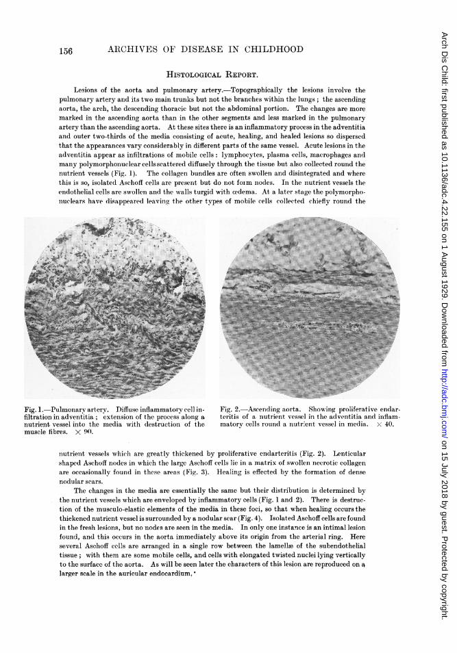
Lesions of the aorta and pulmonary artery.-Topographically the lesions involve thepulmonary artery and its two main trunks but not the branches within the lungs; the ascendingaorta, the arch, the descending thoracic but not the abdominal portion. The changes are moremarked in the ascending aorta than in the other segments and less marked in the pulmonaryartery than the ascending aorta. At these sites there is an inflammatory process in the adventitiaand outer two-thirds of the media consisting of acute, healing, and healed lesions so dispersedthat the appearances vary considerably in different parts of the same vessel. Acute lesions in theadventitia appear as infiltrations of mobile cells: lymphocytes, plasma cells, macrophages andmany polymorphonuclear cells scattered diffusely through the tissue but also collected round thenutrient vessels (Fig. 1). The collagen bundles are often swollen and disintegrated and wherethis is so, isolated Aschoff cells are present but do not foim nodes. In the nutrient vessels theendothelial cells are swollen and the walls turgid with cedema. At a later stage the polymorpho-nuclears have disappeared leaving the other types of mobile cells collected chiefly round the
Z.:0,~ ~
_ l 1 _X sOz!11A_-X-r.. a*I.-. r..el.
Fig. 1. Pulmonarv artery. Diffuse inflammatory cell in- Fig. 2.-Aseendinog aorta. Showin(g proliferative endar-filtration in adventitia; extension of the process along a teritis of a nutrient vessel in the adventitia and inflam-nutrient vessel into the media with destruction of the matorv cells round a nutrient vessel in media. x 40.muscle fibres. X 90.
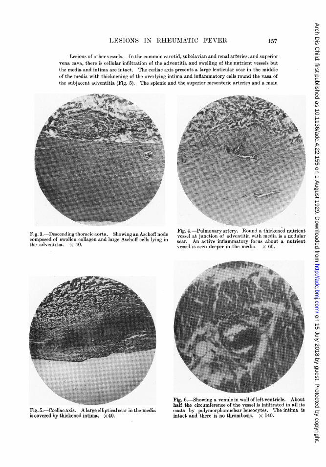
nutrient vessels which are greatly thickened by proliferative endarteritis (Fig. 2). Lenticularshaped Aschoff nodes in which the large Asehoff cells lie in a matrix of swollen necrotic collagenare occasionally found in these areas (Fig. 3). Healing is effected by the formation of densenodular scars.
The changes in the media are essentially the same but their distribution is determined bythe nutrient vessels which are enveloped by inflammatory cells (Fig. 1 and 2). There is destruc-tion of the musculo-elastic elements of the media in these foci, so that when healing occurs thethickened nutrient vessel is surrounded by a nodular scar (Fig. 4). Isolated Aschoff cells are foundin the fresh lesions, but no nodes are seen in the media. In only one instance is an intimal lesionfound, and this occurs in the aorta immediately above its origin from the arterial ring. Hereseveral Aschoff cells are arranged in a single row between the lamellae of the subendothelialtissue; with them are some mobile cells, and cells with elongated twisted nuclei lying verticallyto the surface of the aorta. As will be seen later the characters of this lesion are reproduced on alarger scale in the auricular endocardium.
on 15 July 2018 by guest. Protected by copyright.
http://adc.bmj.com
/A
rch Dis C
hild: first published as 10.1136/adc.4.22.155 on 1 August 1929. D
ownloaded from
LESIONS IN RtHEUMATIC FEVER
Lesions of other vessels. In the common carotid, subelavian and renal arteries, and superiorvena cava, there is cellular infiltration of the adventitia and swelling of the nutrient vessels butthe media and intima are intact. The celiac axis presents a large lenticular scar in the middleof the media with thicknening of the overlying intima and inflammatory cells round the vasa ofthe subjacent adventitia (Fig. 5). The splenic and the superior mesenteric arteries and a main
F 3 ch node Fig. 4.-Pulmonary arterv. Round a thickened nutrientFig.o3. Descendngthoracc aorta. S howing aneAslhoffng vessel at junction of adventitiawith media is a nodularcomposed of swollen collagen and large Aschoff cells lying in scar. An active inflammatory focus about a nutrient
vessel is seen deeper in the media. x 60.
Fig. 5.-Coeliac axis. A large elliptical scar in the mediais covered by thickened intima. x 40.
Fig. 6.-Showing a venule in wall of left ventricle. Abouthalf the circumference of the vessel is infiltrated in all itscoats by polymorphonuclear leucocytes. The intima isintact and there is no thrombosis. x 140.
157
on 15 July 2018 by guest. Protected by copyright.
http://adc.bmj.com
/A
rch Dis C
hild: first published as 10.1136/adc.4.22.155 on 1 August 1929. D
ownloaded from
158 ARCHIVES OF DISEASE IN CHILtAHOOD
pulmonary vein are quite normal and no vascular or other specific lesions are found in the liver,spleen, kidneys, lungs, uterus, ovary, oviduct, pancreas, thyroid, adrenal, urinary bladder,bone-marrow and rib.
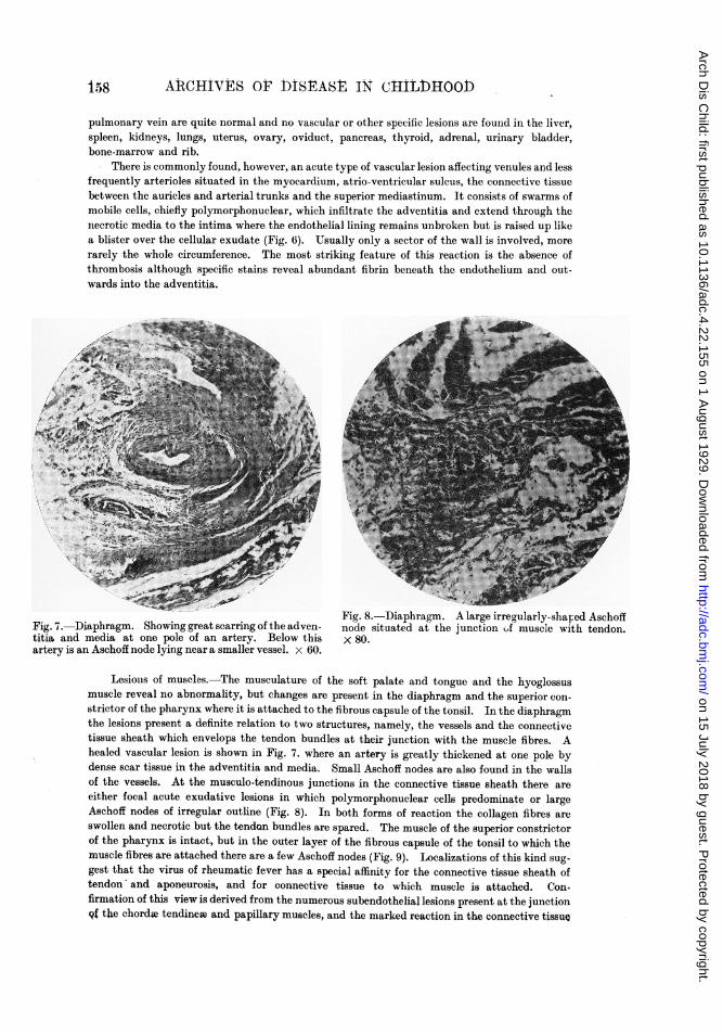
There is commonly found, however, an acute type of vascular lesion affecting venules and lessfrequently arterioles situated in the myocardium, atrio-ventricular sulcus, the connective tissuebetween the auricles and arterial trunks and the superior mediastinum. It consists of swarms ofmobile cells, chiefly polymorphonuclear, which infiltrate the adventitia and extend through thenecrotic media to the intima where the endothelial lining remains unbroken but is raised up likea blister over the cellular exudate (Fig. 6). Usually only a sector of the wall is involved, morerarely the whole circumference. The most striking feature of this reaction is the absence ofthrombosis although specific stains reveal abundant fibrin beneath the endothelium and out-wards into the adventitia.
Fig. 8. Diaphragm. A large irregularly-sharliaphragm. Showing great scarring of the adven- node situated at the junction of muscle wiitmedia at one pole of an artery. Below this x 80.n Aschoff node lying near a smaller vessel. x 60.
Lesions of muscles.-The musculature of the soft palate and tongue and the hyoglossusmuscle reveal no abnormality, but changes are present in the diaphragm and the superior con-strictor of the pharynx where it is attached to the fibrous capsule of the tonsil. In the diaphragmthe lesions present a definite relation to two structures, namely, the vessels and the connectivetissue sheath which envelops the tendon bundles at their junction with the muscle fibres. Ahealed vascular lesion is shown in Fig. 7. where an artery is greatly thickened at one pole bydense scar tissue in the adventitia and media. Small Aschoff nodes are also found in the wallsof the vessels. At the musculo-tendinous junctions in the connective tissue sheath there areeither focal acute exudative lesions in which polymorphonuclear cells predominate or largeAschoff nodes of irregular outline (Fig. 8). In both forms of reaction the collagen fibres areswollen and necrotic but the tendon bundles are spared. The muscle of the superior constrictorof the pharynx is intact, but in the outer layer of the fibrous capsule of the tonsil to which themuscle fibres are attached there are a few Aschoff nodes (Fig. 9). Localizations of this kind sug-gest that the virus of rheumatic fever has a special affinity for the connective tissue sheath oftendon' and aponeurosis, and for connective tissue to which muscle is attached. Con.firmation of this view is derived from the numerous subendotheial lesions present at the junction9i the chordae tendineae and papillary muscles, and the marked reaction in the connective tissue
ed AschoffIi tendon.Fig. 7. D
titia andartery is a]
on 15 July 2018 by guest. Protected by copyright.
http://adc.bmj.com
/A
rch Dis C
hild: first published as 10.1136/adc.4.22.155 on 1 August 1929. D
ownloaded from
LESIONS IN RHEUMATIC FEVER
sheath of the aponeurosis beneath the subcutaneous nodules of the elbow. In these parts too,
the tendon and aponeurosis escape the injury which is located in the connective tissue envelop
alone. Similarly the lesions at the insertion of the pharyngeal muscle may be compared as
regards position to the annulus fibrosus of the heart which is greatly thickened by scar tissue,
diffuse inflammatory infiltration and Aschoff nodes, while at the junction of the auricular andventricular musculature with this structure Aschoff nodes are in evidence. Further, it
seems probable that a topographical survey of the subcutaneous nodules in cases of rheumaticfever would show that their distribution is largely determined by this peculiarity. Of course,
the specific nodes are not confined to the vicinity of tendon or aponeurosis, but wherever they occurthere is always connective tissue. Indeed, the matrix of the node is always necrotic collagenwhich seems to constitute the initial and fundamental injury produced by the rheumatic virus,
the cellular reactions being seconidary. A study of numerous nodes in this case with specific
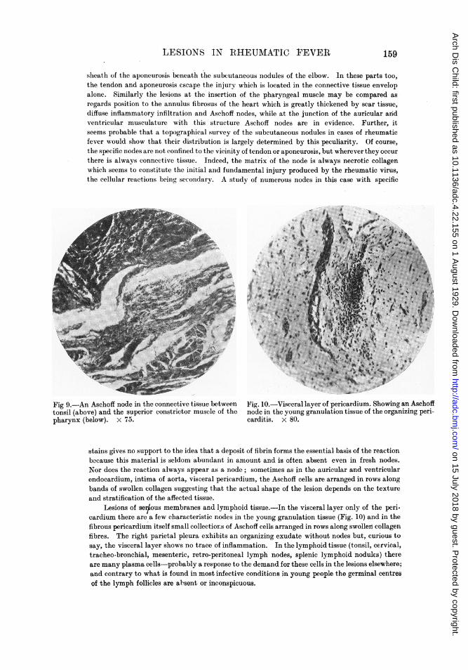
Fig 9.-An Aschoff node in the connective tissue between Fig. 10.-Visceral layer of pericardium. Showing an Aschofftonsil (above) and the superior constrictor muscle of the node in the young granulation tissue of the organizing peri-pharynx (below). X 75. carditis. x 80.
stains gives no support to the idea that a deposit of fibrin forms the essential basis of the reactionbecause this material is seldom abundant in amount and is often absent even in fresh nodes.
Nor does the reaction always appear as a node; sometimes as in the auricular and ventricularendocardium, intima of aorta, visceral pericardium, the Aschoff cells are arranged in rows alongbands of swollen collagen suggesting that the actual shape of the lesion depends on the texture
and stratification of the affected tissue.Lesions of ser/ous membranes and lymphoid tissue.-In the visceral layer only of the peri-
cardium there are a few characteristic nodes in the young granulation tissue (Fig. 10) and in the
fibrous pericardium itself small collectiorns of Aschoff cells arranged in rows along swollerl collagenfibres. The right parietal pleura exhibits an organizing exudate without nodes but, curious to
say, the visceral layer shows no trace of inflammation. In the lymphoid tissue (tonsil, cervical,tracheo-bronchial, mesenteric, retro-peritoneal lymph nodes, splenic lymphoid nodules) there
are many plasma cells-probably a response to the demand for these cells in the lesions elsewhere;and contrary to what is found in most infective conditions in young people the germinal centres
of the lymph follicles are al-sent or inconspicuous.
159
on 15 July 2018 by guest. Protected by copyright.
http://adc.bmj.com
/A
rch Dis C
hild: first published as 10.1136/adc.4.22.155 on 1 August 1929. D
ownloaded from
ARCHIVES OF DISEASE IN CHILDHOOD
Brain.-No adequate anatomical basis for the chorea is found in the meninges, cerebralcortex, caudate nucleus, corpus striatum, optic thalamus, mesencephalon and pons. All theseshow hyperaemia but no vascular or inflammatory lesions.
Certain cardiac lesions.-Nodes are sown thickly in the walls of both ventricles, thosebeneath the endocardium being covered by unbroken endothelium without thrombosis on thesurface. In the atrio-ventricular sulcus are many lesions in various stages of evolution: nodeswhere the sulcus adipose tissue joins the annulus fibrosus; fresh and resolving nodes in and aboutthe coronary sinus and the right and left coronary arteries. Some sections show that healinghas occurred in the circumflex branches of the coronary arteries so that the intima is greatlythickened and the media deeply scarred, the appearances being like those in arterio-sclerosis.Similar changes are present in the connective tissue filling the space between the auricles, aortaand pulmonary artery. On comparing the two sides of the heart it is seen that the sulcus lesionsare symmetrically arranged, and the same phenomenon comes out in other situations, namely,
~~~~~~~~~~~~~~ .... ...... ..:...
7~~~~~~~~~~~~~~~~~~~~~~~~~~~~~~~~~~~~7
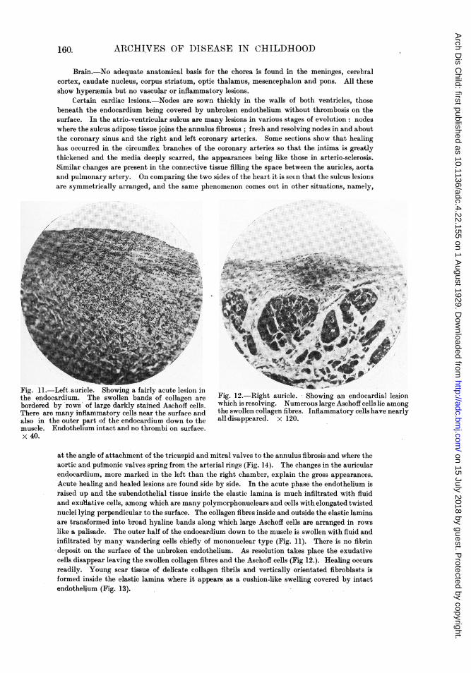
-Left auricle. Showing a fairly acute lesion inocardium. The swollen bands of collagen are Fig. 12.-Right auricle. Showing an endocardby rows of large darkly stained Aschoff cells which is resolving. Numerous large Aschoff cells
re many inflammatory cells near the surface and the swollen collagen fibres. Inflammatory cellshathe outer part of the endocardium down to the alldisappeared. x 120.Endothelium intact and no thrombi on surface.
at the angle of attachment of the tricuspid and mitral valves to the annulus fibrosis and where theaortic and pulmonic valves spring from the arterial rings (Fig. 14). The changes in the auricularendocardium, more marked in the left than the right chamker, explain the gross appearances.Acute healing and healed lesions are found side by side. In the acute phase the endothelium israised up and the subendothelial tissue inside the elastic lamina is much infiltrated with fluidand exu'dative cells, among which are many polymorphonuclears and cells with elongated twistednuclei lying perpendicular to the surface. The collagen fibres inside and outside the elastic laminaare transformed into broad hyaline bands along which large Aschoff cells are arranged in rowslike a palisade. The outer half of the endocardium down to the muscle is swollen with fluid andinfiltrated by many wandering cells chiefly of mononuclear type (Fig. 11). There is no fibrindeposit on the surface of the unbroken endothelium. As resolution takes place the exudativecells disappear leaving the swollen collagen fibres and the Aschoff cells (Fig 12.). Healing occursreadily. Young scar tissue of delicate collagen fibrils and vertically orientated fibroblasts isformed inside the elastic lamina where it appears as a cushion-like swelling covered by intactendothelium (Fig. 13).
dial lesionilie amongave nearly
Fig. I11.-the end(borderedThere aralso inmuscle.x 40.
160.
on 15 July 2018 by guest. Protected by copyright.
http://adc.bmj.com
/A
rch Dis C
hild: first published as 10.1136/adc.4.22.155 on 1 August 1929. D
ownloaded from
L,ESIONS IN RIHEUMATA'1'IC FEVER 1
THE MECHANISM OF RHEUMATIC EN DOCARDITIS.
Koster (1878) appears to have been the first to suggest that enidocarditiswas embolic in origin rather than due to the implantation of bacteria on thesurface of the valve from the passing blood. For many years his theory metwith opposition owing to uncertainty regarding the presence of blood vesselsin the inormal valves of the adult. However in 1917 Baynie-Jones3 was able todemoiistrate by injection methods a vasculature in the valves of nornmal hearts.He found( that the mitral and( tricuspid valves receive arterioles frolm thecorotnary arteries as they pass through the annuluis fibrosus. These arteriolespenetrate the base of the leaflet an(d pass, dowxnuards givino off snall lateralbrainches which rtmify in the upper third. Wheni they reach the liine of closure
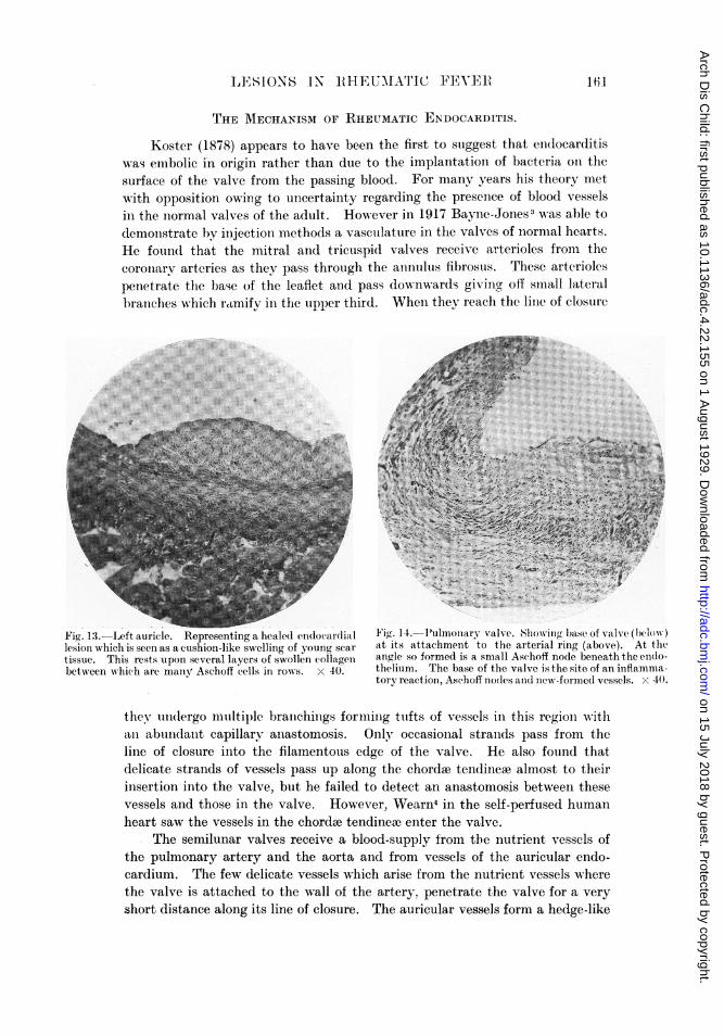
Figy. 13.-Left auricle. Representing a healed endocardiall Fi 11. 1-ulmonary valve. Showing base of valve (below)lesion which is seen as a cushion-like swelling of young scar at its attachment to the arterial ring (above). At thetissue. This rests -upon several layers of swollen collageni anigle so formed is a small Aschoff node beneath the endo-between wi-hich aIr maniy Aschoff cells in rows. x 40. thelium. The base of the valve is the site of an inflamma-
tory reaction, Aschoff nodles anid new-formied vessels. x 40.
they und(lergo multiple branichiiigs foriniiig ttufts of vessels in this regionl withlaii abund(lanit capillary ainastomosis. Only occasional stranlds pass from theline of closure into the filamentous edge of the valve. He also found thatdelicate strands of vessels pass up along the chordae tendineae almost to theirinsertion into the valve, but he failed to detect an anastomosis between thesevessels and those in the valve. However, Wearn4 in the self-perfused humanheart saw the vessels in the chordoe tendineaw enter the valve.
The semilunar valves receive a blood-supply from the nutrient vessels ofthe pulmonary artery and the aorta and from vessels of the auricular endo-cardium. The few delicate vessels which arise from the nutrient vessels wherethe valve is attached to the wall of the artery. penetrate the valve for a veryshort distance along its line of closure. The auricular vessels form a hedge-like
16.1
on 15 July 2018 by guest. Protected by copyright.
http://adc.bmj.com
/A
rch Dis C
hild: first published as 10.1136/adc.4.22.155 on 1 August 1929. D
ownloaded from
6AhCHIVES O( 1)ISEASE IN eIIiLDiHOOD
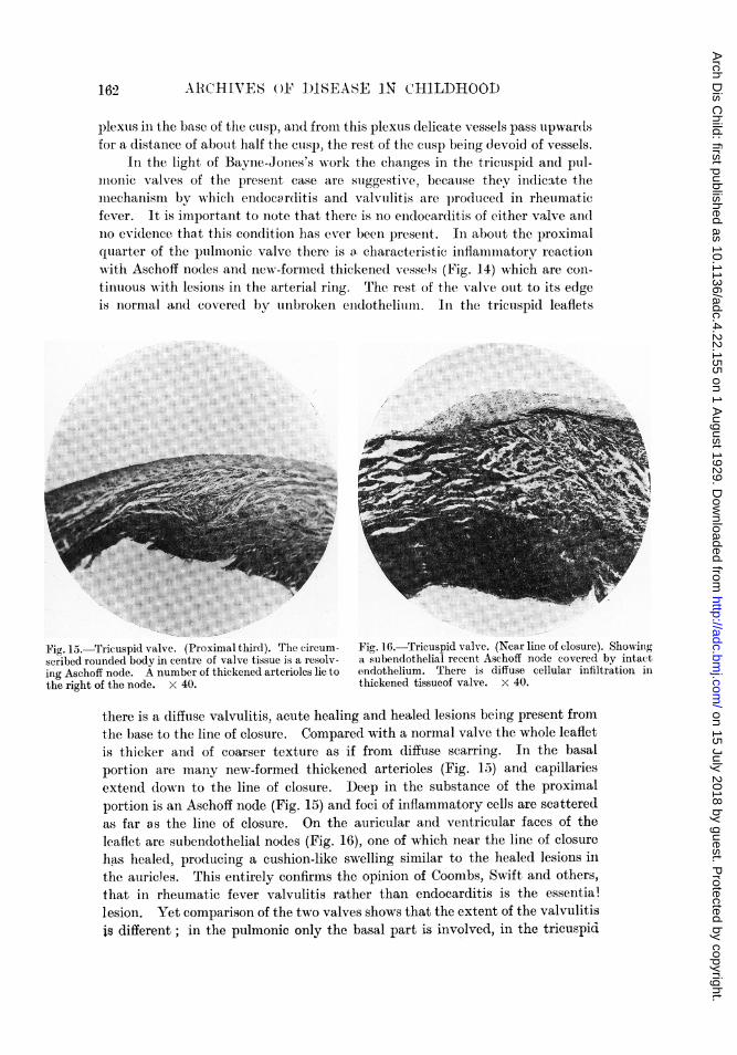
Fig. 15.-scribed rc
ing Aschcthe right
plexuis in the base of the Culsp, andI fromi this plexus delicate vessels pass upwardsfor a distance of abouit half the cusp, the rest of the ctusp being devoid of vessels.
In the light of Bayne-Jones's work the cha.nges in the tricuispid and pu1l-illonie valves of the present case are suggestivre, becauise they indicate them:iechanism by wNhich en(docarditis and valvulitis are produced in rheumaticfever. It is importanit to niote that there is no enidocarditis of either valve alndno evideence that this condition lias ever been preseint. In abotut the proximalqutarter of the pulmonic valve there is a characteristic infla:mi atory reactionxx ith Aschoff no(les anid new-formiied thickened vessels (Fig. 14) which are coni-itnuous with lesions in the arterial ring. The rest of the valve out to its edgeis niormal and covered by unbroken endotheliumil. In the tricuspid leaflets
.. ..: : .:.::. ... .:
..........:.
Tricuspid valve. (Proximal third). The:circum- Fig.16.Tricuspidvalve. (Near line of closure). Showing)unded body in centre of valve tissue is a resolv- a subendothelial recent Aschoff node covered by intact)ff node. A number of thickened arterioles lie to endothelium. There is diffuse cellular infiltration inof the node. x 40. thickened tissucof valve. x 40.
there is a diffuise valvulitis. acute healing and healed lesions being present fromthe base to the line of closure. Compared with a normal valve the whole leafletis thicker and of coarser texture as if from (diffuse scarring, In the basalportion are many new-formed thickened arterioles (Fig. 15-) and capillariesextend (lown to the line of closure. Deep in the substance of the proximalportion is an Aschoff node (Fig. 15) and foci of inflammatory celJs are scatteredas far as the line of closure. On the auricular and ventricular faces of theleaflet are subendothelial nodes (Fig. 16), one of which near the line of closureha~s hiealed, producing a cushion-like swelling similar to the healed lesions inthe auricles. This entirely confirmns the opinion of Coombs, Swift and others,that in rheumatic fever valvulitis rather than endocarditis is the essential,lesion. Yet comparison of the two valves shows that the extent of the valvulitisis different ; in the pulmonic only the basal part is involved, in the tricuspid
1.62
on 15 July 2018 by guest. Protected by copyright.
http://adc.bmj.com
/A
rch Dis C
hild: first published as 10.1136/adc.4.22.155 on 1 August 1929. D
ownloaded from
LESIONS IN RHEUAIATIC PEVt:I 163
the whole length. This difference is governed, we believe, by the degree ofvascularity. In the tricuspid, diffuse permeation with the virus occurs readilyowing to the good blood-supply, and the opportunities for re-infections areincreased by the healing of the inflammatory process which leads to the forma-tion of new vessels from the pre-existing ones. In the pulmonic valve theinitiaJ infection only injures the basal sector, and since the spread of inflamma-tion into the distal non-vascular area takes place by continuity its progressmust be relatively slow.
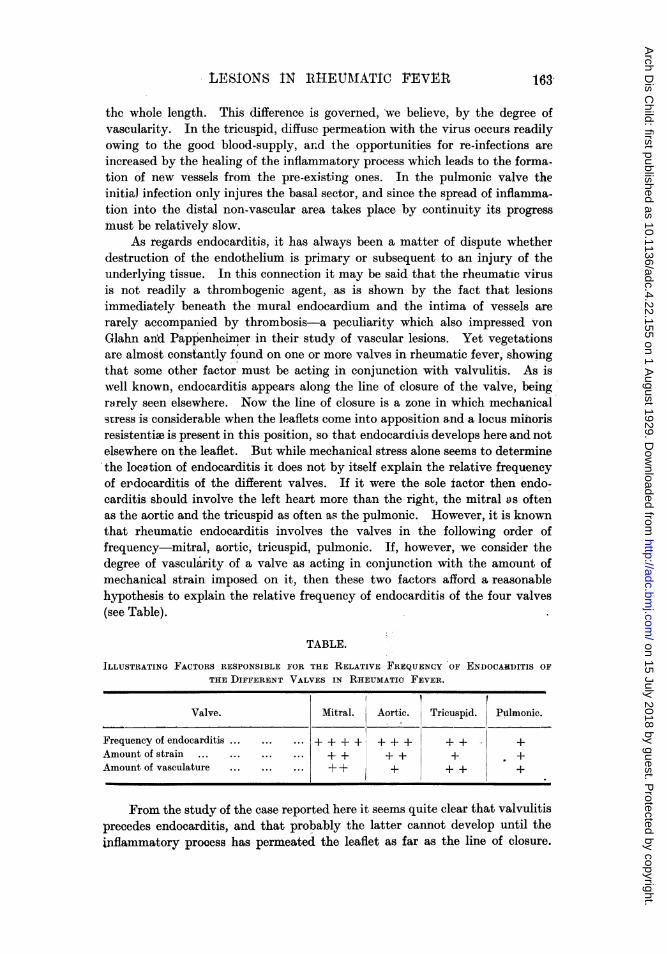
As regards endocarditis, it has always been a matter of dispute whetherdestruction of the endothelium is primary or subsequent to an injury of theunderlying tissue. In this connection it may be said that the rheumatic virusis not readily a thrombogenic agent, as is shown by the fact that lesionsimmediately beneath the mural endocardium and the intima of vessels arerarely accompanied by thrombosis-a peculiarity which also impressed vonGlahn aind Pappenheimer in their study of vascular lesions. Yet vegetationsare almost constantly found on one or more valves in rheumatic fever, showingthat some other factor must be acting in conjunction with valvulitis. As iswell known, endocarditis appears along the line of closure of the valve, being'rarely seen elsewhere. Now the line of closure is a zone in which mechanicalstress is considerable when the leaflets come into apposition and a locus mihorisresistentiae is present in this position, so that endocarciitis develops here and notelsewhere on the leaflet. But while mechanical stress alone seems to deteri=nethe location of endocarditis it does not by itself explain the relative frequiencyof endocarditis of the different valves. If it were the sole factor then endo-carditis sbould involve the left heart more than the right, the mitral as oftenas the aortic and the tricuspid as often as the pulmonic. However, it is knownthat rheumatic endocarditis involves the valves in the following order offrequency-mitral, aortic, tricuspid, pulmonic. If, however, we consider thedegree of vascularity of a valve as acting in conjunction with the amount ofmechanical strain imposed on it, then these two factors afford a reasonablehypothesis to explain the relative frequency of endocarditis of the four valves(see Table).
TABLE.
ILLUSTRATING FACTORS RESPONSIBLE FOR THE RELATIVE FREQUENCY OF ENDOCAlIDITIS OFTHE DIFFERENT VALVES IN RHEUMATIC FEVER.
Valve. Mitral. Aortic. Tricuspid. Pilimonic.
Frequenc1of endocarditis ... ... +++ + ++ + + + +Amount of straia . ..+ ... ... 4. + + + + . +Amount of vasculature ... ... ... ++ + + + +
From the study of the case reported here it seems quite clear that valvulitisprecedes endocarditis, and that probably the latter cannot develop until theinflammatory process has permeated the leaflet as far as the line of closure.
on 15 July 2018 by guest. Protected by copyright.
http://adc.bmj.com
/A
rch Dis C
hild: first published as 10.1136/adc.4.22.155 on 1 August 1929. D
ownloaded from
164 ARCGHIVES OF DISEASE IN CHILDHOOD
That the valvulitis can be of considerable extent and severity before endo-carditis oceurs is shown by the tricuspid valve, in which the anatomical- condi-tions indicate repeated infections of the valvular tissue without the productionof endocarditis before death.
It would be interesting to know how often valvulitis as distinct fromendocarditis occurs in rheumatic fever, because it is evident that valves withoutendocarditis and otherwise normal on naked eye inspection cannot be acquittedof extensive inflammation until examined by the microscope.
My thanks are due to the Newcastle-upon-Tyne and Northern CountiesMedical Society for a contributory grant from its Research Fund in aid ofexpenses incurred in this investigation.
REFERENCES.
1. Alac(Calluin, Bull, Johns Hopkins Hosp, Baltimore, 1924, XXXV, 329.2. Pappenheimer and von Glahn, J. Med. Res., 1924, XLIV, 489. Am. J. Path., Boston, 1926.
ii, 1 and 15 and 235. Ibid., 1927, XVIII, 585.3. Bayne-Jones, An. J. Anat., Philad., 1917, XXI, 449.4. Wearn, J. Exp. Med., N.Y., 1928, XLVII, 273.
on 15 July 2018 by guest. Protected by copyright.
http://adc.bmj.com
/A
rch Dis C
hild: first published as 10.1136/adc.4.22.155 on 1 August 1929. D
ownloaded from