Embed Size (px)
Citation preview
鳥取大学研究成果リポジトリTottori University research result repository
タイトルTitle
Self-assembled Artificial Viral Capsid Decorated withGold Nanoparticles
著者Auther(s)
Matsuura, Kazunori; Ueno, Genki; Fujita, Seiya
掲載誌・巻号・ページCitation
Polymer journal , 47 (2) : 146 - 151
刊行日Issue Date
2015
資源タイプResource Type
学術雑誌論文 / Journal Article
版区分Resource Version
著者版 / Author
権利Rights
注があるものを除き、この著作物は日本国著作権法により保護されています。 / This work is protected underJapanese Copyright Law unless otherwise noted.
DOI 10.1038/pj.2014.99
URL http://repository.lib.tottori-u.ac.jp/5724
Polymer J.
Self-assembled Artificial Viral Capsid Decorated with Gold
Nanoparticles
Kazunori Matsuura,1* Genki Ueno,1 Seiya Fujita1
1 Department of Chemistry and Biotechnology, Graduate School of Engineering, Tottori
University, Koyama-Minami 4-101, Tottori 680-8552, Japan
Corresponding Author, Fax: +81-857-31-6729; Tel: +81-857-31-5262;
E-mail: [email protected]
Running Head: Gold Nanoparticle-decorated Artificial Viral Capsid
Keywords: Self-assembly / Peptide / Spherical virus / Gold nanoparticles / Decoration
Polymer J.
Abstract
The decoration of a peptide-based artificial viral capsid with gold nanoparticles
(AuNPs) is reported. β-Annulus GGGCG-bearing peptide as a binding site of AuNPs
self-assembled into nanocapsules with a diameter of 50 nm. The addition of AuNPs to
the peptide nanocapsules afforded relatively uncontrolled assemblies of AuNPs. In
contrast, the self-assembly of AuNP−peptide conjugates afforded, after dialysis,
controlled assemblies of AuNPs with sizes of 30−60 nm. ζ-Potential measurements
revealed that the surface of the artificial viral capsid self-assembled from β-annulus
peptide was coated with AuNPs.
INTRODUCTION
Fusion materials of gold nanoparticles (AuNPs) and biomacromolecules have
attracted much attention because these materials provide versatile tools for use in
applications such as catalysis, biosensing, and bioimaging systems.1-4 AuNPs have
useful electronic, optical, and plasmonic properties, which depend on the diameter of
the nanoparticles and the structure of the assemblies. To date, the spatial arrangement of
AuNP assemblies has been achieved via the programmed self-assembly of
biomacromolecules such as DNA.5-13 For example, Shultz et al. demonstrated that
Polymer J.
oligodeoxyribonucleotide (ODN) conjugates with tethered AuNPs could be arranged by
hybridization with the complementary ODN templates.8,9 Seeman et al. reported that a
two-dimensional arrangement of AuNPs with nanosized precision was achieved by the
self-assembly of double-crossover DNAs.10,11
Natural viral capsids are also attractive biomolecular nanomaterials. Because viral
capsids are protein assemblies with a discrete size, space, and a specific aggregation
number, they have been used as nanoreactors and nanocontainers for inorganic materials,
drugs, and proteins.14-17 The surface of viral capsids have been utilized as a scaffold to
display biomolecular ligands such as saccharides, DNA aptamers, and antigens.14-17 The
regular arrangement of AuNPs on the surface of spherical viral capsids have also been
reported.18-23 Finn et al. demonstrated that monomaleimide-AuNPs were displayed on
the surface of Cys-mutated cowpea mosaic virus (CPMV) with a diameter of 30 nm.18
Blum et al. reported that 5 nm AuNPs were regularly arranged on GGCGG loops
inserted into subunit proteins of CPMV.19-21 Niikura et al. reported the arrangement of
sialic-acid-modified AuNPs on JC viral capsids through molecular recognition.22
The rational design of self-assembled peptides has been progressively developed for
the construction of peptide-based nano-architectures.24-28 The linear arrangement of
AuNPs on fibrous assemblies of peptides has also been reported.29-31 However, the
Polymer J.
literature contains no reports on the spherical arrangement of AuNPs using a rationally
designed peptide assembly as a template. Recently, we demonstrated that the designed
24-mer peptide fragment (INHVGGTGGAIMAPVAVTRQLVGS), which participates in
the β-annulus motif in tomato bushy stunt virus (TBSV), self-assembles into virus-like
nanocapsules with sizes of 30−50 nm.32-35 Herein, we report the construction of an
artificial viral capsid decorated with AuNPs, which we examined using the following
two methods: (1) modification of the surface of peptide nanocapsules self-assembled
from β-annulus-GGGCG peptide 1 (INHVGGTGGAIMAPVAVTRQLVGSGGGCG)
with AuNPs and (2) self-assembly of AuNP-β-annulus-GGGCG peptide 1 conjugates
(Figure 1).
MATERIALS and METHODS
Reagents were obtained from commercial sources and used without further
purification. A stabilized suspension of AuNPs in citrate buffer (5 nm diameter, 5.5 ×
1013 particles/mL) was purchased from SIGMA-ALDRICH. Reversed-phase HPLC was
performed at ambient temperature on a Shimadzu LC-6AD liquid chromatograph
equipped with a UV–vis detector (220 nm, Shimadzu SPD-20A) and GL Science
Inertsil WP300 C18 (4.6 × 250 mm2 and 20 × 250 mm2) columns. MALDI-TOF mass
Polymer J.
spectra were obtained on an Autoflex II (Bruker Daltonics) spectrometer operated under
the linear/positive mode with α-cyano-4-hydroxy cinnamic acid (α-CHCA) as the
matrix. UV−vis spectra were recorded at 25°C using a JASCO V-630 spectrophotometer.
CD spectra were collected in a 1-mm quartz cell with a JASCO J-820
spectrophotometer at 25°C. Molar concentrations of AuNPs were calculated using the
absorbance at 520 nm.
Synthesis of peptide (1). The peptide H-
-Ile-Asn(Trt)-His-(Trt)-Val-Gly-Gly-Thr(tBu)-Gly-Gly-Ala-Ile-Met-Ala-Pro-Val-Ala-Va
l-Thr(tBu)-Arg(pbf)-Gln(Trt)-Leu-Val-Gly-Ser(tBu)-Gly-Gly-Gly-Cys(Trt)-Gly-Alko-P
EG resin was synthesized on a Fmoc-Gly-Alko-PEG resin (Watanabe Chemical Ind.,
Ltd., 0.22 mmol/g) using Fmoc-based coupling reactions (4 equiv. Fmoc amino acids).
Solutions of 2-(1H-benzotriazole-1-yl)-1,1,3,3-tetramethyluronium hexafluoro-
phosphate (HBTU, 4 equiv.), 1-hydroxybenzotriazole hydrate (HOBt•H2O, 4 equiv.),
and diisopropylamine (8 equiv.) in N-methylpyrrolidone (NMP) were used as coupling
reagents. A solution of 20 % piperidine in N,N-dimethylformamide (DMF) was used for
Fmoc deprotection. The progress of the coupling reaction and Fmoc deprotection was
confirmed using a TNBS and a Chloranil Test Kit (Tokyo Chemical Industry Co., Ltd.).
The peptidyl resin was washed with NMP.
Polymer J.
The peptide was deprotected and cleaved from the resin by treatment with a mixture
of trifluoroacetic acid (TFA)/water/1,2-ethanedithiol/triisopropylsilane =
9.5/0.25/0.25/0.1 at room temperature for 3 h. The reaction mixture was filtered to
remove the resin and the filtrate was concentrated under vacuum. The peptide was
precipitated by the addition of methyl tert-butyl ether (MTBE) to the residue, and the
supernatant was decanted. After the peptide was washed with MTBE three times, the
precipitated peptide was dried under vacuum. The crude product was purified by
reversed-phase HPLC (Inertsil WP300 C18), eluting with a linear gradient of
CH3CN/water containing 0.1% TFA (26/74 to 29/71 over 120 min). The fraction
containing the desired peptide was lyophilized to give 9.5 mg of a flocculent solid (3 %
yield). MALDI-TOF-MS (matrix: α-CHCA): m/z = 2638 [M+H]+.
Modification of the surface of peptide nanocapsules with AuNPs. An aqueous
solution of β-annulus-GGGCG peptide 1 (0.2 mM, pH 4.6) was prepared by simply
dissolving it in deionized water. The formation of spherical assemblies was confirmed
by dynamic light scattering (DLS) measurements and transmission electron microscopy
(TEM). The aqueous solution of peptide 1 (37.5 µL) was mixed with AuNPs in citrate
buffer (5.5 × 10¹³ particles/mL, 30 µL) and incubated for 60 min at 25°C. An aliquot
(7.5 µL) of 20 mM thioctic acid solution in ethanol/water (4/1) was added to the
Polymer J.
mixture of peptide 1 and gold nanoparticles, and the resulting mixture was incubated for
10 min at 25°C. The final concentrations were [peptide 1] = 0.1 mM, [AuNP] = 0.2 µM,
and [thioctic acid] = 2 mM (pH of the final solution was 3.7).
Construction of artificial viral capsid decorated with AuNPs by self-assembly of
AuNP−peptide conjugates. An aqueous solution of β-annulus-GGGCG peptide 1 was
diluted with water to a concentration of 2 µM, which is less than the critical aggregation
concentration. An aliquot (2 mL) of the aqueous solution of peptide 1 (2 µM) was
mixed with a diluted AuNPs dispersion ([AuNP] = 1 µM, 2 mL) and incubated for 60
min at 25°C. An aliquot (0.5 mL) of 20 mM thioctic acid solution in ethanol/water (4/1)
was added to the mixture of peptide 1 and AuNPs, and the resulting mixture was
incubated for 10 min at 25°C. The water in the mixture was evaporated using a Smart
Evaporator® (Bio Chromato, Inc.). The residue was redispersed by the addition of water
(80 µL) to final concentrations of [peptide 1] = 50 µM and [AuNP] = 25 µM. To remove
unassembled AuNPs, peptides, and AuNP−peptide conjugates, the dispersion of
AuNP−peptide conjugates was dialyzed to equilibrium against water using Spectra/Por®
dialysis tubing (cutoff Mw = 50 kDa, Spectrum Laboratories, Inc.). Nanostructures of
the assemblies were evaluated by DLS measurements and TEM observations.
Dynamic light scattering (DLS) measurements. DLS measurements were performed
Polymer J.
using a Zetasizer NanoZS (MALVERN Instruments, Ltd.) instrument at 25 ºC with an
incident He−Ne laser (633 nm). During the measurements, the count rate (the
sample-scattering intensity) was also provided. The correlation time for the scattered
light intensity G(τ) was measured several times, and the averaged results were fitted to
equation 1:
G(τ) = B + A exp(−2q2Dτ) (1)
where B is the baseline, A is the amplitude, q is the scattering vector, τ is the delay time,
and D is the diffusion coefficient. The hydrodynamic radius (RH) of the scattering
particles was calculated using the Stokes−Einstein equation (eq. 2):
(2)
where η is the solvent viscosity, kB is Boltzmann’s constant and T denotes the absolute
temperature.
ζ-Potential measurements. We determined the ζ-potential of peptide assemblies at
pH 4.6 by measuring the electrophoretic mobility at 25 ºC in disposable Zeta cells
using a Zetasizer NanoZS (MALVERN Instruments, Ltd.).
Transmission electron microscopy (TEM). An aliquot (5 µL) of each sample
solution was applied to a carbon-coated grid (ALLANCE Biosystems), left for 60 s,
RH = kBT 6πηD

Polymer J.
and then removed. The grid was subsequently dried in vacuo. In the case of peptide
samples, a drop of 2 wt% aqueous sodium phosphotungstate was placed on each of
the grids. AuNPs were observed without the use of a stain. After the sample-loaded
carbon-coated grids were dried in vacuo, they were observed by TEM (JEOL JEM
1400 Plus) using an acceleration voltage of 80 kV.
RESULTS and DISCUSSION
29-mer β-annulus-GGGCG peptide 1 (INHVGGTGGAIMAPVAVTRQLVGS
GGGCG) was synthesized using the Fmoc-protected solid-phase method, purified by
reversed-phase HPLC, and confirmed by MALDI-TOF-MS (m/z = 2637 [M]+). The
CD spectrum of the aqueous solution of peptide 1 showed a negative peak at 202 nm
and a negative shoulder at approximately 220–230 nm (Figure 2a), which is similar
to the CD spectrum of a 24-mer β-annulus peptide reported previously,32 indicating
the coexistence of random-coil, β-sheet, and turn structures. DLS measurement of
the aqueous solution of peptide 1 showed that peptide 1 formed assemblies with a
hydrodynamic diameter of 51 ± 15 nm (Figure 2b). TEM observation of the
assemblies stained with sodium phosphotungstate also showed the formation of
spherical structures with a diameter of approximately 50 nm (Figure 2c), which is
Polymer J.
comparable to the size (30–50 nm) of nanocapsules self-assembled from 24-mer
β-annulus peptide.32 The concentration dependence of peptide 1 on the scattering
intensity (DLS count rate) revealed that the critical aggregation concentration (CAC)
at 25°C is 29 µM, which is comparable to the CAC (25 µM) of 24-mer β-annulus
peptide under the same conditions.32 These results indicate that the addition of
GGGCG to the terminus of the β-annulus peptide minimally affected the size,
morphology, and stability of the spherical peptide assemblies.
To construct artificial viral capsids decorated with AuNPs, an aqueous dispersion
of AuNPs was added to the aqueous dispersion of nanocapsules self-assembled from
β-annulus-GGGCG peptide 1, and then the surface of the AuNPs was subsequently
protected with thioctic acid to prevent further aggregation (Figure 1a). When a
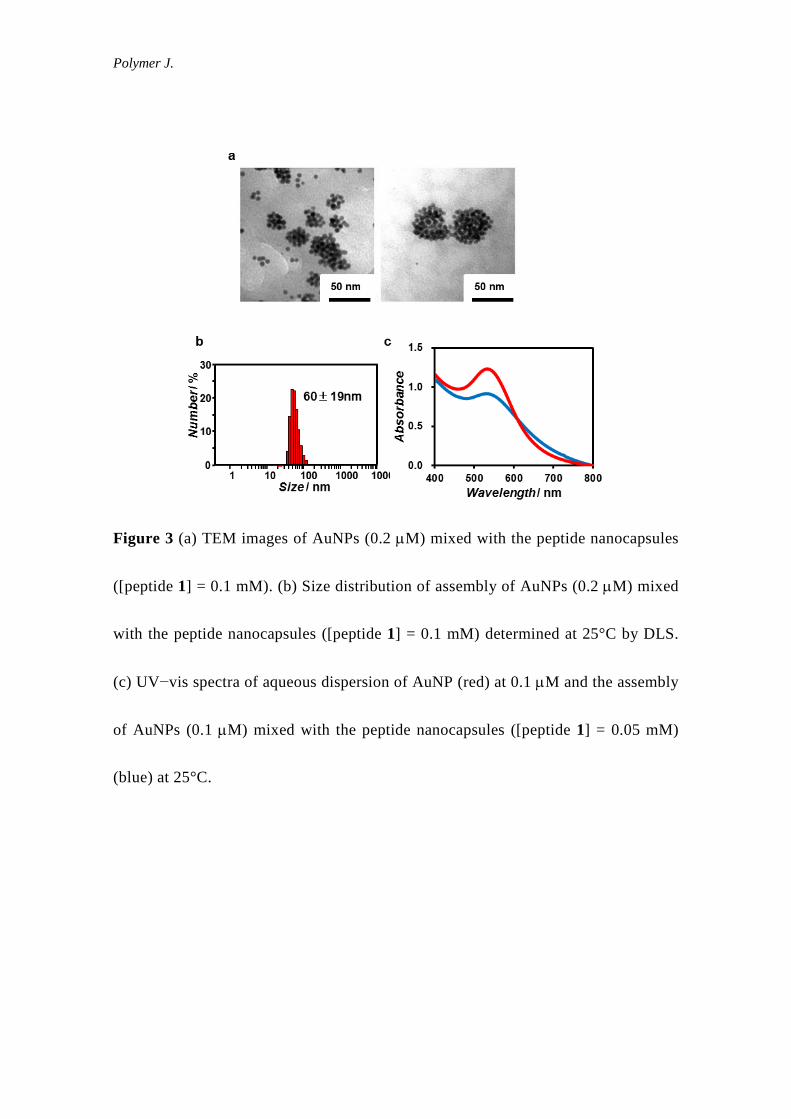
suspension of 0.2 µM AuNPs was mixed with 0.1 mM peptide 1, the TEM
micrographs of the product showed the formation of spherical assemblies with a
diameter of 10−50 nm and which comprised 2−40 AuNPs with a diameter of 5 nm
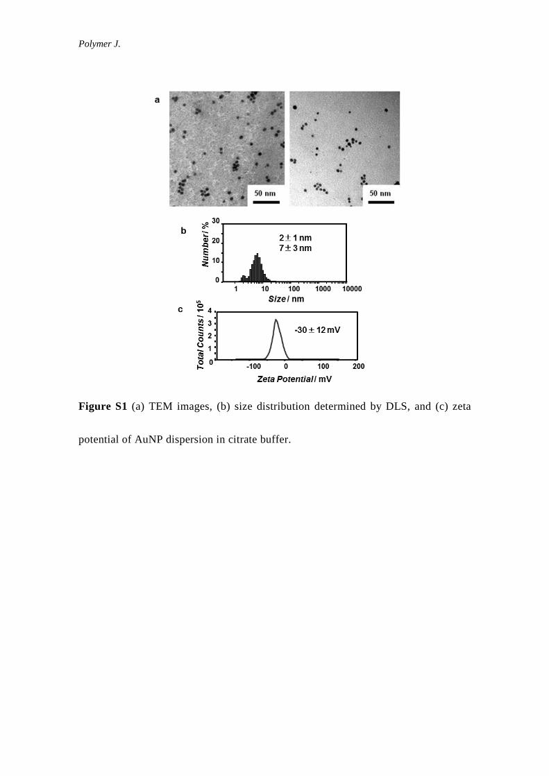
(Figure 3a). In contrast, the TEM micrographs of AuNPs in the absence of peptide 1
showed only individually-dispersed AuNPs (Figure S1). The DLS of the mixture of
AuNPs and peptide 1 also showed the formation of assemblies with a hydrodynamic
diameter of 60 ± 19 nm (Figure 3b). The UV−vis spectrum of AuNPs in the
Polymer J.
presence of the peptide nanocapsule was slightly red-shifted (Figure 3c), which is
ascribed to plasmon coupling among AuNPs self-assembled on the peptide
nanocapsule. Notably, assemblies of AuNPs were observed, although the
concentration of AuNPs was remarkably smaller than that of peptide 1. Niikura et al.
reported the cooperative binding of sialic-acid-modified AuNPs on JC viral capsid.22
In the present study, the AuNPs apparently cooperatively adsorbed onto Cys residues
at the surface of the peptide nanocapsules. However, assemblies consisting of
several AuNPs were also observed in the TEM micrographs (Figure 3a); therefore,
controlling the aggregation number of AuNPs on a peptide nanocapsule is difficult
with this method.
Next, we examined the construction of artificial viral capsid decorated with
AuNPs by the self-assembly of AuNP−peptide 1 conjugates (Figure 1b). We mixed
AuNPs with a diluted solution of β-annulus-GGGCG peptide 1 at a concentration
below the CAC to prepare their conjugate and then protected the surface of the
AuNPs with thioctic acid. The solvent in the dispersion of conjugate was evaporated,
and the conjugate was subsequently redispersed in water to final concentrations of
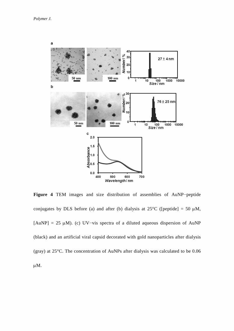
[peptide 1] = 50 µM and [AuNP] = 25 µM. The TEM images of the dispersion of
AuNP−peptide 1 conjugates showed the coexistence of AuNP assemblies with a
Polymer J.
diameter of 30−50 nm and unassembled AuNPs (Figure 4a). After the unassembled
AuNPs were removed using a dialysis membrane (cutoff Mw = 50 kDa), AuNP
assemblies with diameters of 30−60 nm were selectively observed in the TEM
images (Figure 4b). The DLS of the dialyzed dispersion of AuNP−peptide 1
conjugates also indicated the formation of assemblies with a hydrodynamic diameter
of 76 ± 25 nm (Figure 4b). The assemblies observed by TEM consisted of 22−63
AuNPs, which partially corresponds to the ideal aggregation number of 60 for a
dodecahedral peptide assembly. The increase in diameter compared with that of
unmodified peptide nanocapsules (51 ± 15 nm, Figure 2b) suggests that AuNPs
decorate the surface of the peptide nanocapsules. The UV−vis spectrum of the
assembly of AuNP−peptide 1 conjugates was approximately the same as that of
individual AuNPs (Figure 4c), which indicates that the AuNPs are separated from
one another on the peptide nanocapsules. The difference between hydrodynamic
diameters obtained from DLS before and after dialysis (Figure 4a and 4b) might be
caused by pH change of the solutions (The pH before dialysis was 3.7, but that after
dialysis was 4.6).
As previously reported,34 the pH dependence of the ζ-potential of peptide
nanocapsules self-assembled from β-annulus peptide indicates that the C-termini are
Polymer J.
directed toward the exterior of the peptide nanocapsules. Therefore, we expected that
Cys residues of β-annulus-GGGCG peptide 1 and AuNPs are directed toward the
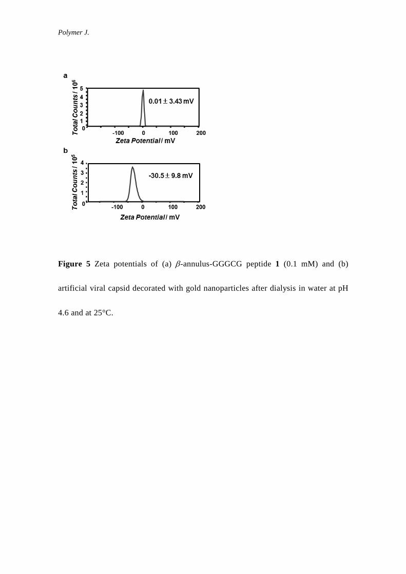
exterior of the peptide nanocapsules. The ζ-potential of unmodified peptide
nanocapsules was approximately neutral (0.01 ± 3.43 mV) at pH 4.6 (Figure 5a). In
contrast, the ζ-potential of the assemblies of AuNP−peptide 1 conjugates was −30.5
± 9.8 mV at pH 4.6 (Figure 5b), corresponding to that of AuNP dispersion in citrate
buffer (−30.0 ± 12 mV, Figure S1). These results clearly indicate that the surface of
artificial viral capsid self-assembled from β-annulus peptide was coated with
AuNPs.
CONCLUSION
We demonstrated that AuNP (5 nm)-β-annulus-GGGCG peptide self-assembled
to afford artificial viral capsids decorated with AuNPs. The AuNP-modified viral
capsids could be applied as scattering imaging capsules in cells.36 The present
strategy extends the design of artificial viral capsids to include decoration with other
functional molecules such as proteins, DNA, and fluorophores.
Polymer J.
Acknowledgements
This research was partially supported by a Grant-in-Aid for Scientific Research on
the Innovative Areas of “Fusion Materials” (No. 2206) from the Ministry of
Education, Science, Sports and Culture of Japan (MEXT) and by the Asahi Glass
Foundation.
Refferences
1 Louis C. & Pluchery, O. Gold nanoparticles for physics, chemistry and biology
(Imperial College Press, London, 2012).
2 Daniel, M.-C. & Astruc, D. Gold Nanoparticles: Assembly, supramolecular
chemistry, quantum-size-related properties, and applications toward biology,
catalysis, and nanotechnology. Chem. Rev. 104, 293–346 (2004).
3 Sperling, R. A., Gil, P. R., Zhang, F., Zanella M., & Parak, W. J. Biological
applications of gold nanoparticles. Chem. Soc. Rev. 37, 1896–1908 (2008).
4 Saha, K., Agasti, S. S., Kim, C., Li, X., & Rotello, V. M. Gold nanoparticles in
chemical and biological sensing. Chem. Rev. 112, pp 2739–2779 (2012).
5 Gothelf, K. V. & LaBean, T. H. DNA-programmed assembly of nanostructures.
Org. Biomol. Chem. 3, 4023–4037 (2005).
Polymer J.
6 Kuzuya A. & Ohya Y. DNA nanostructures as scaffolds for metal nanoparticles.
Polymer J. 44, 452–460 (2012).
7 Mirkin, C. A., Letsinger, R. L., Mucic, R. C., & Storhoff, J. J. A DNA-based
method for rationally assembling nanoparticles into macroscopic materials.
Nature 382, 607–609 (1996).
8 Alivisatos, A. P., Johnsson, K. P., Peng, X., Wilson, T. E., Loweth, C. J.,
Bruchez, M. P., Jr., & Schultz, P. G. Organization of 'nanocrystal molecules'
using DNA. Nature 382, 609–611 (1996).
9 Loweth, C. J., Caldwell, W. B., Peng, X., Alivisatos, A. P., & Schultz, P. G.
DNA-based assembly of gold nanocrystals. Angew. Chem. Int. Ed. 38, 1808–
1812 (1999).
10 Le, J. D., Pinto, Y., Seeman, N. C., Musier-Forsyth, K., Taton, T. A. & Kiehl, R.
A. DNA-templated self-Assembly of metallic nanocomponent arrays on a
surface. Nano Lett. 4, 2343–2347 (2004).
11 Zheng, J., Constantinou, P. E., Micheel, C., Alivisatos, A. P., Kiehl, R. A., &
Seeman, N. C. Two-dimensional nanoparticle arrays show the organizational
power of robust DNA motifs. Nano Lett. 6, 1502–1504 (2006).
12 Mastroianni, A. J., Claridge, S. A., & Alivisatos, A. P. Pyramidal and chiral
Polymer J.
groupings of gold nanocrystals assembled using DNA scaffolds. J. Am. Chem.
Soc. 131, 8455–8459 (2009).
13 Tamaki, T., Miyoshi, N., Uehara, T., & Ohya, Y. Isolation of gold
nanoparticle/oligo-DNA conjugates by the number of oligo-DNAs attached and
their formation of self-assembly. Chem. Lett. 39, 1084–1085 (2010).
14 Douglas, T. & Young, M. Viruses: Making friends with old foes. Science 312,
873–875 (2006).
15 Steinmetz, N. F. & Evans, D. J. Utilisation of plant viruses in
bionanotechnology. Org. Biomol. Chem. 5, 2891–2902 (2007).
16 Bronstein, L. M. Virus-based nanoparticles with inorganic cargo: What does the
future hold? Small 7, 1609–1618 (2011).
17 Witus, L. S. & Francis, M. B. Using synthetically modified proteins to make
new materials. Acc. Chem. Res. 44, 774–783 (2011).
18 Wang, Q., Lin, T., Tang, L., Johnson, J. E., & Finn, M. G. Icosahedral virus
particles as addressable nanoscale building blocks. Angew. Chem. Int. Ed. 41,
459–462 (2002).
19 Blum, A. S., Soto, C. M., Wilson, C. D., Cole, J. D., Kim, M., Gnade, B.,
Chatterji, A., Ochoa, W. F., Lin, T. W., Johnson, J. E., & Ratna, B. R. Cowpea
Polymer J.
mosaic virus as a scaffold for 3-D patterning of gold nanoparticles. Nano Lett. 4,
867–870 (2004).
20 Blum, A. S., Soto, C. M., Wilson, C. D., Brower, T. L., Pollack, S. K., Schull, T.
L., Chatterji, A., Lin, T., Johnson, J. E., Amsinck, C., Franzon, P., Shashidhar,
R., & Ratna, B. R. An engineered virus as a scaffold for three-dimensional
self-assembly on the nanoscale. Small 1, 702–706 (2005).
21 Soto, C. M., Blum, A. S., Wilson, C. D., Lazorcik, J., Kim, M., Gnade, B. &
Ratna, B. R. Separation and recovery of intact gold-virus complex by agarose
electrophoresis and electroelution: Application to the purification of cowpea
mosaic virus and colloidal gold complex. Electrophoresis 25, 2901–2906
(2004).
22 Niikura, K., Nagakawa, K., Ohtake, N., Suzuki, T., Matsuo, Y., Sawa, H., &
Ijiro, K. Gold nanoparticle arrangement on viral particles through carbohydrate
recognition: A non-cross-linking approach to optical virus detection. Bioconj.
Chem. 20, 1848–1852 (2009).
23 Nagakawa, K., Niikura, K., Suzuki, T., Matsuo, Y., Igarashi, Y., Sawa, H., &
Ijiro, K. Virus capsid coating of gold nanoparticles via cysteine-Au interactions
and their effective cellular uptakes. Chem. Lett. 41, 113–115 (2012).
Polymer J.
24 Matsuurua, K. Rational design of self-assembled proteins and peptides for nano-
and micro-sized architectures. RSC Adv. 4, 2942–2953 (2014).
25 Ramakers, B. E. I., van Hest, J. C. M., & Lowik, D. Molecular tools for the
construction of peptide-based materials. Chem. Soc. Rev. 43, 2743–2756 (2014).
26 Boyle, A. L., Bromley, E. H. C., Bartlett, G. J., Sessions, R. B., Sharp, T. H.,
Williams, C. L., Curmi, P. M. G., Forde, N. R., Linke, H. & Woolfson D. N.
Squaring the circle in peptide assembly: From fibers to discrete nanostructures
by de novo design. J. Am. Chem. Soc., 134, 15457–15467 (2012).
27 Fletcher, J. M., Harniman, R. L., Barnes, Fr. R. H., Boyle, A. L., Collins, A.,
Mantell, J., Sharp, T. H., Antognozzi, M., Booth, P. J., Linden, N., Miles, M. J.,
Sessions, R. B., Verkade, P., & Woolfson, D. N. Self-assembling cages from
coiled-coil peptide modules. Science, 340, 595–599 (2013).
28 H. Gradišar, S. Božič, T. Doles, D. Vengust, I. Hafner-Bratkovič, A. Mertelj, B.
Webb, A. Šali, S. Klavžar and R. Jerala, Design of a single-chain polypeptide
tetrahedron assembled from coiled-coil segments. Nat. Chem. Biol., 2013, 9,
362–366.
29 Ryadnov, M. G., Ceyhan, B., Niemeyer, C. M., & Woolfson, D. N. “Belt and
braces”: A peptide-based linker system of de novo design. J. Am. Chem. Soc.
Polymer J.
125, 9388–9394 (2003).
30 Sawada, T., Takahashi, T., & Mihara, H. Affinity-based screening of peptides
recognizing assembly states of self-assembling peptide nanomaterials. J. Am.
Chem. Soc. 131, 14434–14441 (2009).
31 Nonoyama, T., Tanaka, M., Inai, Y., Higuchi, M., & Kinoshita T. Ordered
nanopattern arrangement of gold nanoparticles on β-sheet peptide templates
through nucleobase pairing. ACS Nano 5, 6174–6183 (2011).
32 Matsuura, K., Watanabe, K., Sakurai, K., Matsuzaki, T., & Kimizuka, N.
Self-assembled synthetic viral capsids from a 24-mer viral peptide fragment.
Angew. Chem. Int. Ed. 49, 9662–9665 (2010).
33 Matsuura, K. Construction of spherical virus-inspired peptide nanoassemblies.
Polymer J., 44, 469–474 (2012).
34 Matsuura, K., Watanabe, K., Matsushita, Y., Kimizuka, N. Guest-binding
behavior of peptide nanocapsules self-assembled from viral peptide fragments.
Polymer. J. 45, 529–534 (2013).
35 Fujita, S. & Matsuura, K. Inclusion of zinc oxide nanoparticles into virus-like
peptide nanocapsules self-assembled from viral β-annulus peptide. submitted to
Nanomaterials.
Polymer J.
36 Wan, X., Zheng, L., Gao, P., Yang, X., Li, C., Li, Y. F., & Huang C. Z.
Real-time light scattering tracking of gold nanoparticles-bioconjugated
respiratory syncytial virus infecting HEp-2 cells. Sci. Rep. 4, 4529 (2014).
Polymer J.
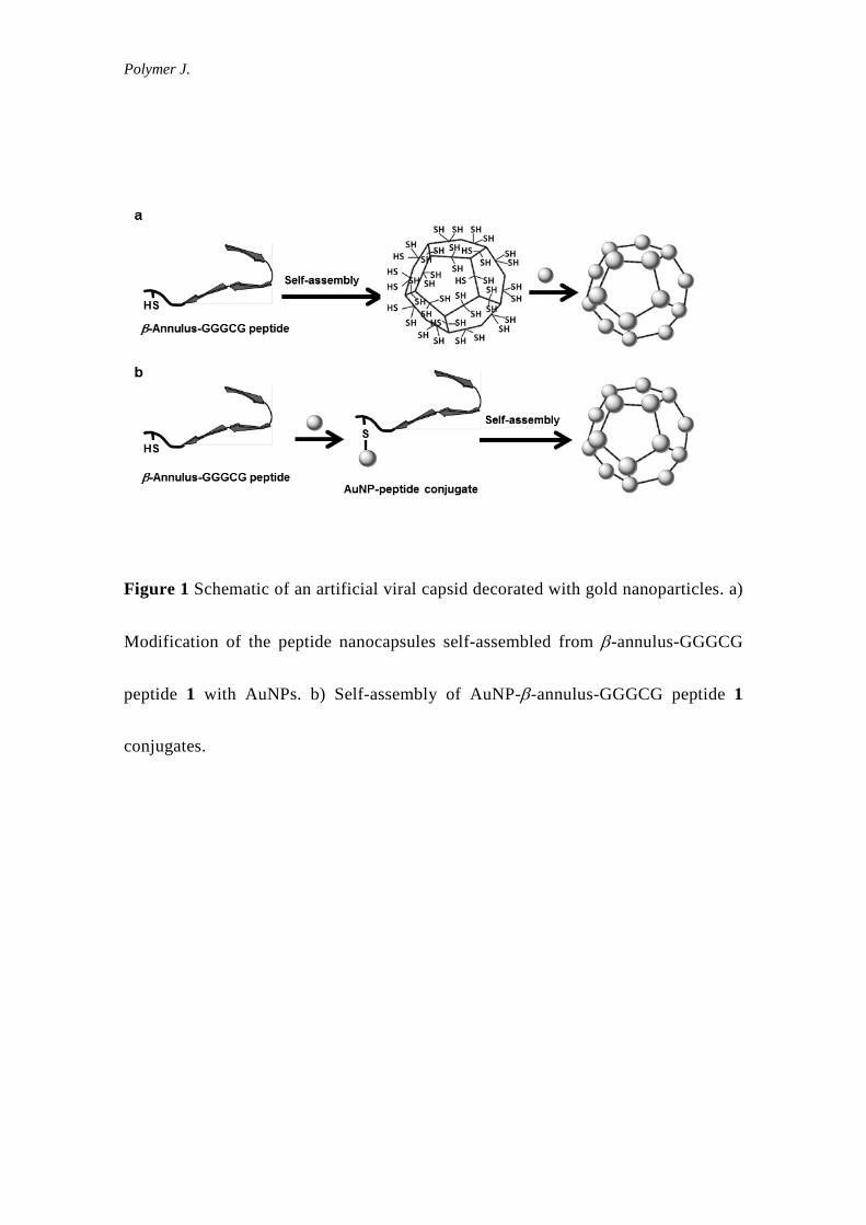
Figure 1 Schematic of an artificial viral capsid decorated with gold nanoparticles. a)
Modification of the peptide nanocapsules self-assembled from β-annulus-GGGCG
peptide 1 with AuNPs. b) Self-assembly of AuNP-β-annulus-GGGCG peptide 1
conjugates.
Polymer J.
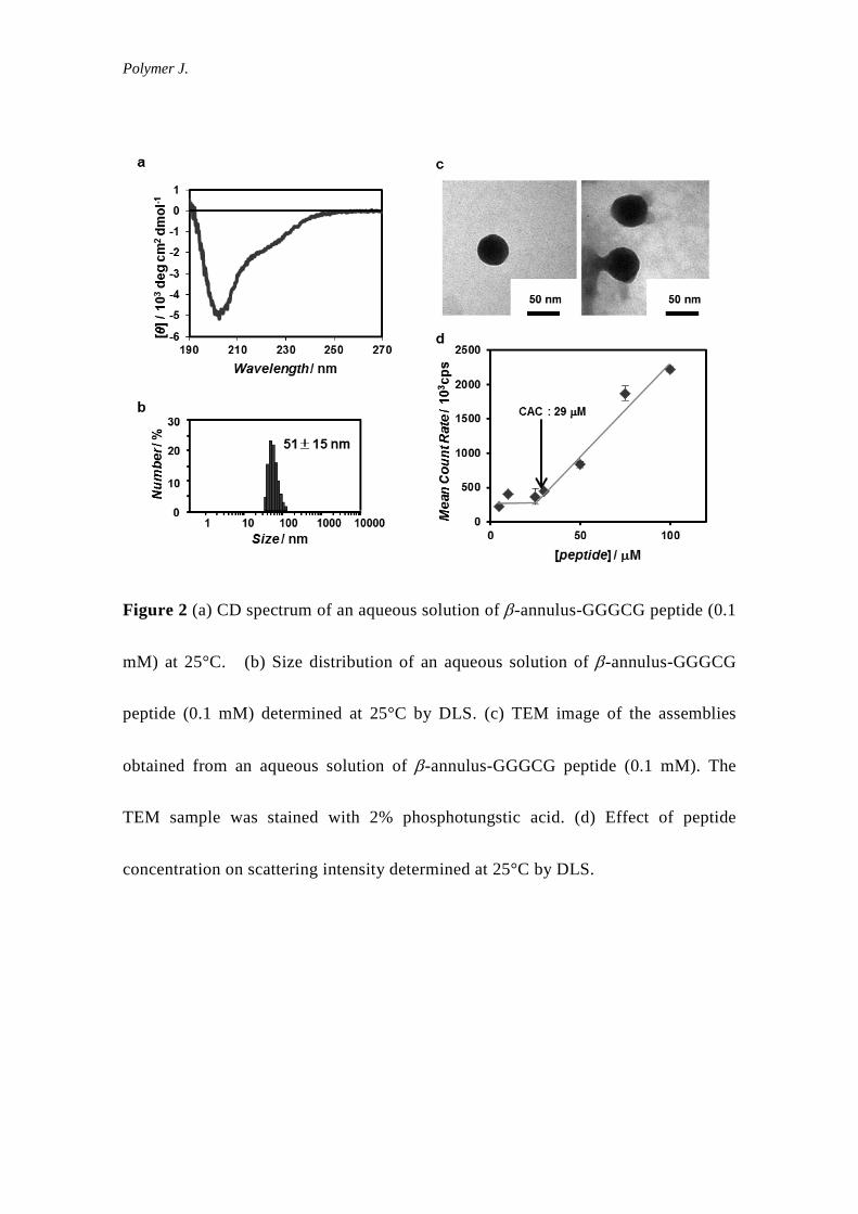
Figure 2 (a) CD spectrum of an aqueous solution of β-annulus-GGGCG peptide (0.1
mM) at 25°C. (b) Size distribution of an aqueous solution of β-annulus-GGGCG
peptide (0.1 mM) determined at 25°C by DLS. (c) TEM image of the assemblies
obtained from an aqueous solution of β-annulus-GGGCG peptide (0.1 mM). The
TEM sample was stained with 2% phosphotungstic acid. (d) Effect of peptide
concentration on scattering intensity determined at 25°C by DLS.
Polymer J.
Figure 3 (a) TEM images of AuNPs (0.2 µM) mixed with the peptide nanocapsules
([peptide 1] = 0.1 mM). (b) Size distribution of assembly of AuNPs (0.2 µM) mixed
with the peptide nanocapsules ([peptide 1] = 0.1 mM) determined at 25°C by DLS.
(c) UV−vis spectra of aqueous dispersion of AuNP (red) at 0.1 µM and the assembly
of AuNPs (0.1 µM) mixed with the peptide nanocapsules ([peptide 1] = 0.05 mM)
(blue) at 25°C.
Polymer J.
Figure 4 TEM images and size distribution of assemblies of AuNP−peptide
conjugates by DLS before (a) and after (b) dialysis at 25°C ([peptide] = 50 µM,
[AuNP] = 25 µM). (c) UV−vis spectra of a diluted aqueous dispersion of AuNP
(black) and an artificial viral capsid decorated with gold nanoparticles after dialysis
(gray) at 25°C. The concentration of AuNPs after dialysis was calculated to be 0.06
µM.
Polymer J.
Figure 5 Zeta potentials of (a) β-annulus-GGGCG peptide 1 (0.1 mM) and (b)
artificial viral capsid decorated with gold nanoparticles after dialysis in water at pH
4.6 and at 25°C.
Polymer J.
Figure S1 (a) TEM images, (b) size distribution determined by DLS, and (c) zeta
potential of AuNP dispersion in citrate buffer.


![Ultrashort self-assembling Fmoc-peptide gelators for anti ... · phase Fmoc peptide synthesis protocols using methods previously demonstrated by our group [4]. Peptides were cleaved](https://img.pdfslide.net/doc/110x75/5f0b640e7e708231d430495b/ultrashort-self-assembling-fmoc-peptide-gelators-for-anti-phase-fmoc-peptide.jpg)