Embed Size (px)
Citation preview

MUHAMMAD FIAZ
TOXICOLOGICAL AND MORPHOLOGICAL EFFECTS OF SQUAMOCIN AND TEBUFENOZIDE ON ANTICARSIA GEMMATALIS (LEPIDOPTERA:
NOCTUIDAE) LARVAE
VIÇOSA MINAS GERAIS – BRASIL
2019
Tese apresentada à Universidade Federal de Viçosa, como parte das exigências do Programa de Pós-Graduação em Entomologia, para obtenção do título de Doctor Scientiae.


ii
DEDICATION
I dedicate this work to my parents, brother and sisters who gave me the motivation, encouragement throughout my studies. May ALLAH, The Almighty, have His mercy,
forgive all their sins and accept all their good deeds.
iii
ACKNOWLEDGEMENTS
First, I would like to bow and praise to ALMIGHTY ALLAH: Who is the eternal sole creator
of whole universe, the most beneficent, the merciful, the gracious, and compassionate, whose
blessings and exaltation flourished my thoughts and thrive my ambitions to have the cherish
fruits of my modest efforts and enabled me to complete this PhD program. At the same time, I
would also praise the perfectness and supremacy of the HOLY PROPHET HAZRAT
MUHAMMAD (S.A.W) with great respect: Who is forever, a troch of guidance and blessing
for the entire humanity.
I deem it my utmost pleasure to express my deepest feelings of regards and the sense of
gratitude to my supervisor Professor Dr. José Eduardo Serrão, Departamento de Biologia
Geral, Universidade Federal de Viçosa, who, despite of his busiest work routine, provided his
precious advices, guidance, and valuable suggestions throughout these research activities. His
continuous believe in my abilities, encouragement, and motivation always energized me
whenever I felt stressed and exhausted.
I am grateful to the laboratory fellows, Pakistani and Brazilian friends, who were supportive
and helpful throughout the years. They have been invaluable in finishing this project. At last
but not the least, I would also delightedly acknowledge the financial and technical support
provided by the CNPq and The World Academy of Science (TWAS) through a collaborative
platform of TWAS-CNPq Fellowship Program (2014) for International PhD students. I also
would like to express my gratitude to the Department of Entomology, the Postgraduate program
and Universidade Federal de Viçosa for their support.

iv
SUMÁRIO
LIST OF FIGURES ................................................................................................................... v
LIST OF TABLES .................................................................................................................... ix
ABSTRACT ............................................................................................................................... x
RESUMO .................................................................................................................................. xi
INTRODUCTION ..................................................................................................................... 1
REVIEW OF LITERATURE .................................................................................................... 2
REFERENCES .......................................................................................................................... 5
CHAPTER 1 ............................................................................................................................ 12
Squamocin induce histological and ultrastructural changes in the midgut cells of Anticarsia gemmatalis (Lepidoptera: Noctuidae) ...................................................................................... 12
CHAPTER 2 ............................................................................................................................ 21
Toxicological and morphological effects of tebufenozide on Anticarsia gemmatalis (Lepidoptera: Noctuidae) larvae .............................................................................................. 21
FINAL CONSIDERATIONS .................................................................................................. 36
v
LIST OF FIGURES CHAPTER 1. Squamocin induce histological and ultrastructural changes in the midgut
cells of Anticarsia gemmatalis (Lepidoptera: Noctuidae)
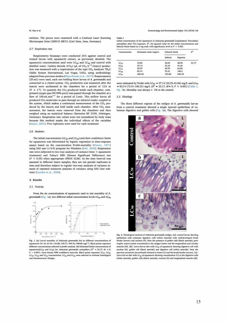
Fig. 1. (a) Larval mortality of Anticarsia gemmatalis fed on different concentrations of
squamocin (31.16; 62.42; 124.85; 249.71; 499.42; 998.85 mgL-1). Black points represent
different concentrations selected in probit analysis. (b) Estimated lethal concentrations of
squamocin (LC50 and LC90) for Anticarsia gemmatalis caterpillars (X2 = 23.17; df = 5; P <
0.001). Lines denote 95% confidence intervals. Black points represent LC25, LC50, LC75, LC90
and LC99 concentration, LC50 and LC90 were selected to evaluate histological and
ultrastructural changes.
Fig. 2. Histological sections of Anticarsia gemmatalis midgut. [a]: control larvae showing
epithelium with columnar digestive cells (white asterisk) with well-developed brush border
(arrow) and nucleus (N). Note the presence of goblet cells (black asterisk), peritrophic matrix
(white arrowhead) in the midgut lumen and the longitudinal and circular muscles (M). [b]:
Larva fed on diet with LC50 of squamocin showing digestive cell with nucleus (N), goblet cell
(black asterisk) and digestive cell (white asterisk). Note the apocrine secretions (arrowhead)
released in lumen (L) and the brush border (arrow). [c]: Larva fed on diet with LC90 of
squamocin showing vacuolization (V) in the digestive cells (white asterisk), goblet cells (black
asterisk), nucleus (N) and longitudinal muscles (M).
Fig. 3. Transmission electron micrographs of the midgut of Anticarsia gemmatalis control
larvae. [a]: Median-apical region of digestive cell showing well developed nucleus (N) with
few crumples of heterochromatin (white arrowhead) and nucleolus (Nu). Note apical well-
developed microvilli (MV) and many mitochondria (black arrowhead). [b]: Median-basal
region of digestive cell showing nucleus (N) and cytoplasm with plasma membrane infoldings
resulting in long and enlarged basal labyrinth (asterisks). [c]: Detail of mitochondria (black
arrowhead) closely associated with basal plasma membrane infoldings. [d]: Detail of digestive
cell basal cell region with enlarged labyrinth (BL) and the basal lamina (white arrow). [e]:
Median region of digestive cells showing profiles of the basal labyrinth (asterisk) associated
with mitochondria (black arrowhead). [f]: General aspect of the goblet cell showing the nucleus
(N) and the internal cavity (C) with microvilli (black arrow). [g]: Detail of the goblet cell cavity
(C) showing microvilli (black arrow) with mitochondria (black arrowhead).
Fig. 4. Transmission electron micrographs of the midgut of Anticarsia gemmatalis larvae fed
on squamocin. [a]: General aspects of epithelium showing cells with disorganized labyrinth
(BL), cluster of vacuoles (V), microvilli (MV) and basal lamina (white arrowhead). [b]:
General aspect of the midgut epithelium of larvae fed on LC50 squamocin showing digestive
vi
cells (DC) with large fragments of the apical cell region (white arrow) releasing to the lumen
(L), disorganized microvilli (MV) and some goblet cell (GC). [c]: Apical region of digestive
cell in larva fed on LC50 squamocin showing dying cell with many autophagic vacuoles (black
arrowheads). [d]: Cytoplasm digestive cell in larva fed on LC90 squamocin showing autophagic
vacuoles (black arrowhead) and an enlarged vacuole with organelle debris and disorganized
microvilli (MV). [e]: Perinuclear region of digestive cell in larva fed on LC90 squamocin
showing many autophagic vacuoles (black arrowheads), nucleus (N) with well-developed
nucleolus (Nu) and condensed chromatin (white arrowhead). [f]: Basal region of digestive cell
in larva fed on LC90 squamocin showing disorganized basal lamina (black arrow) and swelled
basal labyrinth (BL).
Fig. 5. Transmission electron micrographs of goblet cell in the midgut of Anticarsia
gemmatalis fed on squamocin. [a]: General aspect showing multiple vacuoles (V) and the cell
cavity (C) filled with flocculent material. [b]: Detail of cell cavity (C) showing disorganized
microvilli (arrow) without mitochondria.
Fig. 6. Confocal micrographs of Anticarsia gemmatalis larval midgut stained with MitoTracker
fluorescent probe. [a]: Control larvae showing mitochondria (green) and nucleus (red). [b]:
Larvae fed on LC50 squamocin without mitochondria. [c]: Larvae fed on LC90 of squamocin
without mitochondria.
Fig. 7. Respiration rate (means ± se) of Anticarsia gemmatalis larvae after feeding to LC50 and
LC90 squamocin application. Means with by different letters in the same time differ at by
Tukey's mean separation test (P < 0.05).
CHAPTER 2. Toxicological and morphological effects of tebufenozide on Anticarsia
gemmatalis (Lepidoptera: Noctuidae) larvae
Fig. 1. Distance walked and resting time (Means ± SD) of Anticarsia gemmatalis larvae
exposed to level LC50 and LC90 tebufenozide concentrations. [a] Distance walked [b] Resting
time. Bars followed by different letters differ at P < 0.05 (Tukey's mean separation test). n.s. e
non-significant.
Fig. 2. Respiration rate of Anticarsia gemmatalis after exposed to LC50 and LC90
concentrations of tebufenozide on larvae. Lines followed by different letters differ at P < 0.05
(Tukey's mean separation test). Vertical bars represent the standard error of the mean.
Fig. 3. Histological sections of Anticarsia gemmatalis midgut. [a]: Control larvae showing
epithelium with digestive cells (white asterisk) with well-developed brush border (arrow) and
nucleus (N). Note the presence of goblet cells (black asterisk), abundance of vacuoles (arrow
head), midgut lumen (L) and the longitudinal and circular muscles (M). [b]: Larva after 24 h
fed on diet with LC50 of tebufenozide showing digestive cell with nucleus (N), goblet cell
vii
(black asterisk) and digestive cell (white asterisk). Note the lose attachment of brush border
(arrow) and vacuoles (arrowhead). [c]: Larva after 48 h fed on diet with LC50 of tebufenozide
showing abundance of vacuoles (arrowhead) in the digestive cells (white asterisk), goblet cells
(black asterisk), nucleus (N) and longitudinal muscles (M). [d]: Larva after 96 h fed on diet
with LC50 of tebufenozide showing digestive (white asterisk), goblet cells (black asterisk),
nucleus (N), muscles (M). Note the damaged brush border (arrow) cell with few vacuoles
(arrowhead) and abundance of cell fragments (F) released in the midgut lumen (L).
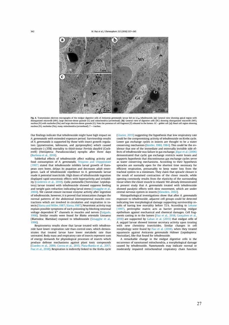
Fig. 4. Transmission electron micrographs of the midgut digestive cells of Anticarsia
gemmatalis larvae fed on LC50 tebufenozide. [a]: General view showing apical region with
disorganized microvilli (MV), large electron-dense granule (G) and mitochondria (arrowhead).
[b]: General view of digestive cells (DC) showing disorganized microvilli (MV), nucleus (N)
with nucleolus (Nu) and large electron-dense granule (G). Note the presence of cell fragment
(F) released to the lumen. GC - goblet cell. [c]: Basal cell region showing nucleus (N),
nucleolus (Nu), many mitochondria (arrowhead). T - trachea.
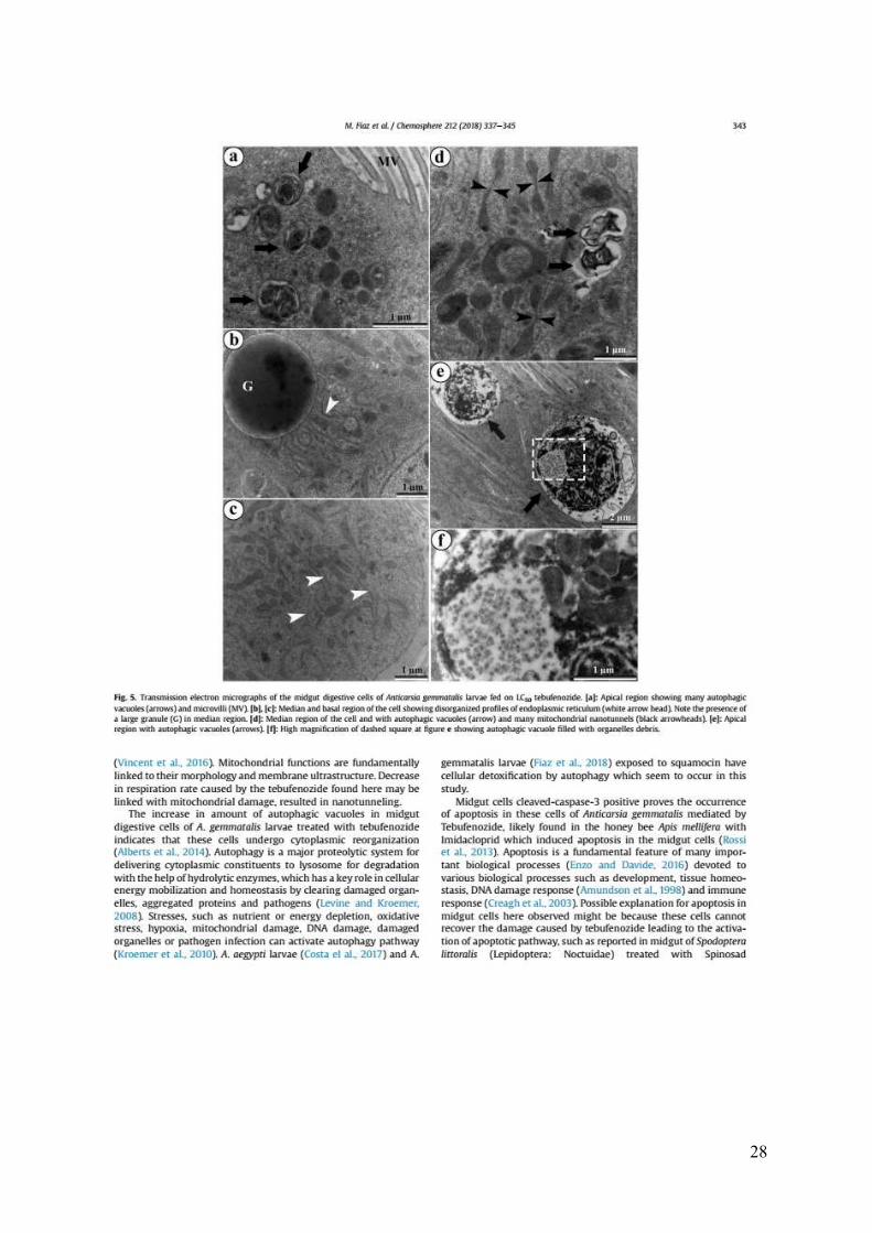
Fig. 5. Transmission electron micrographs of the midgut digestive cells of Anticarsia
gemmatalis larvae fed on LC50 tebufenozide. [a]: Apical region showing many autophagic
vacuoles (arrows) and microvilli (MV). [b], [c]: Median and basal region of the cell showing
disorganized profiles of endoplasmic reticulum (white arrow head). Note the presence of a
large granule (G) in median region. [d]: Median region of the cell and with autophagic vacuoles
(arrow) and many mitochondrial nanotunnels (black arrowheads). [e]: Apical region with
autophagic vacuoles (arrows). [f]: High magnification of dashed square at figure e showing
autophagic vacuole filled with organelles debris.
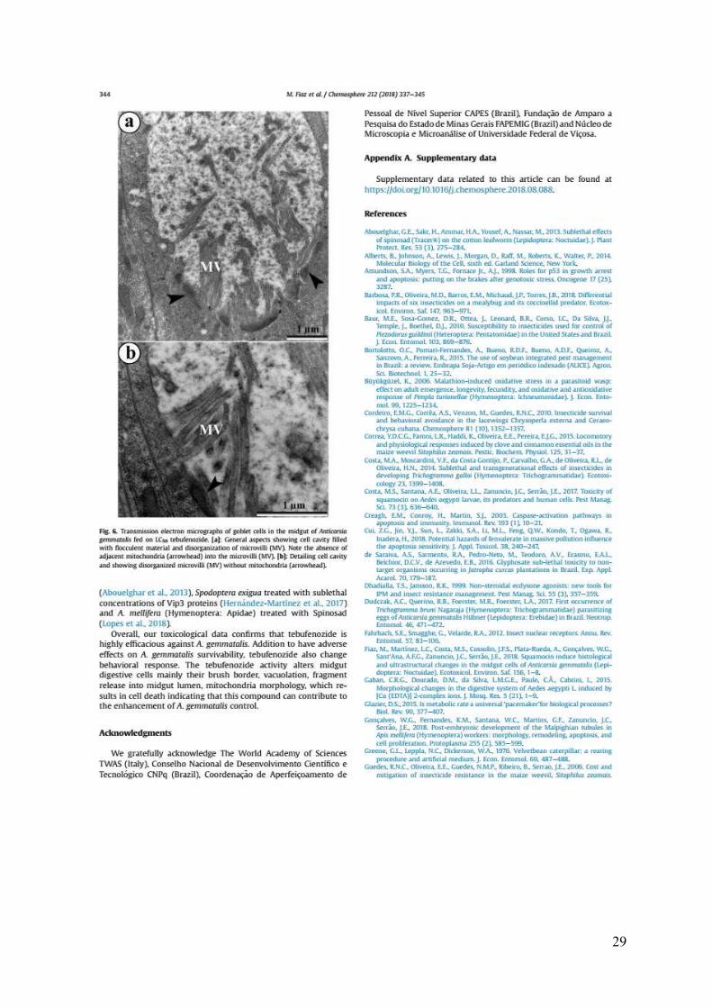
Fig. 6. Transmission electron micrographs of goblet cells in the midgut of Anticarsia
gemmatalis fed on LC50 tebufenozide. [a]: General aspects showing cell cavity filled with
flocculent material and disorganization of microvilli (MV). Note the absence of adjacent
mitochondria (arrowhead) into the microvilli (MV). [b]: Detailing cell cavity and showing
disorganized microvilli (MV) without mitochondria (arrowhead).
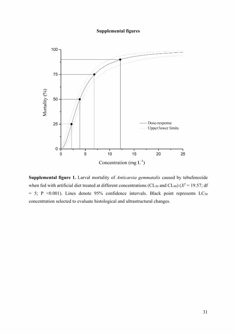
Supplemental figure 1. Larval mortality of Anticarsia gemmatalis caused by tebufenozide
when fed with artificial diet treated at different concentrations (CL50 and CL90) (X2 = 19.57; df
= 5; P <0.001). Lines denote 95% confidence intervals. Black point represents LC50
concentration selected to evaluate histological and ultrastructural changes.
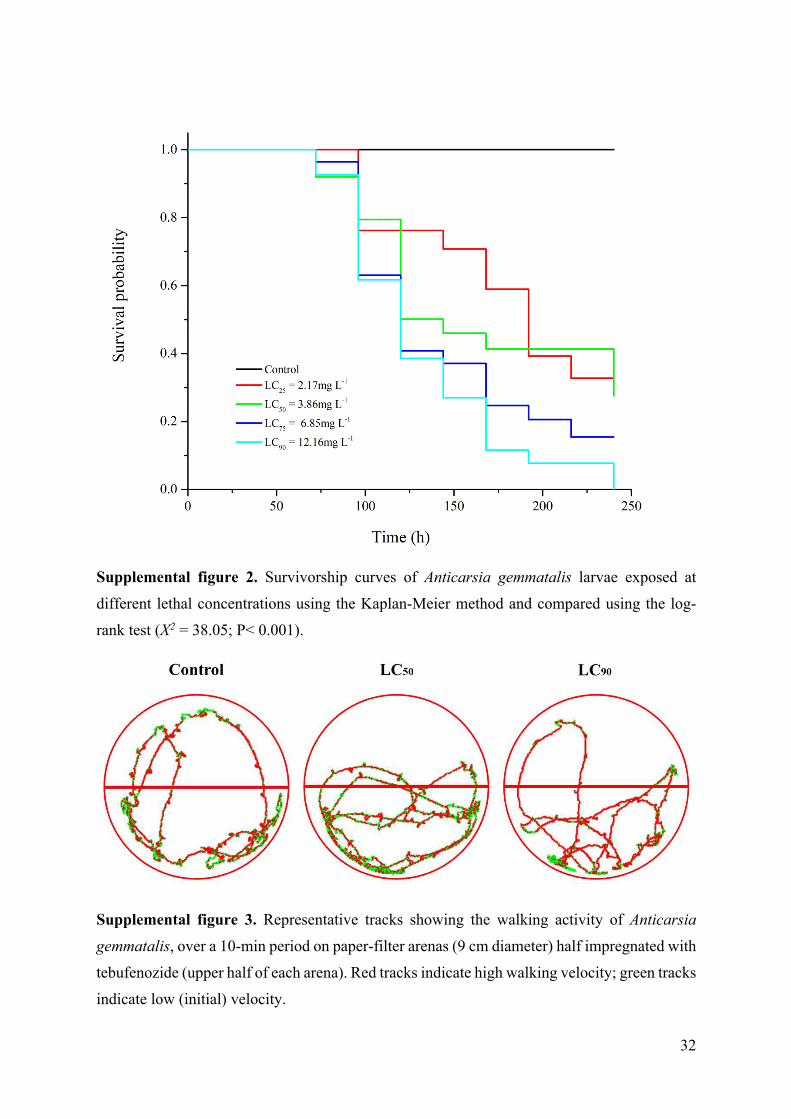
Supplemental figure 2. Survivorship curves of Anticarsia gemmatalis larvae exposed at
different lethal concentrations using the Kaplan-Meier method and compared using the log-
rank test (X2 = 38.05; P< 0.001).
viii
Supplemental figure 3. Representative tracks showing the walking activity of Anticarsia
gemmatalis, over a 10-min period on paper-filter arenas (9 cm diameter) half impregnated with
tebufenozide (upper half of each arena). Red tracks indicate high walking velocity; green tracks
indicate low (initial) velocity.
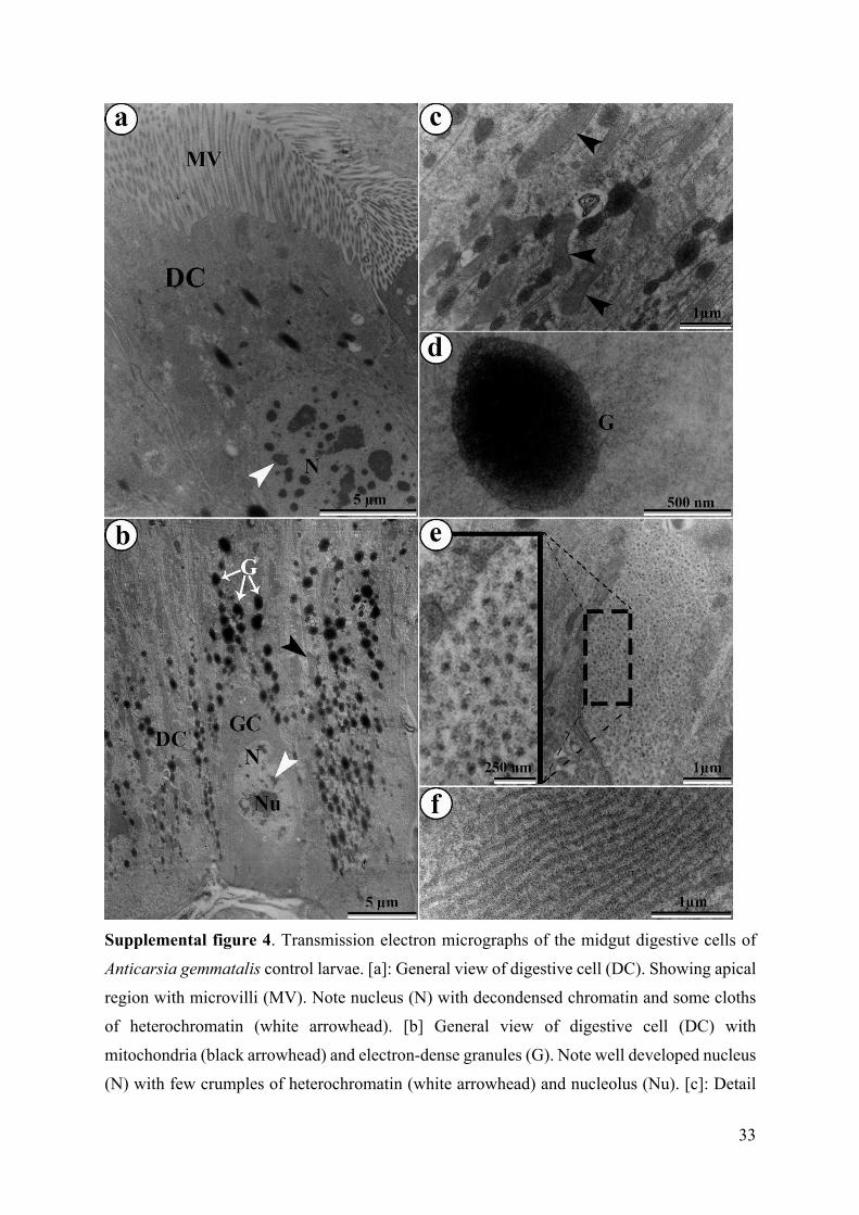
Supplemental figure 4. Transmission electron micrographs of the midgut digestive cells of
Anticarsia gemmatalis control larvae. [a]: General view of digestive cell (DC). Showing apical
region with microvilli (MV). Note nucleus (N) with decondensed chromatin and some cloths
of heterochromatin (white arrowhead). [b] General view of digestive cell (DC) with
mitochondria (black arrowhead) and electron-dense granules (G). Note well developed nucleus
(N) with few crumples of heterochromatin (white arrowhead) and nucleolus (Nu). [c]: Detail
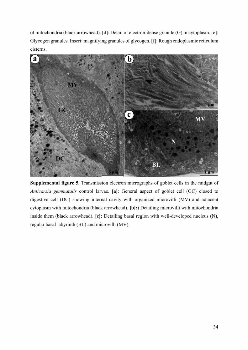
of mitochondria (black arrowhead). [d]: Detail of electron-dense granule (G) in cytoplasm. [e]:
Glycogen granules. Insert: magnifying granules of glycogen. [f]: Rough endoplasmic reticulum
cisterns.
Supplemental figure 5. Transmission electron micrographs of goblet cells in the midgut of
Anticarsia gemmatalis control larvae. [a]: General aspect of goblet cell (GC) closed to
digestive cell (DC) showing internal cavity with organized microvilli (MV) and adjacent
cytoplasm with mitochondria (black arrowhead). [b]:) Detailing microvilli with mitochondria
inside them (black arrowhead). [c]: Detailing basal region with well-developed nucleus (N),
regular basal labyrinth (BL) and microvilli (MV).
Supplemental figure 6. Immunofluorescence staining for cleaved-3-caspase in the midgut of
Anticarsia gemmatalis caterpillars. [a-c-e]: Bright field images. [b]: control larvae showing
negative fluorescence. Apoptotic cells. [d]: larva after 24 h fed on diet with LC50 tebufenozide
concentrations showing apoptotic cells (arrow). [f]: larva after 48 h fed on diet with LC50
tebufenozide concentrations showing apoptotic cells (arrows). Bars=20µm.

ix
LIST OF TABLES
CHAPTER 1. Squamocin induce histological and ultrastructural changes in the midgut
cells of Anticarsia gemmatalis (Lepidoptera: Noctuidae)
Table 1. Lethal concentration of the squamocin in Anticarsia gemmatalis (Lepidoptera:
Noctuidae) caterpillars after 72 h exposure. X2, chi squared value for the lethal concentrations
and fiducial limits based on a log scale with significance level at P < 0.001.
CHAPTER 2. Toxicological and morphological effects of tebufenozide on Anticarsia
gemmatalis (Lepidoptera: Noctuidae) larvae
Table 1. Lethal tebufenozide concentrations to Anticarsia gemmatalis (Lepidoptera:
Noctuidae) larvae after 96 h exposure. X2, chi squared value for the lethal concentrations and
fiducial limits based on a log scale with significance level at P < 0.001.

x
ABSTRACT FIAZ, Muhammad, D.Sc., Universidade Federal de Viçosa, February, 2019. Toxicological and morphological effects of squamocin and tebufenozide on Anticarsia gemmatalis (Lepidoptera: Noctuidae) larvae. Advisor: José Eduardo Serrão.
Anticarsia gemmatalis Hübner (Lepidoptera: Noctuidae) is the main defoliating pest of
soybean (Glycine max L. Merrill, Fabaceae) in Brazil. There are a variety of plant products and
synthetic insecticides used to control A. gemmatalis. The larval midgut is reported to be the
'front line' in creation of local immune defense and detoxification of xenobiotic compounds.
These characteristics make the midgut cells to be the most affected by those xenobiotics.
Squamocin from Annona mucosa and tebufenozide were evaluated to test their toxicity and
their ultrastructural effects in midgut cells of A. gemmatalis. Toxicological results showed that
A. gemmatalis was susceptible to both squamocin and tebufenozide. Larvae exposed to
tebufenozide compromised larval fitness and its survivorship. LC50 and LC90 of squamocin and
tebufenozide against A. gemmatalis were 37.14 mg/L-1, 83.14 mg/L-1 and 3.86 mg/L-1, 12.16
mg/L-1, respectively. Squamocin and tebufenozide intake caused deformities in epithelial cells.
Squamocin damage to midgut cells include enlarged basal labyrinth, highly vacuolated
cytoplasm, damaged apical surface, release of cell protrusions to the gut lumen, autophagy and
cell death. Ingestion of tebufenozide caused damage to striated border with release of
protrusions to the midgut lumen, damaged nuclear membrane and nucleus with condensed
chromatin and increase autophagic vacuolization. Both, squamocin and tebufenozide, damaged
mitochondria and compromised respiration rate, while in case of tebufenozide, severe damage
resulted in to modification of mitochondria into nanotunnels along with compromising
respiration rate. Squamocin and tebufenozide are lethal to larvae, they compromised its fitness
and induced severe morphological changes in midgut cells empowering control program
against A. gemmatalis.
xi
RESUMO
FIAZ, Muhammad, D.Sc., Universidade Federal de Viçosa, fevereiro de 2019. Efeitos toxicológicos e morfológicos da esquamocina e tebufenozida nas larvas de Anticarsia gemmatalis (Lepidoptera: Noctuidae). Orientador: José Eduardo Serrão. Anticarsia gemmatalis Hübner (Lepidoptera: Noctuidae) é uma praga desfolhadora da soja
(Glycine max L. Merrill, Fabaceae) no Brasil, sendo controlada por uma variedade de produtos
naturais e inseticidas sintéticos. O intestino médio das larvas é reportado por fazer parte da
defesa imune primária por participar da detoxificação de compostos xenobióticos. Estas
características fazem do intestino médio o órgão mais afetado por tais xenobióticos. O
composto esquamocina da planta Annona mucosa e tebufenozida foram avaliados para testar
sua toxicidade e possíveis efeitos na ultraestrutura das células do intestino médio de A.
gemmatalis. Resultados toxicológicos mostraram que A. gemmatalis foi suscetível tanto a
esquamocina quanto a tebufenozida. Larvas expostas a tebufenozida tiveram
comprometimento do fitness e sobrevivência. CL50 e CL90 de esquamocina e tebufenozida
contra A. gemmatalis foram 37.14 mg/L-1, 83.14 mg/L-1 e 3.86 mg/L-1, 12.16 mg/L-1,
respectivamente. A ingestão de esquamocina e tebufenozida causaram danos nas células
epiteliais do intestino médio como alargamento do labirinto basal, citoplasma altamente
vacuolizado, superfície apical danificada, liberação de protrusões celulares no lúmen, autofagia
e morte celular. A ingestão de tebufenozida causou danos na borda estriada com liberações de
protrusões para o lumen do intestino, danos na membrana nuclear, núcleo com cromatina
condensada e aumento de vacuolização autofágica. Ambos os compostos esquamocina e
tebufenozida danificaram mitocôndrias e comprometeram a taxa respiratória, enquanto no
tratamento com tebufenozida houve danos severos resultando na modificação das mitocôndrias
em nanotúneis juntamente com o comprometimento da taxa respiratória. Assim sendo,
esquamocina e tebufenozida são letais às larvas, comprometendo seu fitness e induzindo
alterações morfológicas severas nas células do intestino médio, favorecendo o programa de
controle contra A. gemmatalis.

1
INTRODUCTION
Soybean is principal food crop in Brazil, enlisting the country as the second largest
producer in the world. Defoliation of soybean crops is caused by different insect species that
can compromise yield and quality of production, especially when it occurs at reproductive
stages (Begum & Eden 1965). Among those, Anticarsia gemmatalis Hubner 1818 (Noctuidae:
Lepidoptera) is one of the major defoliating soybean pests in the western hemisphere
(Bortolotto et al. 2015), which consumes leaves during all its larval instars, and consequently
can cause complete defoliation of plants (Fugi et al. 2005) diminishing its productivity
(Bortolotto et al. 2015).
Despite availability of several control methods, pest control is still largely based on the
use of pesticides, in the sense of synthetic organic chemical-based ingredients that are applied
on the crops, the commodity, or the household environment. In Brazil A. gemmatalis is
controlled mainly by using synthetic insecticides (Navickiene et al. 2007; Panizzi 2013).
Because of the fact, pesticide made possible a fastest and effective method to control weeds,
diseases and insect in relatively short time (Arain et al. 2018).
Insecticide application is considered to be a necessary component in agricultural
production (Tilman et al. 2002). In addition to have direct killing effect on pests, the application
of pesticide also results in insect physiology, biology, behavior, fertility, fecundity, longevity
and resistance in response to low or sublethal doses of insecticide over time (Guedes et al.
2016; Lee et al. 2000). Those sublethal effects vary depending upon insecticide class and
targeted species. The effects impact on insect growth, development and fecundity, in addition
to affect insect physiological and biochemical processes (Cao et al. 2017; Lee et al. 2000).
Physiological and biochemical changes can be used to assess and predict pesticide efficacy and
potency in context of controlling insect pests (Zhao et al. 2018).
Squamocin, also called anonin I, is derived from plant parts, which is an acetogenin (37
carbon atoms, α,β-unsaturated γ-lactone ring and adjacent bis-tetrahydrofuran (bis-THF) ring)
(Rupprecht et al. 1990) is cytotoxic (Miao et al. 2016), larvicidal (Costa et al. 2014), antitumor
(Chen et al. 2013), neurotoxic (Derbré et al. 2008) and inhibitor of mitochondrial complex I
(Duval et al. 2006). The search of natural compounds for plant protection, especially in organic
agriculture, has increased the interest in botanical insecticides (Martínez et al. 2015; Zanuncio
et al. 2016). Squamocin is ascertained to be gut poisons and effective against insect pests such
as Trichoplusia ni Hübner (Lepidoptera: Noctuidae), Myzus persicae Sulzer (Hemiptera:
Aphididae) (Ribeiro et al. 2014) and Aedes aegypti L. (Diptera: Culicidae) (Costa et al. 2014,
2017).

2
Tebufenozide was first introduced in the early 1990s (Dhadialla & Jansson 1999),
which mimics the biological function of the natural insect hormone JH and interacts with instar
phase. Steroid molting hormone 20E and sesquiterpenoid juvenile hormone in insects plays a
major role in growth regulation, development and reproductive processes (Riddiford 2012).
Tebufenozide halts larval feeding when ingested and induces a premature molt, which is
ultimately lethal (Wing et al. 1988).
The insect digestive system is divided in to three principal regions, foregut and hindgut,
which are derived from the ectoderm, and the midgut, originated from endoderm is the main
organ where digestion and absorption take places (Chapman 2013). In particular with
ephemeropteran and lepidopteran midguts, so called invaginated shaped goblet cells with
enclosed lumen filled with flocculent material are present which encloses a large extracellular
lumen that is continuous with the midgut lumen, via a labyrinthine apical valve formed by the
interdigitating microvilli (Lehane & Billingsley 2012). Among the organs tested against
different insecticides, the medium intestine has been reported to be one of the most affected by
chemicals (Gutiérrez et al. 2016; Catae et al. 2018).
In this work, we aimed to test the plant compound; squamocin from Annona mucosa,
and chemical; tebufenozide against Anticarsia gemmatalis and evaluated their lethal and
sublethal effects on survival and fitness of A. gemmatalis larvae. We report testing of
squamocin and tebufenozide in context with ultrastructural and cytochemical aspects of
midgut, suggesting these agents as tool in managing and controlling A. gemmatalis.
REVIEW OF LITERATURE
Researches are being carried out with crops to find lines and cultivars with relative
levels of resistance to insects. Wild soybeans like, PI 171451, PI 227658 and PI 229358 are
being used since early 1970s to cop with the resistance developed in insects like Epilachna
varivestis (Van Duyn et al. 1972), Trichoplusia ni (Luedders & Dickerson 1977), Diabrotica
speciosa and Colaspis spp. (Rezende & de Miranda 1980), Pseudoplusia includens (Beach &
Todd 1988), Spodoptera spp. (Beach & Todd 1987) and A. gemmatalis (Oliveira et al. 1993).
Piubelli et al. (2003) reported these PIs to possess moderately resistant against some seed
sucking insects. When tested in field, PI 274454 lines were observed to be less defoliated than
those cultivars tested as control (Rezende et al. 1980). Breeding program of the Instituto
Agronômico (Campinas, São Paulo State, Brazil) released a cultivar IAC-100 having
genealogy of PI 274454 and PI 229358 and was particularly resistant to sting bugs and other
leaf feeders (Veiga et al. 1999).
3
Levy et al. (2004) reported the morphology of epithelial cells of Anticarsia gemmatalia
larvae by light and transmission electron microscopy. The midgut of A. gemmatalis has three
different regions: proximal, media and distal. The wall is formed by epithelial tissue which has
four cell types, called columnar, goblet, regenerative and endocrine cells. Columnar cells are
long and numerous in number, with the apical portion rich in microvilli and the basal portion
invaginations forming in to a basal labyrinth. The goblet cells have a large central cavity
encircled by cytoplasmic projections adjacent with mitochondria (Gomes et al. 2013).
Regenerative cells show electron-dense cytoplasm with few organelles and endocrine cells
exhibit electron-dense secretory granules, usually concentrated in the basal region of the
cytoplasm.
Silveira et al. (2004) reported the virus infection of nucleopolyhedro virus in
haemocytes of Anticarsia gemmatalis and characterized intrahaemocoelic infection by A.
gemmatalis M nucleopolyhedrovirus (AgMNPV). Entire cells were phagocytosed by
plasmatocytes only. Infected cells presented a common feature of necrosis, suggesting
cytotoxic effect. There was an exuberant protrusion of budded viruses in granular haemocyte
1 as compared to other haemocytes. Infection to all types of haemocytes were observed,
addition to have intense virus replication in these cells which is indication of importance of
haemolymph for AgMNPV dispersal in natural host, which is a critical factor for
permissiveness.
Knaak & Fiuza (2005) tested the efficacy of Bacillus thuringiensis and
Nucleopolyhedrovirus and their effect on midgut epithelial cells of Anticarsia gemmatalis.
They reported the efficacy of Btk up to 100% when used isolatedly, rather than using the
association of AgNPV-Btk (98.68% of mortality). Btk exhibited changes in larval midgut after
6 hours of treatment.
Piubelli et al. (2005) tested effects of flavonoids from soybean genotypes on biology
and physiology of Anticarsia gemmatalis and quantified those flavonoids. Higher mortality
was observed in larvae fed on extracts of genotypes PI 274454, PI 227687, and “IAC-100”
addition to negative negatively inclined initial larval growth and pupal weight and elongated
larval cycle.
Levy et al. (2011) analyzed the peritrophic membrane of Anticarsia gemmatalis
caterpillar resistant and susceptible to A. gemmatalis multicapsid nucleopolyhedrovirus
(AgMNPV), in viral presence. They concluded that peritrophic membrane’s effectiveness in
protection against pathogens is highly reliant on on the integrity of the epithelial cells and of
the structural preservation of the peritrophic membrane, which is directly implicated in the
resistance of A. gemmatalis larvae to AgMNPV.

4
Fiuza et al. 2013 reported lethal effects and receptors of Bacillus thuringiensis in the
Anticarsia gemmatalis. They bound insecticidal crystal proteins to midgut epithelial cells of
the A. gemmatalis larvae by using streptavidin-mediated detection and presented brush border
as the binding cite of Cry1Aa and Cry1Ac along the entire length of midgut. Contrary to this,
the binding cites of Cry1Ba were not regularly distributed in the microvilli of midgut. The data
of binding cites demonstrated a correlation between receptors and toxicity of tested insecticidal
crystal proteins of insect.
Almeida et al. (2014) reported the ultrastructural analysis of fat body and midgut of
Anticarsia gemmatalis larvae treated with neem seed extract. They described swallow midgut
cells of A. gemmatalis larvae, detachment of basal membrane and complete disruption in severe
conditions in treatments. Lipid and protein reserves were observed to be depleted in treatments.
Overall, A. gemmatalis exhibited negative effects on physiological and biological parameters
when treated with neem seed kernel extract.
Costa et al. (2014) reported acetogenins as secondary metabolites which are exclusively
produced by Annonaceae, having antitumor, cytotoxic, and pesticide properties. Data
estimated with probit analysis suggested that squamocin induced mortality in Aedes aegypti.
Concentrations of squamocin (50, 80, and 100 ppm) were effective in causing 50% mortality
after 360 minutes while 6.4 ppm was effective in causing 50% mortality after 600 of
application. Cytotoxic activity caused by 50 ppm of squamocin after 240 minutes resulted in
midgut cells with low level of vacuolization in cytoplasm while 100 ppm of squamocin
exhibited high level of vacuolization in cytoplasm, damage to apical surface and release of cell
protrusion to lumen.
Schünemann et al. (2014) proposed Bacillus thuringiensis in management of
velvetbean caterpillar and reported that Bt showed toxic activity to caterpillar. They
emphasized that cultures those express Bt can be helpful in global agriculture system with the
aim of reducing number of pests, which results in reduction of synthetic insecticide applications
to control this pest, ultimately boosting final production.
Mushtaq et al. (2017) used insecticidal proteins from Bt to control Anticarsia
gemmatalis and Chrysodeixis includens. High activity of Cry2Ac7 and Vip3Aa11 proteins
were observed in A. gemmatalis and C. includens larvae. In addition to the above results, they
also reported that Cry1Ie2 and Cry7Ab3 has anti-feeding activity in adults of Ceratoma
trifurcate, which is an alternative pest of soybean.
Mageed et al. (2018) tested the efficacy of different chitin synthesis inhibitors
(Flufenoxuron, Chlorfluazuron and Triflumuron) against Spodoptera littoralis (Boisd.). From
all the tested chitin synthesis inhibitors, the highest efficacy was observed with flufenoxuron

5
(LC50 of 0.14 ppm) followed by chlorfluazuron (LC50 of 0.42 ppm) and triflumuron (LC50 of
1661.58 ppm). Variation in activities for each enzyme was observed against those chitin
synthesis inhibitors. Upon ingestion these chitin synthesis inhibitors increase in activity was
observed in AST (Aspartate Transaminase) while they decreased the activity of ALT (Alanine
Transaminase).
Napoleão et al. (2018) reported a comprehensive introduction of insect midgut, its role
in digestion, osmoregulation and defense system. They tested lectins and protease inhibitors
on midgut and reported the interference of PIs with digestion processes, resulting in poor
nutrient absorption and diminishing amino acid bioavailability. The effect of PIs intake can
result in to development of deformities, delay in development and reduction in fertility.
Ingestion of these PIs can also change the set of proteases secreted in insect gut, but this
response is inadequate which can result in malnutrition status. Lectins are protein, known to
interfere with glycoconjugates. The effect of these glycoconjugates on midgut include
disruption of the brush border, peritrophic matrix and secretory cell layer. The effect also
includes induction of oxidative stress and apoptosis, malfunctioning in nutrient absorption and
damaging effects to symbionts.
Ristiati et al. (2018) determined the efficacy of seed extract of custard apple (Annona
squamosa) against Culex vishnui mosquito larvae. Different concentrations had different
mortality in Culex vishnui mosquito larvae. Custard apple seed extract of concentration 150
ppm was the most effective in causing highest mortality of 8.25.
Maciel et al. (2019) used seed extracts of Annona squamosa in microencapsulation to
evaluate its lethal toxicity against Tetranychus urticae. Lethal toxicity was evaluated by
pulverizing microencapsulation and the hexane and ethanol extracts on leaf disks of jack bean
Canavalia ensifomis L. DC (Fabaceae). The hexane, microencapsulated and ethanol extracts
had LC99 of 26.05, 45.26 and 53.27 g/L, respectively. In final consideration, from all tested
formulations, the microencapsulation of Annona squamosa paved to be the efficient in causing
mortality in Tetranychus urticae.
REFERENCES

6
Almeida GD, Zanuncio JC, Senthil-Nathan S, Pratissoli D, Polanczyk RA, Azevedo DO,
Serrão JE (2014) Cytotoxicity in the midgut and fat body of Anticarsia gemmatalis
(Lepidoptera: Geometridae) larvae exerted by neem seeds extract. Invertebr Surv J 11:79-86
Arain M, Brohi KM, Channa A, Brohi ROZ, Mushtaque S, Kumar K, Sameeu A (2018)
Analysis of Chlorpyrifos Pesticide Residues in Surface Water, Ground Water and Vegetables
through Gas Chromatography. J Int Environ Appl Sci 13(3):167-173
Beach RM Todd JW (1988) Foliage consumption and development parameters of the soybean
looper and velvetbean caterpillar (Lepidoptera: Noctuidae) reared on susceptible and resistant
soybean genotypes. J Econ Entomol 81310316
Beach RM, Todd JW (1987) Resistance of the soybean breeding line GatIR 81-296 to foliar
feeding by three Spodoptera sp. J Agric Entomol (USA)
Begum A, Eden WG (1965) Influence of defoliation on yield and quality of soybeans. J Econ
Entomol 58(3):591-592
Bortolotto OC, Pomari-Fernandes A, Bueno RDF, Bueno ADF, Queiroz A, Sanzovo A,
Ferreira R (2015) The use of soybean integrated pest management in Brazil: a review. Embrapa
Soja-Artigo em periódico indexado (ALICE) Agron Sci Biotechnol 1(1)25-32
Cao G, Jia M, Zhao X, Wang L, Tu X, Wang G, Nong X, Zhang Z (2017) Effects of
chlorantraniliprole on detoxification enzymes activities in Locusta migratoria L. J Asia-Pac
Entomol 20(3):741-746
Catae AF, Roat TC, Pratavieira M, da Silva Menegasso AR, Palma MS, Malaspina O (2018)
Exposure to a sublethal concentration of imidacloprid and the side effects on target and
nontarget organs of Apis mellifera (Hymenoptera, Apidae). Ecotoxicology 27(2):109-121
Chapman RF (2013) In: Simpson, S.J. (Ed.), The Insects: Structure and Function. New York,
Cambridge University Press
Chen Y, Chen JW, Zhai JH, Wang Y, Wang SL, Li X (2013) Antitumor activity and toxicity
relationship of annonaceous acetogenins. Food Chem Toxicol 58:394-400

7
Costa MS, Cossolin JF, Pereira MJ, Sant'Ana AE, Lima MD, Zanuncio JC, Serrão JE (2014)
Larvicidal and cytotoxic potential of squamocin on the midgut of Aedes aegypti (Diptera:
Culicidae). Toxins 6(4):1169-1176
Costa MS, Santana AE, Oliveira LL, Zanuncio JC, Serrão JE (2017) Toxicity of squamocin on
Aedes aegypti larvae, its predators and human cells. Pest Manage Sci 73(3):636-640
Derbré S, Gil S, Taverna M, Boursier C, Nicolas V, Demey-Thomas E, Vinh J, Susin SA,
Hocquemiller R, Poupon E (2008) Highly cytotoxic and neurotoxic acetogenins of the
Annonaceae: new putative biological targets of squamocin detected by activity-based protein
profiling. Bioorg Med Chem Lett 18(21):5741-5744
Dhadialla TS, Jansson RK (1999) Non-steroidal ecdysone agonists: new tools for IPM and
insect resistance management. Pestic Sci 55(3):357-359
Duval RA, Lewin G, Peris E, Chahboune N, Garofano A, Dröse S, Cortes D, Brandt U,
Hocquemiller R (2006) Heterocyclic analogues of squamocin as inhibitors of mitochondrial
complex I. On the role of the terminal lactone of annonaceous
acetogenins. Biochemistry 45(8):2721-2728
Fahrbach SE, Smagghe G, Velarde RA (2012) Insect nuclear receptors. Annu Rev
Entomol 57:83-106
Fiuza LM, Knaak N, da Silva RFP, Henriques JAP (2013) Receptors and lethal effect of
Bacillus thuringiensis insecticidal crystal proteins to the Anticarsia gemmatalis (Lepidoptera,
Noctuidae). ISRN microbiology 2013
Fugi CGQ, Lourenção AL, Parra, JRP (2005) Biology of Anticarsia gemmatalis on soybean
genotypes with different degrees of resistance to insects. Sci Agric 62(1):31-35
Gomes FM, Carvalho DB, Machado EA, Miranda K (2013) Ultrastructural and functional
analysis of secretory goblet cells in the midgut of the lepidopteran Anticarsia gemmatalis. Cell
Tissue Res 352(2):313-326
8
Guedes RNC, Smagghe G, Stark JD, Desneux N (2016) Pesticide-induced stress in arthropod
pests for optimized integrated pest management programs. Annu Rev Entomol 61:43-62
Gutiérrez Y, Santos HP, Serrão JE, Oliveira EE (2016) Deltamethrin-mediated toxicity and
cytomorphological changes in the midgut and nervous system of the mayfly Callibaetis
radiatus. PloS one, 11(3):e0152383
Knaak N, Fiuza L M (2005) Histopathology of Anticarsia gemmatalis Hübner (Lepidoptera;
Noctuidae) treated with Nucleopolyhedrovirus and Bacillus thuringiensis serovar
kurstaki. Brazilian J Microbiol 36(2):196-200
Lee CY (2000) Sublethal effects of insecticides on longevity, fecundity and behavior of insect
pests: a review. J Biosci 11:107-112
Lehane M, Billingsley P (Eds.) (2012) Biology of the insect midgut. Springer Science &
Business Media
Levy SM, Falleiros ÂM, Moscardi F, Gregório EA (2011) The role of peritrophic membrane
in the resistance of Anticarsia gemmatalis larvae (Lepidoptera: Noctuidae) during the infection
by its nucleopolyhedrovirus (AgMNPV). Arthropod Struct Dev 40(5):429-434
Levy SM, Falleiros AMF, Gregório EA, Arrebola NR, Toledo LA (2004) The larval midgut of
Anticarsia gemmatalis (Hübner) (Lepidoptera: Noctuidae): light and electron microscopy
studies of the epithelial cells. Brazilian J Biol 64(3B):633-638
Luedders VD, Dickerson WA (1977) Resistance of selected Soybean Genotypes and
Segregating Populations to Cabbage Looper Feeding 1. Crop Sci 17(3):395-397
Maciel AGS, Trindade RC, Júnior IDB, Santana AEG, da Silva JP, Santos LAT, Silva ES,
Freitas JD, Nascimento TG (2019) Microencapsulation of Annona squamosa L. (Annonaceae)
seed extract and lethal toxicity to Tetranychus urticae (Koch, 1836) (Acari:
Tetranychidae). Ind Crops Prod 127:251-259
9
Mageed AA, El-bokl M, Khidr AA, Said R (2018) Disruptive Effects of Selected chitin
synthesis inhibitors on cotton leaf worm Spodoptera littoralis (Boisd.). Aust J Basic Appl Sci
12(1):4-9. DOI: 10.22587/ajbas.2018.12.1.2
Martínez LC, Plata-Rueda A, Zanuncio JC, Serrão JE (2015) Bioactivity of six plant extracts
on adults of Demotispa neivai (Coleoptera: Chrysomelidae). J Insect Sci 15(1):34
Miao YJ, Shi YY, Xu XF, Chen Y, Chen JW, Li X 2016 Three cytotoxic
Annonaceous acetogenins from the seeds of Annona squamosa. Phytochem Lett 16:92–96
Mushtaq R, Behle R, Liu R, Niu L, Song P, Shakoori AR, Jurat-Fuentes JL (2017) Activity of
Bacillus thuringiensis Cry1Ie2, Cry2Ac7, Vip3Aa11 and Cry7Ab3 proteins against Anticarsia
gemmatalis, Chrysodeixis includens and Ceratoma trifurcata. J Invertebr Pathol 150:70-72
Napoleão TH, Albuquerque LP, Santos ND, Nova IC, Lima TA, Paiva PM, Pontual EV (2018)
Insect midgut structures and molecules as targets of plant‐derived protease inhibitors and
lectins. Pest Manag Sci https://doi.org/10.1002/ps.5233
Navickiene HMD, Miranda JE, Bortoli SA, Kato MJ, Bolzani VS, Furlan M (2007) Toxicity
of extracts and isobutyl amides from Piper tuberculatum: potent compounds with potential for
the control of the velvetbean caterpillar, Anticarsia gemmatalis. Pest Manage Sci 63(4):399-
403
Oliveira LJ, Hoffmann-Campo CB, Mazzarin RM (1993) Aspectos biológicos e nutricionais
de Anticarsia gemmatalis (Hübner, 1818) (Lepidoptera: Noctuidae) em diversos genotipos de
soja. An Soc Entomol Bras 22547552
Panizzi AR (2013) History and contemporary perspectives of the integrated pest management
of soybean in Brazil. Neotrop Entomol 42(2):119-127
Piubelli GC, Hoffmann-Campo CB, Arruda ICD, Lara FM (2003) Nymphal development, lipid
content, growth and weight gain of Nezara viridula (L.) (Heteroptera: Pentatomidae) fed on
soybean genotypes. Neotrop Entomol 32(1):127-132
10
Piubelli GC, Hoffmann-Campo CB, Moscardi F, Miyakubo SH, De Oliveira MCN (2005) Are
chemical compounds important for soybean resistance to Anticarsia gemmatalis?. J Chem
Ecol 31(7):1509-1525
Rezende JA, de Miranda MA (1980) Performance of F1 generation of soybean in relation to
Colaspis sp. and Diabrotica speciosa. Soybean Genetics Newslett 7(1):11
Rezende JAM, Miranda MD, Mascarenhas HAA (1980) Comportamento de cultivares de soja
em relação à área foliar comida por lagartas das folhas. Bragantia 39(1):161-165
Ribeiro LP, Akhtar Y, Vendramim JD, Isman MB (2014) Comparative bioactivity of selected
seed extracts from Brazilian Annona species and an acetogenin-based commercial
bioinsecticide against Trichoplusia ni and Myzus persicae. Crop Prot 62:100-106
Riddiford LM (2012) How does juvenile hormone control insect metamorphosis and
reproduction?. Gen Comp Endocrinol 179(3):477-484
Ristiati NP, Widiyanti NLPM, Mulyadiharja S, Nopitayani PP (2018) Toxicity tests custard
apple seed extract (Annona squamosa) to mortality of Culex vishnui mosquito larvae. On Math
Nat Sci (IConMNS) 2017, 1
Rupprecht JK, Hui YH, Mclaughlin JL (1990) Annonaceous acetogenins: a review. J Nat Prod
53:233–278
Schünemann R, Knaak N, Fiuza LM (2014) Mode of action and specificity of Bacillus
thuringiensis toxins in the control of caterpillars and stink bugs in soybean culture. ISRN
Microbiol 2014
Silveira EBD, Cordeiro BA, Ribeiro BM, Báo SN (2004) Morphological characterization of
Anticarsia gemmatalis M nucleopolyhedrovirus infection in haemocytes from its natural larval
host, the velvet bean caterpillar Anticarsia gemmatalis (Hübner) (Lepidoptera:
Noctuidae). Tissue Cell 36(3):171-180
Tilman D, Cassman KG, Matson PA, Naylor R, Polasky S (2002) Agricultural sustainability
and intensive production practices. Nature 418(6898):671

11
Van Duyn JW, Turnipseed SG, Maxwell JD (1972) Resistance in Soybeans to the Mexican
Bean Beetle: II. Reactions of the Beetle to Resistant Plants 1. Crop Sci 12(5):561-562
Veiga RFA, Rossetto CJ, Razera LF, Gallo PB, Bertoletto N, Medina PF, Tisselli Filho O,
Cione J (1999) Caracterização morfológica e agronômica do cultivar de soja ‘IAC-100.’
Instituto Agronômico, Boletim Técnico 177, Campinas
Wing KD, Slawecki RA, Carlson GR (1988) RH 5849, a nonsteroidal ecdysone agonist: effects
on larval Lepidoptera. Science 241(4864):470-472
Zanuncio JC, Mourão SA, Martínez LC, Wilcken CF, Ramalho FS, Plata-Rueda A, Soares
MA, Serrão JE (2016) Toxic effects of the neem oil (Azadirachta indica) formulation on the
stink bug predator, Podisus nigrispinus (Heteroptera: Pentatomidae). Sci Rep 6:30261
Zhao Y, Wang Q, Ding J, Wang Y, Zhang Z, Liu F, Mu W (2018) Sublethal effects of
chlorfenapyr on the life table parameters, nutritional physiology and enzymatic properties of
Bradysia odoriphaga (Diptera: Sciaridae). Pestic Biochem Physiol 148:93-102

12
CHAPTER 1
Squamocin induce histological and ultrastructural changes in the midgut cells of Anticarsia gemmatalis (Lepidoptera: Noctuidae)
Ecotoxicology and Environmental Safety 156: 1-8
Muhammad Fiaza, Luis Carlos Martíneza, Marilza da Silva Costaa,
Jamile Fernanda Silva Cossolinb, Angelica Plata-Ruedaa, Wagner Gonzaga Gonçalvesb,
Antônio Euzébio Goulart Sant’Anac, José Cola Zanuncioa, José Eduardo Serrãob
a Department of Entomology, Federal University of Viçosa, 36570-000 Viçosa, MG, Brazil
b Department of General Biology, Federal University of Viçosa, 36570-000 Viçosa, MG,Brazil
c Institute of Chemistry and Biotechnology, Federal University of Alagoas, Avenida Lourival
Melo Mota, Tabuleiro do Martins, 57072-900, Alagoas, Brazil

13

14
15

16

17

18

19
20

21
CHAPTER 2
Toxicological and morphological effects of tebufenozide on Anticarsia gemmatalis (Lepidoptera: Noctuidae) larvae
Chemosphere 212: 337-345
Muhammad Fiaza, Luis Carlos Martíneza, Angelica Plata-Ruedaa, Wagner Gonzaga
Gonçalvesb, Muhammad Shareefc, José Cola Zanuncioa, José Eduardo Serrãob
a Department of Entomology, Federal University of Viçosa, 36570-000 Viçosa, MG, Brazil
b Department of General Biology, Federal University of Viçosa, 36570-000 Viçosa, MG,Brazil
c Xinjiang Institute of Ecology and Geography, Chinese Academy of Sciences, Urumqi, 830011, China

22
Toxicological and morphological effects of tebufenozide on Anticarsia
gemmatalis (Lepidoptera: Noctuidae) larvae
Muhammad Fiaz a, Luis Carlos Martínez a, Angelica Plata-Rueda a,Wagner Gonzaga Gonçalves b, Muhammad Shareef c, Jos!e Cola Zanuncio a,Jos!e Eduardo Serr~ao b, *
a Department of Entomology, Federal University of Viçosa, 36570-000, Viçosa, MG, Brazilb Department of General Biology, Federal University of Viçosa, 36570-000, Viçosa, MG, Brazilc Xinjiang Institute of Ecology and Geography, Chinese Academy of Sciences, Urumqi, 830011, China
h i g h l i g h t s
! The insecticidal effect of Tebufenozide was tested against A. gemmatalis.
! A. gemmatalis showed significant changes in histology and ultrastructure.
! Induction of mitochondrial nanotunnels by Tebufenozide.
! Tebufenozide compromises respiration and behavior of A. gemmatalis.
a r t i c l e i n f o
Article history:
Received 20 March 2018
Received in revised form
16 August 2018
Accepted 17 August 2018
Available online 20 August 2018
Handling Editor: Willie Peijnenburg
Keywords:
Autophagy
Insecticide
Respirometer rate
Survivorship
Toxicity
Ultrastructure
Velvet bean caterpillar
a b s t r a c t
The velvetbean caterpillar, Anticarsia gemmatalis Hübner (Lepidoptera: Noctuidae), is an important
soybean pest in the Americas. Tebufenozide, a novel nonsteroidal ecdysone agonist is used to control this
pest. Bioassays were conducted to assess tebufenozide toxicity and their ultrastructural effects on midgut
of A. gemmatalis. The toxicity, survivorship, behavior response, and respiration rate for A. gemmatalis
larvae after exposure to tebufenozide were evaluated. Also, A. gemmatalis larvae were treated with LC50
obtained from tebufenozide and changes were observed on their midgut cells after 24, 48 and 96 h.
Tebufenozide was toxic to A. gemmatalis (LC50¼ 3.86mgmL#1 and LC90¼ 12.16mgmL#1) and survivor-
ship was 95% for adults that had not been exposed to tebufenozide, decreasing to 52% with LC50 and 27%
with LC90 estimated value. Damage to midgut cells was increased with exposure time. These cells show
damaged striated border with release of protrusions to the midgut lumen, damaged nuclear membrane
and nucleus with condensed chromatin and increase in amount of autophagic vacuoles. Mitochondria
were modified into nanotunnels which might be an evidence that tebufenozide induces damage to cells,
resulting in cell death, proved by immunofluorescence analyses. This insecticide also caused paralysis
movement with change in homeostasis and compromised larval respiration. Thus, sublethal exposure to
tebufenozide is sufficient to disturb the ultrastructure of A. gemmatalis midgut, which might compromise
insect fitness, confirming tebufenozide a possible controlling insecticide.
© 2018 Elsevier Ltd. All rights reserved.
1. Introduction
Searching for compounds to control pests is one of the mainchallenges for agriculture. Phytosanitary problems were initiallydealt with natural insecticides from plant leaves, barks, flowers ornectar. Advancement of agricultural technology reduced naturalinsect control practices (Santos et al., 2017). Pesticide madepossible a fastest and reliable method to control diseases, weedsand insect, pests are mostly controlled with chemical compounds
* Corresponding author. Federal University of Viçosa, Entomology, Avenida Peter
Henry Rolfs, Campus Universit!ario Viçosa, Minas Gerais, CEP: 36570-000, Brazil.
Tel: þ553187410635.
E-mail addresses: [email protected] (M. Fiaz), [email protected]
(L.C. Martínez), [email protected] (A. Plata-Rueda), wagner2gufv@
gmail.com (W.G. Gonçalves), [email protected] (M. Shareef), zanuncio@
ufv.br (J.C. Zanuncio), [email protected] (J.E. Serr~ao).
Contents lists available at ScienceDirect
Chemosphere
journal homepage: www.elsevier .com/locate/chemosphere
https://doi.org/10.1016/j.chemosphere.2018.08.088
0045-6535/© 2018 Elsevier Ltd. All rights reserved.
Chemosphere 212 (2018) 337e345
23

24

25

26
27
28
29

30
31
Supplemental figures
Supplemental figure 1. Larval mortality of Anticarsia gemmatalis caused by tebufenozide
when fed with artificial diet treated at different concentrations (CL50 and CL90) (X2 = 19.57; df
= 5; P <0.001). Lines denote 95% confidence intervals. Black point represents LC50
concentration selected to evaluate histological and ultrastructural changes.
32
Supplemental figure 2. Survivorship curves of Anticarsia gemmatalis larvae exposed at
different lethal concentrations using the Kaplan-Meier method and compared using the log-
rank test (X2 = 38.05; P< 0.001).
Supplemental figure 3. Representative tracks showing the walking activity of Anticarsia
gemmatalis, over a 10-min period on paper-filter arenas (9 cm diameter) half impregnated with
tebufenozide (upper half of each arena). Red tracks indicate high walking velocity; green tracks
indicate low (initial) velocity.
33
Supplemental figure 4. Transmission electron micrographs of the midgut digestive cells of
Anticarsia gemmatalis control larvae. [a]: General view of digestive cell (DC). Showing apical
region with microvilli (MV). Note nucleus (N) with decondensed chromatin and some cloths
of heterochromatin (white arrowhead). [b] General view of digestive cell (DC) with
mitochondria (black arrowhead) and electron-dense granules (G). Note well developed nucleus
(N) with few crumples of heterochromatin (white arrowhead) and nucleolus (Nu). [c]: Detail
34
of mitochondria (black arrowhead). [d]: Detail of electron-dense granule (G) in cytoplasm. [e]:
Glycogen granules. Insert: magnifying granules of glycogen. [f]: Rough endoplasmic reticulum
cisterns.
Supplemental figure 5. Transmission electron micrographs of goblet cells in the midgut of
Anticarsia gemmatalis control larvae. [a]: General aspect of goblet cell (GC) closed to
digestive cell (DC) showing internal cavity with organized microvilli (MV) and adjacent
cytoplasm with mitochondria (black arrowhead). [b]:) Detailing microvilli with mitochondria
inside them (black arrowhead). [c]: Detailing basal region with well-developed nucleus (N),
regular basal labyrinth (BL) and microvilli (MV).

35
Supplemental figure 6. Immunofluorescence staining for cleaved-3-caspase in the midgut of
Anticarsia gemmatalis caterpillars. [a-c-e]: Bright field images. [b]: control larvae showing
negative fluorescence. Apoptotic cells. [d]: larva after 24 h fed on diet with LC50 tebufenozide
concentrations showing apoptotic cells (arrow). [f]: larva after 48 h fed on diet with LC50
tebufenozide concentrations showing apoptotic cells (arrows). Bars=20µm.

36
FINAL CONSIDERATIONS
Toxicological studies of both squamocin and tebufenozide were evident of having
lethal and sublethal effects on A. gemmatalis proving these agents to be fundamental in
controlling this insect pest. Despite being lethal to A. gemmatalis larvae, tebufenozide is
harmless to non-lepidopteran and adult beneficial insects. Squamocin has already been proven
to be inoffensive for human leukocytes and other beneficial insects (Culex bigoti), falling both
of these agents in the category of integrated pest management (IPM) program.
Ingestion of both toxic substances induced alterations in midgut cells of A. gemmatalis.
Those ultrastructural changes induced were dependent of exposure time and concentrations of
squamocin as well as tebufenozide. Changes in ultrastructure induced by squamocin and
tebufenozide presented apocrine secretions, cell protrusions, disorganization in microvilli and
autophagic vacuoles.
Mitochondria in A. gemmatalis treated by squamocin and tebufenozide presented
molecular and morphological damage, respectively. Molecular damage to mitochondria by
squamocin was the evident of low respiration rate in larvae, ascertained by mitotracker
bioassay. Tebufenozide had adverse morphological effects on mitochondria by reshaping them
in to nano-tunnels ultimately affecting larval respiration rate resulting in death.
Behavioral responses of A. gemmatalis proved that tebufenozide was non-repellent to
tebufenozide. However, tebufenozide induced irritability in larvae resulted in relentless
movement while control larvae exhibited longer resting periods.
Autophagy induced in midgut cells of A. gemmatalis proved the apoptosis occurrence
in these cells of A. gemmatalis mediated by both squamocin and tebufenozide. Apoptosis
observed in midgut cells here might be because these cells could not recover the damage caused
by squamocin and tebufenozide, leading to the activation of apoptotic pathway.
In summary, this study represents the evaluation of squamocin and tebufenozide
toxicity against A. gemmatalis and their effects on behavior and midgut cells. The knowledge
of ultrastructural and cytochemical features of midgut cells of A. gemmatalis enables the
understanding of efficacy of action of these substances to control this defoliator pest.
Therefore, the implications of these findings may have several applications in toxicology,
biochemistry and physiology of insects for betterment of controlling this insect.