Embed Size (px)
Citation preview

Tracheostomy Emergencies. Emerg Med Clin North Am 2019 Feb;37(1):109-19. Bontempo LJ, Manning SL. Overall complication incidence is 40% to 50%, vast majority are minor. 1% of tracheostomy patients suffer a catastrophic tracheostomy-related complication. Of those patients, up to half will die. Intermediate (early postoperative) complications include hemorrhage, tube decannulation, extratracheal air (subcu emphysema, ptx, pneumomediastinum), and infn. Late postoperative complications include hemorrhage, tracheal stenosis, tube decannulation, and fistula formation. Emergent life-threatening complications include tube decannulation, tube obstruction, and hemorrhage. Urgent subacute complications include tracheoesophageal fistula formation, tracheal stenosis, infection, and tracheocutaneous fistula formation. EMERGENT LIFE-THREATENING COMPLICATIONS Tracheostomy Decannulation
• Decannulations in the first week of placement are most concerning because of the lack of mature stoma formation and a narrower tracheocutaneous tract, which increases the risk of airway loss.
• Risk factors for accidental tracheostomy decannulation include mental status change, traumatic brain injury, increased secretions, recent tracheostomy change, increased neck thickness, and pediatric age. Percutaneous technique higher risk for decannulation than surgical approach.
\
ED Approach. First, 2 questions: 1) How long ago was the trach placed? 2) When did the trach become decannulated? • Replacement of trach within the first week only
with direct visualization via fiberoptic endoscopy. • If fiberoptic not available then oral intubation
may be needed. • For trach placed more than 7 days before
dislodgement, a new trach tube may be inserted through the stoma with placement then confirmed by fiberoptic visualization.
• Regardless of the age of the trach, if resistance is met, try smaller trach tube.
• If recannulating unsuccessful, BVM and oral intubation may be needed if the pt fails to oxygenate and ventilate.
• If a replacement trach tube is not readily available, an analogous size ETT may be inserted through the stoma until a trach tube can be obtained.
• Following recannulatio, low-volume, gentle bag-valve ventilation can be used to assist ventilation. If subcu emphysema develops, trach placement must be reevaluated.
• If proper placement is confirmed, the trachea must be evaluated for injury.
• Once recannualted, continuous capnometry is used to monitor ventilation.

Tracheostomy Obstruction • Causes: dried secretions, mucous plugs, clotted blood, partial tube displacement, impingement by the
posterior tracheal wall, granulation tissue buildup, or displacement of the trach into a false lumen. • Can occur at any time and is the leading cause of trach related death in pediatric patients. • Risk factors for obstruction: small trach tube, single cannula trach tube, poor trach care.
ED Approach • Remove obstructions at outer end of trach lumen
(speaking valve, obturator, decan. cap, bandages, or humidify devices).
• Remove inner cannula of double-cannula trachs and suction single or double-cannula trachs.
• If suction catheter can be passed through entire trach into the native trachea, complete obstruction isn’t present.
• If suction catheter can’t be passed, complete obstruction of trach tube is present.
• Stiff instruments, (e.g. bougies and tube changers), not recommended to assess patency b/c they don’t allow for removal of secretions and can create false passage if the trach is obstructed due to partial or complete displacement.
• If trach tube is cuffed, the cuff must be deflated to allow for airflow around the trach tube b/c flow through trach lumen is occluded. If obstruction is relieved by other means or if new trach is placed, reinflate balloon to provide good positive pressure vent.
• If pt still distressed from airway obstruction, must remove trach tube. A nonfunctioning trach no benefit to the pt.
• Following removal, ensure that pt is receiving high-flow O2 via both face and tracheostomy stoma.
• If obstruction is relieved and pt is ventilating, as monitored by capnometry, no need to emergently reinsert an airway.
• Following trach removal, if BVM assistance is used, remember that pt has 2 airway openings (trach and oronasopharynx). If pt is ventilated via mouth, tracheal stoma must be occluded w/ gauze.
• To generate airway positive pressure when ventilating, a pediatric face mask or laryngeal-mask airway can be placed over the stoma (Fig. 4).15 If effective oxygenation and ventilation is not achieved with BVM, intubation should be attempted.
• If intubating by oropharynx, advance ETT beyond stoma to create positive pressure in the lower airway after balloon is inflated.
• If trach > 1 wk old or if expect oral intubation will be difficult, intubation of stoma can be attempted. Smaller trach tube or ETT can be placed directly into stoma.
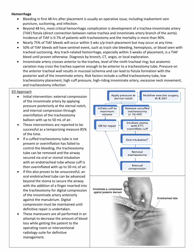
Hemorrhage • Bleeding in first 48 hrs after placement is usually an operative issue, including inadvertent vein
puncture, suctioning, and infection. • Beyond 48 hrs, most critical hemorrhagic complication is development of a trachea-innominate artery
(TIAF) fistula (direct connection between native trachea and innominate artery branch of the aorta). Incidence of TIAF is 0.7% of patients with tracheostomy and the mortality is more than 90%.
• Nearly 75% of TIAF bleeds will occur within 3 wks s/p trach placement but may occur at any time. • 50% of TIAF bleeds will have sentinel event, such as trach site bleeding, hemoptysis, or blood seen with
tracheal suctioning. Any trach-related hemorrhage, especially within 3 weeks of placement, is a TIAF bleed until proven otherwise. Diagnosis by bronch, CT, angio, or local exploration.
• Innominate artery crosses anterior to the trachea, level of the ninth tracheal ring; but anatomic variation may cross the trachea superior enough to be anterior to a tracheostomy tube. Pressure on the anterior tracheal wall results in mucosal ischemia and can lead to fistula formation with the posterior wall of the innominate artery. Risk factors include a cuffed tracheostomy tube, low tracheostomy placement, high cuff pressure, high-riding innominate artery, excessive neck movement, and tracheostomy infection
ED Approach • Initial intervention: external compression
of the innominate artery by applying pressure posteriorly at the sternal notch, and internal compression through overinflation of the tracheostomy balloon with up to 50 mL of air.
• These interventions are reported to be successful as a temporizing measure 85% of the time.
• If a cuffed tracheostomy tube is not present or overinflation has failed to control the bleeding, the tracheostomy tube can be removed and the airway secured via oral or stomal intubation with an endotracheal tube whose cuff is then overinflated with up to 50 mL of air.
• If this also proves to be unsuccessful, an oral endotracheal tube can be advanced beyond the stoma to secure the airway with the addition of a finger inserted into the tracheostomy for digital compression of the innominate artery anteriorly against the manubrium. Digital compression must be maintained until definitive repair is undertaken.
• These maneuvers are all performed in an attempt to decrease the amount of blood loss while getting the patient to the operating room or interventional radiology suite for definitive management.
URGENT COMPLICATIONS Tracheoesophageal Fistula
• Tracheoesophageal fistula (TEF) is a communication between the posterior wall of trachea and anterior wall of esophagus. Prolonged pressure on posteior membranous wall of trachea from a trach balloon results in ischemic necrosis, tissue breakdown, and subsequent fistula formation.
• Risk factors: high airway pressures, prolonged presence of a cuffed tracheostomy tube, high cuff pressures, excessive tracheostomy movement, steroid use, type 1 diabetes mellitus, chronic hypoxia, poor nutritional status, hypotension, anemia, sepsis, the presence of a nasogastric tube, and GERD.
• TEFs may manifest as persistent tracheal air leaks, abdominal distens. (from air entering digestive tract), pulmonary asp. injury, cough with swallowing, copious secretions, and respiratory distress.
• TEF usually 1 to 2 cm distal to the tracheal stoma at the level of the balloon and can be large, measuring 4 to 5 cm. Diagnosis is made by bronchoscopy or, if not available, esophagram.
• Emergency management includes stopping contamination of the airway through tracheal suctioning, discontinuation of oral feeding, and elevation of the patient’s head to 45 degrees. If a gastric tube is present, it should be used to drain the contents of the stomach. However, if a nasogastric tube is in place, it should be removed to pre-vent pressure necrosis and worsening of the fistula. Additionally, any suppurative complications, such as pneumonia, should be treated.
• Long-term treatment consists of conservative management with adjustment of the tracheostomy balloon to a more distal position and nutrition via jejunostomy tube, minimally invasive treatment with tracheal and esophageal stents, or surgery.
Tracheal Stenosis
• Common after prolonged intubation or tracheostomy, with most patients experiencing some degree of tracheal narrowing. However, only 3% to 12% of cases require any intervention and very few of those patients experience critical stenosis requiring urgent intervention.
• Stenosis occurs secondary to granulation tissue and fibrosis of peristomal and tracheal tissues. After granulation tissue forms, it eventually becomes covered with epithelial tissue.
• Trauma to the trachea either from injury during the procedure itself or ischemia from balloon over inflation leads to tracheal inflammation and ulceration. Other risk factors include surgical site infection, GERD, obesity, and hypotension.
• Tracheal stenosis frequently occurs at the site of the stoma, the level of the tracheostomy tube tip, the site of the balloon, or in a suprastomal position.
• Stenosis will not cause symptoms until the diameter of the tracheal lumen is reduced by more than 50%.
• Early symptoms include difficulty clearing secretions, cough, and exertional dyspnea. Dyspnea at rest and stridor are associated with a tracheal diameter of 5 mm. Tracheal stenosis can occur while a patient is still mechanically ventilated or years after decannulation, but most will present within 2 months of decannulation.
• Stenosis can make tube exchanges difficult due to narrowing of the space and bleeding risk. Early stenosis with exposed granulation tissue without overlying epithelium can bleed easily with minor trauma. Stenosis can be treated with dilation, excision with end-to-end anastomosis, or laser excision of granulation or fibrous tissues.

Infection • Tracheostomy is considered a “clean-contaminated” procedure due to the entrance of the upper
aerodigestive tract. Bacterial colonization of the skin and aerodigestive tract combined with the rich environment of blood and secretions at the surgical site place patients at risk of surgical site infection.
• Strict wound care with frequent dressing changes is the cornerstone of preventing infection after tracheostomy.
• Common early infections include cellulitis and tracheitis. These infections frequently occur early in the postoperative course and are more commonly observed after open versus percutaneous tracheostomy. Most early infections are minor, but severe infections, including mediastinitis and necrotizing fasciitis, are possible.
• Delayed presentations of serious infections have been observed, most notably osteomyelitis and septic arthritis of the sternoclavicular joint.
• Aspiration. The presence of a tracheostomy tube, especially a cuffed tube, disrupts swallowing, thus increasing the risk of aspiration. An overinflated tube cuff can compress the esophagus, leading to aspiration. This aspiration is often asymptomatic or silent and therefore unrecognized by patients and caregivers. Aspiration of infected secretions can result in pneumonia or lung abscesses. These infections are most commonly attributed to Staphylococcus aureus, Pseudomonas, and mixed flora.
• Pneumonias and severe skin and soft tissue infections should be treated aggressively. • (Not in paper but discussed in EMOttawablog.com (June 7 2018) Stomal infections often require
admission for IV abx (recommend Vanco and Zosyn to cover staph and pseudomonas). Cutaneous Fistula
• After decannulation, most stomas close in approximately 6 weeks. Persistent epithelialization of the stoma track can result in fistula formation. If the stoma persists after 3 to 6 months, a tracheocutaneous fistula is diagnosed.
• Persistent tracheocutaneous fistulas can lead to skin irritation and infection due to draining secretions, weak cough, and aspiration leading to recurrent pneumonias, poor phonation, poor cosmesis, and submersion intolerance.
• The most notable risk factor is prolonged tracheostomy tube placement with one small series noting tracheostomy placement for at least a year in all observed cases.2 Other risk factors include steroid use, advanced age, and malnutrition. Management options include tract cauterization or excision with healing by secondary intention or surgical closure.