Embed Size (px)
Citation preview
Transcription factor LSF binds two variant bipartite sites within the SV40 late promoter
Hui-Chuan Huang, Rebecca Sundseth, and Ul la Hansen
Laboratory of Eukaryotic Transcription, Dana-Farber Cancer Institute, and Department of Microbiology and Molecular Genetics, Harvard Medical School, Boston, Massachusetts 02115 USA
The HeLa transcription factor LSF has been purified by heparin-agarose and DNA affinity chromatography, and its DNA binding and transcription properties have been characterized. LSF is a 63-kD polypeptide that binds to two distinct bipartite sites within the SV40 promoter region. One binding site consists of GC motifs 2 and 3 within the 21-bp repeats (LSF-GC site), and the other consists of sequences centered 44 bp upstream of the major late initiation site, L325 (LSF-280 site). Four guanine residues within the LSF-GC site, when methylated, strongly interfere with LSF binding. Alteration of the spacing, but not the sequence, between the two directly repeated GC motifs dramatically reduces the binding affinity of LSF for the site. Thus, LSF appears to recognize directly repeated GC motifs, when their center-to-center distance is 10 bp. The LSF-GC and LSF-280 sites share limited sequence homology. Only half of the LSF-280 site contains a short GC-rich sequence homologous to the GC motif. However, the binding affinity of LSF to the two sites is similar. LSF activates transcription from the SV40 late promoter in vitro from initiation site L325, via its binding to the template DNA.
[Key Words: Transcription factor; DNA-binding protein; SV40 late promoter; GC-motif; LSF; GT-IIA]
Received September 4, 1989; revised version accepted November 29, 1989.
SV40 offers an excellent model system for studying tran- scriptional regulatory mechanisms in mammalian cells: The virus uses the host cell transcription machinery for expression of its genes, and both biochemical and ge- netic means can readily be applied in investigating its transcription. During lytic infection, transcription of SV40 is precisely regulated. Before the onset of viral DNA replication, the SV40 early genes (encoding the large-T and small-t antigens) are primarily transcribed; after the onset of viral DNA replication, the SV40 late promoter is activated, and the late genes (encoding the viral capsid proteins) are predominantly expressed (Tooze 1981).
The DNA sequences in the SV40 genome that regulate early and late transcription are located within a region of -400 bp (see Fig. 8, below). The SV40 late promoter ap- pears to be very complex and may actually be a combina- tion of multiple overlapping promoters capable of stimu- lating transcription from the same initiation sites. Many DNA sequence elements have been implicated in pro- moting late transcription in vivo and/or in vitro. These sequences include the origin of DNA replication (Brady and Khoury 1985; Keller and Alwine 1985), the three 21-bp tandem repeats containing six directly repeated GC motifs (GGGCGG) (Fromm and Berg 1982; Hansen and Sharp 1983; Brady et al. 1984; Hartzell et al. 1984a; Rio and Tjian 1984; Vigneron et al. 1984), the 72-bp di- rect repeats containing many enhancer elements
(Fromm and Berg 1982; Hartzell et al. 1984b; Keller and Alwine 1985; Emoult-Lange et al. 1987), and the region from the 72-bp repeats up to and beyond the major in vivo initiation site, L325 (Brady et al. 1982; Keller and Alwine 1985; Emoult-Lange et al. 1987; Ayer and Dynan 1988). The major late initiation site L325 is not preceded by a typical TATA box (Brady et al. 1982). Environ- mental factors may influence the promoter regions that are important in activating SV40 late transcription. In particular, the presence of T antigen dramatically alters the effect of the GC motif region on the SV40 late pro- moter, and the occurrence of SV40 DNA replication alters the relative importance of various elements within the 72-bp repeats for SV40 late transcription {Keller and Alwine 1985; Alwine and Picardi 1986; Omilli et al. 1986; Robbins et al. 1986; Ernoult-Lange et al. 1987).
To elucidate the molecular mechanisms of initiation of transcription by RNA polymerase II, the trans-acti- vating factors that interact with cis-activating DNA se- quences must be purified. Only then can the interac- tions of a specific trans-acting factor with the DNA and with other proteins involved in the initiation of tran- scription at the promoter be studied directly. Multiple DNA-binding proteins specific for sequences within the SV40 promoter region have been identified; however, only recently has the involvement of particular purified proteins in transcription of the major in vivo initiation
GENES & DEVELOPMENT 4:287-298 © 1990 by Cold Spring Harbor Laboratory Press ISSN 0890-9369/90 $1.00 287
Cold Spring Harbor Laboratory Press on November 24, 2018 - Published by genesdev.cshlp.orgDownloaded from
Huang et al.
site of the SV40 late promoter been shown. AP-1 and AP-4 were reported to synergistically stimulate SV40 late transcription from the L325 initiation site three- to fourfold in vitro (Mermod et al. 1988). In addition, we identified previously a cellular transcription factor from HeLa cells, LSF (designated late SV40 factor), that stimu- lated SV40 late transcription and bound specifically se- quences within the SV40 21-bp repeat promoter element (Kim et al. 1987). Here, we present the purification and further characterization of LSF. The protein stimulates SV40 late transcription in vitro from the major late initi- ation site and binds two variant bipartite sites within the SV40 promoter region.
R e s u l t s
Purification of LSF
The LSF purification scheme is described in Methods and summarized in Table 1. HeLa whole-cell extract was applied to a heparin-agarose affinity column, and bound proteins were eluted with a linear salt gradient. Frac- tions containing LSF demonstrated DNA-binding activi- ties that protected both a sequence including GC motifs 2 and 3 and part of GC-motif 1 (LSF-GC site) and a se- quence from nucleotides 272 to 291 (LSF-280 site) from DNase I digestion (Fig. 1B, lanes 3-5 , 11). These two regions were distinct from sequences protected from DNase I cleavage by previously identified proteins. [Spl binds strongly to GC motifs 3, 5, and 6 within the SV40 21-bp repeats (Gidoni et al. 1984). AP-4 protects only se- quences between nucleotides 267 and 276 with high af- finity (Mermod et al. 1988).]
To purify further the LSF transcription activity in the heparin eluate, sequence-specific DNA affinity resins were prepared (Kadonaga and Tjian 1986). The first DNA affinity resin contained the SV40 GC motifs 1, 2, and 3 (GC123, nucleotides 42-70), which includes the LSF- GC site. After purification by two consecutive passes over the column, the fractions with specific DNA- binding activity (Fig. 1B, lanes 6-9) contained a polypep- tide of 63 kD as the major polypeptide species (10% of the eluted protein), excluding the added carrier protein BSA (Fig. 1A, lane 4). Minor polypeptide species, in- cluding those with molecular masses of 105 kD, 60 kD,
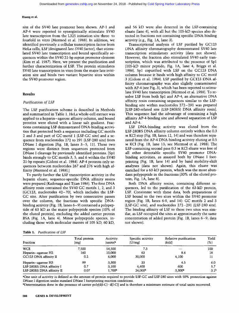
Table 1. Purification of LSF
and 56 kD were also detected in the LSF-containing eluate (lane 4), with all but the 105-kD species also de- tected in fractions not containing specific DNA-binding activity (e.g., Fig. 1A, lane 3).
Transcriptional analysis of LSF purified by GC123 DNA affinity chromatography demonstrated SV40 late transcription stimulatory activity (data not shown). However, the fraction also stimulated SV40 early tran- scription, which was attributed to the presence of Spl (105-kD minor peptide; Fig. 1A, lane 4; Briggs et al. 1986). Spl copurified with LSF on the GC123 DNA column because it binds with high affinity to GC motif 3 (Gidoni et al. 1984). LSF purified by GC123 DNA af- finity chromatography was also slightly contaminated with AP-4 (see Fig. 3), which has been reported to stimu- late SV40 late transcription {Mermod et al. 1988). To re- solve LSF from both Spl and AP-4, an alternative DNA affinity resin containing sequences similar to the LSF- binding site within nucleotides 272-291 was prepared [LSF-280-related site (LSF-280RS) DNA affinity resin]. This sequence had the advantage of containing a high affinity AP-4-binding site and allowed separation of LSF and AP-4.
LSF DNA-binding activity was eluted from the LSF-280RS DNA affinity column entirely within the 0.3 M KC1 step (Fig. 1B, lanes 12, 14) and was therefore sepa- rated from the AP-4 DNA-binding activity eluting at 0.5 M KC1 (Fig. 1B, lane 13; see Mermod et al. 1988). The LSF-containing second pass 0.3 M KC1 eluate was free of all other detectable specific SV40 promoter DNA- binding activities, as assayed both by DNase I foot- printing (Fig. 1B, lane 14) and by band mobility-shift analyses (data not shown). Again, this eluate was enriched for a 63-kD protein, which was the most abun- dant polypeptide in the fractions (30% of the eluted pro- tein; Fig. 1A, lane 8).
Both DNA affinity resins, containing different se- quences, led to the purification of the 63-kD protein, LSF. Consistent with these data, both preparations of LSF bound to the two sites within the SV40 promoter region {Fig. 1B, lanes 6-9, and 14): GC motifs 2 and 3 (LSF-GC site), and nucleotides 272-291 (LSF-280 site). The binding affinity of LSF to these two sites was sim- ilar, as LSF occupied the sites at approximately the same concentration of added protein (Fig. 1B, lanes 6-9 ; data not shown).
Total protein Activity Fraction (mg) (units)~
Specific activity Relative purification Yield (U/mg) (fold) (%)
WCE Heparin-agarose H2 GC 123 DNA affinity II
Heparin-agarose H3 LSF-280RS DNA affinity I LSF-280RS DNA affinity II
7,500 54,500 7.3 - - 160 10,000 63 8.6
0.2 6,000 30,000 4,100
99 3,300 33 4.5 0.7 3,100 4,400 600 0.07 1,700 b 24,000 b 3,300 b
100 18 11
6.0 5.7 3.1 b
aOne unit of activity is defined as the amount of protein required to provide LSF-GC and LSF-280 sites with 50% protection against DNase I digestion under standard DNase I footprinting reaction conditions. bDetermination done in the presence of cartier poly[d(I-C) • d{I-C)] and is therefore a minimum estimate of total units recovered.
288 GENES & DEVELOPMENT
Cold Spring Harbor Laboratory Press on November 24, 2018 - Published by genesdev.cshlp.orgDownloaded from
LSF binds two variant SV40 D N A sequences
H2 2ndp0s~s £ £ o o'0~6Fi.'~5 Es i 2' ¢}
c : v 20 *~ A / T
16C1 . . . .
- I 0 0 -
(178) - 250 -
(178) - 2 5 0
2 7 0 - ~ =. ~ ~ 1" \~' 2 8 0 - ,~ - 2 7 0
,, - 2 8 0
2 0 0 ' -
I I I!!iil!I I16- -66 - ~ ~i~ ; <~ ~<,,,*-~
5 2 5 - o . ~ . , i l ~ , ~
6 6 - ~ ~ - / / - 45 ~ ~ i ~ " i ; ~ i : ~ : ~ : ~ ~
4 5 _ ~ : '
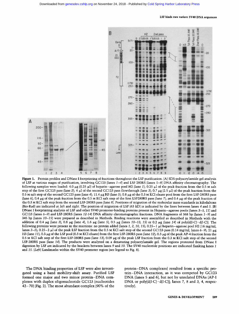
Figure 1. Protein profiles and DNase I footprinting of fractions throughout the LSF purification. {A) SDS-polyacrylamide gel analysis of LSF at various stages of purification, involving GC123 (lanes 1-4) and LSF-280RS (lanes 5-8) DNA affinity chromatography. The following samples were loaded: 4.0 ~g (0.25 wl) of heparin-agarose pool H2 (lane 1); 0.25 ~1 of the peak fraction from the 0.5 M salt step of the first GC123 pass (lane 2); 4 ~1 of the second GC123 pass flowthrough {lane 3); 0.7 ~g (2.5 ~1) of the peak fraction from the 0.5 M salt step of the second GC123 pass (lane 4); 11.4 ~g H3 (lane 5); 0.8 ~g of the 0.3 M KC1 eluate pool from the first LSF-280RS pass (lane 6); 0.4 ~g of the peak fraction from the 0.5 M KCI salt step of the first LSF280RS pass (lane 7); and 0.4 wg of the peak fraction of the 0.3 M KC1 salt step from the second LSF-280RS pass (lane 8). Positions of migration of the molecular mass standards in kilodaltons (Bio-Rad) are indicated at left and right. The position of migration of LSF (63 kD) is indicated by the lines between lanes 4 and 5. (B) DNase I footprinting analysis of LSF and other SV40 promoter-binding proteins present in Heparin-agarose pools (lanes 3-5, 11) and GC123 (lanes 6-9) and LSF-280RS (lanes 12-14) DNA affinity chromatographic fractions. DNA fragments of 368 bp (lanes 1-9) and 345 bp (lanes 10-15) were prepared as described in Methods. Binding reactions were assembled as described in Methods with the addition of 0.4 ~g (lane 3), 0.8 ~g (lane 4), 1.6 ~g (lane 5), 0.5 ~g (lanes 10-13, 15) or 0.2 ~g (lane 14) of poly[d(I-C)- d(I-C)]. The following proteins were present in the reactions: no protein added (lanes 1, 2, 10, 15); 0.25-1 Ixl heparin--agarose pool H2 {16 mg/ml, lanes 3-5); 0.25-2 F1 of the peak LSF fraction from the 0.5 M KC1 salt step of the second GC123 pass (0.14 mg/ml, lanes 6-9); 21 I~g H3 (lane 11); 0.3 v~g of the LSF pool {0.3 M KC1 eluate) from the first LSF-280RS pass (lane 12); 0.3 wg of the peak AP-4 fraction from the 0.5 M KC1 salt step of the first LSF-280RS pass (lane 13); 0.09 wg of the peak LSF fraction from the 0.3 M KC1 salt step of the second LSF-280RS pass (lane 14). The products were analyzed on a denaturing polyacrylamide gel. The regions protected from DNase I digestion by LSF are indicated by the brackets between lanes 9 and 10. The SV40 nucleotide positions are indicated flanking lanes 1 and 15. (Left) Landmarks within the SV40 promoter region {see legend to Fig. 8).
The DNA binding properties of LSF were also investi- gated using a band mobility-shift assay. Purified LSF formed one major and two minor protein-DNA com- plexes with duplex oligonucleotide GC123 (nucleotides 42-70) (Fig. 2). The most abundant complex (90% of the
protein-DNA complexes) resulted from a specific pro- t e in -DNA interaction, as it was competed by GC123 DNA {lanes 5 and 6), but not by unrelated DNAs (AP-1 DNA or poly[d{I-C) • d(I-C)]; lanes 7, 8 and 3, 4, respec- tively).
GENES & DEVELOPMENT 289
Cold Spring Harbor Laboratory Press on November 24, 2018 - Published by genesdev.cshlp.orgDownloaded from
Huang et al.
LSF is a 63-kD polypeptide
To prove that the molecular mass of LSF is 63 kD and to rule out the possibility that purified LSF contains mul- tiple polypeptides that are required for specific DNA- binding activity, the ability of size-fractionated LSF to specifically bind SV40 DNA was tested. A purified prep- aration of LSF was fractionated on an SDS-polyacryl- amide gel, and the proteins from different gel slices were eluted and renatured, as described in Methods. Of pro- teins corresponding to a wide range of molecular masses, only those within the range of 62-64 kD demonstrated specific binding to the GC123 DNA fragment (Fig. 3A, lane 8). This proved that the 63-kD polypeptide was nec- essary and sufficient for formation of the specific LSF/ DNA complex (see Fig. 2).
Protein renatured from the gel slice containing the 63-kD polypeptide also bound to LSF-280 DNA (nucleo- tides 270-290, cf. Fig. 3A and 3B, lanes 8). In addition, the LSF-280 DNA was bound by smaller polypeptides (Fig. 3B; lanes 11-14); however, the mobilities of these p ro te in -DNA complexes were not identical to that of the LSF-DNA complex (lanes 11-13). The complex in the vicinity of 48 kD was likely to be attributable to the presence of AP-4 (lane 12), as the LSF-280 oligonucleo- tide contained 7 out of 10 bp of the consensus AP-4- binding site (Mermod et al. 1988). To investigate any re- lation in DNA-binding activity between AP-4 and LSF, the renatured proteins were tested for their ability to bind to a DNA fragment containing only the AP-4- binding site. The 63-kD polypeptide, which bound LSF-
9
Cornpelitor , ~ ~ D N ~ i - - ' t i , I - - ~ l - -
Free - I 2 3 4 5 6 7 8 9
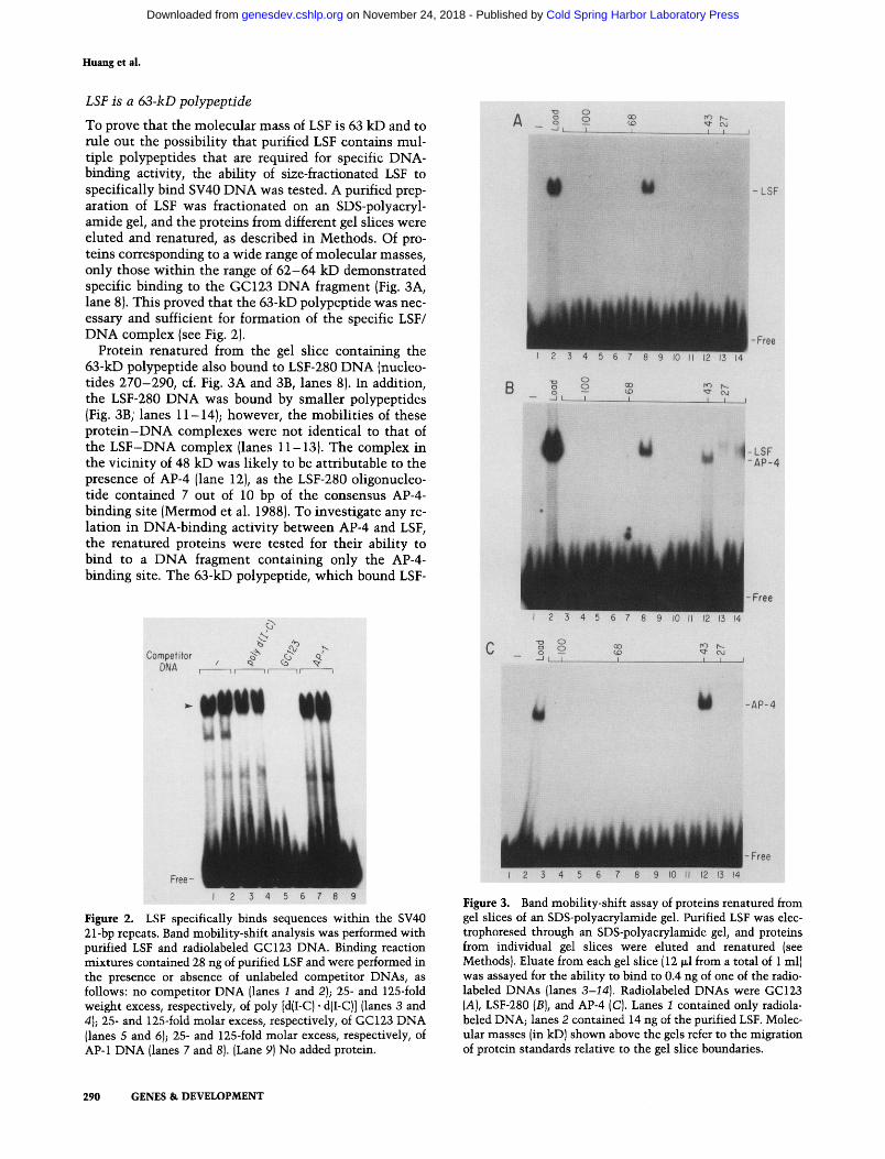
Figure 2. LSF specifically binds sequences within the SV40 21-bp repeats. Band mobility-shift analysis was performed with purified LSF and radiolabeled GC123 DNA. Binding reaction mixtures contained 28 ng of purified LSF and were performed in the presence or absence of unlabeled competitor DNAs, as follows: no competitor DNA [lanes 1 and 2); 25- and 125-fold weight excess, respectively, of poly [dII-C) • dII-C)] {lanes 3 and 41; 25- and 125-fold molar excess, respectively, of GC123 DNA {lanes 5 and 6); 25- and 125-fold molar excess, respectively, of AP-1 DNA {lanes 7 and 81. [Lane 91 No added protein.
0
_, o,, 7 ~ ~ ' ?
-LSF -AP-4
-~ ,T , j , i~, ~
W -AP-4
~ :
[
2
Figure 3. Band mobility-shift assay of proteins renatured from gel slices of an SDS-polyacrylamide gel. Purified LSF was elec- trophoresed through an SDS-polyacrylamide gel, and proteins from individual gel slices were eluted and renatured (see Methods}. Eluate from each gel slice (12 ~1 from a total of 1 ml) was assayed for the ability to bind to 0.4 ng of one of the radio- labeled DNAs (lanes 3-14). Radiolabeled DNAs were GC123 (A), LSF-280 (B), and AP-4 (C). Lanes 1 contained only radiola- beled DNA; lanes 2 contained 14 ng of the purified LSF. Molec- ular masses (in kD) shown above the gels refer to the migration of protein standards relative to the gel slice boundaries.
290 GENES & DEVELOPMENT
Cold Spring Harbor Laboratory Press on November 24, 2018 - Published by genesdev.cshlp.orgDownloaded from
GC and LSF-280 DNAs (Fig. 3A, B, lanes 8), did not bind to the AP-4 DNA (Fig. 3C, lane 8). Conversely, the smaller polypeptide AP-4 (Mermod et al. 1988) only bound to the AP-4 and LSF-280 DNAs but not to the GC123 DNA (Fig. 3A-C, lanes 12). Thus, LSF and AP-4 are different proteins with unrelated DNA-binding prop- erties.
LSF binds to two directly repeated GC-motifs at the LSF-GC site
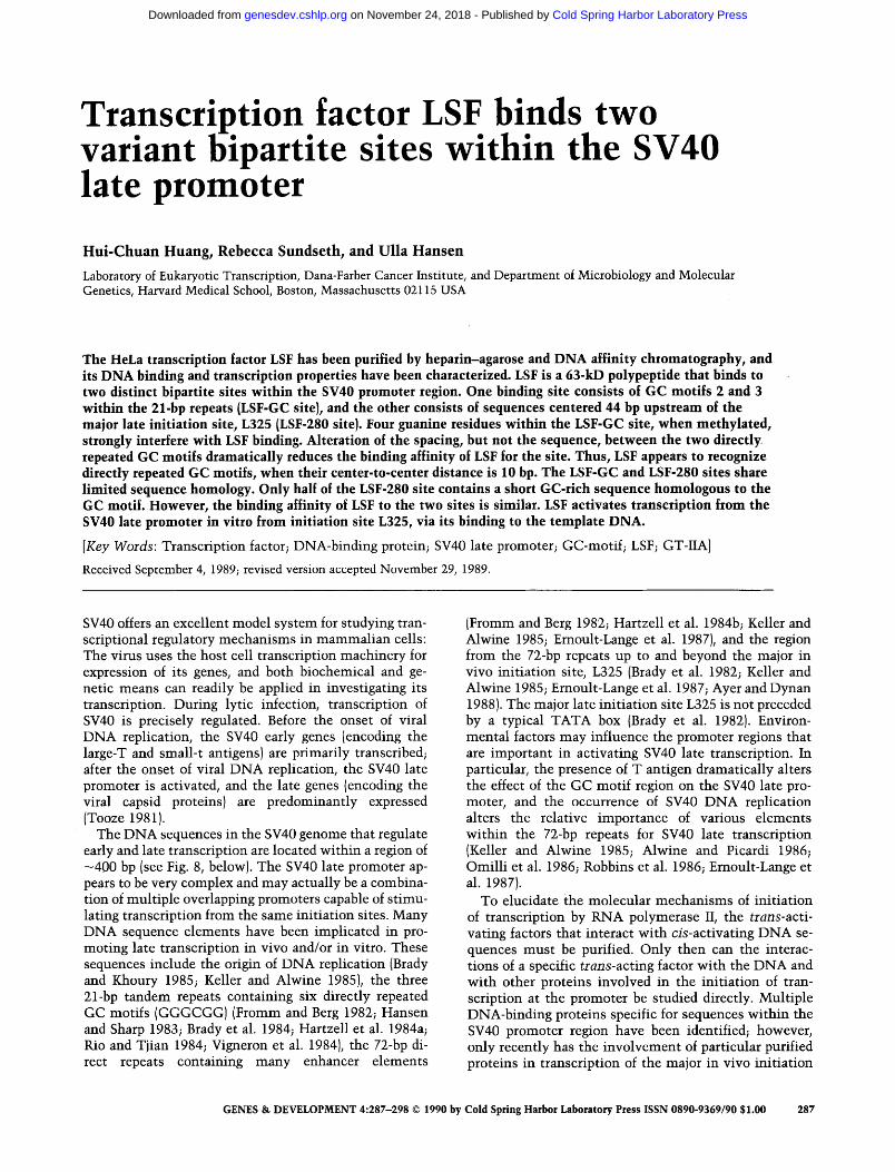
Methylation interference studies (Siebenlist and Gilbert 1980) were performed to characterize the close contact points made by purified LSF bound to the LSF-GC site. Methylated duplex GC123 DNA was incubated with LSF, and the bound form of DNA was separated from the free form by a band mobility-shift experiment. The methylation patterns of the bound and free DNAs are shown in Figure 4. Although the GC123 DNA was ra- diolabeled at both 5' termini, radioactive fragments cleaved at all individual guanine residues from both DNA strands were uniquely separated following electro- phoresis through a denaturing polyacrylamide gel (Fig. 4, lanes 4 and 5). Formation of the LSF complex was strongly inhibited by methylation of four guanine res- idues, located at positions 53, 58, 63, and 68, as bound DNA did not contain methylated bases at these posi- tions (cf. lane 2 with lanes 1 and 3). These four guanine residues represent two from each GC motif, at equiva- lent positions (solid arrows within the LSF-GC site in Fig. 8). In addition, methylation of guanine residues at positions 54, 57, and 64 partially inhibited LSF binding (shaded arrows within the LSF-GC site in Fig. 8). The fact that N7 methylation of guanine residues inhibited binding of LSF suggests that LSF binds within the major groove of the DNA.
F B FM1M2
G68
G65
G58
G53
I 2 3 4 5
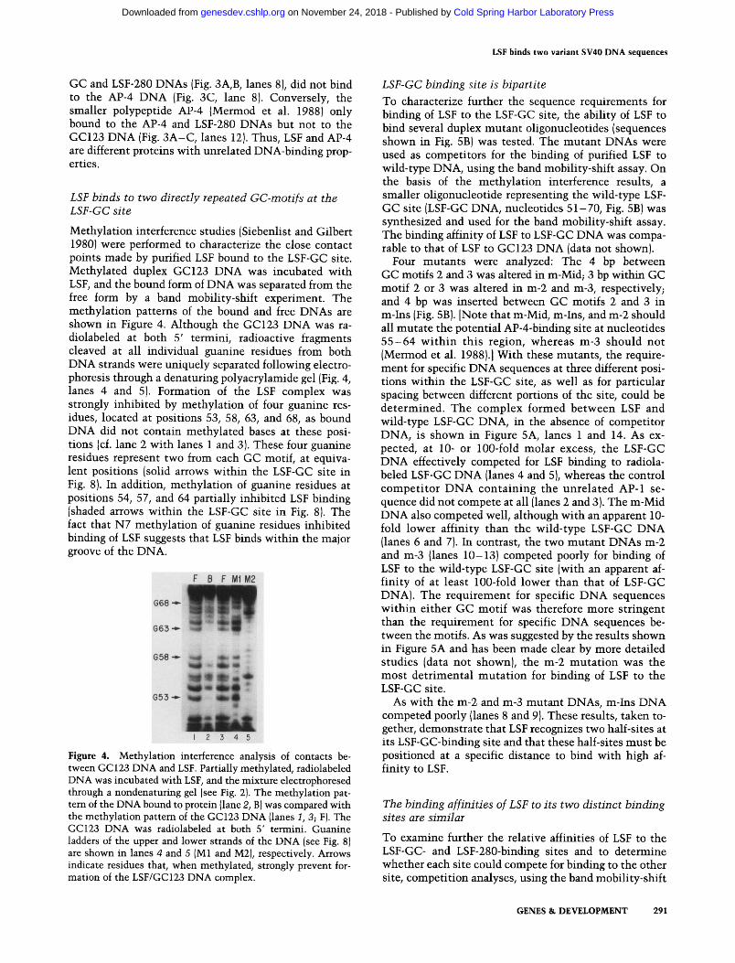
Figure 4. Methylation interference analysis of contacts be- tween GC123 DNA and LSF. Partially methylated, radiolabeled DNA was incubated with LSF, and the mixture electrophoresed through a nondenaturing gel (see Fig. 2). The methylation pat- tern of the DNA bound to protein (lane 2, B) was compared with the methylation pattern of the GC123 DNA (lanes 1, 3; F). The GC123 DNA was radiolabeled at both 5' termini. Guanine ladders of the upper and lower strands of the DNA (see Fig. 8) are shown in lanes 4 and 5 (M1 and M2), respectively. Arrows indicate residues that, when methylated, strongly prevent for- mation of the LSF/GC123 DNA complex.
LSF binds two variant SV40 DNA sequences
LSF-GC binding site is bipartite
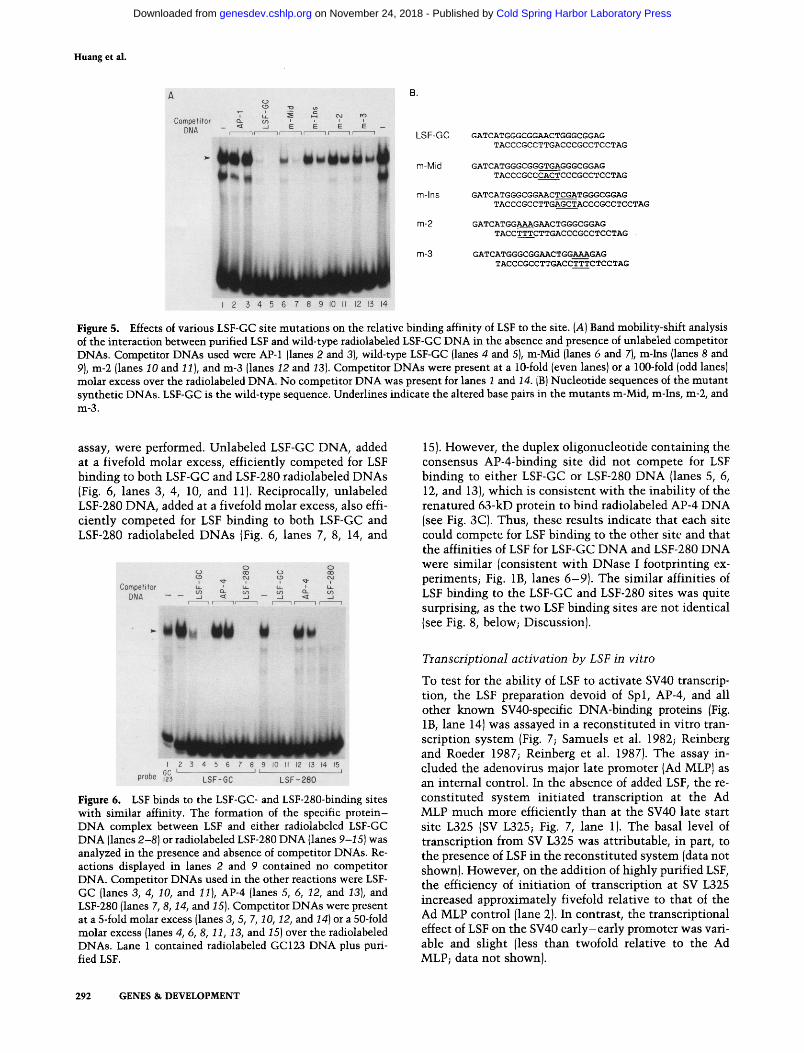
To characterize further the sequence requirements for binding of LSF to the LSF-GC site, the ability of LSF to bind several duplex mutant oligonucleotides (sequences shown in Fig. 5B) was tested. The mutant DNAs were used as competitors for the binding of purified LSF to wild-type DNA, using the band mobility-shift assay. On the basis of the methylation interference results, a smaller oligonucleotide representing the wild-type LSF- GC site (LSF-GC DNA, nucleotides 51-70, Fig. 5B) was synthesized and used for the band mobility-shift assay. The binding affinity of LSF to LSF-GC DNA was compa- rable to that of LSF to GC123 DNA (data not shown).
Four mutants were analyzed: The 4 bp between GC motifs 2 and 3 was altered in m-Mid; 3 bp within GC motif 2 or 3 was altered in m-2 and m-3, respectively; and 4 bp was inserted between GG motifs 2 and 3 in m-Ins (Fig. 5B). [Note that m-Mid, m-Ins, and m-2 should all mutate the potential AP-4-binding site at nucleotides 5 5 - 6 4 wi th in this region, whereas m-3 should not (Mermod et al. 1988}.] With these mutants, the require- ment for specific DNA sequences at three different posi- tions within the LSF-GC site, as well as for particular spacing between different portions of the site, could be determined. The complex formed between LSF and wild-type LSF-GC DNA, in the absence of competitor DNA, is shown in Figure 5A, lanes 1 and 14. As ex- pected, at 10- or 100-fold molar excess, the LSF-GC DNA effectively competed for LSF binding to radiola- beled LSF-GC DNA (lanes 4 and 5), whereas the control compet i tor DNA containing the unrelated AP-1 se- quence did not compete at all (lanes 2 and 3). The m-Mid DNA also competed well, although with an apparent 10- fold lower affinity than the wild-type LSF-GC DNA (lanes 6 and 7). In contrast, the two mutant DNAs m-2 and m-3 (lanes 10-13) competed poorly for binding of LSF to the wild-type LSF-GC site (with an apparent af- finity of at least 100-fold lower than that of LSF-GC DNA). The requirement for specific DNA sequences within either GC motif was therefore more stringent than the requirement for specific DNA sequences be- tween the motifs. As was suggested by the results shown in Figure 5A and has been made clear by more detailed studies (data not shown), the m-2 mutat ion was the most detrimental mutat ion for binding of LSF to the LSF-GG site.
As with the m-2 and m-3 mutant DNAs, m-Ins DNA competed poorly (lanes 8 and 9). These results, taken to- gether, demonstrate that LSF recognizes two half-sites at its LSF-GC-binding site and that these half-sites must be positioned at a specific distance to bind with high af- finity to LSF.
The binddng affinities of LSF to its two distinct binding sites are similar
To examine further the relative affinities of LSF to the LSF-GC- and LSF-280-binding sites and to determine whether each site could compete for binding to the other site, competition analyses, using the band mobility-shift
GENES & DEVELOPMENT 291
Cold Spring Harbor Laboratory Press on November 24, 2018 - Published by genesdev.cshlp.orgDownloaded from
Huang et al.
A
Compefiter ~- ~- ~ ~ - - o ' ~ i i
DNA r '~ ~ E ~= E - - 7 1 --1 r - - - 7 r - - - - 1 r - - I I I L S F - G C
m - M i d
m - I n s
m-2
m-3
GATCATGGGCGGAACTGGGCGGAG TACCCGCCTTGACCCGCCTCCTAG
GATCATGGGCGGGTGAGGGCGGAG TACCCGCCCACTCCCGCCTCCTAG
GATCATGGGCGGAACTCGATGGGCGGAG
TACCCGCCTTGAGCTACCCGCCTCCTAG
GATCATGGAAAGAACTGGGCGGAG TACCTTTCTTGACCCGCCTCCTAG •
GATCATGGGCGGAACTGGAAAGAG TACCCGCCTTGACCTTTCTCCTAG
I 2 3 4 5 6 7 8 9 I0 II 12 13 14
Figure 5. Effects of various LSF-GC site mutations on the relative binding affinity of LSF to the site. (A) Band mobility-shift analysis of the interaction between purified LSF and wild-type radiolabeled LSF-GC DNA in the absence and presence of unlabeled competitor DNAs. Competitor DNAs used were AP-1 (lanes 2 and 3), wild-type LSF-GC (lanes 4 and 5), m-Mid (lanes 6 and 7), m-Ins (lanes 8 and 9), m-2 (lanes 10 and 11), and m-3 (lanes I2 and •3). Competitor DNAs were present at a 10-fold (even lanes) or a 100-fold (odd lanes) molar excess over the radiolabeled DNA. No competitor DNA was present for lanes 1 and 14. [B) Nucleotide sequences of the mutant synthetic DNAs. LSF-GC is the wild-type sequence. Underlines indicate the altered base pairs in the mutants m-Mid, m-Ins, m-2, and m-3.
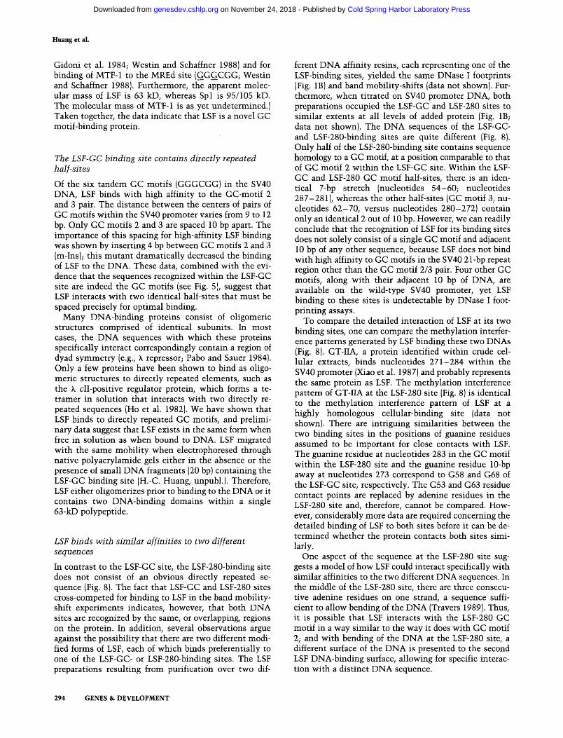
assay, were performed. Unlabeled LSF-GC DNA, added at a fivefold molar excess, efficiently competed for LSF binding to both LSF-GC and LSF-280 radiolabeled DNAs (Fig. 6, lanes 3, 4, 10, and 11). Reciprocally, unlabeled LSF-280 DNA, added at a fivefold molar excess, also effi- ciently competed for LSF binding to both LSF-GC and LSF-280 radiolabeled DNAs (Fig. 6, lanes 7, 8, 14, and
Competitor DNA - - - - "~
I i [ . . . . .
ou , , ~ ,~
- J - - . u , , ~ - . I 11 i i ..... i I - - 1 r - - 1
-roll, im w
I 2 3 4 5 6 7 8 9 JO II 12 J3 14 15 GC L Jt j
probe rz3 LSF-GC LSF-280
Figure 6. LSF binds to the LSF-GC- and LSF-280-binding sites with similar affinity. The formation of the specific protein- DNA complex between LSF and either radiolabeled LSF-GC DNA (lanes 2-8) or radiolabeled LSF-280 DNA (lanes 9-15) was analyzed in the presence and absence of competitor DNAs. Re- actions displayed in lanes 2 and 9 contained no competitor DNA. Competitor DNAs used in the other reactions were LSF- GC (lanes 3, 4, 10, and 11), AP-4 (lanes 5, 6, 12, and 13), and LSF-280 (lanes 7, 8, 14, and 15). Competitor DNAs were present at a 5-fold molar excess (lanes 3, 5, 7, 10, 12, and 14) or a 50-fold molar excess (lanes 4, 6, 8, 11, 13, and 15) over the radiolabeled DNAs. Lane 1 contained radiolabeled GC123 DNA plus puri- fied LSF.
15). However, the duplex oligonucleotide containing the consensus AP-4-binding site did not compete for LSF binding to either LSF-GC or LSF-280 DNA (lanes 5, 6, 12, and 13), which is consistent wi th the inabil i ty of the renatured 63-kD protein to bind radiolabeled AP-4 DNA (see Fig. 3C). Thus, these results indicate that each site could compete for LSF binding to the other site and that the affinities of LSF for LSF-GC DNA and LSF-280 DNA were similar (consistent wi th DNase I footprinting ex- periments; Fig. 1B, lanes 6-9). The similar affinities of LSF binding to the LSF-GC and LSF-280 sites was quite surprising, as the two LSF binding sites are not identical (see Fig. 8, below; Discussion).
Transcriptional activation by LSF in vitro
To test for the ability of LSF to activate SV40 transcrip- tion, the LS£ preparation devoid of Spl, AP-4, and all other known SV40-specific DNA-binding proteins (Fig. 1B, lane 14) was assayed in a reconstituted in vitro tran- scription system (Fig. 7; Samuels et al. 1982; Reinberg and Roeder 1987; Reinberg et al. 1987). The assay in- cluded the adenovirus major late promoter (Ad MLP) as an internal control. In the absence of added LSF, the re- constituted system init iated transcription at the Ad MLP much more efficiently than at the SV40 late start site L325 (SV L325; Fig. 7, lane 1). The basal level of transcription from SV L325 was attributable, in part, to the presence of LSF in the reconstituted system (data not shown). However, on the addition of highly purified LSF, the efficiency of ini t iat ion of transcription at SV L325 increased approximately fivefold relative to that of the Ad MLP control (lane 2). In contrast, the transcriptional effect of LSF on the SV40 ear ly -ear ly promoter was vari- able and slight (less than twofold relative to the Ad MLP; data not shown).
292 G E N E S & DEVELOPMENT
Cold Spring Harbor Laboratory Press on November 24, 2018 - Published by genesdev.cshlp.orgDownloaded from

Competitor ONA
( .9 !
I .L Od ¢ j ) . j E
- - - - I i i i
;if!i:
: :i: ¢
- Ad MLP
I 2 3 4 5 6
Figure 7. Analysis of LSF transcriptional activity on SV40 (SV L325) and adenovirus (Ad MLP) late promoters in a reconsti- tuted in vitro transcription system. General RNA polymerase II transcription factors were combined, as described in Methods, in the absence (lane 1), or presence (lanes 2-6) of 200 ng LSF. This mixture was incubated in the absence (lanes 1 and 2), or presence (lanes 3-6) of competitor DNAs prior to addition of template DNAs. Competitor DNAs used were LSF-GG DNA (30 ng, lane 3; 300 ng, lane 4) and m-2 DNA (30 ng, lane 5; 300 ng, lane 6). Radiolabeled RNAs synthesized in the transcription reaction were selected by hybridization and resolved by electro- phoresis through the denaturing polyacrylamide gel shown. The migration positions of RNAs initiated at SV L325 and Ad MLP are indicated. The 5' terminus of the RNA product mi- grating slightly faster than the SV L325 RNA has been mapped to 18-20 bp downstream of SV L325 (nucleotides 353-355). SV40 16S mRNAs with initiation sites around this position have been observed in vivo when deletions are present in the late leader region, either downstream or upstream of the late initiation sites (Piatak et al. 1981, 1983).
To prove that the transcriptional stimulation of the SV40 late promoter was attributable to the specific binding of LSF to the template DNA, competitor DNAs were added during the transcription reaction. At a 10- fold molar excess of LSF-GC DNA over LSF-binding sites in the SV40 viral DNA transcription template (lane 3), the activation by LSF at SV L325 was decreased 4-fold (cf. lanes 2 and 3), and at 100-fold molar excess, the SV40 L325 signal was decreased 9-fold (cf. lanes 2 and 4). The competitor DNA clearly competed away both the added LSF and the endogenous LSF in the reconstituted tran- scription reaction. In contrast, m-2 DNA had no effect when added at either 10- or 100-fold molar excess (lanes 5,6, respectively).
These results demonstrate that the 63-kD site-specific DNA binding protein LSF is a transcription factor that activates the SV40 late promoter at the major late mRNA start site observed in vivo, L325. In addition, the competition data show that the specific binding of LSF to the transcription template is absolutely required for its transcriptional stimulatory activity.
LSF binds t w o variant SV40 D N A sequences
Discussion
LSF is a GC motif-binding protein
Two GC motif-binding proteins have been characterized previously; Spl and MTF-1 (Gidoni et al. 1984; Westin and Schaffner 1988). Spl binds to GC-motifs in many viral and cellular genes (Dynan 1986; Kadonaga et al. 1986). MTF-1, a zinc-inducible factor, binds to the mouse methallothionein I gene (at the MREd site; Westin and Schaffner 1988). LSF is different from both Spl and MTF-1 by virtue of its DNA binding properties and its molecular mass. Of six GC motifs in the SV40 promoter, LSF only binds strongly to GC motifs 2 and 3, whereas Spl binds strongly to GC motifs 3, 5, and 6 (Gi- doni et al. 1984). The methylation of guanine residues located at the boundaries of each GC-motif (GGGCGG) strongly interferes with binding of LSF to the DNA. In contrast, guanine residues, both at the boundaries and in the center of the GC motifs, are involved as closed con- tact points for binding of Spl to both SV40 and the MREd sites (GGGCGG and GGGCGG, respectively;
LSF-GC
0/5243
EE
m
100 200 300
72 bp 72 bp
LSF-GC LSF-280
L325
TACCCCGCCTCTTACCCGCCTTGACCCGCCTCAATCCCCGCCCTA
I I
L S F - 2 8 0 ACACACATTCCACAGCTGGTTCTTTCCGCCTCAGAAGGTACCTAAC TGTGTGTAAGGTGTCGACCAAGAAAGGCGGAGTCTTCCATGGATTG
F i g u r e 8. Summary of DNase I protection and methylation in- terference analyses of the LSF complexes at LSF-GC and LSF-280 DNA sites within the SV40 promoter region. (Top) Map of the SV40 late and early promoter region. The numbers of nucleotide positions are given above the map. The indicated landmarks of the promoter region include: two 72-bp repeats (shaded boxes), six directly repeated GC motifs [solid boxes) within three copies of direct 21-bp repeats (open boxes), a stretch of 17 adenine and thymine base pairs, including the early TATA sequence (AT box), SV40 early-early and major late initiation sites (EE and L325, respectively}, and the two LSF DNA-binding sites (LSF-GC and LSF-280). DNA sequences containing the LSF-GC- and LSF-280-binding sites are repre- sented below. Brackets mark the overall limits of the DNase I-protected regions by LSF at both LSF-GC and LSF-280 sites. The brackets indicate a minimal protection region, as the boundaries were determined by the outermost cleavage sites protected, and the next unaffected DNase cleavage sites were not at adjacent base pairs. The solid and shaded arrows in the sequence diagram of LSF-GC highlight residues that, when methylated, strongly or partially prevent LSF binding, respec- tively. The solid and shaded arrows in the sequence diagram of LSF-280 indicate residues that, when methylated, strongly or partially prevent GT-IIA binding (Xiao et al. 1987).
GENES & D E V E L O P M E N T 293
Cold Spring Harbor Laboratory Press on November 24, 2018 - Published by genesdev.cshlp.orgDownloaded from
Huang et al.
Gidoni et al. 1984; Westin and Schaffner 1988) and for binding of MTF-1 to the MREd site (GGGCGG; Westin and Schaffner 1988). Furthermore, the apparent molec- ular mass of LSF is 63 kD, whereas Spl is 95/105 kD. The molecular mass of MTF-1 is as yet undetermined.) Taken together, the data indicate that LSF is a novel GC motif-binding protein.
The LSF-GC binding site contains directly repeated half-sites
Of the six tandem GC motifs (GGGCGG) in the SV40 DNA, LSF binds with high affinity to the GC-motif 2 and 3 pair. The distance between the centers of pairs of GC motifs within the $V40 promoter varies from 9 to 12 bp. Only GC motifs 2 and 3 are spaced 10 bp apart. The importance of this spacing for high-affinity LSF binding was shown by inserting 4 bp between GC motifs 2 and 3 {m-Ins); this mutant dramatically decreased the binding of LSF to the DNA. These data, combined with the evi- dence that the sequences recognized within the LSF-GC site are indeed the GC motifs (see Fig. 5), suggest that LSF interacts with two identical half-sites that must be spaced precisely for optimal binding.
Many DNA-binding proteins consist of oligomeric structures comprised of identical subunits. In most cases, the DNA sequences with which these proteins specifically interact correspondingly contain a region of dyad symmetry (e.g., k repressor; Pabo and Sauer 1984). Only a few proteins have been shown to bind as oligo- meric structures to directly repeated elements, such as the k cII-positive regulator protein, which forms a te- trainer in solution that interacts with two directly re- peated sequences (Ho et al. 1982). We have shown that LSF binds to directly repeated GC motifs, and prelimi- nary data suggest that LSF exists in the same form when free in solution as when bound to DNA. LSF migrated with the same mobility when electrophoresed through native polyacrylamide gels either in the absence or the presence of small DNA fragments (20 bp) containing the LSF-GC binding site (H.-C. Huang, unpubl.). Therefore, LSF either oligomerizes prior to binding to the DNA or it contains two DNA-binding domains within a single 63-kD polypeptide.
LSF binds with similar affinities to two different sequences
In contrast to the LSF-GC site, the LSF-280-binding site does not consist of an obvious directly repeated se- quence (Fig. 8). The fact that LSF-GC and LSF-280 sites cross-competed for binding to LSF in the band mobility- shift experiments indicates, however, that both DNA sites are recognized by the same, or overlapping, regions on the protein. In addition, several observations argue against the possibility that there are two different modi- fied forms of LSF, each of which binds preferentially to one of the LSF-GC- or LSF-280-binding sites. The LSF preparations resulting from purification over two dif-
ferent DNA affinity resins, each representing one of the LSF-binding sites, yielded the same DNase I footprints (Fig. 1B) and band mobility-shifts (data not shown). Fur- thermore, when titrated on SV40 promoter DNA, both preparations occupied the LSF-GC and LSF-280 sites to similar extents at all levels of added protein (Fig. 1B; data not shown). The DNA sequences of the LSF-GC- and LSF-280-binding sites are quite different (Fig. 8). Only half of the LSF-280-binding site contains sequence homology to a GC motif, at a position comparable to that of GC motif 2 within the LSF-GC site. Within the LSF- GC and LSF-280 GC motif half-sites, there is an iden- tical 7-bp stretch (nucleotides 54-60; nucleotides 287-281), whereas the other half-sites (GC motif 3, nu- cleotides 62-70, versus nucleotides 280-272) contain only an identical 2 out of 10 bp. However, we can readily conclude that the recognition of LSF for its binding sites does not solely consist of a single GC motif and adjacent 10 bp of any other sequence, because LSF does not bind with high affinity to GC motifs in the $V40 21-bp repeat region other than the GC motif 2/3 pair. Four other GC motifs, along with their adjacent 10 bp of DNA, are available on the wild-type SV40 promoter, yet LSF binding to these sites is undetectable by DNase I foot- printing assays.
To compare the detailed interaction of LSF at its two binding sites, one can compare the methylation interfer- ence patterns generated by LSF binding these two DNAs (Fig. 8). GT-IIA, a protein identified within crude cel- lular extracts, binds nucleotides 271-284 within the SV40 promoter (Xiao et al. 1987) and probably represents the same protein as LSF. The methylation interference pattern of GT-IIA at the LSF-280 site (Fig. 8) is identical to the methylation interference pattern of LSF at a highly homologous cellular-binding site (data not shown). There are intriguing similarities between the two binding sites in the positions of guanine residues assumed to be important for close contacts with LSF. The guanine residue at nucleotides 283 in the GC motif within the LSF-280 site and the guanine residue 10-bp away at nucleotides 273 correspond to G58 and G68 of the LSF-GC site, respectively. The G53 and G63 residue contact points are replaced by adenine residues in the LSF-280 site and, therefore, cannot be compared. How- ever, considerably more data are required concerning the detailed binding of LSF to both sites before it can be de- termined whether the protein contacts both sites simi- larly.
One aspect of the sequence at the LSF-280 site sug- gests a model of how LSF could interact specifically with similar affinities to the two different DNA sequences. In the middle of the LSF-280 site, there are three consecu- tive adenine residues on one strand, a sequence suffi- cient to allow bending of the DNA (Travers 1989). Thus, it is possible that LSF interacts with the LSF-280 GC motif in a way similar to the way it does with GC motif 2; and with bending of the DNA at the LSF-280 site, a different surface of the DNA is presented to the second LSF DNA-binding surface, allowing for specific interac- tion with a distinct DNA sequence.
294 GENES & DEVELOPMENT
Cold Spring Harbor Laboratory Press on November 24, 2018 - Published by genesdev.cshlp.orgDownloaded from
LSF binds two variant SV40 DNA sequences
DNA-binding proteins capable of binding different DNA sequences that share li t t le sequence homology have recently been described (for review, see Johnson and McKnight 1989): TEF-1 from HeLa cells, C/EBP from rat liver, and HAP 1 from yeast. The significance of such different binding sites wi th comparable DNA binding affinities is still unclear. In the case of LSF, one DNA sequence may mediate repression of transcription, whereas the other may mediate activation of transcrip- tion, as has been proposed for different specific DNA- binding sites wi th in different promoters that bind the glucocorticoid receptor (see Johnson and McKnight 1989).
LSF stimulates SV40 late transcription
Mutat ional analyses of the SV40 late promoter, both in vitro and in vivo, have demonstrated the importance of DNA sequences around both the LSF-GC- and the LSF-280-binding sites for efficient transcription from the L325 ini t ia t ion site. Delet ion of sequences that include the LSF-GC site wi th in the 21-bp repeats can result in a 3- to lO-fold decrease in late promoter funct ion in vivo (Fromm and Berg 1982; Hartzell et al. 1984a; Chalifour et al. 1987; Ernoult-Lange et al. 1987; Hertz and Mertz 1988), and similar mutants demonstrate a 2- to 10-fold dependence of SV40 late promoter activity on the GC motif-containing 21-bp repeats in vitro (Hansen and Sharp 1983; Brady et al. 1984; Rio and Tjian 1984; Vig- neron et al. 1984). Delet ion of sequences wi th in or over- lapping the LSF-280 site or mul t iple point mutat ions wi th in the site decrease transcription from L325 at least two- to fivefold, both in vivo and in vitro (Keller and Alwine 1985; Emoult-Lange et al. 1987; Ayer and Dynan 1988). Consistent wi th these previous observations is the abil i ty of LSF, shown here, to s t imulate transcrip- tion from L325 5- to 9-fold in a reconsti tuted in vitro system (Fig. 7). Previously, we reported that crude pro- tein preparations of LSF also s t imulated transcription from L264 and LE (Kim et al. 1987), but this was a result of a contaminat ing transcription factor (R. Sundseth, unpubl.).
Other proteins have been reported to s t imulate SV40 late transcription in vitro. AP-1 and AP-4 apparently act in concert to activate SV40 late transcription in vitro through binding to sites at positions 256-262 and 267-276, respectively (Mermod et al. 1988). This bind- ing site for AP-4, as well as a low affinity AP-4-binding site wi th in the 21-bp repeats, overlaps with the two LSF- binding sites (LSF-280 and LSF-GC, respectively). How- ever, LSF is clearly different from AP-4 in terms of molec- ular mass {Fig. 3), chromatographic properties (Fig. 1B), and specificities of binding to DNA (Fig. 3). The functional significance of the overlapping binding sites of AP-4 and LSF remains unclear. Different factors recognizing over- lapping or adjacent DNA sequences may interact or com- pete wi th each other to produce synergistic or inhibitory effects.
Finally, an impure transcription activity, the late pro- moter activating factor (LAF), binds the SV40 promoter
region (nucleotides 240-300) and also s t imulates tran- scription from the SV40 late promoter in vitro (Beard and Bruggmann 1988). The LSF-binding site LSF-280 is contained wi th in the site protected from DNase I cleavage by LAF. Thus, LSF may be one of the active components wi th in the LAF fraction.
The DNA-binding and transcription properties of LSF suggest that it could be involved in the temporal regula- tion of the SV40 early and late promoters. Spl binding to GC motifs 1, 2, and 3 is most important for mediat ing its activation of the SV40 early promoter (Barrera-Sal- dana et al. 1985; Gidoni et al. 1985). The overlap be- tween the LSF-GC-binding site and two of these func- tional Spl-binding sites suggests that the two proteins may compete for binding wi th in the SV40 promoter. Such competi t ion may be involved in regulating the switch from early to late transcription during an SV40 lyric infection.
M e t h o d s
Purification of LSF
Suspension cultures of HeLa S-3 cells (obtained from the Mas- sachusetts Institute of Technology Cell Culture Facility) were harvested at logarithmic growth phase (4 x l0 s to 6 x l0 s cells/ml). Whole-cell extracts were prepared essentially as de- scribed (Manley et al. 1980) from 180-190 grams of HeLa cells. Whole-cell extract (230 ml) in buffer A [50 mM HEPES (pH 7.9), 12.5 mM MgC12, 1 mM ethylenediaminetetraacetic acid (EDTA), 17% glycerol, 1 mM dithiothreitol (DTT)] with 0.1 M KC1 was loaded onto an 80-ml DEAE-Sepharose (Pharmacia) column to remove nucleic acids. The flowthrough fractions of the DEAE-Sepharose column were loaded onto a 150 ml hep- arin-agarose (LKB: Heparin-Ultrogel) column. Proteins were eluted with a l-liter linear salt gradient (from 0.1 M to 1.0 M KC1) in buffer A. The fractions were pooled from 0.17-0.24 i and 0.24-0.28 M KC1 for H2 and H3 pools, respectively, and proteins precipitated with ammonium sulfate, resuspended in buffer A with 0.1 M KC1, and dialyzed against the same buffer. GC123 DNA: C A T G G G G C G G A G A A T G G G C G G A A C T G G G C G G A G C
C C C G C C T C T T A C C C G C C T T G A C C C G C C T C G G T A C
spanning nucleotides 38-70 at the SV40 promoter region, and LSF-280RS DNA: G A T C A G G T G C A G C C C G C
T C G A C G T C G G G C G C T A G
spanning -74 to -88 of the human proenkephalin promoter and including an AP-4-binding site (Comb et al. 19881, were oli- gomerized to prepare DNA affinity resins, as described [Ka- donaga and Tjian 1986). Heparin-agarose pool H2 (16 mg/ml) was dialyzed against buffer B [25 mM HEPES (pH 7.9), 5 InM MgC12, 0.4 mM EDTA, 0.1% NP-40, 17% glycerol, and 1 mM DTT] plus 0.1 M KC1, and poly[d(I-C), d(I-C)] (Boehringer- Mannheim) was added as carrier nucleic acid to a final concen- tration of 62.5 ~g/ml. The sample was incubated with 1.7 ml of GC123 DNA affinity resin for 16 hr at 4°C. The resin was packed into a column and washed with buffer B containing 0.1 M KC1 and 200 wg/ml BSA. Proteins bound to the resin were successively eluted with buffer B containing 0.5 M KC1 and 200 wg/ml BSA and buffer B plus 1.0 M KC1 plus BSA. Peak fractions from the 0.5 M KC1 eluate (Fig. 1A, lane 2), which contained LSF DNA-binding activity, were pooled and diluted to 0.1 M KC1. Poly[d(I-C) • d(I-C)] was added to 2 v~g/ml, and the sample was applied to a 0.8-ml GC123 DNA affinity column. The second column was eluted as before.
GENES & DEVELOPMENT 295
Cold Spring Harbor Laboratory Press on November 24, 2018 - Published by genesdev.cshlp.orgDownloaded from
Huang et al.
Heparin-agarose pool H3 (99 mg) was diluted in an appro- priate buffer to give the following final concentrations: 5.7 mg/ml protein, 20 mM HEPES (pH 7.8), 80 mM KC1, 4 rnM MgC12, 10% glycerol, 0.1 mM EDTA, and 1 mM DTT. Poly[d(I- C)- d(I-C)] carrier DNA was added to 15 ~g/ml, and the sample was then incubated at 0°C for 15 rain, cleared of insoluble ma- terial, and applied to a 1.2-ml LSF-280RS DNA affinity resin equilibrated in buffer C [20 mM HEPES (pH 7.8), 4 mM MgC12, 0.1 mM EDTA, 10% glycerol, 1 mM DTT, 0.1% NP-40] con- taining 0.08 M KC1. The resin was successively eluted (at 2 ml/hr) with buffer C plus 0.3 M KC1 plus 100 ~g/ml insulin and buffer C plus 0.5 M KC1 and 100 ~g/ml insulin. The 0.3 M KC1 eluate containing LSF DNA-binding activity was diluted to 0.1 M KC1, poly[d(I-C), d(I-C)] added to 1 ~g/ml, and the protein applied to a 0.4-ml LSF-280RS DNA resin. The second column was eluted as before.
Analysis of protein
The protein concentrations for HeLa whole-cell extract, H2, and H3 were measured as described (Bradford 1976). Protein concentrations of DNA affinity chromatographic fractions were measured by comparison with BSA standards and other pro- teins of known concentration, following SDS-PAGE through discontinuous 7.5% or 10% polyacrylamide gels (Laemmli 1970). Silver staining of the proteins within the polyacrylamide gels was performed as described {Wray et al. 1981). For quanti- tation, a photographic negative was made of the stained gel and the relative peak areas of different bands determined by scan- ning with an LKB Ultroscan II scanning densitometer.
Renaturation of gel-purified LSF
Renaturation of protein from a preparative SDS-polyacrylamide gel was performed as described (Hager and Burgess 1980). LSF purified by GC123 DNA chromatography (11.2 ~g total protein) was electrophoresed through a 7.5% SDS-polyacrylamide gel. Prestained protein molecular mass standards (BRL, 14.3-200 kD) were electrophoresed in lanes adjacent to the LSF sample. Gel slices were cut at 6- to 10-ram intervals and the proteins eluted overnight at 22°C into 50 rnM Tris (pH 7.9), 0.1 mM EDTA, 0.1% SDS, 5 mM DTT, 0.15 M NaC1, and 200 ~g/ml BSA. Eluted proteins were precipitated with ice-cold acetone and washed once with 80% ice-cold acetone. The pellets were dissolved in 6 M guanidine hydrochloride and incubated for 20 rain at 22°C before dilution to a final concentration of 0.12 M guanidine hydrochloride. The protein was allowed to renature overnight at 23°C.
Band mobility-shift assays and competition experiments
Band mobility-shift assays were performed essentially as de- scribed (Fried and Crothers 1981, Garner and Revzin 1981), with the following modifications. LSF purified through GC123 DNA affinity chromatography (14-28 ng protein) was incubated for 10 rain at 0°C in a 15 ixl reaction volume containing 20 mM HEPES (pH 7.9), 100 mM KG1, 0.4 rnM MgCI2, 0.2 mM EDTA, 17% glycerol, 2% polyvinyl alcohol, 1 mM DTT, and 100 ~g/ml BSA. Ten to 20 fmoles (20 to 30 x 10 a cpm) radiolabeled DNA fragment was then added, and the incubation continued for 15 rain at 0°C. DNA-protein complexes were separated by electrophoresis through a 5% polyacrylamide gel (60 : 1 acryla- mide/bis-acrylamide) containing 44.5 mM Tris-base, 44.5 mM boric acid and 1 mM EDTA. In competition experiments, the competitor DNAs were preincubated with the protein samples for 10 min on ice before radiolabeled DNAs were added.
The radiolabeled and competitor DNAs, except poly[d(I- C). d(I-C)], were double-stranded synthetic oligonucleotides. DNAs were labeled using T4 polynucleotide kinase and [~-a~P]ATP. The sequences of the DNAs are as follows:
GC123: see above, Pur i f i ca t ion of LSF
AP- 1: GATCCGCGCTGAGT CAC GCGCGACTCAGTGCTAG
LSF-180: GATCCAGCTGGTTCTTTCCGCCTCA GT CGACCAAGAAAGGCGGAGTCTAG
AP-4: GATCATTCCACAGCTGGT TAAGGTGTCGACCACTAG
LSF-GC and mutants thereof: see Fig 5B
Methylation interference experiments
Methylation interference studies were performed as described previously (Siebenlist and Gilbert 1980). Eleven nanograms of duplex GC123 DNA radiolabeled at both 5' termini, was modi- fied by dimethylsulfate and incubated with 0.8 }xg GC123 DNA affinity-purified LSF. Reaction mixtures were electrophoresed as described above for the band mobility-shift analyses. Free and complexed DNAs were visualized by autoradiography of the wet gel. Labeled DNA was excised and purified, as de- scribed (Baldwin 1988). The recovered DNA was cleaved with piperidine {Maxam and Gilbert 1980) and electrophoresed as de- scribed below for DNase I footprinting.
DNase I footprinting
DNase I footprinting probes were prepared by restricting pSEG0 plasmid DNA (Barrera-Saldana et al. 1985) at the CIaI or EcoRI sites {pBR322 nucleotides 23 and 4362), radiolabeling the 3' ter- mini with [~-s2p]NTPs using either AMV reverse transcriptase or the Klenow fragment of DNA polymerase I and then di- gesting with HindIII. The labeled SV40 promoter-containing DNA fragments, 368 or 345 bp, respectively, were then purified from a 5% polyacrylamide gel. DNA-binding reactions were carried out in an 18- to 26-~1 volume with 0.5-5 ng (2-20 fmole) of the radiolabeled DNA fragment, indicated amounts of carrier DNA (poly [d(I-C) • d(I-C)]), and variable amounts of pro- tein in a final buffer containing 25 mM HEPES (pH 7.9), 3-4 mM MgC12, 0.5 mM EDTA, 50 mM KC1, 1 mM DTT, 10% glyc- erol, and 2% polyvinyl alcohol. Reactions were incubated for 10-15 rain on ice or at room temperature and digested for 1 rain at room temperature with 0.13-4.0 ~g/ml DNase I (Worthington) in the presence of an additional 1.25-2.0 mM CaC12 and 0.5-7.4 mM MgC12. Guanine ladders of DNA probes were prepared as described (Maxam and Gilbert 1980). All samples were subjected to electrophoresis through an 8% poly- acrylamide gel containing 7.3 M urea, 89 mM Tris-base, 89 mM boric acid, and 2 rnM EDTA.
In vitro transcription analysis
The reconstituted transcription system included the HeLa transcription factors IIA, IIB, liE, and IID and calf thymus RNA polymerase U. These proteins were partially purified over phos- phocellulose and DEAE-cellulose columns as described (Hodo and Blatti 1977; Samuels et at. 1982, Reinberg and Roeder 1987; Reinberg et al. 1987). Transcription factors IIA (7.7 ~g), IIB (1.7 ~g), lie (0.8 ~g), and liD (0.9 ~g) and RNA polymerase II { 1.0 ~g) were combined with SV40 DNA (16.75 ~g/ml) and pFLBH plasmid DNA {3.25 ~g/ml 1, containing the Ad MLP (Samuels et al. 1982), in the presence of 20 mM HEPES (pH 7.9), 4 MgC12, 40 rnM KC1, 1 mM DTT, and 0.1 mM EDTA in a 16-~1 reaction. Reactions were preincubated at 30°C for 40 rain prior
296 GENES & DEVELOPMENT
Cold Spring Harbor Laboratory Press on November 24, 2018 - Published by genesdev.cshlp.orgDownloaded from
LSF binds two variant SV40 DNA sequences
to addition of 2 ~1 of 10 x pulse nucleotide mixture (for final concentrations of 60 ~M GTP, ATP, and CTP, 2 mM creatine phosphate, 1 ~M UTP, and 15 ~Ci [a-a2P]UTP). Protein fractions being analyzed for transcription activity were added at the start of the 40-rain preincubation step. Incubation at 30°C was con- tinued for 10 min, followed by a 10-min chase (addition of 2 ~1 of 10 x chase nucleotide mixture, for final concentrations of 330 ~M GTP, ATP, and CTP and 1.0 mM UTP). Reactions were then terminated, and specific radiolabeled RNAs were selected by hybridization to complementary single-stranded DNAs, as described (Hansen and Sharp 1983; Kim et al. 1987). Following digestion with T1 RNase, resistant RNAs were resolved by electrophoresis through denaturing 5% polyacrylamide gels (29 : 1 acrylamide/bis-acrylamide, 8 M urea, 89 mlvi Tris-base, 89 m_M boric acid, 2 mM EDTA). The gels were soaked in buffer to remove urea, dried, and exposed to preflashed XAR-5 X-ray film at - 70°C.
For competition analyses, general factors and LSF were as- sembled under the conditions described above, including the appropriate competitor but omitting the template DNAs. This mixture was then incubated at 0°C for 20 min. Subsequently, template DNAs were added, and the reactions were preincu- bated at 30°C, as described above, prior to adding nucleotides. Quantitation of the relative transcriptional stimulation was performed by scanning the autoradiographs with an LKB Ultro- scan II scanning densitometer.
A c k n o w l e d g m e n t s
This paper is dedicated to the memory of Eva Paucha, an in- sightful scientist and a dear friend. We wish to express our ap- preciation to the laboratory of P. Chambon for plasmid pSEGO, S. Batson and P. Casaz for preparation of extracts and some chromatographic fractions, D. O'Brien for technical assistance, and P. Kaplan and J. Licht for help in preparation of the figures. We also wish to acknowledge the invaluable assistance of J. Hirsh and P. Klein for critical reading of the manuscript, and S. Sherman for preparing the manuscript. This research was sup- ported by the National Institutes of Health (R01-CA38038), the March of Dimes Birth Defects Foundation (1-1061), and the Whitaker Health Sciences Fund. R.S. was funded in part, by the National Institutes of Health (5T32-CA09031) and, in part, by the American Cancer Society (PF-3233). U.H. was funded, in part, by the American Cancer Society (JFRA-1361.
R e f e r e n c e s
Alwine, J.C. and J. Picardi. 1986. Activity of simian virus 40 late promoter elements in the absence of large T antigen: Evidence for repression of late gene expression. J. Virol. 60: 400-404.
Ayer, D.E. and W.S. Dynan. 1988. Simian virus 40 major late promoter: a novel tripartite structure that includes intra- genic sequences. Mol. Cell. Biol. 8: 2021-2033.
Baldwin, A.S. Jr. 1988. Methylation interference assay for anal- ysis of DNA-protein interactions. In Current protocols in molecular biology. (ed. F.M. Ausubel, R. Brent, R.E. Kingston, D.D. Moore, J.G. Seidman, J.A. Smith, and K. Struhl), Vol. 2, Chapter 12.3. Greene Publishing John Wiley, New York.
Barrera-Saldana, H., K. Takahashi, M. Vigueron, A. Wildeman, I. Davidson, and P. Chambon. 1985. All six GC motifs of the SV40 early upstream element contribute to promoter ac- tivity in vivo and in vitro. EMBO J. 4: 3839-3849.
Beard, P. and H. Bruggmann. 1988. A transcription factor from
simian virus 40 chromosomes which activates the viral late promoter in vitro. ]. Virol. 62: 4296-4302.
Bradford, M.M. 1976. A rapid and sensitive method for the quantitation of microgram quantities of protein utilizing the principle of protein-dye binding. A n a l Biochem. 72: 248- 254.
Brady, ]., M. Radonovich, M. Vodkin, V. Natarajan, M. Thoren, G. Das, J. ]anik, and N.P. Salzman. 1982. Site-specific base substitution and deletion mutations that enhance or sup- press transcription of the SV40 major late RNA. Cell 31: 625-633.
Brady, ]., M. Radonovich, M. Thoren, G. Das, and N.P. Salzman. 1984. Simian virus 40 major late promoter: An up- stream DNA sequence required for efficient in vitro tran- scription. Mol. Cell. Biol. 4: 133-141.
Brady, ]. and G. Khoury. 1985. Trans-activation of the simian virus 40 late transcription unit by T-antigen. Mol. Cell. Biol. 5: 1391-1399.
Briggs, M.R., ].T. Kadonaga, S.P. Bell, and R. Tjian. 1986. Purifi- cation and biochemical characterization of the promoter- specific transcription factor, Spl. Science 234: 47-52.
Chalifour, L.E., D.O. Wirak, U. Hansen, P.M. Wassarman, and M.L. DePamphilis. 1987. cis- and trans-acting sequences re- quired for expression of simian virus 40 genes in mouse oo- cytes. Genes Dev. 1: 1096-1106.
Comb, M., N. Mermod, S.E. Hyman, J. Pearlberg, M.E. Ross, and H.M. Goodman. 1988. Proteins bound at adjacent DNA elements act synergistically to regulate human proenke- phalin cAMP inducible transcription. EMBO J. 7: 3793- 3805.
Dynan, W.S. 1986. Promoters for housekeeping genes. Trends Genet. 2: 196-197.
Ernoult-Lange, M., F. Omilli, D. O'Reilly, and E. May. 1987. Characterization of the simian virus 40 late promoter: rela- tive importance of sequences within the 72-base-pair repeats differs before and after viral DNA replication. ]. Virol. 6: 167-176.
Fried, M., and D.M. Crothers. 1981. Equilibria and kinetics of lac repressor-operator interactions by polyacrylamide gel electrophoresis. Nucleic Acids Res. 9: 6505-6525.
Fromm, M. and P. Berg. 1982. Deletion mapping of DNA re- gions required for SV40 early region promoter function in vivo. J. Mol. Appl. Genet. 1: 457-481.
Garner, M.M., and A. Revzin. 1981. A gel electrophoresis method for quantifying the binding of proteins to specific DNA regions: Application to components of the Escherichia coli lactose operon regulatory system. Nucleic Acids Res. 9: 3047-3060.
Gidoni, D., W.S. Dynan, and R. Tjian. 1984. Multiple specific contacts between a mammalian transcription factor and its cognate promoters. Nature 312: 409-413.
Gidoni, D., J.T. Kadonaga, H. Barrera-Saldana, K. Takahashi, P. Chambon, and R. Tjian. 1985. Bidirectional SV40 transcrip- tion mediated by tandem Spl binding interactions. Science 230: 511-517.
Hager, D.A.. and R.R. Burgess. 1980. Elution of proteins from sodium dodecyl sulfate-polyacrylamide gels, removal of so- dium dodecyl sulfate, and renaturation of enzymatic ac-
t ivi ty: Results with sigma subunit of Escherichia coli RNA polymerase, wheat germ DNA topoisomerase, and other en- zymes. Anal. Biochem. 109: 76-86.
Hansen, U. and P.A. Sharp. 1983. Sequences controlling in vitro transcription of SV40 promoters. EMBO J. 2: 2293-2303.
Hartzell, S.W., B.J. Byrne, and K.N. Subramanian. 1984a. Map- ping of the late promoter of simian virus 40. Proc. Natl. Acad. Sci. 81: 23-27.
GENES & DEVELOPMENT 297
Cold Spring Harbor Laboratory Press on November 24, 2018 - Published by genesdev.cshlp.orgDownloaded from
Httang et al.
• 1984b. The simian virus 40 minimal origin and the 72- base-pair repeats are required simultaneously for efficient induction of late gene expression with large tumor antigen. Proc. Natl. Acad. Sci. 81: 6335-6339.
Hertz, G.Z., and J.E. Mertz. 1988. The enhancer elements and GGGCGG boxes of SV40 provide similar functions in bidir- ectionally promoting transcription. Virology 163:579- 590.
Ho, Y.-S., M. Lewis, and M. Rosenberg. 1982. Purification and properties of a transcriptional activator: The cII protein of phage h. J. Biol. Chem. 257: 9128-9134.
Hodo, H.G. and S.P. Blatti. 1977. Purification using polyethy- lenimine precipitation and low molecular weight subunit analyses of calf thymus and wheat germ DNA-dependent RNA polymerase II. Biochemistry 16: 2334-2343.
Johnson, P.F. and S.L. McKnight. 1989. Eukaryotic transcrip- tional regulatory proteins. Annu. Rev. Biochem. 58: 799- 839.
Kadonaga, J.T. and R. Tjian. 1986. Affinity purification of se- quence-specific DNA binding proteins. Proc. Natl. Acad. Sci. 83: 5889-5893.
Kadonaga, J.T., K.A. Jones, and R. Tjian. 1986. Promoter-spe- cific activation of RNA polymerase II transcription by Spl. Trends Biochem. 11: 20-23.
Keller, J.M. and J.C. Alwine. 1985. Analysis of an activatable promoter: sequences in the simian virus 40 late promoter required for T-antigen-mediated trans activation. Mol. Cell. Biol. 5: 1859-1869.
Kim, C.H., C. Heath, A. Bertuch, and U. Hansen. 1987. Specific stimulation of simian virus 40 late transcription in vitro by a cellular factor binding the simian virus 40 21-base-pair re- peat promoter element. Proc. Nat1. Acad. Sci. 84: 6025- 6029, ~
Laemmll, U.K. 1970. Cleavage of structural proteins during the assembly of the head of bacteriophage T4. Nature 227: 680- 685.
Manley, J.L., A. Fire, A. Cano, P.A. Sharp, and M.L. Gefter. 1980. DNA-dependent transcription of adenovirus genes in a soluble whole-cell extract. Proc. Natl. Acad. Sci. 77: 3855- 3859.
Maxam, A.M, and W. Gilbert. 1980. Sequencing end-labeled DNA with base-specific chemical cleavages. Methods En- zymol. 65: 499-560.
Mermod, N., T.J. Williams, and R. Tjian. 1988. Enhancer binding factors AP-4 and AP-1 act in concert to activate SV40 late transcription in vitro. Nature 332: 557-561.
Omilli, F., M. Ernoult-Lange, J. Borde, and E. May. 1986. Se- quences involved in initiation of simian virus 40 late tran- scription in the absence of T antigen. Mol. Cell. Biol. 6: 1875-1885.
Pabo, C.O. and R.T. Saner. 1984. Protein-DNA recognition. Annu. Rev. Biochem. 53: 293-321.
Piatak, M., P.K. Ghosh, L.C. Norkin, and S.M. Weissman. 1983. Sequences locating the 5' ends of the major simian virus 40 late mRNA forms. J. Virol. 48: 503-520.
Piatak, M., K.N. Subramanian, P. Roy, and S.M. Weissman. 1981. Late messenger RNA production by viable simian virus 40 mutants with deletions in the leader region. J. Mol. Biol. 153: 589-618.
Reinberg, D. and R.G. Roeder. 1987. Factors involved in specific transcription by mammalian RNA polymerase II: Purifica- tion and functional analysis of initiation factors IIB and IIE. J. Biol. Chem. 262: 3310-3321.
Reinberg, D., M. Horikoshi, and R.G. Roeder. 1987. Factors in- volved in specific transcription in mammalian RNA poly- merase II: Functional analysis of initiation factors IIA and IID and identification of a new factor operating at sequences
298 GENES & DEVELOPMENT
downstream of the initiation site. J. Biol. Chem. 262: 3322- 3330.
Rio, D.C., and R. Tjian. 1984. Multiple control elements in- volved in the initiation of SV40 late transcription. J. Mol. Appl. Genet. 2: 423-435.
Robbins, P.D., D.C. Rio, and M.R. Botchan. 1986. Transactiva- tion of the simian virus 40 enhancer. Mol. Cell. Biol. 6: 1283-1295.
Samuels, M., A. Fire, and P.A. Sharp. 1982. Separation and char- acterization of factors mediating accurate transcription by RNA polymerase II. J. Biol. Chem. 257: 14419-14427.
Siebenlist, U. and Gilbert, W. 1980. Contacts between Esche- richia coli RNA polymerase and an early promoter of phage T7. Proc. Natl. Acad. Sci. 77: 122-126.
Tooze, J., ed. 1981. DNA tumor viruses: Molecular biology of tumor viruses, 2nd ed. Cold Spring Harbor Laboratory, Cold Spring Harbor, New York.
Travers, A.A. 1989. DNA conformation and protein binding. Annu. Rev. Biochem. 58: 427-452.
Vigneron, M., H.A. Barrera-Saldana, D. Baty, R.E. Everett, and P. Chambon. 1984. Effect of the 21-bp repeat upstream ele- ment on in vitro transcription from the early and late SV40 promoters. EMBO J. 3: 2373-2382.
Westin, G. and W. Schaffner. 1988. A zinc-responsive factor in- teracts with a metal-regulated enhancer element (MRE) of the mouse metallothionein-I gene. EMBO J. 7: 3763-3770.
Wray, W., T. Boulikas, V.P. Wray, and R. Hancock. 1981. Silver staining of proteins in polyacrylamide gels. Anal. Biochem. 118: 197-203.
Xiao, J.H., I. Davidson, D. Ferrandon, R. Rosales, M. Vigneron, M. Macchi, F. Ruffenach and P. Chambon. 1987. One cell- specific and three ubiquitous nuclear proteins bind in vitro to overlapping motifs in the domain B1 of the SV40 en- hancer. EMBO J. 6: 3005-3013.
Cold Spring Harbor Laboratory Press on November 24, 2018 - Published by genesdev.cshlp.orgDownloaded from
10.1101/gad.4.2.287Access the most recent version at doi: 4:1990, Genes Dev.
H C Huang, R Sundseth and U Hansen SV40 late promoter.Transcription factor LSF binds two variant bipartite sites within the
References
http://genesdev.cshlp.org/content/4/2/287.full.html#ref-list-1
This article cites 49 articles, 22 of which can be accessed free at:
License
ServiceEmail Alerting
click here.right corner of the article or
Receive free email alerts when new articles cite this article - sign up in the box at the top
Copyright © Cold Spring Harbor Laboratory Press
Cold Spring Harbor Laboratory Press on November 24, 2018 - Published by genesdev.cshlp.orgDownloaded from