Embed Size (px)
Citation preview
JOURNAL OF VIROLOGY, June 2011, p. 5581–5592 Vol. 85, No. 110022-538X/11/$12.00 doi:10.1128/JVI.02609-10Copyright © 2011, American Society for Microbiology. All Rights Reserved.
Transgenic Expression of Polyomavirus Middle T Antigen in theMouse Prostate Gives Rise to Carcinoma�†
Sang Hyun Lee,1,2 Shidong Jia,1,2 Yanni Zhu,3 Tamara Utermark,1,2 Sabina Signoretti,2,4
Massimo Loda,2,4 Brian Schaffhausen,3* and Thomas M. Roberts1,2*Departments of Cancer Biology1 and Medical Oncology,4 Dana-Farber Cancer Institute, Boston, Massachusetts 02115; Department of
Pathology, Harvard Medical School, Boston, Massachusetts 021152; and Department of Biochemistry,Tufts University School of Medicine, Boston, Massachusetts 021123
Received 15 December 2010/Accepted 10 March 2011
The middle T (MT) antigen of polyomavirus has provided fundamental insights into the regulation ofmammalian cell growth in vitro and important animal models for the analysis of tumor induction. The mousemammary tumor virus (MMTV)-MT model of breast cancer has been important for probing the cellularsignaling pathways in mammary tumorigenesis. MT itself has no intrinsic enzymatic activity but, rather,transforms by binding to and activating key intracellular signaling molecules, phosphatidylinositol 3-kinase(PI3-kinase) being the best studied of these. Thus, MT mimics a constitutively activated receptor tyrosinekinase (RTK). Our recent work suggests that MT signaling, like that of RTKs, is often quite dependent oncellular context in vitro. Here, we examine contextual effects on signaling in animal models as well. In thisstudy, we generated transgenic mice in which MT is expressed in the mouse prostate under the control of an(ARR)2-Probasin promoter. All male transgenic mice displayed mouse prostatic intraepithelial neoplasia(mPIN) in the ventral and dorsal/lateral prostate as early as 8 weeks of age. Notably, during the course oftumor development over time, invasive cancer, reactive stroma, and infiltration of inflammatory cells wereseen. Transcriptional profiling analyses show regulation of multiple pathways, with marked upregulation ofboth the NF-�B and inflammatory pathways. Comparison of expression profiles of our MT prostate model withthose from an MMTV-MT breast model (23) shows both tissue-specific and tissue-independent MT effects. Thesignature of genes regulated by MT in a tissue-independent manner may have prognostic value.
In the male population of the United States, prostate canceris the second most commonly diagnosed cancer after derma-tologic cancer. Typically developing slowly with age, it rankssecond among all cancers as a cause of mortality in men in theUnited States. Considerable insight concerning the effects ofgenetic alterations on prostate tissue has been gleaned from anumber of genetically engineered mouse models (GEMs). Forexample, the activation of receptor tyrosine kinase (RTK)pathways has been modeled via transgenic mice expressingFgf8b (15, 56), Neu (34), and Erbb2 (6). Loss-of-function mod-els for tumor suppressor genes include Nkx3.1 (30) and PTEN(11, 61, 63, 68). Models of oncogene expression include theexpression of Akt (40), PIK3CB (32), c-myc (14), H-ras (52),and two models expressing simian virus 40 (SV40) large Tantigen (the transgenic adenocarcinoma of mouse prostate[TRAMP] model [22] and the large probasin-large T antigen[LPB-Tag] model [41]). The resulting lesions vary according tothe target gene, displaying histological abnormalities rangingfrom hyperplasia to metastatic cancer. Tumor development in
most transgenic mice created via the expression of a singlegene is limited to hyperplasia or mouse prostatic intraepithelialneoplasia (mPIN), while high levels of expression of myc andSV40 T antigen resulted in metastatic cancers (14, 22). Genedeletion of Nkx3.1 or PTEN did not result in metastatic adeno-carcinoma (11, 30, 61, 63) except in one PTEN study (68).However, double knockout models, such as the deletion ofPTEN together with p53 (9), p27 (10), or Nkx3.1 (31), developmore aggressive phenotypes, suggesting that multiple genetichits may be required to accelerate prostate cancer progressioninitiated by PTEN loss.
Polyomavirus (PyV) middle T antigen (MT) is one of threeearly gene products encoded by the single early mRNA of thissmall DNA tumor virus (21, 51). It is the only protein encodedby the PyV genome that induces transformation in vitro. MTutilizes a hydrophobic C-terminal membrane anchor to asso-ciate with the cytoplasmic surface of the plasma membrane(and other membranes in contact with the cytoplasm) andsubsequently binds and activates a subset of the src tyrosinekinase family (src, yes, and fyn), resulting in the phosphoryla-tion of several key tyrosine residues on MT. These phosphor-ylated residues, along with adjacent amino acids, provide dock-ing sites for cellular signaling molecules bearing SH2 or PTBdomains, such as Tyr 250 for SHC (5, 12), Tyr 315 for phos-phatidylinositol 3-kinase (PI3-kinase) (29, 71), and Tyr 322 forphospholipase C gamma (PLC-�) (58). This phenomenon ishighly similar to signal transduction triggered through RTKsthat are activated by ligand binding, leading to autophosphor-ylation of several tyrosine residues on the activated receptor
* Corresponding author. Mailing address for Brian Schaffhausen:Department of Biochemistry, Tufts University School of Medicine,Jaharis 606, Boston, MA 02112. Phone: (617) 636-6876. Fax: (617)636-2409. E-mail: [email protected]. Mailing address forThomas M. Roberts: Department of Cancer Biology, Dana-FarberCancer Institute, 44 Binney Street, Boston, MA 02115. Phone: (617)632-3049. Fax: (617) 632-4770. E-mail: [email protected].
† Supplemental material for this article may be found at http://jvi.asm.org/.
� Published ahead of print on 16 March 2011.
5581
on June 28, 2018 by guesthttp://jvi.asm
.org/D
ownloaded from
and subsequent activation of downstream signaling. Thus, MThas provided considerable insight into the mechanism by whichconstitutively activated receptor tyrosine kinases can directcellular transformation. In particular, much of the originalwork on PI3-kinase signaling was done using MT as a model(51).
MT has been used to create mouse models to investigateseveral different types of cancers. The most extensively studiedmodel is the mammary tumor model of Guy and colleagues.When expressed in the mammary gland under the control ofthe mouse mammary tumor virus long terminal repeat(MMTV-LTR), MT rapidly induces multifocal mammary ad-enocarcinomas and subsequently induces metastasis in 3months in MMTV-MT634 mice (23). In addition, the roles ofseveral signaling cascades associated with MT have been in-vestigated. Mice with mutant forms of MT defective for bind-ing to Shc (replacement of Tyr 250 by phenylalanine) and/orPI3-kinase (replacement of Tyr 315 by phenylalanine) exhibitsignificantly delayed tumor onset and do not develop meta-static cancer (70). Another well-characterized MT model is thepancreatic tumor model, which uses the RCAS-TVA system tointroduce MT into the pancreas (33). By histological analyses,the resulting tumors appeared to arise from either acinar orductal cells. Finally, two MT-driven prostate cancer modelshave also been described. In the first study, the C-3 promoterwas utilized to express MT in the mouse prostate, resulting inmPIN (62). However, this promoter also drives gene expres-sion in other tissues, causing other types of cancers in variousorgans, which limited its usefulness in prostate cancer studies.The second system utilized the LoxP-stop-LoxP cassette sys-tem to limit the expression of an MT transgene to only thosetissues in which cre was expressed via either a second transgeneor a viral vector (7). Upon deletion of the stopper sequencesthrough cre recombinase expression (Probasin-Cre) in theprostate, MT expression was induced, again ultimately result-ing in mPIN. However, this study did not detail disease pro-gression or the molecular characteristics of the mPIN. There-fore, these two models have, to date, provided only limitedunderstanding of the molecular mechanisms of MT-drivenprostate cancer.
In this study, we used a modified rat Probasin compositepromoter containing two copies of androgen-responsive re-gions [(ARR)2PB] cassette to drive the expression of MT inthe mouse prostate. MT expression produced progressive tu-mor development that ultimately resulted in an invasive tumorphenotype. We have explored this process in detail by standardhistological techniques, as well as molecular methods.
MATERIALS AND METHODS
Plasmid construction and generation of transgenic mice. The (ARR)2PB-Crevector was obtained from William Sellers’s laboratory. This clone includes aminimal promoter and two androgen receptor (AR) binding sites, a cDNAencoding the Cre recombinase and a poly(A) signal. The probasin promoterregion was excised from (ARR)2PB-Cre with XhoI and NotI, while the poly(A)tail region was digested with EcoRI and BamHI. The excised fragments weresubcloned into the corresponding sites of pcDNA 3.1(�) to generate(ARR)2PB-Poly(A). MT was PCR amplified with oligonucleotides containingEcoRV restriction enzyme recognition sites. The resulting PCR products weredigested with EcoRV and then subcloned into (ARR)2PB-Poly(A) to generate(ARR)2PB-MT. A restriction fragment containing the whole (ARR)2PB-MTcassette was then isolated for pronuclear injection to generate transgenic animalsby standard means in the DFCI Core Facility. All procedures were performed
according to protocols approved by the Institutional Animal Care and UseCommittee of the Dana-Farber Cancer Institute.
Genotyping. Founders and positive transgenic animals were screened bygenomic DNA PCR using genomic DNA prepared from tail clips. Oligonucle-otides for PCR primers were from the probasin promoter region (5�GACACTGCCCATGCCAATCAT-3�) and an internal region of MT (5�-ATATACAACTACATGAGCTG-3�). DNA isolation and testing were performed as previouslydescribed (27).
IHC analysis. Prostate tissues were isolated and fixed in 10% buffered forma-lin (Fisher Scientific) at 4°C overnight, followed by phosphate-buffered saline(PBS) washing. Tissues were then paraffin embedded, sectioned at 5 �m, andmounted on slides. Hematoxylin and eosin (H&E) staining and immunohisto-chemistry (IHC) assays were carried out as previously reported (27). In summary,slides were deparaffinized and immersed in 0.5% hydrogen peroxide in TBS (50mM Tris and 150 mM NaCl). Next, antigens were retrieved with sodium citratebuffer (10 mM sodium citrate, 0.05% Tween 20, pH 6.0) in a microwave oven for10 min and blocked in 3% goat serum diluted in TBS for 1 h. Primary antibodieswere diluted in 3% goat serum in TBS as follows: anti-MT (1:10,000; polyclonalantibody 18-8 generated by the Roberts laboratory), Ki-67 (1:100; Dako), CK8/TROMA-1 (1:50; Developmental Studies Hybridoma Bank, The University ofIowa), anti-AR (1:50; Millipore), anti-phospho-AKT (Ser 473) (1:100; Cell Sig-naling Technology), anti-phospho-S6 ribosomal protein (Ser 240/244) (1:50; CellSignaling Technology), anti-phospho-GSK� (glycogen synthase kinase-�) (Ser 9)(1:400; Cell Signaling Technology), anti-phospho-ERK1/2 (extracellular signal-regulated kinase 1/2) (Thr 202/Tyr 204) (1:100; Cell Signaling Technology),anti-p65 (1:50; Cell Signaling Technology), anti-CXCL5 (1:250; R&D System),anti-p63 (1:100; Santa Cruz), anti-CD3 (1:1,000; Cell Marque), anti-B220 (1:200;BD Pharmingen), and anti-Mac2 (1:32,000; Cedarlane) antibodies. After either1 h of incubation at room temperature or overnight incubation at 4°C, slides werewashed with TBS three times, and secondary antibodies (1:200 diluted in 1.5%goat serum diluted in TBS) were added and incubated for 30 min at roomtemperature. After washing with TBS, slides were incubated with avidin biotincomplex in TBS for 30 min at room temperature, and the stain was developedwith 3,3�-diaminobenzidine (DAB) solution (Sigma). The slides were thenwashed and counterstained with Gill’s hematoxylin solution (Sigma-Aldrich).The slides were washed again, and sequential dehydration steps were followedthrough 70%, 80%, 95%, and 100% ethanol. The slides were later soaked inxylene and mounted.
Masson’s trichrome staining. Paraffin-embedded sections were stained withMasson’s trichrome (Accustain trichrome; Sigma-Aldrich). Staining procedureswere carried out according to the manufacturer’s protocol. In summary, sectionswere deparaffinized, incubated in preheated Bouin’s solution (Sigma-Aldrich)for 15 min at 56°C, and then washed in running tap water. Then, sections werestained for 5 min each in the following staining solutions: Weigert’s iron hema-toxylin solution, Biebrich scarlet-acid fucshin, phosphotungstic-phosphomolyb-dic acid solution, aniline blue solution, and (for 2 min) 1% acetic acid solution.After each staining, sections were washed in running tap water for 5 min. Afterstaining in phosphotungstic-phosphomolybdic acid solution, sections were di-rectly stained in aniline blue solution. Finally, slides were dehydrated andmounted as described above.
Microarray analysis. Total RNA samples were extracted from freshly isolatedventral prostate tissues of 8-week-old mice, four of the wild type and four(ARR)2PB-MT transgenic, and from freshly isolated mammary glands of 11- to12-week-old mice, three of the wild type and three MMTV-MT transgenic, withTrizol (Invitrogen) and additionally cleaned using RNeasy MiniPrep columns(Qiagen). The integrity of the RNA was evaluated both by agarose gel visual-ization and by spectrophotometric measurement. RNA samples were submittedto the DFCI Microarray Core Facility for assays. DNA array experiments wereconducted on mouse expression array 430A2.0 chips (Affymetrix). The geneexpression cell files (.CEL) were normalized with the robust multiarray average(RMA) method implemented in the Limma package in the R/Bioconductorpackage. In addition, BRB Array Tools (developed by Richard Simon and theBRB-ArrayTools Development Team [http://linus.nci.nih.gov/BRB-ArrayTools.html]) were also used. Differential expression of genes was computed using theempirical Bayes method (eBayes), which was included in the Limma package inR (55). To compare pathways that could be differentially involved in tumorigen-esis in prostate cancer and breast cancer in mice, we utilized Ingenuity PathwayAnalysis software (Ingenuity) and Gene Set Enrichment Analysis (GSEA) (http://www.broad.mit.edu/gsea/) (59). Gene expression profile data were obtainedfrom Gene Expression Omnibus (GEO); GEO accession numbers are as follows:MMTV-PyV MT (GSE3165) (26), TRAMP (GSE10525) (25), AKT transgenicmice (GSE1413) (39), and myristylated PI3-kinase p110� transgenic mice(GSE21543) (32). Myc data were from the authors’ website (http://doe-mbi.ucla
5582 LEE ET AL. J. VIROL.
on June 28, 2018 by guesthttp://jvi.asm
.org/D
ownloaded from
.edu/myc_driven_prostate_cancer/), and gene expression data of PTENknockout and MMTV-MT mice are unpublished data from our laboratory. Eachdata set was separately processed to find differentially expressed genes by t testusing BRB Array tools. Human clinical data were obtained from Glinsky et al.for prostate cancer (20) and from the Gene Expression Omnibus for breastcancer (GSE1456) (46). Genes consistently up- or downregulated in bothprostate and breast tumors of mice were selected, and then human orthologswere identified from the Mouse Genome Informatics (MGI) database (http://www.informatics.jax.org/) and used for survival analyses. Kaplan-Meier plotswere generated using GraphPad Prism (GraphPad Software). Microarray datawere deposited in GEO under accession number GSE26234.
Real-time PCR. All oligonucleotide pairs used for real-time PCR experimentswere designed using the GenScript Real-Time PCR Primers Designs tool and arelisted in Table S1 in the supplemental material. Total RNA was extracted fromprostate lobes of both wild-type and transgenic mice using TRIzol reagent (In-vitrogen). One microgram of total RNA was reverse transcribed using a Super-Script II RT-PCR kit (Invitrogen) with random hexamer primers according tothe manufacturer’s protocol. The resulting cDNA (25 ng) was used for PCRswith SYBR green master mix (Applied Biosystems). Thermocycling programswere 45 cycles of 95°C for 15 s, 60°C for 1 min, with an initial step of 95°C for 10min. Expression levels were calculated according to the Pfaffl method (48). Thelevels of amplified transcripts were normalized to the levels of beta actin ob-tained.
RESULTS
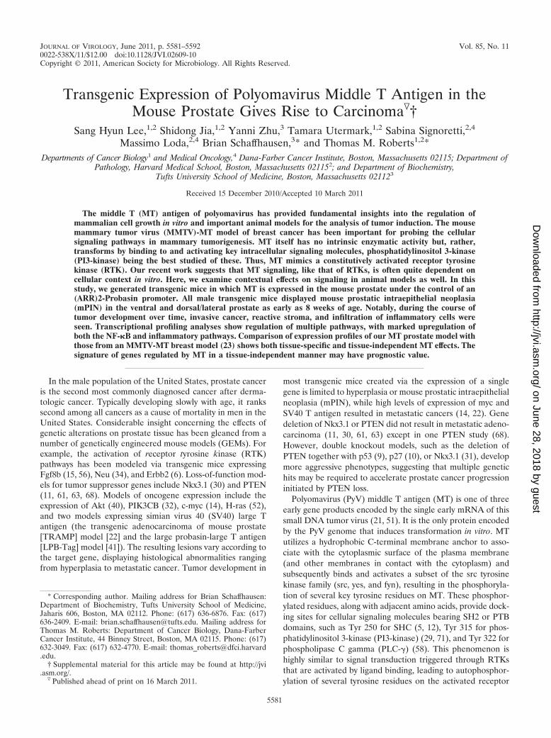
An (ARR)2PB-MT transgene directs the expression of MTin the mouse prostate. To effect prostate-specific expression ofMT in the mouse, we utilized the well-known (ARR)2PB pro-moter cassette, which drives gene expression upon androgenstimulation. Within this cassette, there are two androgen-re-sponsive regions (ARR) containing androgen-responsiveelements (AREs), which drive hormone-dependent gene ex-pression during mouse prostate development (73). A PCR-amplified cDNA encoding MT was subcloned into the(ARR)2PB cassette to produce the (ARR)2PB-MT construct(Fig. 1A). This plasmid was then linearized and microinjectedinto the pronucleus of fertilized mouse eggs to generate trans-genic animals. PCR analyses of genomic DNA from tail clipswere used to screen for founders, and representative resultsare shown in Fig. 1B. By this approach, three transgenic lineswere obtained: GG982, FF23, and 36B. All three models ulti-mately displayed generally similar characteristics. However,there were certain phenotypic differences among them, pre-sumably arising from prostate lobe-specific differences in MTexpression, with the FF23 line expressing MT in all three lobeswhile the GG982 and 36B lines failed to express MT in theanterior prostate (see below). Since the GG982 and 36B lineswere roughly equivalent, we used the GG982 and FF23 linesfor subsequent experiments.
First, we confirmed the expression of MT in the transgenicmouse prostates. Total RNA was purified from the ventralprostates of 10-week-old mice, and real-time PCR was per-formed with oligonucleotides targeted to the poly(A) signalregion (Fig. 1A). As shown in Fig. 1C, MT expression wasdetected in transgenic mice but not in wild-type animals. Wenext confirmed protein expression. Total cellular lysates wereprepared from the ventral prostate (VP), dorsal/lateral pros-tate (DLP), and anterior prostate (AP) lobes of transgenic andcontrol mice, and immunoblotting experiments were per-formed using an anti-MT antibody. As shown in Fig. 1D, MTexpression was detected in the ventral and dorsal/lateral lobesof the transgenic prostates in all lines but not in the anteriorlobes of line GG982 (Fig. 1D) or line 36B3 (data not shown).
However, in the FF23 line, MT is also expressed in the anteriorlobe (Fig. 1D).
MT transduces oncogenic signals via interactions betweenseveral phosphorylated tyrosine residues on MT and SH2 orphosphotyrosine binding (PTB) domain-containing proteins,such as SHC, PI3-kinase, and PLC-�. These associations leadto clear activation of PI3-kinase and, in some cases, RASpathways, while activation of PLC-� is usually difficult to de-tect. To verify the molecular function of MT in mouse prostatecells, we performed a series of immunoblotting experiments,examining the dorsal/lateral prostate, where MT is consistentlyexpressed. We first probed with anti-phospho-AKT (Ser 473)and (Thr 308) antibodies to see if the PI3-kinase pathway was
FIG. 1. Expression of functional polyomavirus middle T in the mouseprostate. (A) Schematic diagram of the DNA construct used for thecreation of transgenic mice. PCR1 represents the expected PCR productfrom genomic DNA for genotyping. PCR2 represents the PCR product ofreal-time PCR for measuring expression levels. Arrows indicate oligonu-cleotides used for PCRs. (B) An example of a genotyping PCR. TG,transgenic mouse, WT, wild type. (C) The table summarizes the averagereal-time cycle threshold (Ct) values. ud, undetected. The real-time PCRproducts were run on an agarose gel. (D) Detection of MT expression inthe mouse prostate. Cellular lysates were prepared from the anterior(AP), ventral (VP), and dorsal/lateral prostate (DLP) from 10-week-oldTG (GG982) and WT littermates and 30-week-old TG (FF23) and WTlittermates. Transgene expression was confirmed by immunoblotting ex-periments using an anti-MT antibody. (E) MT induces functional signaltransduction. Total lysates from VP and DLP from WT and TG micewere analyzed by immunoblotting using antibodies directed against anti-phospho-AKT (Ser473), anti-phospho-AKT (Thr308), anti-phospho-S6(Ser240/244), anti-phospho-Shc (Tyr239/240), anti-PRAS40 (Thr246), anti-AKT, anti-S6, and antitubulin antibodies.
VOL. 85, 2011 MT EXPRESSION IN MOUSE PROSTATE INDUCES PROSTATE CANCERS 5583
on June 28, 2018 by guesthttp://jvi.asm
.org/D
ownloaded from
activated. As shown in Fig. 1E, phospho-AKT signals weremarginally but reproducibly increased in dorsal/lateral lobes(Fig. 1E). Since phospho-AKT levels are only weakly in-creased, we also examined the phosphorylation of AKT sub-strates. Phosphorylation of both a proximal AKT target,PRAS40, and a downstream target, S6 ribosomal protein(S6RP) (Ser 240/244), was clearly increased in transgenic mice,further supporting the activation of the PI3K/AKT/mTORpathway (Fig. 1E). In addition, we examined the activation ofthe ras pathway, measuring tyrosine phosphorylation of SHC(Tyr 239), which was also significantly increased comparedwith that of controls (Fig. 1E).
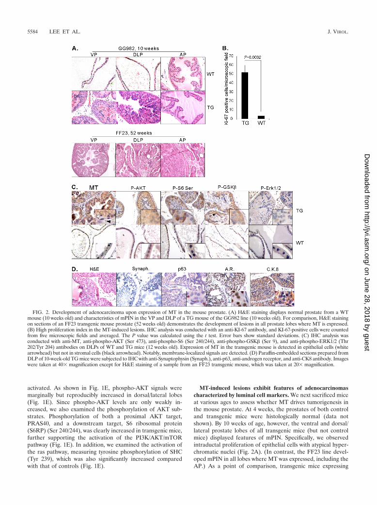
MT-induced lesions exhibit features of adenocarcinomascharacterized by luminal cell markers. We next sacrificed miceat various ages to assess whether MT drives tumorigenesis inthe mouse prostate. At 4 weeks, the prostates of both controland transgenic mice were histologically normal (data notshown). By 10 weeks of age, however, the ventral and dorsal/lateral prostate lobes of all transgenic mice (but not controlmice) displayed features of mPIN. Specifically, we observedintraductal proliferation of epithelial cells with atypical hyper-chromatic nuclei (Fig. 2A). (In contrast, the FF23 line devel-oped mPIN in all lobes where MT was expressed, including theAP.) As a point of comparison, transgenic mice expressing
FIG. 2. Development of adenocarcinoma upon expression of MT in the mouse prostate. (A) H&E staining displays normal prostate from a WTmouse (10 weeks old) and characteristics of mPIN in the VP and DLP of a TG mouse of the GG982 line (10 weeks old). For comparison, H&E stainingon sections of an FF23 transgenic mouse prostate (52 weeks old) demonstrates the development of lesions in all prostate lobes where MT is expressed.(B) High proliferation index in the MT-induced lesions. IHC analysis was conducted with an anti-KI-67 antibody, and KI-67-positive cells were countedfrom five microscopic fields and averaged. The P value was calculated using the t test. Error bars show standard deviations. (C) IHC analysis wasconducted with anti-MT, anti-phospho-AKT (Ser 473), anti-phospho-S6 (Ser 240/244), anti-phospho-GSK� (Ser 9), and anti-phospho-ERK1/2 (Thr202/Tyr 204) antibodies on DLPs of WT and TG mice (12 weeks old). Expression of MT in the transgenic mouse is detected in epithelial cells (whitearrowhead) but not in stromal cells (black arrowhead). Notably, membrane-localized signals are detected. (D) Paraffin-embedded sections prepared fromDLP of 10-week-old TG mice were subjected to IHC with anti-Synaptophysin (Synaph.), anti-p63, anti-androgen receptor, and anti-CK8 antibody. Imageswere taken at 40� magnification except for H&E staining of a sample from an FF23 transgenic mouse, which was taken at 20� magnification.
5584 LEE ET AL. J. VIROL.
on June 28, 2018 by guesthttp://jvi.asm
.org/D
ownloaded from
activated PI3-kinase p110� or activated Akt in the prostatealso display mPIN at this age (39, 40). However, the mPINinduced by MT tends to involve a larger proportion of theglands than the activated-PI3-kinase p110� and activated-AKTmodels. We further analyzed the mitotic index by performingIHC staining for the proliferation marker KI-67. As shown inFig. 2B, significantly more Ki-67-positive cells were detected inthe prostates of transgenic mice than in those of wild-typemice.
Next, we examined which cell types feature MT expressionby IHC staining with anti-MT antibody. As shown in Fig. 2C,the expression of MT was clearly evident in epithelial cells butwas not observed in stromal cells. We then determinedwhether activation of the PI3-kinase pathway is detected inPIN lesions. Increased levels of phosphorylation were detectedfor AKT and its substrate GSK�, as well as S6RP (Fig. 2C).These results, together with those of the immunoblotting ex-periments shown in Fig. 1E, suggest that activation of thePI3-kinase pathway does indeed occur in the prostates of thetransgenic mice. In contrast, phosphorylation of the down-stream target Erk1/2 was not significantly altered in transgenicmice compared with that in wild-type mice, as is often seen inMT-transformed cells (51).
Mouse prostate epithelial cells consist of three major celltypes: neuroendocrine cells, secretory luminal cells, and basalcells. IHC analysis experiments were performed to identify thenature of the neoplastic cells. We analyzed the expression ofthe following markers specific for the various cell types: anantisynaptophysin antibody for neuroendocrine cells, anti-p63antibody for basal cells, and anti-Ck8/18 antibody and anti-androgen receptor (AR) antibody for luminal cells. As shownin Fig. 2D, abnormal cells within the lesions express AR andCK8/18 but are negative for synaptophysin and p63 expression(normal basal cells surrounding the lesions can be p63 posi-tive). We examined even older mice, but they did not expresssynaptophysin in the mPIN (data not shown). These resultsindicate that, similar to human PIN, the prostate lesionscaused by MT overexpression display a luminal phenotype.
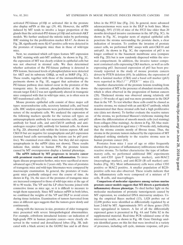
The mPIN induced by MT progresses to invasive cancerwith prominent reactive stroma and inflammation. To inves-tigate disease progression further, mice were sacrificed at moreadvanced ages (30 weeks to 2 years old). The sizes of prostatesof transgenic mice and wild-type littermates were compared bymacroscopic examination. In general, the prostates of trans-genic mice gradually enlarged over the course of time. Asshown in Fig. 3A, the sizes of the prostates of transgenic miceare increased compared to those of wild-type mice at the age of80 to 90 weeks. The VP and the LP often become joined withconnective tissue as mice age, so it is difficult to measure toweigh them separately. Since MT induces mPIN in both lobes,we combined the weights of the VP and DLP to avoid errorsduring tissue isolation. Examination of tumors harvested frommice at different ages suggests that the tumors grew slowly andcontinuously.
Along with the increase in size, prostates of older transgenicmice presented with severe histopathological abnormalities.For example, cribriform intraductal lesions—an indication ofhigh-grade PIN in human prostate cancer—were clearly ob-served in the ventral and dorsal/lateral lobes (Fig. 3B, indi-cated with a black arrow) in the GG982 line and in all three
lobes in the FF23 line (Fig. 2A). In general, more advancedadenocarcinomas were seen in the DLP in both lines. Morestrikingly, 50% (5/10) of mice of the FF23 line older than 20months developed invasive carcinomas in the AP (Fig. 3C). Asshown in Fig. 3C, irregular nests of atypical epithelial cellspenetrate the stroma surrounding the prostate ducts, a clearindication of invasion. To confirm the invasion of prostatecancer cells, we performed IHC assays with anti-CK8/18 andanti-p63. As shown in Fig. 3C, the expression of p63 is nolonger confined to the basement membrane area (comparewith Fig. 2D) but is now randomly dispersed within the stro-mal compartment. In addition, the invasive tumor compo-nent contained cells expressing CK8 markers, as well as cellsexpressing p63. Increased expression of p63 has been pre-viously reported in the study of mouse prostate cancersdriven by PTEN deletion (69). In addition, the expression ofboth a luminal marker (CK8) and a basal cell marker (p63)were reported in Nkx3.1�/�;PTEN�/� mice (1).
Another characteristic of the prostate lesions resulting fromthe expression of MT is the presence of abundant stromal cells,which is often observed in the progression of human cancers(28). Thickened stroma was observed among relatively oldmice (�30 weeks of age) and was more prominent in the DLPthan in the VP. To test whether these cells could be classed asreactive stroma, we stained with an anti-KI-67 antibody, whichdemonstrated that these stromal cells had a high proliferativerate (data not shown). To further evaluate the reactive natureof the stroma, we performed Masson’s trichrome staining thatallows the differentiation of smooth muscle cells (red staining)from collagen (blue staining). As shown in Fig. 3D, blue stain-ing is readily detected in aged mice (�30 weeks old), indicatingthat the stroma consists mostly of fibrous tissue. Thus, thestroma in the prostate tumors induced by the expression of MTdisplayed striking similarity to the reactive stroma seen inhuman prostate cancer (64).
Prostates from mice 1 year of age or older frequentlyshowed the presence of inflammatory infiltrations within thereactive stroma. To further characterize the type of inflam-matory cells, we performed additional IHC experimentswith anti-CD3 (pan-T lymphocyte marker), anti-MAC2(macrophage marker), and anti-B220 (B cell marker) anti-bodies (Fig. 3E). Most inflammatory cells were either CD3positive or MAC2 positive. A smaller population of B220-positive cells was also observed. These results indicate thatthe inflammatory cells were composed of a mixture of Tcells, B cells, and macrophages.
Comparison of molecular signatures among various murineprostate cancer models suggests that MT directs a particularlyinflammatory disease phenotype. To shed further light on themolecular mechanisms of prostatic tumorigenesis caused byMT expression, we conducted DNA microarray analyses. Us-ing the empirical Bayes method (13), 1,028 probes out of22,690 probes were identified as differentially regulated by atleast 2-fold by MT. Approximately 58% of these genes (583)were upregulated in tumors. A list of all the differentiallyexpressed genes is presented in Fig. 4A and Table S2 in thesupplemental material. Real-time PCR validated some of themicroarray results, as shown in Fig. 4B. Gene Ontology anal-ysis identified genes on this list that were involved in a varietyof processes, including cell cycle, immune response, cell pro-
VOL. 85, 2011 MT EXPRESSION IN MOUSE PROSTATE INDUCES PROSTATE CANCERS 5585
on June 28, 2018 by guesthttp://jvi.asm
.org/D
ownloaded from
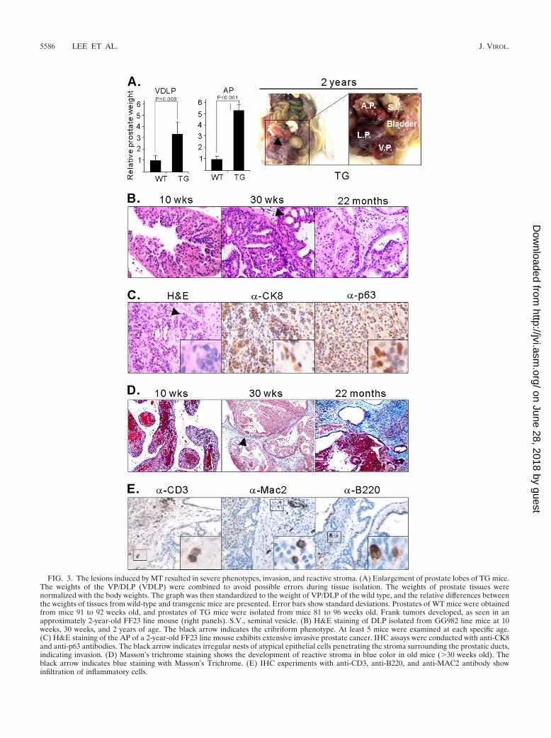
FIG. 3. The lesions induced by MT resulted in severe phenotypes, invasion, and reactive stroma. (A) Enlargement of prostate lobes of TG mice.The weights of the VP/DLP (VDLP) were combined to avoid possible errors during tissue isolation. The weights of prostate tissues werenormalized with the body weights. The graph was then standardized to the weight of VP/DLP of the wild type, and the relative differences betweenthe weights of tissues from wild-type and transgenic mice are presented. Error bars show standard deviations. Prostates of WT mice were obtainedfrom mice 91 to 92 weeks old, and prostates of TG mice were isolated from mice 81 to 96 weeks old. Frank tumors developed, as seen in anapproximately 2-year-old FF23 line mouse (right panels). S.V., seminal vesicle. (B) H&E staining of DLP isolated from GG982 line mice at 10weeks, 30 weeks, and 2 years of age. The black arrow indicates the cribriform phenotype. At least 5 mice were examined at each specific age.(C) H&E staining of the AP of a 2-year-old FF23 line mouse exhibits extensive invasive prostate cancer. IHC assays were conducted with anti-CK8and anti-p63 antibodies. The black arrow indicates irregular nests of atypical epithelial cells penetrating the stroma surrounding the prostatic ducts,indicating invasion. (D) Masson’s trichrome staining shows the development of reactive stroma in blue color in old mice (�30 weeks old). Theblack arrow indicates blue staining with Masson’s Trichrome. (E) IHC experiments with anti-CD3, anti-B220, and anti-MAC2 antibody showinfiltration of inflammatory cells.
5586 LEE ET AL. J. VIROL.
on June 28, 2018 by guesthttp://jvi.asm
.org/D
ownloaded from

liferation, apoptosis, and metabolism (see Tables S3 and S4 inthe supplemental material). As will be discussed below, chemo-kines were the single most prominently overexpressed group ofgenes.
Next, we performed Gene Set Enrichment Analysis(GSEA), which helps to identify gene sets enriched in relevantbiological processes. We used the C2 (curated gene sets) andC3 (motif gene sets) gene sets (59). Among the gene setsenriched, both curated and motif analyses identified NF-Btarget genes and inflammatory genes as consistently enrichedin MT-induced prostate cancer (Fig. 4C; also see Tables S5 andS6 in the supplemental material). As shown in Fig. 4D, IHCexperiments corroborated the array results showing increasedlevels of the p65 subunit of the NF-B complex and increasedlevels of p65 in the transgenic mouse (Fig. 4D).
Finally, we compared the molecular signatures obtainedfrom the MT transgenic mice to those from previously re-ported prostate models. Notably, the various mouse modelsresult in diseases of distinctly different severities. The lesionsdeveloped in the PIK3CB (32) and Akt mice (40) are charac-terized as mPIN, which never proceed to invasive prostatecancer, in contrast to the PTEN null tumors used in our lab-oratory; furthermore, the MT and PTEN null tumors in otherbackgrounds (37, 63, 68) invade, and the myc (14) andTRAMP mice (22) progress from invasive tumors to franklymetastatic lesions. While this comparison provides some in-
sight into the differences in tumor severity phenotypes, anotheraspect of gene expression proved to be most striking. Notably,the microarray data identified chemokines as the gene familymost highly overexpressed in MT tumors when compared withthe other models (Fig. 5A). Principal among these are CXCL5,CXCL2, CCL5, and CXCL15. The overexpression of CXCL5and CXCL2 was confirmed by real-time PCR (Fig. 4B). Wefurther examined the expression of one of the chemokine fam-ily proteins, CXCL5, by IHC assay (Fig. 5B). IHC analysisshows high signals for CXCL5 in an MT transgenic mouse.Recent studies have suggested that extracellular matrix genes(versican and tanscin) and chemokines (BDFN, CCL5,CXCL5, and CXCL16) may be induced by the interactionbetween stromal and epithelial cells during human cancer pro-gression (60, 66). The upregulation of chemokine families inthis highly stromal mouse prostate cancer model further sug-gests a possible link between overexpression of chemokinesand the development of proliferative stromal cells. Chemokineexpression may also explain the infiltration of inflammatorycells seen in our histological studies (Fig. 3E).
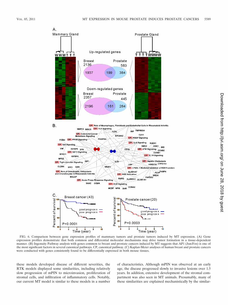
Comparisons of molecular signatures of breast cancer andprostate cancer driven by MT expression. We previously re-ported that activation of the PI3-kinase pathway is required forMT-mediated transformation in both human and mouse mam-mary epithelial cells but that the mechanism of action couldvary in a cell type-dependent manner (65). We wanted to
FIG. 4. Analyses of DNA microarrays identify enrichment of genes associated with the NF-B signaling pathway. (A) Hierarchical clusteringof gene expression data from WT and TG prostate tissues. Genes differentially expressed by at least 2-fold were selected for hierarchical clustering.The heat map depicts differential gene expression between the WT and TG groups. Each row represents an individual gene, and each columnrepresents an individual tissue sample. The red and green colors represent relatively higher and lower expression levels, respectively. (B) Summaryof real-time PCR data of selected genes identified from DNA array analyses. (C) GSEA with the C3 motif database identified gene sets enrichedin the NF-B pathway and the interferon signaling cascade. FDR, false discovery rate. (D) IHC assays and immunoblotting experiments withanti-p65 antibodies demonstrate increased levels of p65 in the prostate of transgenic mice.
VOL. 85, 2011 MT EXPRESSION IN MOUSE PROSTATE INDUCES PROSTATE CANCERS 5587
on June 28, 2018 by guesthttp://jvi.asm
.org/D
ownloaded from
determine whether there might be a core signature of MTtransformation common to different cellular contexts. Wecompared gene expression data obtained from breast and pros-tate tumors induced by transgenic expression of MT. As shownin Fig. 6A, we identified 360 genes that were consistently up- ordownregulated in both tissues. These include SAA1, Stathmin,Jun, Fos, CD44, Ki67, and others (see Table S7 in the supple-mental material). However, many genes are regulated by MTin a tissue-specific manner (see Tables S8 and S9 in the sup-plemental material). Interestingly, chemokine genes are al-most exclusively upregulated in prostate cancer (see Table S9).Notably, downregulation of chemokine genes (except CXCL5)was also observed in MMTV-MT transgenic mice in a previousgene expression study using a different platform of arrays(GSE3165) (26). Likewise, some genes, including Car6, Car12,Skp2, and FoxC1, were uniquely identified in breast tumors.This result indicates that the molecular mechanisms of tumor-igenesis by MT expression may differ depending on the cellularcontext. Notably, a number of the commonly regulated genes,including SAA1/2, Tacstd2, Jun, CD24a, Tspan8, andTMPRSS4, were also identified as PI3-kinase pathway-depen-dent genes (32), consistent with the idea that activation of the
PI3-kinase pathway is intrinsically important in MT transfor-mation. To delineate the possible pathways associated with MTexpression, we used Ingenuity Pathway Analysis software andsought canonical pathways. As shown in Fig. 6B, the AP1complex (Jun/Fos) was mapped at the center of the pathways.This confirms that MT activates the AP1 pathway (57). Inaddition, AP1 has been used a prognostic marker for advancedtypes of breast cancer (67) and prostate cancer (45).
A previous study using comparative analysis of MMTV-MTmodels demonstrated that a key gene set for predicting meta-static human breast cancers was also found in MT-associatedcancer genes identified from mouse models (49). This resultdemonstrated the utility of the signature of MT-associatedcancer. We therefore wondered whether the common gene setrepresenting the molecular signature of the intrinsic MT func-tion (Fig. 6A; also see Table S7 in the supplemental material)might be clinically correlated in breast and prostate cancers.Using human orthologs of the signature genes, we appliedunsupervised clustering to both human prostate cancer data(20) and human breast cancer data (46). In both cases, humancancer samples were separated into two clusters. We thenperformed Kaplan-Meier plots to see whether these two clus-ters have different survival or recurrent disease patterns. Asshown in Fig. 6C, for breast cancer cohorts, the common sig-nature is significantly associated with survival, which is consis-tent with the results of previous studies (49). Furthermore, thesignature can also be used for predicting the outcome of hu-man prostate cancers. This indicates that molecular mecha-nisms associated with MT in developing cancers might haverelevance to clinical outcomes, although MT is not directlyinvolved in human cancers.
DISCUSSION
For the studies described here, we have created transgenicmice using polyomavirus MT under the control of an androgenreceptor-driven promoter. MT expressed in the mouse pros-tate drives the slow development of invasive cancer, mimickingthe slow progression of the human disease. Ordinarily, themajority of tumors arising from epithelial cells are caused bythe cumulative effects of multiple genetic or epigenetic alter-ations (24). These alterations result in gain of function ofoncogenes, loss of function of tumor suppressors, or epigeneticchanges. These changes disturb the cellular homeostasis andeventually induce diseases, including cancers. MT affects mul-tiple signaling pathways directly, in large part through tyrosinekinase activation. Most importantly, the MT model also dis-plays significant histophysiological similarities with humanprostate cancers, including a large component of actively di-viding stromal cells and infiltration by inflammatory cells.
Activation of RTKs is often seen in human prostate cancer,although the correlation of insulinlike growth factor (IGF)levels in plasma and clinical outcomes remains controversial(8, 50). However, several clinical studies demonstrated that thelevel of ErbB2 expression is often elevated in advanced pros-tate cancer patients, especially those who have undergone ra-diation or hormone deprivation therapies (18, 42, 44, 54).Modeling of RTK activation in the prostate has been carriedout via transgenic expression of Erbb2-delta (a C-terminaltruncation mutant) (6), Neu (34), or FGF8b (15, 56). Although
FIG. 5. Expression of chemokine family genes is predominantlyinduced in transgenic mice arisen by expression of MT. (A) Compar-ison of chemokine gene expression in mouse prostate cancers amongdifferent genetically engineered models (GEMs): MT transgenic mice,myr-AKT transgenic mice, myr-flag-PIK3CB transgenic mice, VP andAP of PTEN knockout mice, TRAMP transgenic mice, and high-Myctransgenic mice. Differential expression of chemokines was obtainedfrom the results of each study separately by a t test (P 0.001)between wild types and tumor models using BRB tools, and the folddifferences are summarized. (B) Paraffin-embedded sections were pre-pared from WT and MT transgenic mice, and IHC assays with anti-CXCL5 antibody were conducted.
5588 LEE ET AL. J. VIROL.
on June 28, 2018 by guesthttp://jvi.asm
.org/D
ownloaded from
these models developed disease of different severities, theRTK models displayed some similarities, including relativelyslow progression of mPIN to microinvasion, proliferation ofstromal cells, and infiltration of inflammatory cells. Notably,our current MT model is similar to these models in a number
of characteristics. Although mPIN was observed at an earlyage, the disease progressed slowly to invasive lesions over 1.5years. In addition, extensive development of the stromal com-partment was also seen in MT animals. Presumably, many ofthese similarities are explained mechanistically by the similar-
FIG. 6. Comparison between gene expression profiles of mammary tumors and prostate tumors induced by MT expression. (A) Geneexpression profiles demonstrate that both common and differential molecular mechanisms may drive tumor formation in a tissue-dependentmanner. (B) Ingenuity Pathway analysis with genes common to breast and prostate cancers induced by MT suggests that AP1 (Jun/Fos) is one ofthe most significant factors in several canonical pathways. CP, canonical pathway. (C) Kaplan-Meier analyses of human breast and prostate cancerswere conducted with genes consistently found to be differentially expressed in both mouse tissues.
VOL. 85, 2011 MT EXPRESSION IN MOUSE PROSTATE INDUCES PROSTATE CANCERS 5589
on June 28, 2018 by guesthttp://jvi.asm
.org/D
ownloaded from
ity of the signaling pathways, such as the AKT and Raf path-ways, activated by both MT and RTKs.
The infiltration of inflammatory cells seen in the histologicalstudies recalls the importance of interactions of tumor cellswith the underlying stromal cells seen in the MT breast model,where the stroma contributes to the migratory and invasivebehavior of MT tumor cells (17). Tumor-associated proteases,which are important for metastasis, come mostly from thebreast stroma (47). Particular attention has been paid to mac-rophages, and loss of macrophage infiltration in CSF-1 nullmice results in tumors that do not invade well and results in adramatic decrease in metastasis (35). These studies prove thatthe tumor microenvironment in the MT-driven breast tumorscan promote tumor progression to invasion or metastasis.Since similar stromal phenotypes were observed in the MT-driven prostate tumor model described here, further studies ofthis model may help in understanding the mechanisms of in-vasive prostate cancer.
The high levels of chemokine production seen in our MTprostate tumor model make it a unique animal model to studythe function of chemokines in prostate cancer development.
DNA microarray experiments identified chemokine ligandsas the most highly expressed class of genes in MT transgenictumors. These ligands act through G protein-coupled recep-tors, and their signaling cascades are known to be involved inpromoting angiogenesis and to affect the constitution of tumormicroenvironments (2). Increased expression of CXCL5 waspreviously associated with macrophage and myeloid cell infil-tration in MMTV-MT tumors (4, 72). The addition of CCL5increased the migratory behavior of tumor-associated myeloidcells, while antagonists of CXCR2 reduced metastasis (72).The expression levels of chemokines, such as CXCL5, CCL5,or CXCL16, are significantly associated with prostate cancerprogression, including invasive and bone metastatic prostatetumors (3, 36, 60). It is quite possible that chemokine produc-tion by the MT-driven tumor epithelia may be driving infiltra-tion of both stromal and immune cells. It is also known thatMT-transformed mouse mammary epithelial cells produceCCL2 and CCL5 (Y. Zhu and B. Schaffhausen, unpublishedresults). It is also possible that the stromal production ofchemokines subsequently leads to infiltration by inflammatorycells. Notably, a recent study from the Hanahan laboratoryshowed that cancer-associated fibroblasts can play a key role intumor inflammation by expressing inflammatory genes, such aschemokines (16). Perhaps MT elicits stromal infiltration inde-pendently of chemokines and it is the stroma that produces thechemokines that subsequently lead to infiltration by inflamma-tory cells.
There are several possible origins for the stromal cells thatare so prevalent in these tumors. Since prostate stromal cellsalso express substantial levels of androgen receptor, transgeneexpression from the (ARR)2-probasin promoter in those cellsis possible. However, MT expression was seen only in luminalepithelial cells by IHC experiments, as shown in Fig. 2C.Therefore, the development of the cancer-associated stromalphenotype, as shown in Fig. 3, is more likely being induced byparacrine factors arising in epithelial cells expressing MT. In-deed, the observation that the abnormal cells appear onlywithin the prostate lumen, with no increased stromal involve-ment at young ages, is consistent with the hypothesis that MT
acts solely in the prostate epithelium. However, we cannotcompletely rule out the possibility that some of the activatedstroma may have arisen from cells expressing low levels of MT,which could be below the detection level in IHC. Alternatively,the reactive stroma could have arisen by epithelial-mesenchy-mal transition (EMT) in MT-expressing epithelial cells late intumor progression.
Regardless of which cells are expressing the chemokines anddriving infiltration of both stromal cells and white cells, thequestion arises as to what aspect of MT signaling initiates theseevents. Most of MT’s effects on host cells are thought to arisevia two pathways: first, the PI3-kinase pathway, which is acti-vated by binding of the p85 adaptor subunit of PI3-kinase tothe phospho-YMPM motif at Tyr 315, and second, the RAS/RAF/ERK pathway, activated by binding of the RAS regula-tor, Shc, to the NPXY motif at Tyr 250. Since prostate tumorsdriven by p110� and AKT transgenes do not express chemo-kines abundantly, it seems unlikely that the PI3-kinase pathwayis driving their expression. However, both MT expression and,to a lesser degree, ablation of PTEN activate the RAS/RAF/MEK/ERK pathway, making it a reasonable candidate forchemokine induction. Another key tyrosine residue in the in-teraction with cellular signaling molecules is Tyr 322, which isthe site for PLC-� association. Notably, previous work pointedto the importance of PLC-� in metastasis in MMTV-MT formammary tumors and TRAMP for prostate tumors (53), fur-ther suggesting a potential role for PLC-� in MT-driven pros-tate cancer. However, since we did not observe metastasis inthe MT prostate cancer model, the role of Tyr 322 here mightbe different from the breast case. We are currently generatingtransgenic animals expressing MT mutant alleles at Tyr 250,Tyr 315, or Tyr 322 to test these hypotheses.
To explore the common molecular mechanisms in tumori-genesis induced by MT expression, we compared the geneexpression data from breast and prostate cancers. The expres-sion of the majority of genes was regulated in a tissue-depen-dent manner. This suggests that viral oncogenes activate po-tentially different molecular pathways in different cell types.This is not surprising, since it is well known that the impor-tance of individual MT signaling pathways differs among tis-sues. There are also differences in gene expression profilesobtained for the MMTV-MT model when MMTV-MT634 tu-mors are derived in different mouse strain backgrounds and,indeed, even in passage of different MMTV-MT634-derivedtumors in FVB mice (38, 43, 49). Such differences could ariseas a function of aging, mouse strain differences, gender differ-ences, and disease-specific stages. Our current study did notaddress these variances. Differences could also result from thedifferential contribution of stromal components. Indeed, theMMTV-MT model displayed a greater ratio of a luminal cellmarker (Krt8) and a stromal cell marker (Desmin) than of(ARR)2PB-MT (data not shown). Taken together, these con-straints limit our ability to interpret the tissue-specific differ-ences we observed to be shared by the two MT models. How-ever, this same logic adds increased importance to the featuresthat we observed. Among the commonly regulated genes weregenes present in PI3-kinase pathways, suggesting that MT uti-lized this pathway for inducing cancer. This is consistent withthe observation that polyomavirus that is unable to activatePI3-kinase is defective in tumorigenesis in many tissues (19).
5590 LEE ET AL. J. VIROL.
on June 28, 2018 by guesthttp://jvi.asm
.org/D
ownloaded from
Functional genomic annotation analyses identified many com-monly upregulated genes involved in cell cycle regulation andAP1 signaling, whereas downregulated genes are negativelyassociated with RXR signaling. Consistent with these findings,higher AP1 levels were clinically correlated with poorer prog-nosis in breast (67) and prostate cancer (45). Indeed, analysisof commonly expressed genes allowed us to construct a genesignature which appeared to have prognostic utility in a retro-spective examination of a limited set of prostate tumors.Therefore, molecular pathways inducing the expression ofthese genes would be potential targets for therapeutic appli-cations, even though MT itself does not induce breast or pros-tate cancer in humans.
In summary, we report a new mouse model of prostaticadenocarcinoma that shows several interesting features, in-cluding invasiveness and infiltration of inflammatory cells.These features are also found in human prostate cancers. Inthe most recent decade, several chemical compounds havebeen synthesized to target PI3-kinase and MAP kinase path-ways, probably the most activated pathways in cancer. SinceMT effectively activates these pathways, our MT transgenicmice can be utilized as an appropriate model for studyingadvanced prostate cancers and for evaluating therapeuticchemical compounds.
ACKNOWLEDGMENTS
We thank A. Upadhyaya and L. Clayton for helpful comments onthe manuscript and E. Li and J. Horner for creating transgenic mice.
This work was supported in part by grants from the National Insti-tutes of Health (T.M.R., B.S., and M.L.), the Prostate Cancer Foun-dation (M.L.), the Linda and Arthur Gelb Center for TranslationalResearch (M.L.), and the DFHCC Prostate SPORE (T.M.R. andM.L.). In compliance with Harvard Medical School guidelines, wedisclose the consulting relationships: Novartis Pharmaceuticals, Inc.(T.M.R. and M.L.).
REFERENCES
1. Abate-Shen, C., et al. 2003. Nkx3.1; Pten mutant mice develop invasiveprostate adenocarcinoma and lymph node metastases. Cancer Res. 63:3886–3890.
2. Balkwill, F. 2004. Cancer and the chemokine network. Nat. Rev. Cancer4:540–550.
3. Begley, L. A., et al. 2008. CXCL5 promotes prostate cancer progression.Neoplasia 10:244–254.
4. Bierie, B., et al. 2008. Transforming growth factor-beta regulates mammarycarcinoma cell survival and interaction with the adjacent microenvironment.Cancer Res. 68:1809–1819.
5. Campbell, K. S., et al. 1994. Polyoma middle tumor antigen interacts withSHC protein via the NPTY (Asn-Pro-Thr-Tyr) motif in middle tumor anti-gen. Proc. Natl. Acad. Sci. U. S. A. 91:6344–6348.
6. Casimiro, M., et al. 2007. ErbB-2 induces the cyclin D1 gene in prostateepithelial cells in vitro and in vivo. Cancer Res. 67:4364–4372.
7. Cecena, G., F. Wen, R. D. Cardiff, and R. G. Oshima. 2006. Differentialsensitivity of mouse epithelial tissues to the polyomavirus middle T onco-gene. Am. J. Pathol. 168:310–320.
8. Chan, J. M., et al. 1998. Plasma insulin-like growth factor-I and prostatecancer risk: a prospective study. Science 279:563–566.
9. Chen, Z., et al. 2005. Crucial role of p53-dependent cellular senescence insuppression of Pten-deficient tumorigenesis. Nature 436:725–730.
10. Di Cristofano, A., M. De Acetis, A. Koff, C. Cordon-Cardo, and P. P. Pan-dolfi. 2001. Pten and p27KIP1 cooperate in prostate cancer tumor suppres-sion in the mouse. Nat. Genet. 27:222–224.
11. Di Cristofano, A., B. Pesce, C. Cordon-Cardo, and P. P. Pandolfi. 1998. Ptenis essential for embryonic development and tumour suppression. Nat. Genet.19:348–355.
12. Dilworth, S. M., et al. 1994. Transformation by polyoma virus middle T-an-tigen involves the binding and tyrosine phosphorylation of Shc. Nature 367:87–90.
13. Efron, B., R. Tisbshirani, J. D. Storey, and V. Tusher. 2001. Empirical Bayesanalysis of a microarray experiment. J. Am. Stat. Assoc. 96:1151–1160.
14. Ellwood-Yen, K., et al. 2003. Myc-driven murine prostate cancer sharesmolecular features with human prostate tumors. Cancer Cell 4:223–238.
15. Elo, T. D., et al. 2010. Stromal activation associated with development ofprostate cancer in prostate-targeted fibroblast growth factor 8b transgenicmice. Neoplasia 12:915–927.
16. Erez, N., M. Truitt, P. Olson, S. T. Arron, and D. Hanahan. 2010. Cancer-associated fibroblasts are activated in incipient neoplasia to orchestrate tu-mor-promoting inflammation in an NF-kappaB-dependent manner. CancerCell 17:135–147.
17. Fluck, M. M., and B. S. Schaffhausen. 2009. Lessons in signaling and tumor-igenesis from polyomavirus middle T antigen. Microbiol. Mol. Biol. Rev.73:542–563.
18. Fossa, A., et al. 2002. Independent prognostic significance of HER-2 onco-protein expression in pN0 prostate cancer undergoing curative radiotherapy.Int. J. Cancer 99:100–105.
19. Freund, R., C. J. Dawe, J. P. Carroll, and T. L. Benjamin. 1992. Changes infrequency, morphology, and behavior of tumors induced in mice by a poly-oma virus mutant with a specifically altered oncogene. Am. J. Pathol. 141:1409–1425.
20. Glinsky, G. V., A. B. Glinskii, A. J. Stephenson, R. M. Hoffman, and W. L.Gerald. 2004. Gene expression profiling predicts clinical outcome of prostatecancer. J. Clin. Invest. 113:913–923.
21. Gottlieb, K. A., and L. P. Villarreal. 2001. Natural biology of polyomavirusmiddle T antigen. Microbiol. Mol. Biol. Rev. 65:288–318.
22. Greenberg, N. M., et al. 1995. Prostate cancer in a transgenic mouse. Proc.Natl. Acad. Sci. U. S. A. 92:3439–3443.
23. Guy, C. T., R. D. Cardiff, and W. J. Muller. 1992. Induction of mammarytumors by expression of polyomavirus middle T oncogene: a transgenicmouse model for metastatic disease. Mol. Cell. Biol. 12:954–961.
24. Hahn, W. C., and R. A. Weinberg. 2002. Modelling the molecular circuitry ofcancer. Nat. Rev. Cancer. 2:331–341.
25. Haram, K. M., et al. 2008. Gene expression profile of mouse prostate tumorsreveals dysregulations in major biological processes and identifies potentialmurine targets for preclinical development of human prostate cancer ther-apy. Prostate 68:1517–1530.
26. Herschkowitz, J. I., et al. 2007. Identification of conserved gene expressionfeatures between murine mammary carcinoma models and human breasttumors. Genome Biol. 8:R76.
27. Jia, S., et al. 2008. Essential roles of PI(3)K-p110beta in cell growth, me-tabolism and tumorigenesis. Nature 454:776–779.
28. Kalluri, R., and M. Zeisberg. 2006. Fibroblasts in cancer. Nat. Rev. Cancer6:392–401.
29. Kaplan, D. R., et al. 1986. Phosphatidylinositol metabolism and polyoma-mediated transformation. Proc. Natl. Acad. Sci. U. S. A. 83:3624–3628.
30. Kim, M. J., et al. 2002. Nkx3.1 mutant mice recapitulate early stages ofprostate carcinogenesis. Cancer Res. 62:2999–3004.
31. Kim, M. J., et al. 2002. Cooperativity of Nkx3.1 and Pten loss of function ina mouse model of prostate carcinogenesis. Proc. Natl. Acad. Sci. U. S. A.99:2884–2889.
32. Lee, S. H., et al. 2010. A constitutively activated form of the p110betaisoform of PI3-kinase induces prostatic intraepithelial neoplasia in mice.Proc. Natl. Acad. Sci. U. S. A. 107:11002–11007.
33. Lewis, B. C., D. S. Klimstra, and H. E. Varmus. 2003. The c-myc and PyMToncogenes induce different tumor types in a somatic mouse model for pan-creatic cancer. Genes Dev. 17:3127–3138.
34. Li, Z., M. Szabolcs, J. D. Terwilliger, and A. Efstratiadis. 2006. Prostaticintraepithelial neoplasia and adenocarcinoma in mice expressing a probasin-Neu oncogenic transgene. Carcinogenesis 27:1054–1067.
35. Lin, E. Y., A. V. Nguyen, R. G. Russell, and J. W. Pollard. 2001. Colony-stimulating factor 1 promotes progression of mammary tumors to malig-nancy. J. Exp. Med. 193:727–740.
36. Lu, Y., et al. 2008. CXCL16 functions as a novel chemotactic factor forprostate cancer cells in vitro. Mol. Cancer Res. 6:546–554.
37. Ma, X., et al. 2005. Targeted biallelic inactivation of Pten in the mouseprostate leads to prostate cancer accompanied by increased epithelial cellproliferation but not by reduced apoptosis. Cancer Res. 65:5730–5739.
38. Maglione, J. E., et al. 2004. Polyomavirus middle T-induced mammary in-traepithelial neoplasia outgrowths: single origin, divergent evolution, andmultiple outcomes. Mol. Cancer Ther. 3:941–953.
39. Majumder, P. K., et al. 2004. mTOR inhibition reverses Akt-dependentprostate intraepithelial neoplasia through regulation of apoptotic and HIF-1-dependent pathways. Nat. Med. 10:594–601.
40. Majumder, P. K., et al. 2003. Prostate intraepithelial neoplasia induced byprostate restricted Akt activation: the MPAKT model. Proc. Natl. Acad. Sci.U. S. A. 100:7841–7846.
41. Masumori, N., et al. 2001. A probasin-large T antigen transgenic mouse linedevelops prostate adenocarcinoma and neuroendocrine carcinoma with met-astatic potential. Cancer Res. 61:2239–2249.
42. Myers, R. B., S. Srivastava, D. K. Oelschlager, and W. E. Grizzle. 1994.Expression of p160erbB-3 and p185erbB-2 in prostatic intraepithelial neo-plasia and prostatic adenocarcinoma. J. Natl. Cancer Inst. 86:1140–1145.
VOL. 85, 2011 MT EXPRESSION IN MOUSE PROSTATE INDUCES PROSTATE CANCERS 5591
on June 28, 2018 by guesthttp://jvi.asm
.org/D
ownloaded from
43. Namba, R., et al. 2006. Heterogeneity of mammary lesions represent mo-lecular differences. BMC Cancer 6:275.
44. Osman, I., et al. 2005. Serum levels of shed Her2/neu protein in men withprostate cancer correlate with disease progression. J. Urol. 174:2174–2177.
45. Ouyang, X., et al. 2008. Activator protein-1 transcription factors are associ-ated with progression and recurrence of prostate cancer. Cancer Res. 68:2132–2144.
46. Pawitan, Y., et al. 2005. Gene expression profiling spares early breast cancerpatients from adjuvant therapy: derived and validated in two population-based cohorts. Breast Cancer Res. 7:R953–R964.
47. Pedersen, T. X., et al. 2005. Extracellular protease mRNAs are predomi-nantly expressed in the stromal areas of microdissected mouse breast carci-nomas. Carcinogenesis 26:1233–1240.
48. Pfaffl, M. W. 2001. A new mathematical model for relative quantification inreal-time RT-PCR. Nucleic Acids Res. 29:e45.
49. Qiu, T. H., et al. 2004. Global expression profiling identifies signatures oftumor virulence in MMTV-PyMT-transgenic mice: correlation to humandisease. Cancer Res. 64:5973–5981.
50. Schaefer, C., G. D. Friedman, and J. C. P. Quesenberry. 1998. IGF-1 andprostate cancer. Science 282:199a.
51. Schaffhausen, B. S., and T. M. Roberts. 2009. Lessons from polyoma middleT antigen on signaling and transformation: a DNA tumor virus contributionto the war on cancer. Virology 384:304–316.
52. Scherl, A., J. F. Li, R. D. Cardiff, and N. Schreiber-Agus. 2004. Prostaticintraepithelial neoplasia and intestinal metaplasia in prostates of probasin-RAS transgenic mice. Prostate 59:448–459.
53. Shepard, C. R., J. Kassis, D. L. Whaley, H. G. Kim, and A. Wells. 2007. PLCgamma contributes to metastasis of in situ-occurring mammary and prostatetumors. Oncogene 26:3020–3026.
54. Signoretti, S., et al. 2000. Her-2-neu expression and progression towardandrogen independence in human prostate cancer. J. Natl. Cancer Inst.92:1918–1925.
55. Smyth, G. K. 2004. Linear models and empirical Bayes methods for assessingdifferential expression in microarray experiments. Stat Appl. Genet. Mol.Biol. 3:Article3.
56. Song, Z., et al. 2002. Fibroblast growth factor 8 isoform B overexpression inprostate epithelium: a new mouse model for prostatic intraepithelial neo-plasia. Cancer Res. 62:5096–5105.
57. Srinivas, S., A. Schonthal, and W. Eckhart. 1994. Polyomavirus middle-sizedtumor antigen modulates c-Jun phosphorylation and transcriptional activity.Proc. Natl. Acad. Sci. U. S. A. 91:10064–10068.
58. Su, W., W. Liu, B. S. Schaffhausen, and T. M. Roberts. 1995. Association ofpolyomavirus middle tumor antigen with phospholipase C-gamma 1. J. Biol.Chem. 270:12331–12334.
59. Subramanian, A., et al. 2005. Gene set enrichment analysis: a knowledge-based approach for interpreting genome-wide expression profiles. Proc. Natl.Acad. Sci. U. S. A. 102:15545–15550.
60. Sung, S. Y., et al. 2008. Coevolution of prostate cancer and bone stroma inthree-dimensional coculture: implications for cancer growth and metastasis.Cancer Res. 68:9996–10003.
61. Suzuki, H., et al. 1998. Interfocal heterogeneity of PTEN/MMAC1 genealterations in multiple metastatic prostate cancer tissues. Cancer Res. 58:204–209.
62. Tehranian, A., et al. 1996. Neoplastic transformation of prostatic and uro-genital epithelium by the polyoma virus middle T gene. Am. J. Pathol.149:1177–1191.
63. Trotman, L. C., et al. 2003. Pten dose dictates cancer progression in theprostate. PLoS Biol. 1:E59.
64. Tuxhorn, J. A., G. E. Ayala, and D. R. Rowley. 2001. Reactive stroma inprostate cancer progression. J. Urol. 166:2472–2483.
65. Utermark, T., B. S. Schaffhausen, T. M. Roberts, and J. J. Zhao. 2007. Thep110alpha isoform of phosphatidylinositol 3-kinase is essential for polyoma-virus middle T antigen-mediated transformation. J. Virol. 81:7069–7076.
66. Vindrieux, D., P. Escobar, and G. Lazennec. 2009. Emerging roles of chemo-kines in prostate cancer. Endocr. Relat. Cancer 16:663–673.
67. Vleugel, M. M., A. E. Greijer, R. Bos, E. van der Wall, and P. J. van Diest.2006. c-Jun activation is associated with proliferation and angiogenesis ininvasive breast cancer. Hum. Pathol. 37:668–674.
68. Wang, S., et al. 2003. Prostate-specific deletion of the murine Pten tumorsuppressor gene leads to metastatic prostate cancer. Cancer Cell 4:209–221.
69. Wang, S., et al. 2006. Pten deletion leads to the expansion of a prostaticstem/progenitor cell subpopulation and tumor initiation. Proc. Natl. Acad.Sci. U. S. A. 103:1480–1485.
70. Webster, M. A., et al. 1998. Requirement for both Shc and phosphatidylino-sitol 3� kinase signaling pathways in polyomavirus middle T-mediated mam-mary tumorigenesis. Mol. Cell. Biol. 18:2344–2359.
71. Whitman, M., D. R. Kaplan, B. Schaffhausen, L. Cantley, and T. M. Roberts.1985. Association of phosphatidylinositol kinase activity with polyoma mid-dle-T competent for transformation. Nature 315:239–242.
72. Yang, L., et al. 2008. Abrogation of TGF beta signaling in mammary carci-nomas recruits Gr-1�CD11b� myeloid cells that promote metastasis. Can-cer Cell 13:23–35.
73. Zhang, J., T. Z. Thomas, S. Kasper, and R. J. Matusik. 2000. A smallcomposite probasin promoter confers high levels of prostate-specific geneexpression through regulation by androgens and glucocorticoids in vitro andin vivo. Endocrinology 141:4698–4710.
5592 LEE ET AL. J. VIROL.
on June 28, 2018 by guesthttp://jvi.asm
.org/D
ownloaded from