Embed Size (px)
Citation preview
10.1101/gad.14.15.1920Access the most recent version at doi: 2000 14: 1920-1932 Genes Dev.
Cor F. Calkhoven, Christine Müller and Achim Leutz
isoform expressionβ and C/EBPαTranslational control of C/EBP
References
http://genesdev.cshlp.org/content/14/15/1920.full.html#ref-list-1
This article cites 100 articles, 67 of which can be accessed free at:
ServiceEmail Alerting
click here.right corner of the article orReceive free email alerts when new articles cite this article - sign up in the box at the top
http://genesdev.cshlp.org/subscriptionsgo to: Genes & Development To subscribe to
Cold Spring Harbor Laboratory Press
Cold Spring Harbor Laboratory Press on October 17, 2013 - Published by genesdev.cshlp.orgDownloaded from Cold Spring Harbor Laboratory Press on October 17, 2013 - Published by genesdev.cshlp.orgDownloaded from Cold Spring Harbor Laboratory Press on October 17, 2013 - Published by genesdev.cshlp.orgDownloaded from Cold Spring Harbor Laboratory Press on October 17, 2013 - Published by genesdev.cshlp.orgDownloaded from Cold Spring Harbor Laboratory Press on October 17, 2013 - Published by genesdev.cshlp.orgDownloaded from Cold Spring Harbor Laboratory Press on October 17, 2013 - Published by genesdev.cshlp.orgDownloaded from Cold Spring Harbor Laboratory Press on October 17, 2013 - Published by genesdev.cshlp.orgDownloaded from Cold Spring Harbor Laboratory Press on October 17, 2013 - Published by genesdev.cshlp.orgDownloaded from Cold Spring Harbor Laboratory Press on October 17, 2013 - Published by genesdev.cshlp.orgDownloaded from Cold Spring Harbor Laboratory Press on October 17, 2013 - Published by genesdev.cshlp.orgDownloaded from Cold Spring Harbor Laboratory Press on October 17, 2013 - Published by genesdev.cshlp.orgDownloaded from Cold Spring Harbor Laboratory Press on October 17, 2013 - Published by genesdev.cshlp.orgDownloaded from Cold Spring Harbor Laboratory Press on October 17, 2013 - Published by genesdev.cshlp.orgDownloaded from Cold Spring Harbor Laboratory Press on October 17, 2013 - Published by genesdev.cshlp.orgDownloaded from
Translational control of C/EBP�and C/EBP� isoform expressionCor F. Calkhoven,1 Christine Muller, and Achim Leutz1
Max Delbruck Center for Molecular Medicine, 13092 Berlin, Germany
Transcription factors derived from CCAAT/enhancer binding protein (C/EBP)� and C/EBP� genes controldifferentiation and proliferation in a number of cell types. Various C/EBP isoforms arise from unique C/EBP�and C/EBP� mRNAs by differential initiation of translation. These isoforms retain different parts of theamino terminus and therefore display different functions in gene regulation and proliferation control. Weshow that PKR and mTOR signaling pathways control the ratio of C/EBP isoform expression through theeukaryotic translation initiation factors eIF-2� and eIF-4E, respectively. An evolutionary conserved upstreamopen reading frame in C/EBP� and C/EBP� mRNAs is a prerequisite for regulated initiation from the differenttranslation initiation sites and integrates translation factor activity. Deregulated translational control leadingto aberrant C/EBP� and C/EBP� isoform expression or ectopic expression of truncated isoforms disruptsterminal differentiation and induces a transformed phenotype in 3T3-L1 cells. Our results demonstrate thatthe translational controlled ratio of C/EBP� and C/EBP� isoform expression determines cell fate.
[Key Words: CCAAT; enhancer binding protein; translational control; upstream open reading frame; cellulartransformation; 3T3-L1 differentiation; proliferation]
Received March 31, 2000; revised version accepted June 12, 2000.
Transcription factors of the CCAAT/enhancer bindingprotein (C/EBP) family have decisive roles during differ-entiation in a number of cell types, including adipocytes,hepatocytes, enterocytes, keratinocytes, certain cells ofthe lung, mammary gland, the hematopoietic system, aswell as in ovulation (Birkenmeier et al. 1989; Cao et al.1991; Samuelsson et al. 1991; Lin and Lane 1992; Scott etal. 1992; Chandrasekaran and Gordon 1993; Piontkewitzet al. 1993; Freytag et al. 1994; Muller et al. 1995; Screp-anti et al. 1995; Tanaka et al. 1995; Flodby et al. 1996;Pall et al. 1997; Sterneck et al. 1997; Swart et al. 1997;Zhang et al. 1997; Nerlov et al. 1998; Radomska et al.1998). C/EBPs exert their function by regulating both theexpression of tissue-specific genes and cell proliferation(Christy et al. 1989; Kaestner et al. 1990; Park et al. 1990;Umek et al. 1991; Lin et al. 1993; Ness et al. 1993; Bucket al. 1994; Constance et al. 1996; Oelgeschlager et al.1996; Timchenko et al. 1997; McNagny et al. 1998; Bucket al. 1999; Muller et al. 1999). The importance of C/EBPproteins initially has been demonstrated in tissue cul-ture model systems of adipogenesis and hematopoiesis(Lin and Lane 1992; Freytag et al. 1994; Hu et al. 1995;Muller et al. 1995; Nerlov et al. 1998) and has now beenfirmly established through analysis of the respectiveknockout mice (Screpanti et al. 1995; Tanaka et al. 1995;Wang et al. 1995; Flodby et al. 1996; Sterneck et al. 1997;Zhang et al. 1997).
Several C/EBP� and C/EBP� protein isoforms corre-sponding to full-length and amino-terminally extendedand truncated proteins can be detected in the cell. Theseisoforms display contrasting functions in gene activationand cell proliferation (Descombes and Schibler 1991; Linet al. 1993; Ossipow et al. 1993; Buck et al. 1994; Calk-hoven et al. 1994, 1997; Freytag et al. 1994; Sears andSealy 1994; Nerlov et al. 1998; Kowenz-Leutz and Leutz1999). Changes in the isoform ratio were observed ininductive cellular processes such as acute phase response(An et al. 1996), in liver development and liver regenera-tion (Diehl et al. 1994; Rana et al. 1995; Timchenko et al.1998), in mammary glands during lactation (Raught et al.1995), and in tumorigenic conversion (Raught et al.1996). These findings suggest that the expression of theC/EBP protein isoforms is regulated and that the ratio ofisoforms is important in proliferation and differentiationcontrol.
Initially, differential translation initiation from inter-nal AUG codons via a mechanism called leaky scanningof ribosomes was proposed to be responsible for thegeneration of truncated cellular C/EBP isoforms(Descombes and Schibler 1991; Lin et al. 1993; Ossipowet al. 1993). Recently, however, it has been suggestedthat limited proteolytic cleavage accounts for amino-ter-minally truncated C/EBP isoforms (Baer et al. 1998;Welm et al. 1999). In view of the importance of C/EBPproteins in the determination of cell fate, it is of consid-erable interest to reveal how C/EBP isoforms are gener-ated and how this process is regulated.
1Corresponding authors.E-MAIL [email protected]; FAX 49-(0)-30-9406-3298.E-MAIL [email protected]; FAX 49 (0) 30 9406 3298.
1920 GENES & DEVELOPMENT 14:1920–1932 © 2000 by Cold Spring Harbor Laboratory Press ISSN 0890-9369/00 $5.00; www.genesdev.org
Here we demonstrate that regulated initiation of trans-lation from different sites is the prevailing mechanismfor the generation of different protein isoforms fromC/EBP� and C/EBP� mRNAs. The regulated expressionof different C/EBP isoforms depends on the integrity ofevolutionary conserved upstream open reading frames(uORF) in both C/EBP� and C/EBP� mRNAs. Signaltransduction pathways regulating the function of thetranslation initiation factors eIF-2 and eIF-4E determinethe ratio of C/EBP isoforms. Deregulated expression oftruncated C/EBP� and C/EBP� isoforms interferes withterminal differentiation and induces cell transformationin 3T3-L1 adipocytes. Hence, regulation of translationinitiation that determines C/EBP isoform ratio has a cru-cial role in the control of cell proliferation and differen-tiation in C/EBP�- and C/EBP�-expressing cells.
Results
Multiple C/EBP� and C/EBP� protein isoforms aregenerated from different translation initiation sites
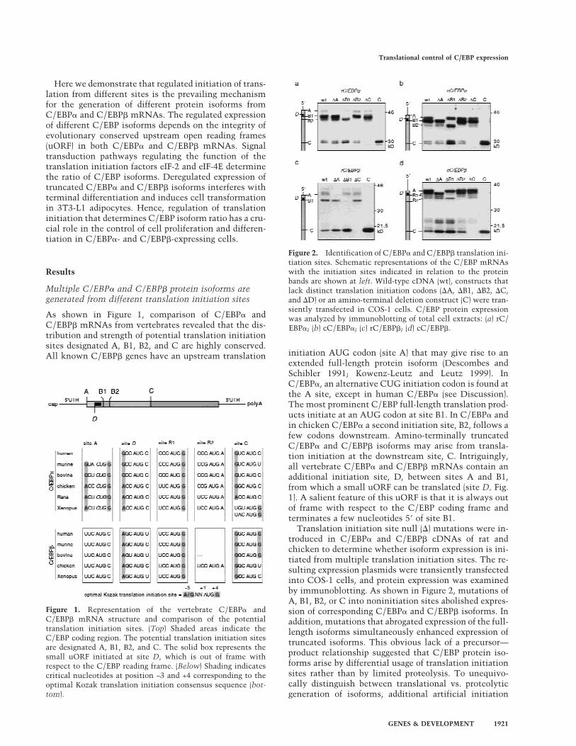
As shown in Figure 1, comparison of C/EBP� andC/EBP� mRNAs from vertebrates revealed that the dis-tribution and strength of potential translation initiationsites designated A, B1, B2, and C are highly conserved.All known C/EBP� genes have an upstream translation initiation AUG codon (site A) that may give rise to an
extended full-length protein isoform (Descombes andSchibler 1991; Kowenz-Leutz and Leutz 1999). InC/EBP�, an alternative CUG initiation codon is found atthe A site, except in human C/EBP� (see Discussion).The most prominent C/EBP full-length translation prod-ucts initiate at an AUG codon at site B1. In C/EBP� andin chicken C/EBP� a second initiation site, B2, follows afew codons downstream. Amino-terminally truncatedC/EBP� and C/EBP� isoforms may arise from transla-tion initiation at the downstream site, C. Intriguingly,all vertebrate C/EBP� and C/EBP� mRNAs contain anadditional initiation site, D, between sites A and B1,from which a small uORF can be translated (site D, Fig.1). A salient feature of this uORF is that it is always outof frame with respect to the C/EBP coding frame andterminates a few nucleotides 5� of site B1.
Translation initiation site null (�) mutations were in-troduced in C/EBP� and C/EBP� cDNAs of rat andchicken to determine whether isoform expression is ini-tiated from multiple translation initiation sites. The re-sulting expression plasmids were transiently transfectedinto COS-1 cells, and protein expression was examinedby immunoblotting. As shown in Figure 2, mutations ofA, B1, B2, or C into noninitiation sites abolished expres-sion of corresponding C/EBP� and C/EBP� isoforms. Inaddition, mutations that abrogated expression of the full-length isoforms simultaneously enhanced expression oftruncated isoforms. This obvious lack of a precursor—product relationship suggested that C/EBP protein iso-forms arise by differential usage of translation initiationsites rather than by limited proteolysis. To unequivo-cally distinguish between translational vs. proteolyticgeneration of isoforms, additional artificial initiation
Figure 1. Representation of the vertebrate C/EBP� andC/EBP� mRNA structure and comparison of the potentialtranslation initiation sites. (Top) Shaded areas indicate theC/EBP coding region. The potential translation initiation sitesare designated A, B1, B2, and C. The solid box represents thesmall uORF initiated at site D, which is out of frame withrespect to the C/EBP reading frame. (Below) Shading indicatescritical nucleotides at position −3 and +4 corresponding to theoptimal Kozak translation initiation consensus sequence (bot-tom).
Figure 2. Identification of C/EBP� and C/EBP� translation ini-tiation sites. Schematic representations of the C/EBP mRNAswith the initiation sites indicated in relation to the proteinbands are shown at left. Wild-type cDNA (wt), constructs thatlack distinct translation initiation codons (�A, �B1, �B2, �C,and �D) or an amino-terminal deletion construct (C) were tran-siently transfected in COS-1 cells. C/EBP protein expressionwas analyzed by immunoblotting of total cell extracts: (a) rC/EBP�; (b) cC/EBP�; (c) rC/EBP�; (d) cC/EBP�.
Translational control of C/EBP expression
GENES & DEVELOPMENT 1921
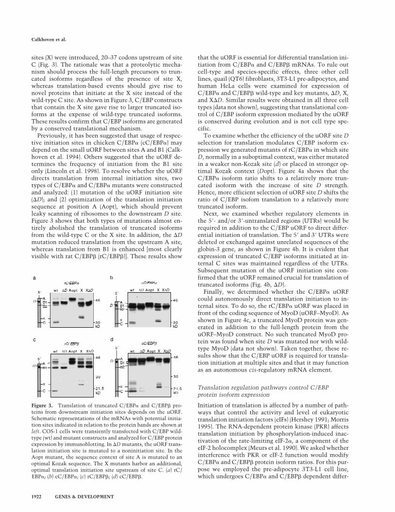
sites (X) were introduced, 20–37 codons upstream of siteC (Fig. 3). The rationale was that a proteolytic mecha-nism should process the full-length precursors to trun-cated isoforms regardless of the presence of site X,whereas translation-based events should give rise tonovel proteins that initiate at the X site instead of thewild-type C site. As shown in Figure 3, C/EBP constructsthat contain the X site gave rise to larger truncated iso-forms at the expense of wild-type truncated isoforms.These results confirm that C/EBP isoforms are generatedby a conserved translational mechanism.
Previously, it has been suggested that usage of respec-tive initiation sites in chicken C/EBP� (cC/EBP�) maydepend on the small uORF between sites A and B1 (Calk-hoven et al. 1994). Others suggested that the uORF de-termines the frequency of initiation from the B1 siteonly (Lincoln et al. 1998). To resolve whether the uORFdirects translation from internal initiation sites, twotypes of C/EBP� and C/EBP� mutants were constructedand analyzed: (1) mutation of the uORF initiation site(�D); and (2) optimization of the translation initiationsequence at position A (Aopt), which should preventleaky scanning of ribosomes to the downstream D site.Figure 3 shows that both types of mutations almost en-tirely abolished the translation of truncated isoformsfrom the wild-type C or the X site. In addition, the �Dmutation reduced translation from the upstream A site,whereas translation from B1 is enhanced [most clearlyvisible with rat C/EBP� (rC/EBP�)]. These results show
that the uORF is essential for differential translation ini-tiation from C/EBP� and C/EBP� mRNAs. To rule outcell-type and species-specific effects, three other celllines, quail (QT6) fibroblasts, 3T3-L1 pre-adipocytes, andhuman HeLa cells were examined for expression ofC/EBP� and C/EBP� wild-type and key mutants, �D, X,and X�D. Similar results were obtained in all three celltypes (data not shown), suggesting that translational con-trol of C/EBP isoform expression mediated by the uORFis conserved during evolution and is not cell type spe-cific.
To examine whether the efficiency of the uORF site Dselection for translation modulates C/EBP isoform ex-pression we generated mutants of rC/EBP� in which siteD, normally in a suboptimal context, was either mutatedin a weaker non-Kozak site (d) or placed in stronger op-timal Kozak context (Dopt). Figure 4a shows that theC/EBP� isoform ratio shifts to a relatively more trun-cated isoform with the increase of site D strength.Hence, more efficient selection of uORF site D shifts theratio of C/EBP isofom translation to a relatively moretruncated isoform.
Next, we examined whether regulatory elements inthe 5�- and/or 3�-untranslated regions (UTRs) would berequired in addition to the C/EBP uORF to direct differ-ential initiation of translation. The 5� and 3� UTRs weredeleted or exchanged against unrelated sequences of theglobin-3 gene, as shown in Figure 4b. It is evident thatexpression of truncated C/EBP isoforms initiated at in-ternal C sites was maintained regardless of the UTRs.Subsequent mutation of the uORF initiation site con-firmed that the uORF remained crucial for translation oftruncated isoforms (Fig. 4b, �D).
Finally, we determined whether the C/EBP� uORFcould autonomously direct translation initiation to in-ternal sites. To do so, the rC/EBP� uORF was placed infront of the coding sequence of MyoD (uORF–MyoD). Asshown in Figure 4c, a truncated MyoD protein was gen-erated in addition to the full-length protein from theuORF–MyoD construct. No such truncated MyoD pro-tein was found when site D was mutated nor with wild-type MyoD (data not shown). Taken together, these re-sults show that the C/EBP uORF is required for transla-tion initiation at multiple sites and that it may functionas an autonomous cis-regulatory mRNA element.
Translation regulation pathways control C/EBPprotein isoform expression
Initiation of translation is affected by a number of path-ways that control the activity and level of eukaryotictranslation initiation factors (eIFs) (Hershey 1991; Morris1995). The RNA-dependent protein kinase (PKR) affectstranslation initiation by phosphorylation-induced inac-tivation of the rate-limiting eIF-2�, a component of theeIF-2 holocomplex (Meurs et al. 1990). We asked whetherinterference with PKR or eIF-2 function would modifyC/EBP� and C/EBP� protein isoform ratios. For this pur-pose we employed the pre-adipocyte 3T3-L1 cell line,which undergoes C/EBP� and C/EBP� dependent differ-
Figure 3. Translation of truncated C/EBP� and C/EBP� pro-teins from downstream initiation sites depends on the uORF.Schematic representations of the mRNAs with potential initia-tion sites indicated in relation to the protein bands are shown atleft. COS-1 cells were transiently transfected with C/EBP wild-type (wt) and mutant constructs and analyzed for C/EBP proteinexpression by immunoblotting. In �D mutants, the uORF trans-lation initiation site is mutated to a noninitiation site. In theAopt mutant, the sequence context of site A is mutated to anoptimal Kozak sequence. The X mutants harbor an additional,optimal translation initiation site upstream of site C. (a) rC/EBP�; (b) cC/EBP�; (c) rC/EBP�; (d) cC/EBP�.
Calkhoven et al.
1922 GENES & DEVELOPMENT
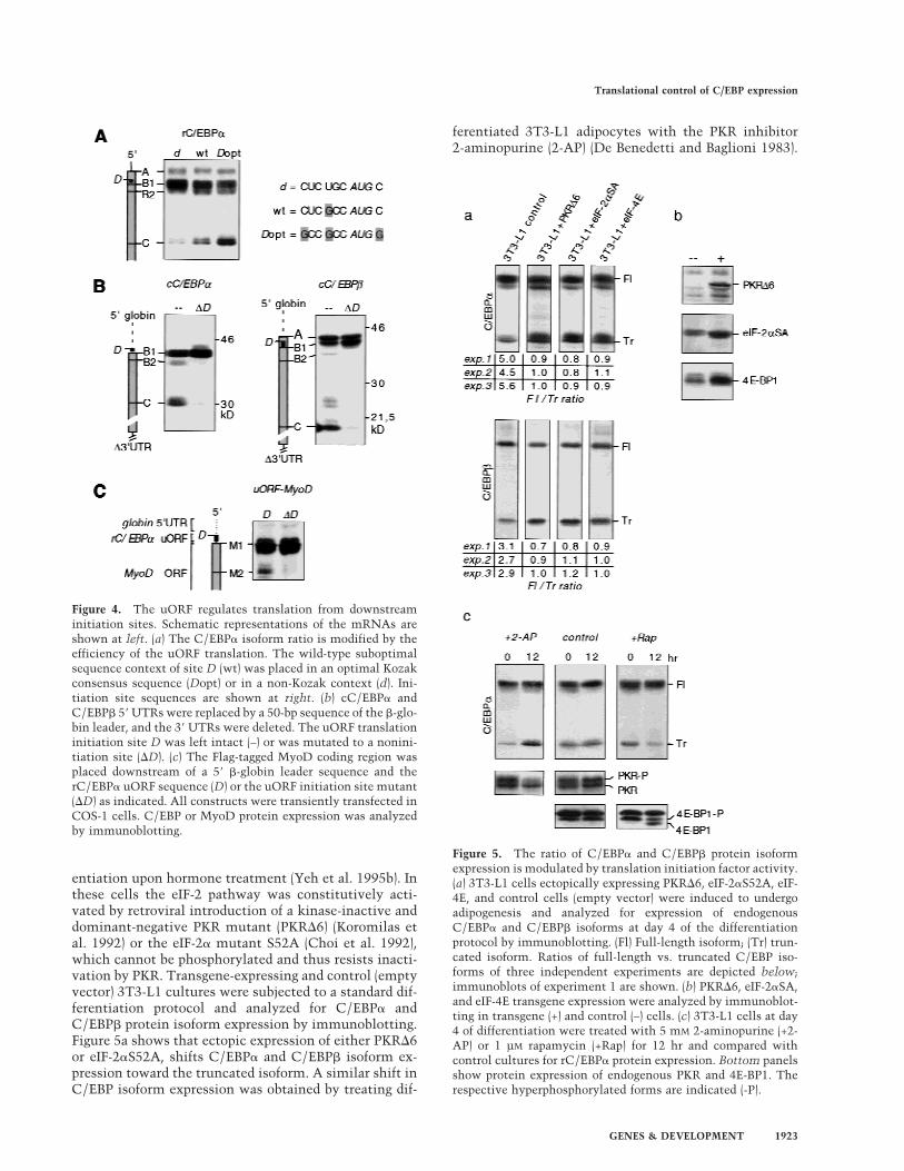
entiation upon hormone treatment (Yeh et al. 1995b). Inthese cells the eIF-2 pathway was constitutively acti-vated by retroviral introduction of a kinase-inactive anddominant-negative PKR mutant (PKR�6) (Koromilas etal. 1992) or the eIF-2� mutant S52A (Choi et al. 1992),which cannot be phosphorylated and thus resists inacti-vation by PKR. Transgene-expressing and control (emptyvector) 3T3-L1 cultures were subjected to a standard dif-ferentiation protocol and analyzed for C/EBP� andC/EBP� protein isoform expression by immunoblotting.Figure 5a shows that ectopic expression of either PKR�6or eIF-2�S52A, shifts C/EBP� and C/EBP� isoform ex-pression toward the truncated isoform. A similar shift inC/EBP isoform expression was obtained by treating dif-
ferentiated 3T3-L1 adipocytes with the PKR inhibitor2-aminopurine (2-AP) (De Benedetti and Baglioni 1983).
Figure 4. The uORF regulates translation from downstreaminitiation sites. Schematic representations of the mRNAs areshown at left. (a) The C/EBP� isoform ratio is modified by theefficiency of the uORF translation. The wild-type suboptimalsequence context of site D (wt) was placed in an optimal Kozakconsensus sequence (Dopt) or in a non-Kozak context (d). Ini-tiation site sequences are shown at right. (b) cC/EBP� andC/EBP� 5� UTRs were replaced by a 50-bp sequence of the �-glo-bin leader, and the 3� UTRs were deleted. The uORF translationinitiation site D was left intact (–) or was mutated to a nonini-tiation site (�D). (c) The Flag-tagged MyoD coding region wasplaced downstream of a 5� �-globin leader sequence and therC/EBP� uORF sequence (D) or the uORF initiation site mutant(�D) as indicated. All constructs were transiently transfected inCOS-1 cells. C/EBP or MyoD protein expression was analyzedby immunoblotting.
Figure 5. The ratio of C/EBP� and C/EBP� protein isoformexpression is modulated by translation initiation factor activity.(a) 3T3-L1 cells ectopically expressing PKR�6, eIF-2�S52A, eIF-4E, and control cells (empty vector) were induced to undergoadipogenesis and analyzed for expression of endogenousC/EBP� and C/EBP� isoforms at day 4 of the differentiationprotocol by immunoblotting. (Fl) Full-length isoform; (Tr) trun-cated isoform. Ratios of full-length vs. truncated C/EBP iso-forms of three independent experiments are depicted below;immunoblots of experiment 1 are shown. (b) PKR�6, eIF-2�SA,and eIF-4E transgene expression were analyzed by immunoblot-ting in transgene (+) and control (–) cells. (c) 3T3-L1 cells at day4 of differentiation were treated with 5 mM 2-aminopurine (+2-AP) or 1 µM rapamycin (+Rap) for 12 hr and compared withcontrol cultures for rC/EBP� protein expression. Bottom panelsshow protein expression of endogenous PKR and 4E-BP1. Therespective hyperphosphorylated forms are indicated (-P).
Translational control of C/EBP expression
GENES & DEVELOPMENT 1923
As shown in Figure 5c, concomitantly with 2-AP-in-duced dephosphorylation and inactivation of PKR, ex-pression of truncated C/EBP� isoform was enhanced.These results show that high eIF-2 activity shifts theratio of C/EBP isoform expression toward a more trun-cated isoform.
The FKBP12–rapamycin-associated protein (FRAP)/mammalian target of rapamycin (mTOR) (Brown et al.1994) enhances the level of accessible eIF-4E (Lawrenceand Abraham 1997): mTOR phosphorylates and inhibitsphosphatase PP2A, which keeps the inhibitory 4E-bind-ing protein 1 (4E-BP1, also called PHAS1) in an activeunphosphorylated state (Lin et al. 1994; Pause et al.1994; Brunn et al. 1997; Peterson et al. 1999). We gener-ated 3T3-L1 cells that overexpressed eIF-4E by retroviraltransfer and subjected them to the standard differentia-tion protocol. As shown in Figure 5a, enhanced levels ofeIF-4E shift C/EBP� isoform expression toward the trun-cated isoform. On the other hand, when mTOR was in-hibited by rapamycin, expression of the truncatedC/EBP� isoform was reduced concomitantly with thedephosphorylation of 4E-BP1 (Fig. 5c). Similar resultswere obtained with C/EBP� (data not shown). In conclu-sion, two rate-limiting translation initiation factors con-trol the ratio of C/EBP isoforms: At high eIF-2 and eIF-4Eactivity relatively more truncated C/EBP isoforms areexpressed, whereas at lower eIF activity expression of thefull-length isoforms dominates.
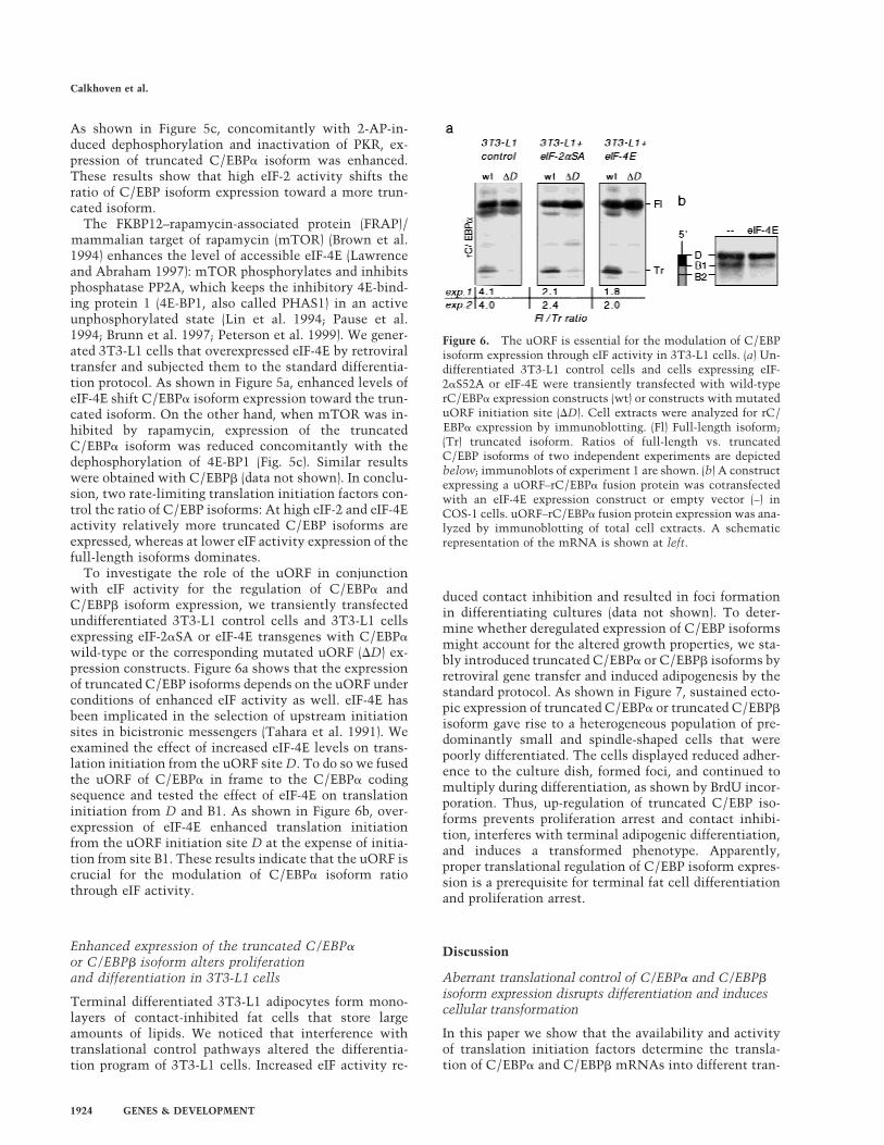
To investigate the role of the uORF in conjunctionwith eIF activity for the regulation of C/EBP� andC/EBP� isoform expression, we transiently transfectedundifferentiated 3T3-L1 control cells and 3T3-L1 cellsexpressing eIF-2�SA or eIF-4E transgenes with C/EBP�wild-type or the corresponding mutated uORF (�D) ex-pression constructs. Figure 6a shows that the expressionof truncated C/EBP isoforms depends on the uORF underconditions of enhanced eIF activity as well. eIF-4E hasbeen implicated in the selection of upstream initiationsites in bicistronic messengers (Tahara et al. 1991). Weexamined the effect of increased eIF-4E levels on trans-lation initiation from the uORF site D. To do so we fusedthe uORF of C/EBP� in frame to the C/EBP� codingsequence and tested the effect of eIF-4E on translationinitiation from D and B1. As shown in Figure 6b, over-expression of eIF-4E enhanced translation initiationfrom the uORF initiation site D at the expense of initia-tion from site B1. These results indicate that the uORF iscrucial for the modulation of C/EBP� isoform ratiothrough eIF activity.
Enhanced expression of the truncated C/EBP�or C/EBP� isoform alters proliferationand differentiation in 3T3-L1 cells
Terminal differentiated 3T3-L1 adipocytes form mono-layers of contact-inhibited fat cells that store largeamounts of lipids. We noticed that interference withtranslational control pathways altered the differentia-tion program of 3T3-L1 cells. Increased eIF activity re-
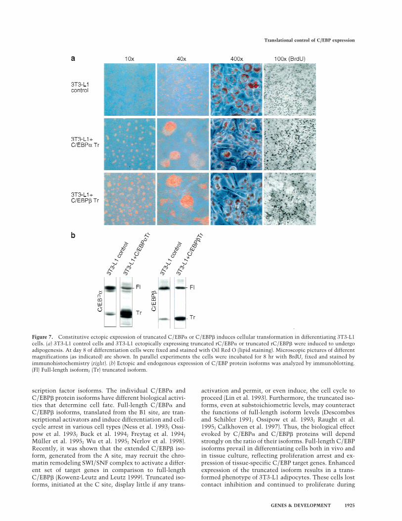
duced contact inhibition and resulted in foci formationin differentiating cultures (data not shown). To deter-mine whether deregulated expression of C/EBP isoformsmight account for the altered growth properties, we sta-bly introduced truncated C/EBP� or C/EBP� isoforms byretroviral gene transfer and induced adipogenesis by thestandard protocol. As shown in Figure 7, sustained ecto-pic expression of truncated C/EBP� or truncated C/EBP�isoform gave rise to a heterogeneous population of pre-dominantly small and spindle-shaped cells that werepoorly differentiated. The cells displayed reduced adher-ence to the culture dish, formed foci, and continued tomultiply during differentiation, as shown by BrdU incor-poration. Thus, up-regulation of truncated C/EBP iso-forms prevents proliferation arrest and contact inhibi-tion, interferes with terminal adipogenic differentiation,and induces a transformed phenotype. Apparently,proper translational regulation of C/EBP isoform expres-sion is a prerequisite for terminal fat cell differentiationand proliferation arrest.
Discussion
Aberrant translational control of C/EBP� and C/EBP�isoform expression disrupts differentiation and inducescellular transformation
In this paper we show that the availability and activityof translation initiation factors determine the transla-tion of C/EBP� and C/EBP� mRNAs into different tran-
Figure 6. The uORF is essential for the modulation of C/EBPisoform expression through eIF activity in 3T3-L1 cells. (a) Un-differentiated 3T3-L1 control cells and cells expressing eIF-2�S52A or eIF-4E were transiently transfected with wild-typerC/EBP� expression constructs (wt) or constructs with mutateduORF initiation site (�D). Cell extracts were analyzed for rC/EBP� expression by immunoblotting. (Fl) Full-length isoform;(Tr) truncated isoform. Ratios of full-length vs. truncatedC/EBP isoforms of two independent experiments are depictedbelow; immunoblots of experiment 1 are shown. (b) A constructexpressing a uORF–rC/EBP� fusion protein was cotransfectedwith an eIF-4E expression construct or empty vector (–) inCOS-1 cells. uORF–rC/EBP� fusion protein expression was ana-lyzed by immunoblotting of total cell extracts. A schematicrepresentation of the mRNA is shown at left.
Calkhoven et al.
1924 GENES & DEVELOPMENT
scription factor isoforms. The individual C/EBP� andC/EBP� protein isoforms have different biological activi-ties that determine cell fate. Full-length C/EBP� andC/EBP� isoforms, translated from the B1 site, are tran-scriptional activators and induce differentiation and cell-cycle arrest in various cell types (Ness et al. 1993; Ossi-pow et al. 1993; Buck et al. 1994; Freytag et al. 1994;Muller et al. 1995; Wu et al. 1995; Nerlov et al. 1998).Recently, it was shown that the extended C/EBP� iso-form, generated from the A site, may recruit the chro-matin remodeling SWI/SNF complex to activate a differ-ent set of target genes in comparison to full-lengthC/EBP� (Kowenz-Leutz and Leutz 1999). Truncated iso-forms, initiated at the C site, display little if any trans-
activation and permit, or even induce, the cell cycle toproceed (Lin et al. 1993). Furthermore, the truncated iso-forms, even at substoichiometric levels, may counteractthe functions of full-length isoform levels (Descombesand Schibler 1991; Ossipow et al. 1993; Raught et al.1995; Calkhoven et al. 1997). Thus, the biological effectevoked by C/EBP� and C/EBP� proteins will dependstrongly on the ratio of their isoforms. Full-length C/EBPisoforms prevail in differentiating cells both in vivo andin tissue culture, reflecting proliferation arrest and ex-pression of tissue-specific C/EBP target genes. Enhancedexpression of the truncated isoform results in a trans-formed phenotype of 3T3-L1 adipocytes. These cells lostcontact inhibition and continued to proliferate during
Figure 7. Constitutive ectopic expression of truncated C/EBP� or C/EBP� induces cellular transformation in differentiating 3T3-L1cells. (a) 3T3-L1 control cells and 3T3-L1 ectopically expressing truncated rC/EBP� or truncated rC/EBP� were induced to undergoadipogenesis. At day 8 of differentiation cells were fixed and stained with Oil Red O (lipid staining). Microscopic pictures of differentmagnifications (as indicated) are shown. In parallel experiments the cells were incubated for 8 hr with BrdU, fixed and stained byimmunohistochemistry (right). (b) Ectopic and endogenous expression of C/EBP protein isoforms was analyzed by immunoblotting.(Fl) Full-length isoform; (Tr) truncated isoform.
Translational control of C/EBP expression
GENES & DEVELOPMENT 1925
differentiation, which suggests that down-regulation oftruncated C/EBP� isoforms is essential for cessation ofproliferation and terminal differentiation. Constitutiveactivation of eIF-2 or eIF-4E, which deregulated C/EBP�and C/EBP� isoform expression, induced a similar trans-formed phenotype in 3T3-L1 adipocytes.
Deregulated eIF activity has been shown to transformfibroblasts (Koromilas et al. 1992; Donze et al. 1995).Although deregulated eIF activity will affect translationof many genes, it is tempting to speculate that inad-equate adjustment of C/EBP isoform ratios contributesto neoplastic conversion in tissues that express C/EBP�and/or C/EBP�. A correlation has been found betweenenhanced eIF-2 activity and enhanced truncated C/EBP�expression in mammary epithelial cancer cells (Raughtet al. 1996). In addition, up-regulation of eIF-4E is anearly event in colon cancer cells known to expressC/EBPs (Rosenwald et al. 1999). Also, the myxoid lipo-sarcoma-specific chromosomal rearrangement of theCHOP gene, TLS–CHOP, which is required for onco-genic transformation, interferes with C/EBP function(Zinszner et al. 1994).
Differential initiation of translation generates differentC/EBP isoforms
Initially, truncated C/EBP� and C/EBP� isoforms wereproposed to arise by a ribosomal scanning mechanism toalternative translation initiation sites in their mRNAs(Descombes and Schibler 1991; Lin et al. 1993; Ossipowet al. 1993; An et al. 1996). In contrast, limited proteoly-sis recently has been suggested to account for the gen-eration of truncated isoforms (Baer et al. 1998; Welm etal. 1999). We showed by a mutagenesis approach thatdifferent C/EBP� and C/EBP� protein isoforms originatefrom all evolutionary conserved translation initiationsites (termed A, B1, B2, and C; Fig. 1). Elimination ofupstream initiation sites increases expression of trun-cated C/EBP isoforms. Moreover, introduction of novelinitiation sites (X) between B and C sites results in pro-duction of novel isoforms initiated at the X rather thanat the C site. Shift of translation initiation to down-stream initiation sites once upstream sites were re-moved, and substitution of the wild-type truncated prod-ucts (C) by alternative products once an extra initiationcodon (X) was introduced argue strongly in favor of atranslational mechanism in the generation of truncatedC/EBP isoforms. Although we cannot rule out that lim-ited proteolysis of C/EBPs might occur under specificconditions (Welm et al. 1999), our results are incompat-ible with precursor–product relationships between full-length and truncated proteins and consistent with obser-vations reported by Sears and Sealy (1994), who alsofailed to find a precursor–product relationship by pulse-chase labeling. Additional experiments, using bicistroniccontructs also ruled out the possibility that truncatedproteins are generated by internal ribosomal entry sitesin C/EBP mRNAs (data not shown). We conclude thattranslational control is the prevailing mechanism ofC/EBP� and C/EBP� isoform expression in vertebrates.
The conservation of translation initiation site distri-bution and sequence context further supports the experi-mental data. The sequence context of initiation codonsdetermines the fidelity and frequency of their selection,as has been described by Kozak (1989). Only two devia-tions from the conserved initiation sites were found.First, the A site in human C/EBP� is apparently absent.However, an immunoreactive band corresponding to anextended isoform was detected in extracts of the humanHL-60 leukemia cell line, indicating that translationmay be initiated from a functionally equivalent A site (aGUG codon; C.F. Calkhoven, unpubl.). The second ex-ception is that a B2 site is present in all C/EBP� isoformsexamined and in cC/EBP� but not in mammalian andXenopus laevis C/EBP�.
The C/EBP uORF mediates differential translationinitiation
A small uORF is located between the A and the B sites inall vertebrate C/EBP� and C/EBP� mRNAs. The uORFis out of frame with respect to the C/EBP reading frameand terminates just upstream of the major initiation siteB1. Fusion of the uORF to the C/EBP reading frame re-vealed that site D is selected for translation. Our datashow that the uORF has a crucial role in the regulationof isoform expression. Three lines of evidence show thatthe uORF is paramount for translation initiation atdownstream initiation sites. First, different types of mu-tations that disrupt the function of the uORF concomi-tantly abrogate translation initiation at downstreamsites. Second, the efficiency of translation from site C isproportional to the efficiency of site D selection. Third,the uORF mediates translation from downstream initia-tion sites in a different mRNA context (MyoD transcript)and thus displays an autonomous function.
The observation that removal of the uORF initiationcodon abolished initiation at the C site (or X site) andoptimization of the uORF site D enhances initiation at Cindicates that uORF translation is required for its func-tion. This implies that translation reinitiation ratherthan leaky scanning is the mechanism of downstreaminitiation at C (or X). Translation from site B1 is notdependent on the uORF. In contrast, it is inversely regu-lated to the strength of the D site. This indicates thattranslation from B1 mainly results from leaky scanningover the wild-type D site (or the weak mutant site d).However, the optimization of the D site (Dopt), whichdoes not allow leaky scanning of ribosomes (data notshown), diminishes but does not completely abolishtranslation from B1 (as one would expect with leakyscanning as the only mechanism). Hence, immediatereinitiation after uORF translation seems to occur aswell. Taken together, translation from site C appears todepend strictly on uORF translation, whereas initiationat site B1 can occur by either leaky scanning or by im-mediate reinitiation.
Removal of the D site also revealed that the uORF isinvolved in regulation of initiation at site A. We presumethat under certain cellular conditions translation of the
Calkhoven et al.
1926 GENES & DEVELOPMENT
uORF may cause cueing of ribosomes and thus increasethe probability of initiation at the weak initiation site A.Although several details need to be solved, the data showthat a conserved uORF in vertebrate C/EBP� andC/EBP� mRNAs directs the translation from multipletranslation sites.
Translational control and cell fate
Kinases PKR and mTOR, implicated in C/EBP isoformtranslation, are involved in many cellular processes. Amajor function of PKR is to mediate translational repres-sion after virus infection and interferon signaling (Wek1994). However, regulating PKR activity also appears tobe essential for normal cell proliferation and differentia-tion. Others showed that both the inactivation of PKR inquiescent cells or ectopic expression of the PKR phos-phorylation-defective eIF-2�S52A mutant induces prolif-eration and even transformation (Koromilas et al. 1992;Meurs et al. 1993; Barber et al. 1994; Donze et al. 1995).We also observed loss of contact inhibition and persis-tent proliferation in differentiating 3T3-L1 cells express-ing dominant-negative PKR�6 or eIF-2�S52A (data notshown). Augmentation of rate-limiting eIF-2� enhancedexpression of truncated C/EBP forms in an uORF-depen-dent manner. A paradigm of eIF-2 function and transla-tional control is provided by the yeast transcription fac-tor GCN4. In yeast, high levels of eIF-2 mediate multipletranslation reinitiation events after initial uORF trans-lation (Mueller and Hinnebusch 1986; Hinnebusch1994). In a similar fashion, elevated eIF-2 levels mightenhance translation reinitiation at C/EBP C sites aftertranslation of the uORF.
The FRAP/mTOR kinases are involved in the regu-lation of several cellular processes, including cell pro-liferation, transcriptional response to nutrients, andmRNA translation (Brown et al. 1994; Zheng andSchreiber 1997; Peterson and Schreiber 1998; Beckand Hall 1999; Cardenas et al. 1999; Dennis et al. 1999;Hardwick et al. 1999; Kuruvilla and Schreiber 1999). Ourdata show that inhibition of FRAP/mTOR function byrapamycin reduced expression of the truncated C/EBPisoforms, whereas overexpression of eIF-4E enhanced ex-pression of the truncated isoforms in an uORF-depen-dent manner. Others have shown that eIF-4E is impli-cated in selection of upstream initiation sites in bicis-tronic messengers (Tahara et al. 1991). Accordingly, weobserved enhanced translation initiation from uORF siteD upon overexpression of eIF-4E (Fig. 6b). As the trans-lation of the uORF determines reinitiation at the down-stream site C (Fig. 4a), it is presumably eIF-4E-mediatedenhanced D-site usage that leads to increased expressionof truncated C/EBP isoforms. Our data are also in accor-dance with that of Yeh et al. (1995a), who showed thattreatment of 3T3-L1 cells with rapamycin at the onset ofthe differentiation program inhibits their clonal expan-sion, a prerequisite for 3T3-L1 differentiation. It is pos-sible that the observed repression of truncated C/EBPisoforms is responsible for failure of clonal expansion.
Taken together, pathways that modulate the activity
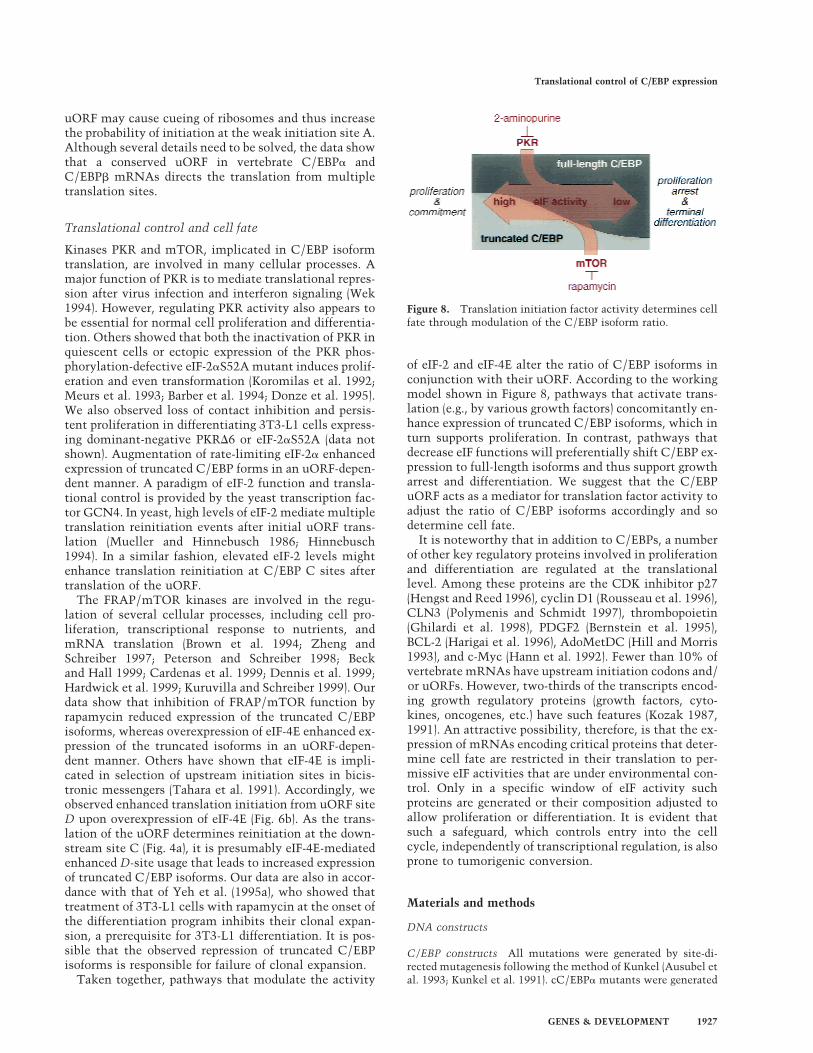
of eIF-2 and eIF-4E alter the ratio of C/EBP isoforms inconjunction with their uORF. According to the workingmodel shown in Figure 8, pathways that activate trans-lation (e.g., by various growth factors) concomitantly en-hance expression of truncated C/EBP isoforms, which inturn supports proliferation. In contrast, pathways thatdecrease eIF functions will preferentially shift C/EBP ex-pression to full-length isoforms and thus support growtharrest and differentiation. We suggest that the C/EBPuORF acts as a mediator for translation factor activity toadjust the ratio of C/EBP isoforms accordingly and sodetermine cell fate.
It is noteworthy that in addition to C/EBPs, a numberof other key regulatory proteins involved in proliferationand differentiation are regulated at the translationallevel. Among these proteins are the CDK inhibitor p27(Hengst and Reed 1996), cyclin D1 (Rousseau et al. 1996),CLN3 (Polymenis and Schmidt 1997), thrombopoietin(Ghilardi et al. 1998), PDGF2 (Bernstein et al. 1995),BCL-2 (Harigai et al. 1996), AdoMetDC (Hill and Morris1993), and c-Myc (Hann et al. 1992). Fewer than 10% ofvertebrate mRNAs have upstream initiation codons and/or uORFs. However, two-thirds of the transcripts encod-ing growth regulatory proteins (growth factors, cyto-kines, oncogenes, etc.) have such features (Kozak 1987,1991). An attractive possibility, therefore, is that the ex-pression of mRNAs encoding critical proteins that deter-mine cell fate are restricted in their translation to per-missive eIF activities that are under environmental con-trol. Only in a specific window of eIF activity suchproteins are generated or their composition adjusted toallow proliferation or differentiation. It is evident thatsuch a safeguard, which controls entry into the cellcycle, independently of transcriptional regulation, is alsoprone to tumorigenic conversion.
Materials and methods
DNA constructs
C/EBP constructs All mutations were generated by site-di-rected mutagenesis following the method of Kunkel (Ausubel etal. 1993; Kunkel et al. 1991). cC/EBP� mutants were generated
Figure 8. Translation initiation factor activity determines cellfate through modulation of the C/EBP isoform ratio.
Translational control of C/EBP expression
GENES & DEVELOPMENT 1927
on the cC/EBP�wt–pSG5 template (Calkhoven et al. 1994) witholigonucleotide primers �A (accctgg → ccccggg), 5�-ccgccccgtc-cgaccccgggtttgccggagccc-3�; �B1 (ttcatgg → ttcctcg), 5�-ggctgtag-gtgcttcctcgagcaagccaacttc-3�; �B2 (ccgatga → cggggag), 5�-cccg-gcccccggggagcagcggccagcaccacc-3�; �C (ggcatgc → gggatcc), 5�-ttccacgggatccacggggcc-3�; �D (accatgc → aggatcc), 5�-ccgggccct-tcaggatccccggcaggctg-3�; Aopt (cgaaccctgg → gccaccatgg), 5�-cg-cgccccgtcgccaccatggattgccggagc-3�; and X (gacctct → gccatgg),5�-cgagttcctggccgccatggtccagcacagcaagc-3�. The C mutant wasgenerated by introducing an EcoRI site with primer 5�-ggggatttc-gaattccacggcatg-3� and subsequent removal of the EcoRI frag-ment containing the 5� UTR and amino-terminal C/EBP se-quences. For the cC/EBP�–5� �-globin/�3� UTR and corre-sponding �D mutants a HindIII site was introduced upstream ofsite D with primer 5�-gcagagccgccgcaagcttgtccgaaccctgg-3�, andthe KpnI–HindIII 5� UTR fragment was exchanged for the KpnI–HindIII fragment of pBAT (Annweiler et al. 1991) containing the�-globin leader sequence. The 3� UTR was removed by ApaI–BamHI digestion. A cC/EBP� (NF-M) EcoRI cDNA fragment(Katz et al. 1993) was cloned into pSG5, and mutants were gen-erated on the cC/EBP�wt–pSG5 template with the primers �A(ttcatgc → tgaattc), 5�-ccccctttgcttgaattcaacgcctggtgg-3�; �B1(tccatgg → tccattgaa), 5�-cgcctttaaatccattgaagtggctaatttctattacg-aggcgg-3�; �B2 (tccatga → tctagaa), 5�-gggccgctctagaaccgaacttac-cgtagg-3�; �C (ggcatgt → ggaattc), 5�-ggaccggggggaattccctcgc-cctacggc-3�; �D (agcatgc → agcaagc), 5�-ggcctgggacgcagcttgcctc-cccattcagcc-3�; Aopt (gctttcatgc → gccaccatgg), 5�-gcatcccccttt-gccaccatggaacgcctggtggcc-3�; Dopt (cgcagcatgc → gccaccatgg),5�-ggtggcctgggcgccaccatggctccccattcagcc-3�; and X (gagaccct-gg → gccaccatgg), 5�-ggagccggtcttcgccaccatggactcttgcaaagg-3�.The C mutant was generated by introducing an EcoRI site withprimer 5�-ccgtaaggaagaattcggagcggggccagg-3� and subsequent re-moval of the EcoRI fragment containing the 5� UTR and amino-terminal cC/EBP� sequences. For cC/EBP�–5� �-globin/�3�
UTR and corresponding �D mutants a HindIII site was intro-duced upstream of site D with primer 5�-ccgtcttctcctccaagcttc-cccctttgc-3�, and the KpnI–HindIII 5� UTR fragment was ex-changed for the KpnI–HindIII fragment of pBAT (Annweiler etal. 1991) containing the �-globin leader sequence. The 3� UTRwas removed by BamHI–EcoRI digestion after introduction of aBamHI site downstream of the C/EBP� stop codon with primer5�-cgctgctgaccccggatccggccgcgc-3�. A rC/EBP� wild-type clonecontaining the cDNA sequence was generated by cloning a 590-bp EcoRI–NotI PCR fragment (primers, 5�-ccggaattccattcgcgac-ccaaagctgcg-3� and 5�-cgcggatccgatctggaactgcaagtgaggg-3�) fromgenomic DNA containing the 5�UTR, together with the NotI–BamHI rC/EBP� cDNA fragment (Landschulz et al. 1988) intopSG5, which was used as template for further mutagenesis withthe primers �A (gtactgg → cggatcc), 5�-gggcgagttgggcggatccgtgg-gcggcgg-3�; �B1 (cccatgg → cccatcg), 5�-ctctaactcccccatcgagtcg-gccgac-3�; �B2 (ccgatga → cggatcc), 5�-cggccccggatccgcagccacct-cc-3�; �C (gtcatgt → gtgaatt), 5�-ggcggtgcggtgaattccgcgggggcgca-cgg-3�; �D (gccatgc → gggatcc), 5�-ccgaggctcgggatcccgggagaactc-taactccc-3�; Aopt (ggggtactgg → gccgccatgg), 5�-gggcgagttgccgc-catgggtgggcggcgg-3�; d (gccatgc → tgcatgc), 5�-ccgccgaggctctg-catgccgggagaactc-3�; Dopt (ctcgccatgc → gccgccatgg), 5�-gctgga-ggccgtcgacggccgccatggcgggagaactctaactcc-3�; X (gccgacctct →gccgccatgg), 5�-cgagttcctggccgccatggtccagcacagccggc-3�; anduORF+C/EBP� fusion (taactc → aactc), 5�-ccatgccgggagagct-caactcccccatgg-3�. The C mutant was generated by cloning a725-bp EcoRI–BamHI PCR fragment (primers 5�-gcgaattcatgtc-cgcgggggcgcacggacc-3� and 5�-gcggatcctcacgcgcagttgcccatg-gccttgacc-3�) into pcDNA3. A rat C/EBP�wt–pSG5 vector wasgenerated by cloning the IL-6–DBP EcoRI cDNA fragment (Poliet al. 1990) into pSG5 and used as template for further muta-genesis with the primers �A (ttcatgc → tgaattc), 5�-ggccccgcgt-
gaattcaccgcctgctggcc-3�; �B1 (cccatgg → cccattg), 5�-gcctttagac-ccattgaagtggccaacttc-3�; �C (gccatgg → gcgatcg), 5�-cgacgcgccc-gcgatcgcggccggcttccc-3�; �D (agcatgc → agaattc), 5�-ggcctg-ggacgcagaattcctcccgccgcc-3�; Aopt (gcgttcatgc → gccaccatgg),5�-gggccccgccaccatggcccgcctgctggc-3�; and X (gccgcactca → gcc-gccatgg), 5�-gcctcccgccgccatggaggccgagccggg-3�. The C mutantwas generated by removal of the EcoRI fragment containing the5� UTR and amino-terminal C/EBP sequences from the rC/EBP�X–pSG5 construct. The MyoD–uORF–pSG5 [gccatgccgg-gagaactctaa (uORF) ctcccccatgg (MyoD)] and MyoD–�uORF–pSG5 [ggg atc (�D uORF) ccgggagaactctaactcccccatgg (MyoD)]vectors were generated by combined cloning of a KpnI–NcoIfragment containing the �-globin leader and rC/EBP�–uORF or–�D sequences (from the cC/EBP�–5� �-globin/�3� UTR mu-tants), together with a NcoI–XbaI fragment containing amino-terminal Flag-tagged human MyoD coding region into pSG5.
pBabe–puro retroviral constructs rC/EBP�Tr–pBabe–puro wasgenerated by cloning the EcoRI–BamHI fragment from rC/EBP�C–pSG5 into pBabe–puro (Morgenstern and Land 1990).rC/EBP�pBabe–puro was generated by cloning the EcoRI frag-ment from rC/EBP�C–pSG5 into pBabe–puro. A human eIF-2�
EcoRI–HindIII 1.6-kb fragment was cloned from SP65-2a (Ernstet al. 1987) into pSG5. eIF-2�wt–pSG5 was used for creating theS52A mutant (Choi et al. 1992) with the primer 5�-cttagtgaat-tggccagaaggcgtatccg-3�. The BamHI eIF-2�S52A fragment fromeIF-2�S52A–pSG5 was cloned into pBabe–puro. Human PKR(Meurs et al. 1990) was cloned by PCR from HeLa cells using theprimers 5�-gggaatcaacatccacacttccg-3� and 5�-gggagactgtgtcattg-cactcc-3�, tagged with BamHI sites and cloned into pSG5. Thedominant-negative PKR�6 mutant (Koromilas et al. 1992) wasgenerated on PKR–pSG5 with primer 5�-ggtcaaagactaagtgcttct-gtgataaagggaccttgg-3�, and the BamHI fragment was cloned intopBabe–puro. Human eIF-4E (Rychlik et al. 1987) was cloned byPCR from HeLa cells using the primers 5�-gattcagatcgatctaa-gatgg-3� and 5�-cctatgagaatactcagaagg-3�, tagged with BamHIsites, and cloned into pBabe–puro.
Cells and tissue culture
COS-1 cells (ATCC, CRL-1650) were propagated in DMEM,F12, and 5% FCS (GIBCO); Hela cells (ATCC, CCL-2) in DMEMand 10% FCS; 3T3-L1 cells (ATCC, CL-173) in DMEM and 10%FCS (Seromed); QT6 cells (ATCC, CRL 1708) in DMEM, 8%FCS, and 2% heat-inactivated chicken serum; and Phoenix Ecells (G.P. Nolan, Stanford University School of Medicine, Stan-ford, CA; ATCC, SD 3444) in DMEM and 10% FCS in a hu-midified atmosphere with 5% CO2 at 37°C. Induction of adipo-genetic differentiation in 3T3-L1 cells was induced in 2-dayconfluent cultures (designated day 0) with 2 days of incubationin medium containing 10 µg/ml insulin (Sigma), 1 µM dexa-methasone (Sigma), and 0.5 mM 3-isobutyl-1-methylxanthine(Sigma) (days 1–2), followed by incubation in 10 µg/ml insulinwith medium exchange every second day (days 3–8) (Yeh et al.1995b). Medium for pBabe–puro-infected 3T3-L1 cells con-tained an additional 0.5 µg/ml puromycin (Sigma). Rapamycin(Calbiochem) was used in a concentration of 1 µM, and2-amunopurine (Calbiochem) was used in a concentration of5 mM.
Oil-Red-O staining 3T3-L1 cells were washed with PBS, fixedwith 4% paraformaldehyde overnight at 4°C, stained with Oil-Red-O solution for 5 min [2:3, 0.3% (wt/vol) Oil Red O (Sigma)in isopropanol and water before filtering], and analyzed bybright-field microscopy.
Calkhoven et al.
1928 GENES & DEVELOPMENT
BrdU labeling 3T3-L1 cells were labeled with BrdU for 8 hrfollowing the manufacturer’s protocol (BrdU Labeling and De-tection Kit II, Boehringer Mannheim).
Retroviral methods
The ecotropic-packaging cell line Phoenix E was transientlytransfected with the calcium phosphate–DNA precipitationmethod, and infectious virus was harvested after 48 hr. 3T3-L1target cells (5 × 10⁵) were infected as described in Pear et al.(1993) and selected for puromycin (2 µg/ml) resistance.
Transfections
COS-1 cells were transfected with 5 µg of pSG5-based expres-sion vector using DEAE–dextran/chloroquine as described byGonzalez and Joly (1995). HeLa and 3T3-L1 cells were trans-fected with 0.75 µg of pSG5-based expression vector using Ge-nePORTER (Gene Therapy Systems, Inc) following the protocolof the manufacturer in six-well culture trays. Transfected cellswere harvested 24 hr after transfection. QT6 cells were trans-fected with 5 µg of pSG5-based expression vector using the cal-cium phosphate–DNA precipitation method as described in Au-subel et al. (1993).
Western blot analysis
Cells were lysed rapidly in 0.5 M NaOH, neutralized by adding0.5 M HCl, or directly lysed in RIPA buffer (150 mM NaCl, 50mM Tris-HCl at pH 7.5, 1% NP-40, 0.1% SDS), supplementedwith SDS loading buffer, sonicated, and boiled. The proteinswere separated on a 12.5% SDS–polyacrylamide gel and electro-blotted on PVDF membrane (Immobilon-P, Millipore). Westernblot analysis was performed, as described in Calkhoven et al.(1994), followed by luminescent detection according to themanufacturer’s protocol (Amersham Life Technologies, ECLsystem). The following antisera were used: 1:1500 cC/EBP�
(Calkhoven et al. 1994) and 1:3000 cC/EBP�/NF-M (Katz et al.1993); 0.5 µg/ml rC/EBP� (14AA), rC/EBP� (C-19), PKR (M-515), PKR (K-17), 4E-BP1 (R-113), and eIF-2� (C-20); 1:2000 anti-goat immunoglobulin HRP (Sc-2020) (all from Santa Cruz Bio-technology Inc.); 0.5 µg/ml eIF-4E (E27620) (Transduction Labo-ratories); 10 µg/ml anti-Flag M2 (IB13026) (Eastman KodakCompany); 1:2000 anti-mouse immunoglobulin HRP (NA931)and 1:5000 anti-rabbit immunoglobulin HRP (NA934) (Amer-sham Life Technologies). Protein bands were quantified usingthe FUJIFILM Science Lab/Image Gauge computer program.
Acknowledgments
We offer special thanks to Marion Bengs for extensive technicalassistance. We thank Dr. Garry P. Nolan for providing the Phoe-nix E cells, Dr. Adri A.M. Thomas for providing an eIF-2� clone,Dr. Steven L. McKnight for providing a rC/EBP� clone, ValeriaPoli for providing an IL-6DBP clone, Dr. Barbara Winter for pro-viding a MyoD clone, Dr. Stephane Ansieau for the Flag-taggedMyoD construct, and Dr. Dipak Ramji for X. laevis C/EBP�
sequence information. This research is supported by the Deut-sche Forschungsgemeinschaft by grants to A.L. (LE77072-2/LE770/3-1). C.F.C was supported by a Marie Curie /TMR fel-lowship (ERBFMBICT961254).
The publication costs of this article were defrayed in part bypayment of page charges. This article must therefore be herebymarked “advertisement” in accordance with 18 USC section1734 solely to indicate this fact.
References
An, M.R., Hsieh, C.C., Reisner, P.D., Rabek, J.P., Scott, S.G.,Kuninger, D.T., and Papaconstantinou, J. 1996. Evidence forposttranscriptional regulation of C/EBP� and C/EBP� iso-form expression during the lipopolysaccharide-mediatedacute-phase response. Mol. Cell. Biol. 16: 2295–2306.
Annweiler, A., Hipskind, R.A., and Wirth, T. 1991. A strategyfor efficient in vitro translation of cDNAs using the rabbit�-globin leader sequence. Nucleic Acids Res. 19: 3750.
Ausubel, F.M., Brent, R., Kingston, R.E., Moore, D.D., Seidman,J.G., Smith, J.A., and Struhl, K. 1993. Current Protocols inMolecular Biology. Green Publishing Associates/John Wiley& Sons, New York, NY.
Baer, M., Williams, S.C., Dillner, A., Schwartz, R.C., andJohnson, P.F. 1998. Autocrine signals control CCAAT/en-hancer binding protein beta expression, localization, and ac-tivity in macrophages. Blood 92: 4353–4365.
Barber, G.N., Thompson, S., Lee, T.G., Strom, T., Jagus, R.,Darveau, A., and Katze, M.G. 1994. The 58-kilodalton in-hibitor of the interferon-induced double-stranded RNA-acti-vated protein kinase is a tetratricopeptide repeat proteinwith oncogenic properties. Proc. Natl. Acad. Sci. 91: 4278–4282.
Beck, T. and Hall, M.N. 1999. The TOR signalling pathwaycontrols nuclear localization of nutrient-regulated transcrip-tion factors. Nature 402: 689–692.
Bernstein, J., Shefler, I., and Elroy-Stein, O. 1995. The transla-tional repression mediated by the platelet-derived growthfactor 2/c-sis mRNA leader is relieved during megakaryo-cytic differentiation. J. Biol. Chem. 270: 10559–10565.
Birkenmeier, E.H., Gwynn, B., Howard, S., Jerry, J., Gordon, J.I.,Landschulz, W.H., and McKnight, S.L. 1989. Tissue-specificexpression, developmental regulation, and genetic mappingof the gene encoding CCAAT/enhancer binding protein.Genes & Dev. 3: 1146–1156.
Brown, E.J., Albers, M.W., Shin, T.B., Ichikawa, K., Keith, C.T.,Lane, W.S., and Schreiber, S.L. 1994. A mammalian proteintargeted by G1-arresting rapamycin-receptor complex. Na-ture 369: 756–758.
Brunn, G.J., Hudson, C.C., Sekulic, A., Williams, J.M., Hosoi,H., Houghton, P.J., Lawrence Jr., J.C., and Abraham, R.T.1997. Phosphorylation of the translational repressor PHAS-1by the mammalian target of rapamycin. Science 277: 99–101.
Buck, M., Turler, H., and Chojkier, M. 1994. LAP (NF-IL-6), atissue-specific transcriptional activator, is an inhibitor ofhepatoma cell proliferation. EMBO J. 13: 851–860.
Buck, M., Poli, V., van der Geer, P., Chojkier, M., and Hunter, T.1999. Phosphorylation of rat serine 105 or mouse threonine217 in C/EBP� is required for hepatocyte proliferation in-duced by TGF alpha. Mol. Cell 4: 1087–1092.
Calkhoven, C.F., Bouwman, P.R., Snippe, L., and Ab, G. 1994.Translation start site multiplicity of the CCAAT/enhancerbinding protein alpha mRNA is dictated by a small 5� openreading frame. Nucleic Acids Res. 22: 5540–5547.
Calkhoven, C.F., Snippe, L., and Ab, G. 1997. Differentialstimulation by CCAAT/ enhancer-binding protein alphaisoforms of the estrogen-activated promoter of the very-low-density apolipoiprotein II gene. Eur. J. Biochem. 249: 113–120.
Cao, Z., Umek, R.M., and McKnight, S.L. 1991. Regulated ex-pression of three C/EBP isoforms during adipose conversionof 3T3-L1 cells. Genes & Dev. 5: 1538–1552.
Cardenas, M.E., Cutler, N.S., Lorenz, M.C., Di Como, C.J., andHeitman, J. 1999. The TOR signaling cascade regulates gene
Translational control of C/EBP expression
GENES & DEVELOPMENT 1929
expression in response to nutrients. Genes & Dev. 13: 3271–3279.
Chandrasekaran, C. and Gordon, J.I. 1993. Cell lineage-specificand differentiation-dependent patterns of CCAAT/enhancerbinding protein alpha expression in the gut epithelium ofnormal and transgenic mice. Proc. Natl. Acad. Sci. 90: 8871–8875.
Choi, S.Y., Scherer, B.J., Schnier, J., Davies, M.V., Kaufman, R.J.,and Hershey, J.W.B. 1992. Stimulation of protein synthesisin COS cells transfected with variants of the alpha- subunitsof initiation factor eIF-2. J. Biol. Chem. 267: 286–293.
Christy, R.J., Yang, V.W., Ntambi, J.M., Geiman, D.E., Landsch-ulz, W.H., Friedman, A.D., Nakabeppu, Y., Kelly, T.J., andLane, M.D. 1989. Differentiation-induced gene expression in3T3-L1 preadipocytes: CCAAT/enhancer binding protein in-teracts with and activates the promoters of two adipocyte-specific genes. Genes & Dev. 3: 1323–1335.
Constance, C.M., Morgan, J.I.T., and Umek, R.M. 1996. C/EBP�
regulation of the growth-arrest-associated gene gadd45. Mol.Cell Biol. 16: 3878–3883.
De Benedetti, A. and Baglioni, C. 1983. Phosphorylation of ini-tiation factor eIF-2 alpha, binding of mRNA to 48S com-plexes, and its reutilization in initiation of protein synthesis.J. Biol. Chem. 258: 14556–14562.
Dennis, P.B., Fumagalli, S., and Thomas, G. 1999. Target ofrapamycin (TOR): Balancing the opposing forces of proteinsynthesis and degradation. Curr. Opin. Genet. Dev. 9: 49–54.
Descombes, P. and Schibler, U. 1991. A liver-enriched transcrip-tional activator protein, LAP, and a transcriptional inhibi-tory protein, LIP, are translated from the same mRNA. Cell67: 569–579.
Diehl, A.M., Michaelson, P., and Yang, S.Q. 1994. Selective in-duction of CCAAT/enhancer binding protein isoforms oc-curs during rat liver development. Gastroenterology106: 1625–1637.
Donze, O., Jagus, R., Koromilas, A.E., Hershey, J.W., and Sonen-berg, N. 1995. Abrogation of translation initiation factoreIF-2 phosphorylation causes malignant transformation ofNIH 3T3 cells. EMBO J. 14: 3828–3834.
Ernst, H., Duncan, R.F., and Hershey, J.W. 1987. Cloning andsequencing of complementary DNAs encoding the alpha-subunit of translational initiation factor eIF-2. Characteriza-tion of the protein and its messenger RNA. J. Biol. Chem.262: 1206–1212.
Flodby, P., Barlow, C., Kyleford, H., Ahrlund-Richter, L., andXanthopoulos, G. 1996. Increased hepatic cell proliferationand lung abnormalities in mice deficient in CCAAT/en-hancer binding protein alpha. J. Biol. Chem. 271: 24753–24760.
Freytag, S.O., Paielli, D.L., and Gilbert, J.D. 1994. Ectopic ex-pression of the CCAAT/enhancer-binding protein a pro-motes the adipogenic program in a variety of mouse fibro-blastic cells. Genes & Dev. 8: 1654–1663.
Ghilardi, N., Wiestner, A., and Skoda, R.C. 1998. Thrombopoi-etin production is inhibited by a translational mechanism.Blood 92: 4023–4030.
Gonzalez, A.L. and Joly, E. 1995. A simple procedure to increaseefficiency of DEAE-dextran transfection of COS cells.Trends Genet. 11: 216–217.
Hann, S.R., Sloan-Brown, K., and Spotts, G.D. 1992. Transla-tional activation of the non-AUG-initiated c-myc 1 proteinat high cell densities due to methionine deprivation. Genes& Dev. 6: 1229–1240.
Hardwick, J.S., Kuruvilla, F.G., Tong, J.K., Shamji, A.F., andSchreiber, S.L. 1999. Rapamycin-modulated transcriptiondefines the subset of nutrient-sensitive signaling pathways
directly controlled by the Tor proteins. Proc. Natl. Acad. Sci.96: 14866–14870.
Harigai, M., Miyashita, T., Hanada, M., and Reed, J.C. 1996. Acis-acting element in the BCL-2 gene controls expressionthrough translational mechanisms. Oncogene 12: 1369–1374.
Hengst, L. and Reed, S.I. 1996. Translational control of p27kip1accumulation during the cell cycle. Science 271: 1861–1864.
Hershey, J.W. 1991. Translational control in mammalian cells.Annu. Rev. Biochem. 60: 717–755.
Hill, J.R. and Morris, D.R. 1993. Cell-specific translational regu-lation of S-adenosylmethionine decarboxylase mRNA. De-pendence on translation and coding capacity of the cis-actingupstream open reading frame. J. Biol. Chem. 268: 726–731.
Hinnebusch, A.G. 1994. Translational control of GCN4: An invivo barometer of initiation-factor activity. Trends Bio-chem. Sci. 19: 409–414.
Hu, E., Tontonoz, P., and Spiegelman, B.M. 1995. Transdiffer-entiation of myoblasts by the adipogenic transcription fac-tors PPAR� and C/EBP�. Proc. Natl. Acad. Sci. 92: 9856–9860.
Kaestner, K.H., Christy, R.J., and Lane, M.D. 1990. Mouse in-sulin-responsive glucose transporter gene: Characterizationof the gene and trans-activation by the CCAAT/enhancerbinding protein. Proc. Natl. Acad. Sci. 87: 251–255.
Katz, S., Kowenz, L.E., Muller, C., Meese, K., Ness, S.A., andLeutz, A. 1993. The NF-M transcription factor is related toC/EBP� and plays a role in signal transduction, differentia-tion and leukemogenesis of avian myelomonocytic cells.EMBO J. 12: 1321–1332.
Koromilas, A.E., Roy, S., Barber, G.N., Katze, M.G., and Sonen-berg, N. 1992. Malignant transformation by a mutant of theIFN-inducible dsRNA-dependent protein kinase. Science257: 1685–1689.
Kowenz-Leutz, E. and Leutz, A. 1999. A C/EBP beta isoformrecruits the SWI/SNF complex to activate myeloid genes.Mol. Cell 4: 735–743.
Kozak, M. 1987. An analysis of 5�-noncoding sequences from699 vertebrate messenger RNAs. Nucleic Acids Res. 15:8125–8148.
———. 1989. The scanning model for translation: An update. J.Cell Biol. 108: 229–241.
———. 1991. An analysis of vertebrate mRNA sequences: Inti-mations of translational control. J. Cell Biol. 115: 887–903.
Kunkel, T.A., Bebenek, K., and McClary, J. 1991. Efficient site-directed mutagenesis using uracil-containing DNA. Meth-ods Enzymol. 204: 125–139.
Kuruvilla, F.G. and Schreiber, S.L. 1999. The PIK-related ki-nases intercept conventional signaling pathways. Chem.Biol. 6: R129–R136.
Landschulz, W.H., Johnson, P.F., Adashi, E.Y., Graves, B.J., andMcKnight, S.L. 1988. Isolation of a recombinant copy of thegene encoding C/EBP. Genes & Dev. 2: 786–800.
Lawrence, J.C.J. and Abraham, R.T. 1997. PHAS/4E-BPs as regu-lators of mRNA translation and cell proliferation. TrendsBiochem. Sci. 22: 345–349.
Lin, F.-T. and Lane, M.D. 1992. Antisense CCAAT/enhancer-binding protein RNA suppresses coordinate gene expressionand triglyceride accumulation during differentiation of 3T3-L1 preadipocytes. Genes & Dev. 6: 533–544.
Lin, F.T., MacDougald, O.A., Diehl, A.M., and Lane, M.D. 1993.A 30-kDa alternative translation product of the CCAAT/enhancer binding protein alpha message: Transcriptional ac-tivator lacking antimitotic activity. Proc. Natl. Acad. Sci.90: 9606–9610.
Lin, T.A., Kong, X., Haystead, T.A., Pause, A., Belsham, G.,
Calkhoven et al.
1930 GENES & DEVELOPMENT
Sonenberg, N., and Lawrence, J.C.J. 1994. PHAS-I as a linkbetween mitogen-activated protein kinase and translationinitiation. Science 266: 653–656.
Lincoln, A.J., Monczak, Y., Williams, S.C., and Johnson, P.F.1998. Inhibition of CCAAT/enhancer-binding protein alphaand beta translation by upstream open reading frames. J.Biol. Chem. 273: 9552–9560.
McNagny, K.M., Sieweke, M.H., Doderlein, G., Graf, T., andNerlov, C. 1998. Regulation of eosinophil-specific gene ex-pression by a C/EBP-Ets complex and GATA-1. EMBO J.17: 3669–3680.
Meurs, E., Chong, K., Galabru, J., Thomas, N.S.B., Kerr, I.M.,Williams, B.R.G., and Hovanessian, A.G. 1990. Molecularcloning and characterization of the human double-strandedRNA-activated protein kinase induced by interferon. Cell62: 379–390.
Meurs, E.F., Galabru, J., Barber, G.N., Katze, M.G., and Hovan-essian, A.G. 1993. Tumor suppressor function of the inter-feron-induced double-stranded RNA-activated protein ki-nase. Proc. Natl. Acad. Sci. 90: 232–236.
Morgenstern, J.P. and Land, H. 1990. Advanced mammaliangene transfer: High titre retroviral vectors with multipledrug selection markers and a complementary helper-freepackaging cell line. Nucleic Acids Res. 18: 3587–3596.
Morris, D.R. 1995. Growth control of translation in mammaliancells. Prog. Nucleic Acid Res. Mol. Biol. 51: 339–363.
Mueller, P.P. and Hinnebusch, A.G. 1986. Multiple upstreamAUG codons mediate translational control of GCN4. Cell45: 201–207.
Muller, C., Kowenz-Leutz, E., Grieser-Ade, S., Graf, T., andLeutz, A. 1995. NF-M (chicken C/EBPb) induces eosinophil-ic differentiation and apoptosis in a hematopoietic progeni-tor cell line. EMBO J. 14: 6127–6135.
Muller, C., Alunni-Fabbroni, M., Kowenz-Leutz, E., Mo, X.,Tommasino, M., and Leutz, A. 1999. Separation of C/EBP�-mediated proliferation arrest and differentiation pathways.Proc. Natl. Acad. Sci. 96: 7276–7281.
Nerlov, C., McNagny, K.M., Doderlein, G., Kowenz-Leutz, E.,and Graf, T. 1998. Distinct C/EBP functions are required foreosinophil lineage commitment and maturation. Genes &Dev. 12: 2413–2423.
Ness, S.A., Kowenz, L.E., Casini, T., Graf, T., and Leutz, A.1993. Myb and NF-M: Combinatorial activators of myeloidgenes in heterologous cell types. Genes & Dev. 7: 749–759.
Oelgeschlager, M., Nuchprayoon, I., Luscher, B., and Friedman,A.D. 1996. C/EBP, c-Myb, and PU.1 cooperate to regulatethe neutrophil elastase promotor. Mol. Cell. Biol. 16: 4717–4725.
Ossipow, V., Descombes, P., and Schibler, U. 1993. CCAAT/enhancer-binding protein mRNA is translated into multipleproteins with different transcription activation potentials.Proc. Natl. Acad. Sci. 90: 8219–8223.
Pall, M., Hellberg, P., Brannstrom, M., Mikuni, M., Peterson,C.M., Sundfeldt, K., Norden, B., Hedin, L., and Enerback, S.1997. The transcription factor C/EBP-� and its role in ovar-ian function; evidence for direct involvement in the ovula-tory process. EMBO J. 16: 5273–5279.
Park, E.A., Roesler, W.J., Liu, J., Klemm, D.J., Gurney, A.L.,Thatcher, J.D., Shuman, J., Friedman, A., and Hanson, R.W.1990. The role of the CCAAT/enhancer-binding protein inthe transcriptional regulation of the gene for phosphoenol-pyruvate carboxykinase (GTP). Mol. Cell. Biol. 10: 6264–6272.
Pause, A., Belsham, G.J., Gingras, A.C., Donze, O., Lin, T.A.,Lawrence, J.C., Jr, , and Sonenberg, N. 1994. Insulin-depen-dent stimulation of protein synthesis by phosphorylation of
a regulator of 5�-cap function. Nature 371: 762–767.Pear, W.S., Nolan, G.P., Scott, M.L., and Baltimore, D. 1993.
Production of high-titer helper-free retroviruses by transienttransfection. Proc. Natl. Acad. Sci. 90: 8392–8396.
Peterson, R.T. and Schreiber, S.L. 1998. Translation control:Connecting mitogens and the ribosome. Curr. Biol. 8: R248–R250.
Peterson, R.T., Desai, B.N., Hardwick, J.S., and Schreiber, S.L.1999. Protein phosphatase 2A interacts with the 70-kDa S6kinase and is activated by inhibition of FKBP12-rapamyci-nassociated protein. Proc. Natl. Acad. Sci. 96: 4438–4442.
Piontkewitz, Y., Enerback, S., and Hedin, L. 1993. Expressionand hormonal regulation of the CCAAT enhancer bindingprotein-alpha during differentiation of rat ovarian follicles.Endocrinology 133: 2327–2333.
Poli, V., Mancini, F.P., and Cortese, R. 1990. IL-6DBP, a nuclearprotein involved in interleukin-6 signal transduction, de-fines a new family of leucine zipper proteins related toC/EBP. Cell 63: 643–653.
Polymenis, M. and Schmidt, V. 1997. Coupling of cell divisionto cell growth by translational control of the G1 cyclin CLN3in yeast. Genes & Dev. 11: 2522–2531.
Radomska, H.S., Huettner, C.S., Zhang, P., Cheng, T., Scadden,D.T., and Tenen, D.G. 1998. CCAAT/enhancer binding pro-tein alpha is a regulatory switch sufficient for induction ofgranulocytic development from bipotential myeloid progeni-tors. Mol. Cell. Biol. 18: 4301–4314.
Rana, B., Xie, Y., Mischoulon, D., Bucher, N.L., and Farmer, S.R.1995. The DNA binding activity of C/EBP transcription fac-tor is regulated in the G1 phase of the hepatocyte cell cycle.J. Biol. Chem. 270: 18123–18132.
Raught, B., Liao, W.S., and Rosen, J.M. 1995. Developmentallyand hormonally regulated CCAAT/enhancer-binding pro-tein isoforms influence �-casein gene expression. Mol. En-docrinol. 9: 1223–1232.
Raught, B., Gingras, A.C., James, A., Medina, D., Sonenberg, N.,and Rosen, J.M. 1996. Expression of a translationally regu-lated, dominant-negative CCAAT/enhancer-binding proteinbeta isoform and up-regulation of the eukaryotic translationinitiation factor 2� are correlated with neoplastic transfor-mation of mammary epithelial cells. Cancer Res. 56: 4382–4386.
Rosenwald, I.B., Chen, J.J., Wang, S., Savas, L., London, I.M., andPullman, J. 1999. Upregulation of protein synthesis initia-tion factor eIF-4E is an early event during colon carcinogen-esis. Oncogene 18: 2507–2517.
Rousseau, D., Kaspar, R., Rosenwald, I.B., Gehrke, L., andSonenberg, N. 1996. Translation initiation of ornithine de-carboxilase and nucleocytoplasmic transport of cyclin D1mRNA are increased in cells overexpressing eukaryotic ini-tiation factor 4E. Proc. Natl. Acad. Sci. 93: 1065–1070.
Rychlik, W., Domier, L.L., Gardner, P.R., Hellmann, G.M., andRhoads, R.E. 1987. Amino acid sequence of the mRNA cap-binding protein from human tissues (published erratum ap-pears in Proc. Natl. Acad. Sci. 1992, 89: 1148). Proc. Natl.Acad. Sci. 84: 945–949.
Samuelsson, L., Stromberg, K., Vikman, K., Bjursell, G., andEnerback, S. 1991. The CCAAT/enhancer binding proteinand its role in adipocyte differentiation: Evidence for directinvolvement in terminal adipocyte development. EMBO J.10: 3787–3793.
Scott, L.M., Civin, C.I., Rorth, P., and Friedman, A.D. 1992. Anovel temporal expression pattern of three C/EBP familymembers in differentiating myelomonocytic cells. Blood80: 1725–1735.
Screpanti, I., Romani, L., Musiani, P., Modesti, A., Fattori, E.,
Translational control of C/EBP expression
GENES & DEVELOPMENT 1931
Lazzaro, D., Sellitto, C., Scarpa, S., Bellavia, D., Lattanzio,G., et al. 1995. Lymphoproliferative disorder and imbalancedT-helper response in C/EBP beta-deficient mice. EMBO J.14: 1932–1941.
Sears, R.C. and Sealy, L. 1994. Multiple forms of C/EBP betabind the EFII enhancer sequence in the Rous sarcoma viruslong terminal repeat. Mol. Cell. Biol. 14: 4855–4871.
Sterneck, E., Tessarollo, L., and Johnson, P.F. 1997. An essentialrole for C/EBP� in female reproduction. Genes & Dev.11: 2153–2162.
Swart, G.W., van Groningen, J.J., van Ruissen, F., Bergers, M.,and Schalkwijk, J. 1997. Transcription factor C/EBPalpha:Novel sites of expression and cloning of the human gene.Biol. Chem. 378: 373–379.
Tahara, S.M., Dietlin, T.A., Dever, T.E., Merrick, W.C., andWorrilow, L.M. 1991. Effect of eukaryotic initiation factor 4Fon AUG selection in a bicistronic mRNA. J. Biol. Chem.266: 3594–3601.
Tanaka, T., Akira, S., Yoneda, Y., Shirafuji, N., Fujiwara, H.,Suematsu, S., Yoshida, N., and Kishimoto, T. 1995. Targeteddisruption of the NF-IL6 gene discloses its essential role inbacteria killing and tumor cytotoxity by macrophages. Cell80: 353–361.
Timchenko, N.A., Harris, T.E., Wilde, M., Bilyeu, T.A., Burgess-Beusse, B.L., Finegold, M.J., and Darlington, G.J. 1997.CCAAT/enhancer binding protein alpha regulates p21 pro-tein and hepatocyte proliferation in newborn mice. Mol.Cell. Biol. 17: 7353–7361.
Timchenko, N.A., Wilde, M., Kosai, K.I., Heydari, A., Bilyeu,T.A., Finegold, M.J., Mohamedali, K., Richardson, A., andDarlington, G.J. 1998. Regenerating livers of old rats containhigh levels of C/EBPalpha that correlate with altered expres-sion of cell cycle associated proteins. Nucleic Acids Res.26: 3293–3299.
Umek, R.M., Friedman, A.D., and McKnight, S.L. 1991.CCAAT-enhancer binding protein: A component of a differ-entiation switch. Science 251: 288–292.
Wang, N.D., Finegold, M.J., Bradley, A., Ou, C.N., Abdelsayed,S.V., Wilde, M.D., Taylor, L.R., Wilson, D.R., and Darling-ton, G.J. 1995. Impaired energy homeostasis in C/EBP�
knockout mice. Science 269: 1108–1112.Wek, R.C. 1994. eIF-2 kinases: Regulators of general and gene-
specific translation initiation. Trends Biochem. Sci. 19: 491–496.
Welm, A.L., Timchenko, N.A., and Darlington, G.J. 1999.C/EBPalpha regulates generation of C/EBPbeta isoformsthrough activation of specific proteolytic cleavage. Mol.Cell. Biol. 19: 1695–1704.
Wu, Z., Xie, Y., Bucher, N.L., and Farmer, S.R. 1995. Condi-tional ectopic expression of C/EBP� in NIH-3T3 cells in-duces PPAR� and stimulates adipogenesis. Genes & Dev.9: 2350–2363.
Yeh, W.C., Bierer, B.E., and McKnight, S.L. 1995a. Rapamycininhibits clonal expansion and adipogenic differentiation of3T3-L1 cells. Proc. Natl. Acad. Sci. 92: 11086–11090.
Yeh, W.C., Cao, Z., Classon, M., and McKnight, S.L. 1995b.Cascade regulation of terminal adipocyte differentiation bythree members of the C/EBP family of leucine zipper pro-teins. Genes & Dev. 9: 168–181.
Zhang, D.E., Zhang, P., Wang, N.D., Hetherington, C.J., Dar-lington, G.J., and Tenen, D.G. 1997. Absence of granulocytecolony-stimulating factor signaling and neutrophil develop-ment in CCAAT enhancer binding protein alpha-deficientmice. Proc. Natl. Acad. Sci. 94: 569–574.
Zheng, X.F. and Schreiber, S.L. 1997. Target of rapamycin pro-teins and their kinase activities are required for meiosis.
Proc. Natl. Acad. Sci. 94: 3070–3075.Zinszner, H., Albalat, R., and Ron, D. 1994. A novel effector
domain from the RNA-binding protein TLS or EWS is re-quired for oncogenic transformation by CHOP. Genes &Dev. 8: 2513–2526.
Calkhoven et al.
1932 GENES & DEVELOPMENT

![C/EBPα in normal and malignant myelopoiesisC/EBPα is abundant in several cell lineages, including adi-pocytes, hepatocytes, and type II pneumocytes [57]. 56, Within hematopoiesis,](https://img.pdfslide.net/doc/110x75/60098e9e238fa53637374667/cebp-in-normal-and-malignant-myelopoiesis-cebp-is-abundant-in-several-cell.jpg)


![MSc in Translational (Neuroscience) · PDF fileMSc in Translational Pathology [Neuroscience] Why Translational Pathology? The MSc Translational Pathology (Neuroscience) course combines](https://img.pdfslide.net/doc/110x75/5a7454947f8b9a0d558bb440/msc-in-translational-neuroscience-a-msc-in-translational-pathology-neuroscience.jpg)