Embed Size (px)
Citation preview
4
THE NEW ARMENIAN MEDICAL JOURNAL Vol .8 (2014) , Nо 1, p . 4-15
TRAUMA ANALYSIS IN PALEOPATHOLOGY: DISTRIBUTION, STRUCTURE, INTERPRETATION (BRONZE AND IRON AGES, ARMENIAN UPLAND)
Khudaverdyan a.yu.1*, yengibaryan a.a.2, vardanyan Sh.a. 3, MatevoSyan h.Sh.2, Karalyan Z.a.2, 4
1 Institute of Archaeology and Ethnography, National Academy of Science of the Republic of Armenia, Yerevan, Armenia
2 Department of Medical Biology, Yerevan State Medical University, Yerevan, Armenia3 Department of Forensic Medicine, Yerevan State Medical University, Yerevan, Armenia
4 Institute of Molecular Biology, National Academy of Science of the Republic of Armenia, Yerevan, Armenia
AbstractThe purpose of this paper was to report on the incidence of traumatic bone lesions among the
Bronze and Iron Ages population of the Armenian Upland. A total of 51 traumatic injuries were observed in 147 human skeletal remains. Injuries were observed on the cranium, humerus, ulna, rib, tibia, fibula, etc. The injuries were distributed among 37 male (46.3%, n=80), 13 female (28.9%, n=45) and 1 child (5%, n=20) skeletons.
Injuries observed in skeletal samples from Nerkin Getashen, Noraduz, Shirakavan and Lori Berd sites were identified in remains of subjects “aged” up to 29 years; in Sarukhan and Artsva-kar injuries belonged to those at the “age” of 50-65 years.
At this moment we cannot determine with certainty, whether the intentional violence occurred within a community (e.g., domestic violence) or the fights occurred between communities due to disputes over sources of potable water, plots of fertile land or places for grazing livestock. The analyzed skeletal samples are characterized by the presence of pathological changes, which are frequently associated with stressful episodes such as anaemia, inadequate nutrition, infectious diseases and the occurrence of parasites.
In ancient inhabitants of the Armenian Upland different paleopathological changes were identified. The increased frequency of such signs as porotic hyperstosis (anaemia, cribra orbita-lia), enamel hypoplasias, osteomyelitis, etc. signify to internal and extermnal stressful impacts, though different in strength and duration.
Keywords: Armenian Upland, Bronze and Iron Ages, bioarchaeology, skeletal trauma, warfare, violence.
AddreSS for correSPoNdeNce:Institute of Archaeology and EthnographyNational Academy of Science of the Republic of Armenia15 Charents Street, Yerevan, 0025, ArmeniaTel.: (+374 77) 13 32 54; (+374 10) 56 58 84E-mail: [email protected]
introduction
Bioarchaeological Trauma Studies: The evi-dence of traumatic injury to the skeleton is often cited as one of the most prevalent paleopathological lesions encountered in archaeological samples [Lovell N., 1997; Ortner D., 2003]. These lesions have the potential to reveal much information re-garding conditions of life in the past, including the types of risks that people might have been exposed
to and the ways in which they interacted with their physical and social environments [Aufderhide A., Rodriguez-Martin C., 1998; Ortner D., 2003; Brick-ley M., 2006; Brickley M., Smith M., 2006]. Through the investigation of injuries and their associated morbidity and mortality bioarchaeologists can eval-uate behavior in past populations and the environ-mental and sociocultural factors that may have in-fluenced it [Larsen C., 1999]. Beginning with C. Lovejoy and K. Heiple (1981), most researchers shifted to the use of a paleoepidemiological ap-proach with the focus on quantitative analysis and patterns of trauma within an archaeological sample.
Received 8/15/2013; accepted in final form 1/20/2014
5
The New ArmeNiAN medicAl JourNAl, Vol.8 (2014), No 1, p. KhudAverdyAN A.yu. et al. 4-15
Within anthropology, bioarchaeological research has especially highlighted the importance of eco-geographical factors on trauma frequencies in an-cient populations. Anthropologists have long sought to understand the origins of conflict, violence, and physical injury. Variables include the effect of local economy [Walker P., 1989], resource competition [Milner G. et al., 1991; Standen V., Arriaza B., 2000], and uneven geophysical terrain [Alvrus A., 1999] on skeletal trauma. Unfortunately, written sources are frequently scarce for the recent histori-cal past and unavailable for more distant eras.
A bone fracture can be defined as a break in the continuity of a bone, either complete or incomplete [Lovell N., 1997; Ortner D., 2003]. The type of fracture produced in any situation will depend on the type of force applied to the bone, which interacts with the biomechanical properties of bone and the morphological properties of the element affected [Lynn K., Fairgrieve S., 2009]. Forces causing frac-ture can be either direct or indirect, and the severity of the break produced will differ based on the type of force, as well as its magnitude and direction.
Direct trauma tends to produce fracture types such as transverse, penetrating, and comminuted, whereas indirect trauma caused by blunt or acute tool tends to produce spiral, oblique, and impacted fracture types.
Chronic lesions can also result from more subtle forces, such as the lower-impact repetitive forces that cause stress or fatigue fractures, or can occur second-arily to a pathological condition that weakens the bone and turns to a pathological fracture [Ortner D., 2003]. Fractures may be accompanied by complica-tions such as hemorrhage, extensive soft tissue dam-age, nerve disruption or damage, excessive inflam-mation, and infection [Ortner D., 2003].
Blunt force trauma is the result of impact on the skeleton with a blunt object; this injury tends to cause fractures that differ depending on the skele-tal element. In long bones, blunt force trauma often results in direct force injuries, whereas in the cra-nial vault depressed fractures might be produced. Sharp force trauma results from attack with a bladed weapon, such as a knife, hatchet, sword, or bayonet; the resultant lesion is typically a straight incision with very sharp edges.
Material and MethodS
Skeletal material from 8 archaeological sites (Kaps, Black Fortress, Shirakavan, Lori Berd, Ner-kin Getashen, Sarukhan, Artsvakar, Noraduz) in Armenia was examined for the presence of trau-matic bone lesions. The material of the present study consisted of 147 skulls of the Armenian Up-land ancient inhabitants (80 males, 45 females, and 20 children; 2 subjects were of undetermined sex).
The Early Bronze Age sample is represented exclusively by burials from Kaps, a location on the Shirak plateau, inhabited since the Early Bronze Age: from the fourth to the third millennium B.C. (Kura-Araxes culture). A multiple burial was exca-vated there and contained the remains of 3 indi-viduals together with rich grave goods [Petrosyan L. et al., 2009; Eganyan L., 2010].
The site of the Black Fortress is remarkable owing to its archaeological features spanning two periods of ancient Armenian history (Late Bronze Age and Late Antiquity, i.e. from the 1st century B.C. to the 3rd century A.D.) [Ter-Markaryan S., Avagyan I., 2000; Avagyan I., 2003]. Several set-tlements were found here in association with a very large cemetery. All the burials were typical of the Late Bronze Age (the 14th–12th centuries B.C.), they were oriented in the east-west direc-tion. In total, the remains of 13 individuals were recovered at this site.
The material excavated sheds light on various aspects of ancient life in this region convincingly testifying to the complex culture all over the Shira-kavan area [Torosyan R. et al., 2002]. Altogether, 21 individuals were found from Shirakavan site. All the burials appeared typical of the Iron Age in-terments (the 9th–6th centuries B.C.), being ori-ented with the east-west axis. The cemeteries com-prised mostly single burials, but double and triple burials were also present. Skeletal remains were recovered as isolated bones and skulls, largely in disarticulated positions.
Excavations at Lori Berd have begun in 1969 by S. Devejyan and are still in progress. This orig-inal and interesting site consists of an immense cemetery near the town of Stepanavan in the vil-lage of Lori Berd. The materials discovered in these tombs included a large number of richly or-namented ritual vessels, beads made of stone and
6
The New ArmeNiAN medicAl JourNAl, Vol.8 (2014), No 1, p. KhudAverdyAN A.yu. et al. 4-15
precious metals, and other items [Devejyan S., 1981]. As reported by S. Devejyan the discovery involved weapons in graves of males, especially spear points and arrowheads, as well as bronze and iron knives and axes that were probably used in warfare. This suggests that warlike activities were also important in this population. The Lori Berd skeletal sample is fragmentary, often with only a few skeletal elements per individual, and the pres-ervation of bone tissue is generally poor. A total of 16 skeletons were excavated at this site.
The examined human remains from the Sevan region (Nerkin Getashen, Sarukhan, Artsvakar, Noraduz) consisted mainly of skulls and, to a lesser degree, of postcranial elements. The preservation of crania is satisfactory. Nerkin Getashen is a Late Bronze Age site inhabited from the 15th–11th cen-turies B.C. The skeletal remains of 32 individuals were identified at this site. From these burial de-posits only the skulls and some postcranial ele-ments were studied. Late Bronze/Early Iron Age (the 11th–8th centuries B.C.) burials were located within settlements in well-defined burial areas. In-dividual interments were accompanied by metal-work grave goods, such as jewelry, weaponry, and pottery. The Noraduz site is a burial location that includes at least 35 adult individuals of both sexes and all ages, accompanied by many stone and bone tools, as well as ornamental objects. Various settle-ment sites were found surrounding this large cem-etery, which was excavated in 1960 by H. Martiro-syan. Skeletons from Noraduz, Nerkin Getashen, Sarukhan, and Artsvakar are a part of collection
gathered by Anna Palikyan (1990).Traumatic lesions were observed both in cranial
and postcranial skeletal remains. Antemortem trauma was distinguished from perimortem trauma by the appearance of new bone deposits, resulting in callus formation or beveled edges [Aufderheide А., Rodriguez-Martin С., 1998].
reSultS and diScuSSion
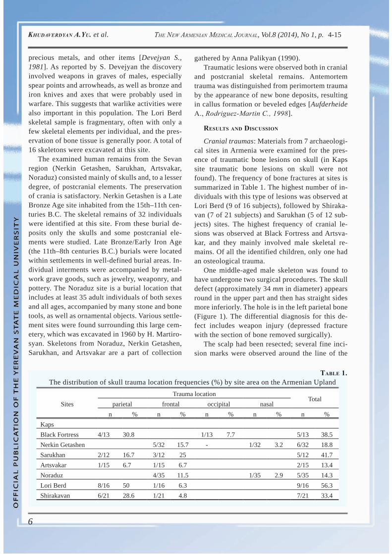
Cranial traumas: Materials from 7 archaeologi-cal sites in Armenia were examined for the pres-ence of traumatic bone lesions on skull (in Kaps site traumatic bone lesions on skull were not found). The frequency of bone fractures at sites is summarized in Table 1. The highest number of in-dividuals with this type of lesions was observed at Lori Berd (9 of 16 subjects), followed by Shiraka-van (7 of 21 subjects) and Sarukhan (5 of 12 sub-jects) sites. The highest frequency of cranial le-sions was observed at Black Fortress and Artsva-kar, and they mainly involved male skeletal re-mains. Of all the identified children, only one had an osteological trauma.
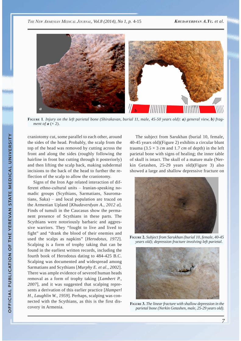
One middle-aged male skeleton was found to have undergone two surgical procedures. The skull defect (approximately 34 mm in diameter) appears round in the upper part and then has straight sides more inferiorly. The hole is in the left parietal bone (Figure 1). The differential diagnosis for this de-fect includes weapon injury (depressed fracture with the section of bone removed surgically).
The scalp had been resected; several fine inci-sion marks were observed around the line of the
table 1. The distribution of skull trauma location frequencies (%) by site area on the Armenian Upland
SitesTrauma location
Totalparietal frontal occipital nasal
n % n % n % n % n %KapsBlack Fortress 4/13 30.8 1/13 7.7 5/13 38.5Nerkin Getashen 5/32 15.7 - 1/32 3.2 6/32 18.8Sarukhan 2/12 16.7 3/12 25 5/12 41.7Artsvakar 1/15 6.7 1/15 6.7 2/15 13.4Noraduz 4/35 11.5 1/35 2.9 5/35 14.3Lori Berd 8/16 50 1/16 6.3 9/16 56.3Shirakavan 6/21 28.6 1/21 4.8 7/21 33.4
7
The New ArmeNiAN medicAl JourNAl, Vol.8 (2014), No 1, p. KhudAverdyAN A.yu. et al. 4-15
craniotomy cut, some parallel to each other, around the sides of the head. Probably, the scalp from the top of the head was removed by cutting across the front and along the sides (roughly following the hairline in front but cutting through it posteriorly) and then lifting the scalp back, making subdermal incisions to the back of the head to further the re-flection of the scalp to allow the craniotomy.
Signs of the Iron Age related interaction of dif-ferent ethno-cultural units ‒ Iranian-speaking no-madic groups (Scythians, Sarmatians, Sauroma-tians, Saka) ‒ and local population are traced on the Armenian Upland [Khudaverdyan A., 2012 a]. Finds of tumuli in the Caucasus show the perma-nent presence of Scythians in these parts. The Scythians were notoriously barbaric and aggres-sive warriors. They “fought to live and lived to fight” and “drank the blood of their enemies and used the scalps as napkins” [Herodotus, 1972]. Scalping is a form of trophy taking that can be found in the earliest written records, including the fourth book of Herodotus dating to 484-425 B.C. Scalping was documented and widespread among Sarmatians and Scythians [Murphy E. et al., 2002]. There was ample evidence of severed human heads removal as a form of trophy taking [Lambert P., 2007], and it was suggested that scalping repre-sents a derivation of this earlier practice [Hamperl H., Laughlin W., 1959]. Perhaps, scalping was con-nected with the Scythians, as this is the first dis-covery in Armenia.
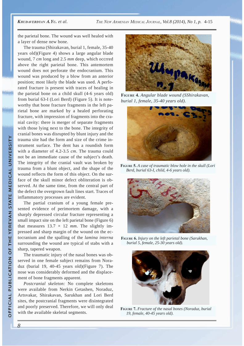
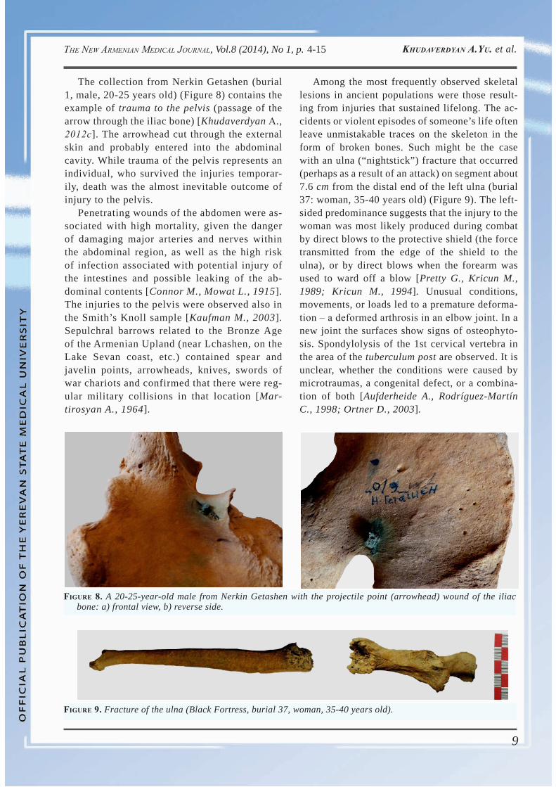
The subject from Sarukhan (burial 10, female, 40-45 years old)(Figure 2) exhibits a circular blunt trauma (3.5 × 3 cm and 1.7 cm of depth) in the left parietal bone with signs of healing; the inner table of skull is intact. The skull of a mature male (Ner-kin Getashen, 25-29 years old)(Figure 3) also showed a large and shallow depressive fracture on
Figure 1. Injury on the left parietal bone (Shirakavan, burial 11, male, 45-50 years old): a) general view, b) frag-ment of a (× 2).
Figure 2. Subject from Sarukhan (burial 10, female, 40-45 years old); depression fracture involving left parietal.
Figure 3. The linear fracture with shallow depression in the parietal bone (Nerkin Getashen, male, 25-29 years old).
ba
8
The New ArmeNiAN medicAl JourNAl, Vol.8 (2014), No 1, p. KhudAverdyAN A.yu. et al. 4-15
the parietal bone. The wound was well healed with a layer of dense new bone.
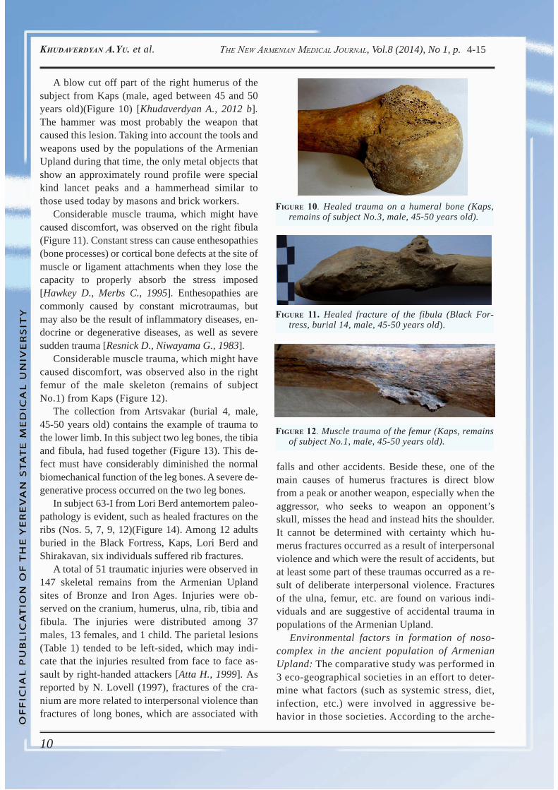
The trauma (Shirakavan, burial 1, female, 35-40 years old)(Figure 4) shows a large angular blade wound, 7 cm long and 2.5 mm deep, which occrred above the right parietal bone. This antemortem wound does not perforate the endocranium. This wound was produced by a blow from an anterior position; most likely the blade was used. A perfo-rated fracture is present with traces of healing in the parietal bone on a child skull (4-6 years old) from burial 63-I (Lori Berd) (Figure 5). It is note-worthy that bone fracture fragments in the left pa-rietal bone are marked by a healed perforating fracture, with impression of fragments into the cra-nial cavity: there is merger of separate fragments with those lying next to the bone. The integrity of cranial bones was disrupted by blunt injury and the trauma site had the form and size of the crime in-strument surface. The dent has a roundish form with a diameter of 4.2-3.5 cm. The trauma could not be an immediate cause of the subject’s death. The integrity of the cranial vault was broken by trauma from a blunt object, and the shape of the wound reflects the form of this object. On the sur-face of the skull minor defect obliteration is ob-served. At the same time, from the central part of the defect the overgrown fault lines start. Traces of inflammatory processes are evident.
The partial cranium of a young female pre-sented evidence of perimortem damage, with a sharply depressed circular fracture representing a small impact site on the left parietal bone (Figure 6) that measures 13.7 × 12 mm. The slightly im-pressed and sharp margin of the wound on the ec-tocranium and the spalling of the lamina interna surrounding the wound are typical of stabs with a sharp, tapered weapon.
The traumatic injury of the nasal bones was ob-served in one female subject remains from Nora-duz (burial 19, 40-45 years old)(Figure 7). The nose was considerably deformed and the displace-ment of bone fragments apparent.
Postcranial skeleton: No complete skeletons were available from Nerkin Getashen, Noraduz, Artsvakar, Shirakavan, Sarukhan and Lori Berd sites, the postcranial fragments were disintegrated and poorly preserved. Therefore, we will only deal with the available skeletal segments.
Figure 4. Angular blade wound (SShirakavan,burial 1, female, 35-40 years old).
Figure 5. A case of traumatic blow hole in the skull (Lori Berd, burial 63-I, child, 4-6 years old).
Figure 6. Injury on the left parietal bone (Sarukhan, burial 5, female, 25-30 years old).
Figure 7. Fracture of the nasal bones (Noraduz, burial 19, female, 40-45 years old).
9
The New ArmeNiAN medicAl JourNAl, Vol.8 (2014), No 1, p. KhudAverdyAN A.yu. et al. 4-15
The collection from Nerkin Getashen (burial 1, male, 20-25 years old) (Figure 8) contains the example of trauma to the pelvis (passage of the arrow through the iliac bone) [Khudaverdyan А., 2012с]. The arrowhead cut through the external skin and probably entered into the abdominal cavity. While trauma of the pelvis represents an individual, who survived the injuries temporar-ily, death was the almost inevitable outcome of injury to the pelvis.
Penetrating wounds of the abdomen were as-sociated with high mortality, given the danger of damaging major arteries and nerves within the abdominal region, as well as the high risk of infection associated with potential injury of the intestines and possible leaking of the ab-dominal contents [Connor M., Mowat L., 1915]. The injuries to the pelvis were observed also in the Smith’s Knoll sample [Kaufman M., 2003]. Sepulchral barrows related to the Bronze Age of the Armenian Upland (near Lchashen, on the Lake Sevan coast, etc.) contained spear and javelin points, arrowheads, knives, swords of war chariots and confirmed that there were reg-ular military collisions in that location [Mar-tirosyan A., 1964].
Among the most frequently observed skeletal lesions in ancient populations were those result-ing from injuries that sustained lifelong. The ac-cidents or violent episodes of someone’s life often leave unmistakable traces on the skeleton in the form of broken bones. Such might be the case with an ulna (“nightstick”) fracture that occurred (perhaps as a result of an attack) on segment about 7.6 cm from the distal end of the left ulna (burial 37: woman, 35-40 years old) (Figure 9). The left-sided predominance suggests that the injury to the woman was most likely produced during combat by direct blows to the protective shield (the force transmitted from the edge of the shield to the ulna), or by direct blows when the forearm was used to ward off a blow [Pretty G., Kricun M., 1989; Kricun M., 1994]. Unusual conditions, movements, or loads led to a premature deforma-tion ‒ a deformed arthrosis in an elbow joint. In a new joint the surfaces show signs of osteophyto-sis. Spondylolysis of the 1st cervical vertebra in the area of the tuberculum post are observed. It is unclear, whether the conditions were caused by microtraumas, a congenital defect, or a combina-tion of both [Aufderheide A., Rodríguez-Martín C., 1998; Ortner D., 2003].
Figure 8. A 20-25-year-old male from Nerkin Getashen with the projectile point (arrowhead) wound of the iliac bone: a) frontal view, b) reverse side.
Figure 9. Fracture of the ulna (Black Fortress, burial 37, woman, 35-40 years old).
10
The New ArmeNiAN medicAl JourNAl, Vol.8 (2014), No 1, p. KhudAverdyAN A.yu. et al. 4-15
A blow cut off part of the right humerus of the subject from Kaps (male, aged between 45 and 50 years old)(Figure 10) [Khudaverdyan A., 2012 b]. The hammer was most probably the weapon that caused this lesion. Taking into account the tools and weapons used by the populations of the Armenian Upland during that time, the only metal objects that show an approximately round profile were special kind lancet peaks and a hammerhead similar to those used today by masons and brick workers.
Considerable muscle trauma, which might have caused discomfort, was observed on the right fibula (Figure 11). Constant stress can cause enthesopathies (bone processes) or cortical bone defects at the site of muscle or ligament attachments when they lose the capacity to properly absorb the stress imposed [Hawkey D., Merbs C., 1995]. Enthesopathies are commonly caused by constant microtraumas, but may also be the result of inflammatory diseases, en-docrine or degenerative diseases, as well as severe sudden trauma [Resnick D., Niwayama G., 1983].
Considerable muscle trauma, which might have caused discomfort, was observed also in the right femur of the male skeleton (remains of subject No.1) from Kaps (Figure 12).
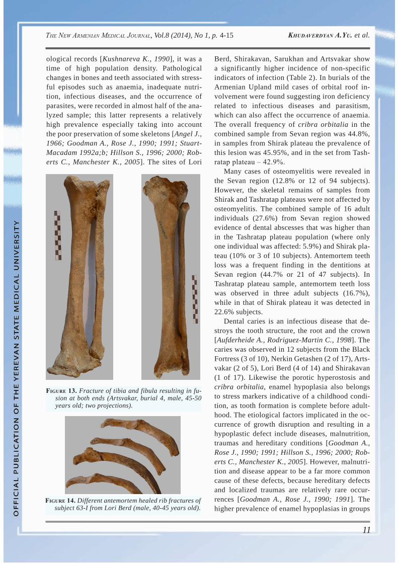
The collection from Artsvakar (burial 4, male, 45-50 years old) contains the example of trauma to the lower limb. In this subject two leg bones, the tibia and fibula, had fused together (Figure 13). This de-fect must have considerably diminished the normal biomechanical function of the leg bones. A severe de-generative process occurred on the two leg bones.
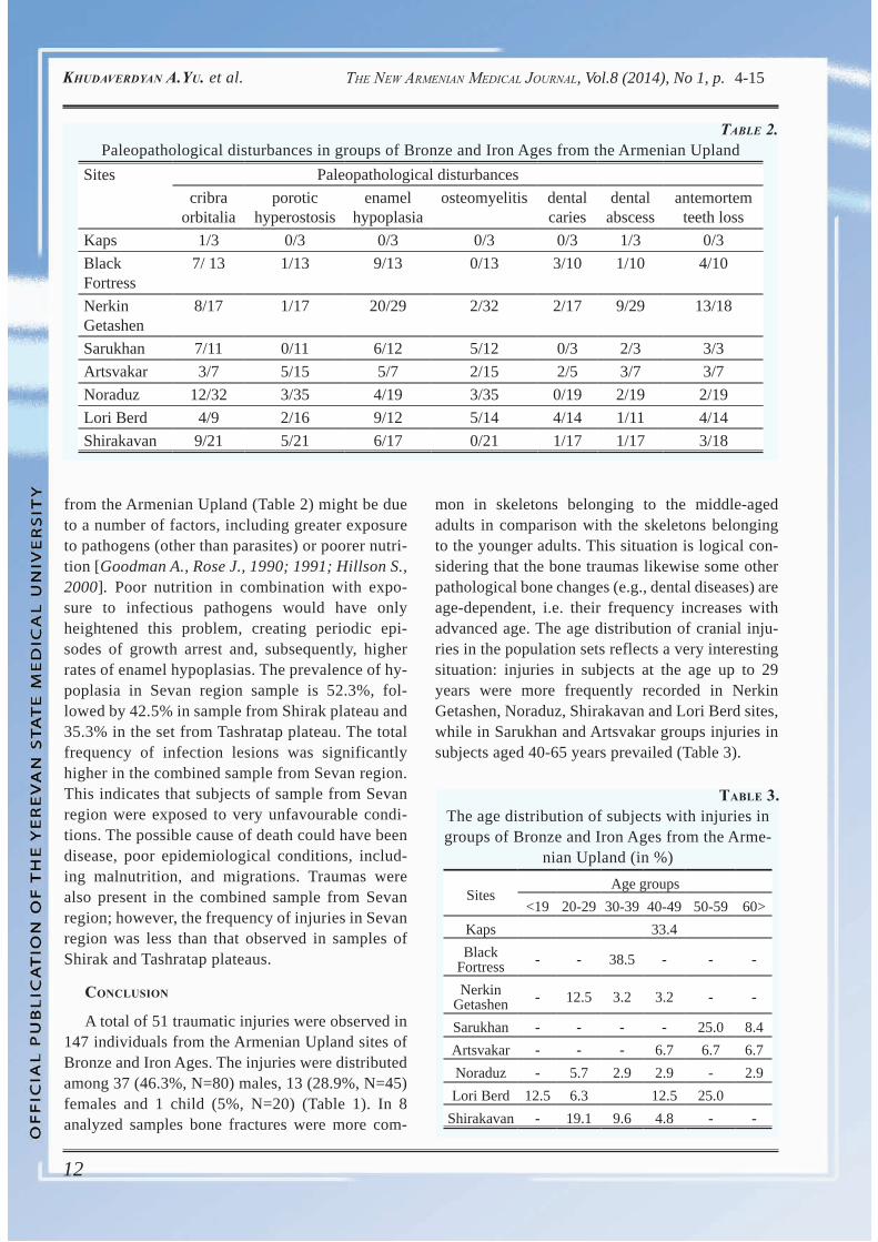
In subject 63-I from Lori Berd antemortem paleo-pathology is evident, such as healed fractures on the ribs (Nos. 5, 7, 9, 12)(Figure 14). Among 12 adults buried in the Black Fortress, Kaps, Lori Berd and Shirakavan, six individuals suffered rib fractures.
A total of 51 traumatic injuries were observed in 147 skeletal remains from the Armenian Upland sites of Bronze and Iron Ages. Injuries were ob-served on the cranium, humerus, ulna, rib, tibia and fibula. The injuries were distributed among 37 males, 13 females, and 1 child. The parietal lesions (Table 1) tended to be left-sided, which may indi-cate that the injuries resulted from face to face as-sault by right-handed attackers [Atta H., 1999]. As reported by N. Lovell (1997), fractures of the cra-nium are more related to interpersonal violence than fractures of long bones, which are associated with
Figure 11. Healed fracture of the fibula (Black For-tress, burial 14, male, 45-50 years old).
Figure 12. Muscle trauma of the femur (Kaps, remains of subject No.1, male, 45-50 years old).
falls and other accidents. Beside these, one of the main causes of humerus fractures is direct blow from a peak or another weapon, especially when the aggressor, who seeks to weapon an opponent’s skull, misses the head and instead hits the shoulder. It cannot be determined with certainty which hu-merus fractures occurred as a result of interpersonal violence and which were the result of accidents, but at least some part of these traumas occurred as a re-sult of deliberate interpersonal violence. Fractures of the ulna, femur, etc. are found on various indi-viduals and are suggestive of accidental trauma in populations of the Armenian Upland.
Environmental factors in formation of noso-complex in the ancient population of Armenian Upland: The comparative study was performed in 3 eco-geographical societies in an effort to deter-mine what factors (such as systemic stress, diet, infection, etc.) were involved in aggressive be-havior in those societies. According to the arche-
Figure 10. Healed trauma on a humeral bone (Kaps, remains of subject No.3, male, 45-50 years old).
11
The New ArmeNiAN medicAl JourNAl, Vol.8 (2014), No 1, p. KhudAverdyAN A.yu. et al. 4-15
ological records [Kushnareva K., 1990], it was a time of high population density. Pathological changes in bones and teeth associated with stress-ful episodes such as anaemia, inadequate nutri-tion, infectious diseases, and the occurrence of parasites, were recorded in almost half of the ana-lyzed sample; this latter represents a relatively high prevalence especially taking into account the poor preservation of some skeletons [Angel J., 1966; Goodman A., Rose J., 1990; 1991; Stuart-Macadam 1992a;b; Hillson S., 1996; 2000; Rob-erts C., Manchester K., 2005]. The sites of Lori
Figure 14. Different antemortem healed rib fractures of subject 63-I from Lori Berd (male, 40-45 years old).
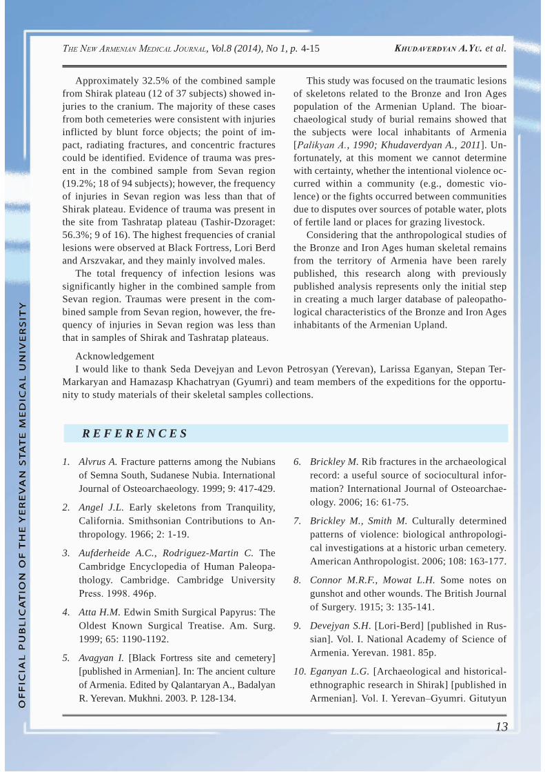
Berd, Shirakavan, Sarukhan and Artsvakar show a significantly higher incidence of non-specific indicators of infection (Table 2). In burials of the Armenian Upland mild cases of orbital roof in-volvement were found suggesting iron deficiency related to infectious diseases and parasitism, which can also affect the occurrence of anaemia. The overall frequency of cribra orbitalia in the combined sample from Sevan region was 44.8%, in samples from Shirak plateau the prevalence of this lesion was 45.95%, and in the set from Tash-ratap plateau ‒ 42.9%.
Many cases of osteomyelitis were revealed in the Sevan region (12.8% or 12 of 94 subjects). However, the skeletal remains of samples from Shirak and Tashratap plateaus were not affected by osteomyelitis. The combined sample of 16 adult individuals (27.6%) from Sevan region showed evidence of dental abscesses that was higher than in the Tashratap plateau population (where only one individual was affected: 5.9%) and Shirak pla-teau (10% or 3 of 10 subjects). Antemortem teeth loss was a frequent finding in the dentitions at Sevan region (44.7% or 21 of 47 subjects). In Tashratap plateau sample, antemortem teeth loss was observed in three adult subjects (16.7%), while in that of Shirak plateau it was detected in 22.6% subjects.
Dental caries is an infectious disease that de-stroys the tooth structure, the root and the crown [Aufderheide A., Rodriguez-Martin C., 1998]. The caries was observed in 12 subjects from the Black Fortress (3 of 10), Nerkin Getashen (2 of 17), Arts-vakar (2 of 5), Lori Berd (4 of 14) and Shirakavan (1 of 17). Likewise the porotic hyperostosis and cribra orbitalia, enamel hypoplasia also belongs to stress markers indicative of a childhood condi-tion, as tooth formation is complete before adult-hood. The etiological factors implicated in the oc-currence of growth disruption and resulting in a hypoplastic defect include diseases, malnutrition, traumas and hereditary conditions [Goodman A., Rose J., 1990; 1991; Hillson S., 1996; 2000; Rob-erts C., Manchester K., 2005]. However, malnutri-tion and disease appear to be a far more common cause of these defects, because hereditary defects and localized traumas are relatively rare occur-rences [Goodman A., Rose J., 1990; 1991]. The higher prevalence of enamel hypoplasias in groups
Figure 13. Fracture of tibia and fibula resulting in fu-sion at both ends (Artsvakar, burial 4, male, 45-50 years old; two projections).
12
The New ArmeNiAN medicAl JourNAl, Vol.8 (2014), No 1, p. KhudAverdyAN A.yu. et al. 4-15
TAble 2. Paleopathological disturbances in groups of Bronze and Iron Ages from the Armenian Upland
Sites Paleopathological disturbancescribra
orbitaliaporotic
hyperostosisenamel
hypoplasiaosteomyelitis dental
cariesdental
abscessantemortem
teeth lossKaps 1/3 0/3 0/3 0/3 0/3 1/3 0/3BlackFortress
7/ 13 1/13 9/13 0/13 3/10 1/10 4/10
Nerkin Getashen
8/17 1/17 20/29 2/32 2/17 9/29 13/18
Sarukhan 7/11 0/11 6/12 5/12 0/3 2/3 3/3Artsvakar 3/7 5/15 5/7 2/15 2/5 3/7 3/7Noraduz 12/32 3/35 4/19 3/35 0/19 2/19 2/19Lori Berd 4/9 2/16 9/12 5/14 4/14 1/11 4/14Shirakavan 9/21 5/21 6/17 0/21 1/17 1/17 3/18
from the Armenian Upland (Table 2) might be due to a number of factors, including greater exposure to pathogens (other than parasites) or poorer nutri-tion [Goodman A., Rose J., 1990; 1991; Hillson S., 2000]. Poor nutrition in combination with expo-sure to infectious pathogens would have only heightened this problem, creating periodic epi-sodes of growth arrest and, subsequently, higher rates of enamel hypoplasias. The prevalence of hy-poplasia in Sevan region sample is 52.3%, fol-lowed by 42.5% in sample from Shirak plateau and 35.3% in the set from Tashratap plateau. The total frequency of infection lesions was significantly higher in the combined sample from Sevan region. This indicates that subjects of sample from Sevan region were exposed to very unfavourable condi-tions. The possible cause of death could have been disease, poor epidemiological conditions, includ-ing malnutrition, and migrations. Traumas were also present in the combined sample from Sevan region; however, the frequency of injuries in Sevan region was less than that observed in samples of Shirak and Tashratap plateaus.
concluSion
A total of 51 traumatic injuries were observed in 147 individuals from the Armenian Upland sites of Bronze and Iron Ages. The injuries were distributed among 37 (46.3%, N=80) males, 13 (28.9%, N=45) females and 1 child (5%, N=20) (Table 1). In 8 analyzed samples bone fractures were more com-
mon in skeletons belonging to the middle-aged adults in comparison with the skeletons belonging to the younger adults. This situation is logical con-sidering that the bone traumas likewise some other pathological bone changes (e.g., dental diseases) are age-dependent, i.e. their frequency increases with advanced age. The age distribution of cranial inju-ries in the population sets reflects a very interesting situation: injuries in subjects at the age up to 29 years were more frequently recorded in Nerkin Getashen, Noraduz, Shirakavan and Lori Berd sites, while in Sarukhan and Artsvakar groups injuries in subjects aged 40-65 years prevailed (Table 3).
table 3. The age distribution of subjects with injuries in groups of Bronze and Iron Ages from the Arme-
nian Upland (in %)
SitesAge groups
<19 20-29 30-39 40-49 50-59 60>Kaps 33.4Black
Fortress - - 38.5 - - -
Nerkin Getashen - 12.5 3.2 3.2 - -
Sarukhan - - - - 25.0 8.4Artsvakar - - - 6.7 6.7 6.7Noraduz - 5.7 2.9 2.9 - 2.9Lori Berd 12.5 6.3 12.5 25.0
Shirakavan - 19.1 9.6 4.8 - -
13
The New ArmeNiAN medicAl JourNAl, Vol.8 (2014), No 1, p. KhudAverdyAN A.yu. et al. 4-15
Approximately 32.5% of the combined sample from Shirak plateau (12 of 37 subjects) showed in-juries to the cranium. The majority of these cases from both cemeteries were consistent with injuries inflicted by blunt force objects; the point of im-pact, radiating fractures, and concentric fractures could be identified. Evidence of trauma was pres-ent in the combined sample from Sevan region (19.2%; 18 of 94 subjects); however, the frequency of injuries in Sevan region was less than that of Shirak plateau. Evidence of trauma was present in the site from Tashratap plateau (Tashir-Dzoraget: 56.3%; 9 of 16). The highest frequencies of cranial lesions were observed at Black Fortress, Lori Berd and Arszvakar, and they mainly involved males.
The total frequency of infection lesions was significantly higher in the combined sample from Sevan region. Traumas were present in the com-bined sample from Sevan region, however, the fre-quency of injuries in Sevan region was less than that in samples of Shirak and Tashratap plateaus.
This study was focused on the traumatic lesions of skeletons related to the Bronze and Iron Ages population of the Armenian Upland. The bioar-chaeological study of burial remains showed that the subjects were local inhabitants of Armenia [Palikyan А., 1990; Khudaverdyan A., 2011]. Un-fortunately, at this moment we cannot determine with certainty, whether the intentional violence oc-curred within a community (e.g., domestic vio-lence) or the fights occurred between communities due to disputes over sources of potable water, plots of fertile land or places for grazing livestock.
Considering that the anthropological studies of the Bronze and Iron Ages human skeletal remains from the territory of Armenia have been rarely published, this research along with previously published analysis represents only the initial step in creating a much larger database of paleopatho-logical characteristics of the Bronze and Iron Ages inhabitants of the Armenian Upland.
AcknowledgementI would like to thank Seda Devejyan and Levon Petrosyan (Yerevan), Larissa Eganyan, Stepan Ter-
Markaryan and Hamazasp Khachatryan (Gyumri) and team members of the expeditions for the opportu-nity to study materials of their skeletal samples collections.
R E F E R E N C E S
1. Alvrus A. Fracture patterns among the Nubians of Semna South, Sudanese Nubia. International Journal of Osteoarchaeology. 1999; 9: 417-429.
2. Angel J.L. Early skeletons from Tranquility, California. Smithsonian Contributions to An-thropology. 1966; 2: 1-19.
3. Aufderheide A.C., Rodriguez-Martin C. The Cambridge Encyclopedia of Human Paleopa-thology. Cambridge. Cambridge University Press. 1998. 496р.
4. Atta H.M. Edwin Smith Surgical Papyrus: The Oldest Known Surgical Treatise. Am. Surg. 1999; 65: 1190-1192.
5. Avagyan I. [Black Fortress site and cemetery] [published in Armenian]. In: The ancient culture of Armenia. Edited by Qalantaryan A., Badalyan R. Yerevan. Mukhni. 2003. P. 128-134.
6. Brickley M. Rib fractures in the archaeological record: a useful source of sociocultural infor-mation? International Journal of Osteoarchae-ology. 2006; 16: 61-75.
7. Brickley M., Smith M. Culturally determined patterns of violence: biological anthropologi-cal investigations at a historic urban cemetery. American Anthropologist. 2006; 108: 163-177.
8. Connor M.R.F., Mowat L.H. Some notes on gunshot and other wounds. The British Journal of Surgery. 1915; 3: 135-141.
9. Devejyan S.H. [Lori-Berd] [published in Rus-sian]. Vol. I. National Academy of Science of Armenia. Yerevan. 1981. 85p.
10. Eganyan L.G. [Archaeological and historical-ethnographic research in Shirak] [published in Armenian]. Vol. I. Yerevan‒Gyumri. Gitutyun
14
The New ArmeNiAN medicAl JourNAl, Vol.8 (2014), No 1, p. KhudAverdyAN A.yu. et al. 4-15
(Science Publishers) and Shirak Center of Ar-menian Studies. 2010. 280p.
11. Goodman A.H., Rose J.C. Assessment of sys-temic physiological perturbations from dental enamel hypoplasias and associated histologi-cal structures. Am. J. Phys. Anthropol. 1990; 33: 59-110.
12. Goodman A.H., Rose J.C. Dental enamel hypo-plasias as indicators of nutritional status. In: Advances in dental anthropology. Edited by Kelley M.A., Larsen C.S. New York. Wiley-Liss. 1991. P. 279-293.
13. Hamperl H., Laughlin W.S. Osteological Conse-quences of Scalping. Hum. Biol. 1959; 31: 80-89.
14. Hawkey D.E., Merbs C.F. Activity-induced musculoskeletal stress markers (MSM) and subsistence strategy changes among ancient Hudson Bay Eskimos. International Journal of Osteoarchaeology. 1995; 5: 324-338.
15. Herodotus. [History in Nine Books] [published in Russian]. Translation and Notes by Stratanovsky G.A. Leningrad. 1972. Vol. IV. 600р.
16. Hillson S. Dental anthropology. Cambridge. Cambridge University Press. 1996. 392p.
17. Hillson S. Dental pathology. In: Biological an-thropology of the human skeleton. Edited by Katzenberg M.A., Saunders S.R. New York. Wiley-Liss. 2000. P. 249-286.
18. Kaufman M.H. Musket-ball and sabre injuries from the first half of the nineteenth century. Edinburgh. The Royal College of Surgeons of Edinburgh. 2003. 255p.
19. Khudaverdyan A.Yu. [Ancient communities of the Caucasus ‒ in the worlds’ dialogs (an an-thropological etude)] [published in Russian]. Germany: LAP LAMBERT Academic Publish-ing AG & Co. KG. 2011. 299p.
20. Khudaverdyan A.Yu. A bioarchaeological anal-ysis of the population of the Armenian High-land and Transcaucasus in the Antiquity Age. The Mankind Quarterly (Washington). 2012a; 53(1): 3-35.
21. Khudaverdyan A.Yu. Osteological analysis of human skeletal remains in Bronze Age from
Armenian Highland. Archaeological Science Journal. 2012b; 1(3): 21-36.
22. Khudaverdyan A.Yu. The population of the Ar-menian Uplands: the paleopathology and pa-leoecology. The New Armenian Medical Jour-nal. 2012с; 6(1): 4-14.
23. Kricun M.E. Paleoradiology of the Prehistoric Australian Aborigines. Am. J. Roentgenol. 1994; 163: 241-247.
24. Kushnareva K.K. [Cultural and economic unity of Southern Caucasus in IV-III millenium B.C.]. In: Interdisciplinary research on culture genesis and ethnogenesis of the Armenian Uplands and adjacent areas] [published in Russian]. Edited by Areshyan G., Esayan S. Yerevan. Yerevan State University, 1990. P. 194-204.
25. Lambert P. Paraguay. In: Countries at the crossroads. Maryland. Edited by Kelly S., Walker C., Dizard J. U.S.A. Rowman & Little-field. 2007. P. 467-488.
26. Larsen C.S. Bioarchaeology: interpreting be-havior from the human skeleton. Port Hope. Cambridge University Press. 1999. 476p.
27. Lovejoy C.O., Heiple K.G. The analysis of frac-tures in skeletal populations with an example from the Libben site, Ottowa County, Ohio. Am. J. Phys. Anthropol. 1981; 55: 529-541.
28. Lovell N.C. Trauma analysis in paleopathol-ogy. Yearbook of Physical Anthropology. 1997; 40: 139-170.
29. Lynn K.S., Fairgrieve S.I. Macroscopic analy-sis of axe and hatchet trauma in fleshed and defleshed mammalian long bones. J. Forensic Sci. 2009; 54: 786-792.
30. Martirosyan A.A. [Armenia in the Bronze Age and Early Iron Age] [published in Russian]. Yerevan. National Academy of Science of Ar-menia. 1964. 346р.
31. Milner G.R., Anderson E., Smith V.G. Warfare in late prehistoric west-central Illinois. Ameri-can Antiquity. 1991; 56(4): 581-603.
32. Murphy E., Gokhman I., Chistov Y., Barkova L. Prehistoric Old World Scalping: New Cases from Cemetery of Aymyrlyg, South Siberia. Am. J. Phys. Anthropol. 2002; 106: 1-10.
15
The New ArmeNiAN medicAl JourNAl, Vol.8 (2014), No 1, p. KhudAverdyAN A.yu. et al. 4-15
33. Ortner D.J. Identification of pathological con-ditions in human skeletal remains. 2nd edition. London. Academic Press. 2003. 645p.
34. Palikyan A.K. [New paleoanthropological materi-als from Armenia] [Article in Russian]. Biological Journal of Armenia. 1990; 4(43): 296-300.
35. Petrosyan L., Eganyan L., Khachatryan H. [Early Bronze Age monuments from Kaps] [published in Armenian]. In: Shirak’s histori-cal and cultural heritage. Edited by S. Haira-petyan. Vol. 12. Gyumri. Gitutyun (Science Publishers). 2009. P. 154-165.
36. Pretty G.L., Kricun M.E. Prehistoric health status of the Roonka population. World Ar-chaeology. 1989; 21: 198-224.
37. Resnick D., Niwayama G. Entheses and enthe-sopathies. Radiology. 1983; 146: 1-9.
38. Roberts C., Manchester K. The Archaeology of Disease. 3rd edition. Ithaca. Cornell Univer-sity Press. 2005. 352p.
39. Standen V.G., Arriaza B.T. Trauma in the pre-ceramic coastal populations of northern Chile: violence or occupational hazards? Am. J. Phys. Anthropol. 2000; 112: 239-249.
40. Stuart-Macadam P. Anemia in past human populations. In: Diet, demography, and dis-ease: changing perspectives on anemia. Edited by Stuart-Macadam P., Kent S. New York. Al-dine de Gruyter. 1992a. P. 151-170.
41. Stuart-Macadam P. Porotic hyperostosis: a new perspective. Am. J. Phys. Anthropol. 1992b; 87: 39-47.
42. Ter-Markaryan S., Avagyan I. [Site and ceme-tery Black Fortress] [published in Armenian]. In: Shirak’s historical and cultural heritage. Edited by S. Hairapetyan. Gyumri. Gitutyun (Science Publishers). 2000. P. 9-11.
43. Torosyan R.M., Hnkikyan O.S., Petrosyan L.A. [Ancient Shirakavan (the results of excavations 1977-1981)] [published in Armenian]. Archaeo-logical Excavations in Armenia. Yerevan. Gitu-tyun (Science Publishers). 2002. 256p.
44. Walker P.L. Cranial injuries as evidence of vio-lence in prehistoric Southern California. Am. J. Phys. Anthropol. 1989; 80: 313-323.