Embed Size (px)
Citation preview

Traumatic rhabdomyolysis: causes, pathophysiology and
management strategies
By Sharon Fish

Overview
• Definitions
• Historically
• Causes
• Pathophysiology
• Clinical
• Management

Definitions
• Rhabdomyolysis - destruction of striated muscle
• A crush injury is direct injury resulting from a crush
• A crush syndrome is the systemic manifestation of muscle cell damage, resulting from pressure or crushing.– Also known as traumatic rhabdomyolysis

• Based on 3 criteria:1. Involvement of a muscle mass
2. Prolonged compression
3. Compromised local circular

History• In 1910 Myer-Betz Syndrome, German
physician.– Triad: Muscle Pain, Weakness, Brown Urine.
• World War II– Dr Bywaters described patients during London
Bombings (Battle of Britain 1941).– Oliguria, pigmented casts, limb oedema, shock and
death.
• In 1943, in animal models, Bywaters & Stead identified myoglobin as the offending agent, and formulated the first treatment plan.
History• In 1950 Korean War, dialysis reduces mortality
rate from 84% to 53%.• Natural Disasters – Earthquakes
– 1976 Tangshan (near Beijing): 20% of 242,000 deaths due to crush syndrome.
– 1988 Spitak (Armenia)– In 1995, British nephrologists introduced the Disaster
Relief Task Force with the goal to prevent acute renal failure.
– 1999 Marmara (Turkey): 7.2 Richter scale earthquake. 12% hospitalised patients had renal failure, 76% received dialysis, 19% fatality rate.

Causes - Traumatic
• Trauma and compression– Crush injuries– MVA– Long-term confinement without changing
position– Physical torture and abuse– Prolonged hours of surgery without changing
position

Causes – Non-traumatic• Strainful muscle exercise• Electrical current
• Lightning• Cardioversion• Electric shock
• Hyperthermia• Neuromuscular malignant syndrome• Heat stroke
• Metabolic disorders• Mcardles disease• Palmitoyotransferase def
• Drugs• Cocaine• statins
• Sepsis

pathogenesis• Compressive forces leads to cellular
hypoperfusion and hypoxia • Decrease in ATPasefailure of ATPase
pump and sacrolemma leakage• Lysed cell release inflammatory mediators • platelet aggregation• vasoconstriction• inc vasc permeability
Lysed cell release• Potassium• Phospate• Creatine kinase• Myoglobin
Electrolyte disturbances• Hyperkalaemia • Hypocalcaemia• Hyperphosphatemia• Hyperuricaemia• Metabolic acidosis
Revascularization
• Fluids trapped in damaged tissue
• Oedema of affected limb
• Haemoconcentration and shock
• Myoglobin, potassium, phosphate enter venous circulation
Mechanisms of ARF in rhabdomyolysis
• Renal vasoconstriction with diminished renal perfusion
• Cast formation leads to tubular obstruction
Direct Myoglobin nephrotoxicity- Haeme produced free radicles
Clinical manifestations• Range from asymptomatic to acute renal
failure and DIC• Triad : muscle pain weakness ,dark urine• Musculoskeletal signs• General manifestations• Complications
– early– late

Musculoskeletal signs
• Pain
• Weakness
• Swelling

General manifestation
• Malaise
• Fever
• Tachycardia
• Nausea
• vomiting

Complications
Early• Hypovolaemia• Hyperkalaemia• Hypocalcaemia• Cardiac arrhythmias• Cardiac arrest• Compartment
syndrome
Late(12-72hrs)• Acute renal failure • DIC• ARDS• sepsis

Lab findings
• CK n 45-260U/L
• Rises within 12hours
• Peaks 1-3 days
• Declines 3-5days after cessation of muscle injury
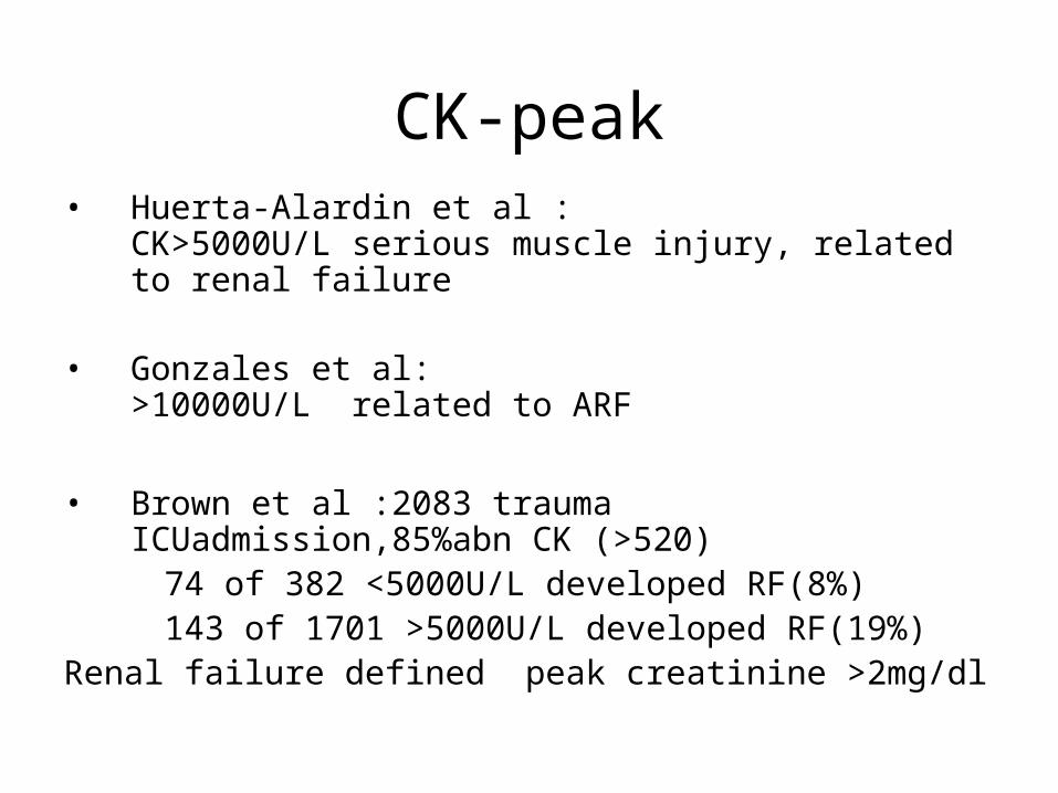
CK-peak• Huerta-Alardin et al :
CK>5000U/L serious muscle injury, related to renal failure
• Gonzales et al:
>10000U/L related to ARF
• Brown et al :2083 trauma ICUadmission,85%abn CK (>520)
74 of 382 <5000U/L developed RF(8%) 143 of 1701 >5000U/L developed RF(19%)Renal failure defined peak creatinine >2mg/dl

CK-peak• Oda et al: 372 crush injury pts at Hanshin
earthquake
CK < 75000 45 of 115 (39%) developed RF requiring dialysis
CK > 75000 43 of 51 (84.3%) developed renal failure requiring dialysis
Note different definitions of renal failure

Other muscle markers• Measuring myoglobin level in serum or
urine• Appears in urine when plasma
concentration exceeds 1.5mg/dl• Urine becomes dark red –brown colour
>100mg/dl• Myoglobin has short T1/2 (2-3hours)• Serum level return to normal after 6-
8hours
• Carbonic anhydrase 3
• Aldolase
• Trop T I
Lab tests
• Raised U&E
Hyperkalaemia
hypocalcaemia
hyperphosphataemia
uric acid
Treatment
• A B C
• Fluids
• Treat hyperkalaemia
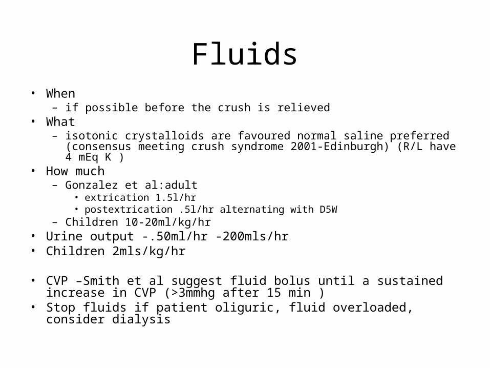
Fluids• When
– if possible before the crush is relieved• What
– isotonic crystalloids are favoured normal saline preferred (consensus meeting crush syndrome 2001-Edinburgh) (R/L have 4 mEq K )
• How much– Gonzalez et al:adult
• extrication 1.5l/hr• postextrication .5l/hr alternating with D5W
– Children 10-20ml/kg/hr• Urine output -.50ml/hr -200mls/hr• Children 2mls/kg/hr
• CVP –Smith et al suggest fluid bolus until a sustained increase in CVP (>3mmhg after 15 min )
• Stop fluids if patient oliguric, fluid overloaded, consider dialysis

Alkalinisation of urine• Alkalinisation increases the solubility of
myoglobin and promotes its excretion .• Bicarbonate is used to raise the urine pH to 6.5
thereby increasing solubility of Haeme pigments• Add 50 ml 8.5%sodium bicarbonate to each litre• HOWEVER little clinical evidence to support use• Brown and colleagues CK >5000U/L
– 154(40%) received mannitol and bicarbonate – 228 (60%) didn’t– No significant difference in renal failure ,dialysis,or
mortality between the groups.

Mannitol

• It was postulated that treatment with mannitol was more efficacious than isotonic volume expansion alone.
• It is argued that it forces an osmotic diuresis, thereby diluting nephrotoxic agents and encouraging their excretion.
• little evidence to prove mannitol alone • Brown et al –Failed to show benefit of
bic/man

Dialysis
• Despite optimal treatment ,daily haemodialysis or haemofiltration may be necessary
• Remove urea and potassium

Free radical scavengers and antioxidants
• The magnitude of muscle necrosis caused by ischemia-reperfusion injury has been reduced in experimental models by the administration of free-radical scavengers .
• Many of these agents have been used in the early treatment of crush syndrome to minimize the amount of nephrotoxic material released from the muscle
• Pentoxyphylline is a xanthine derivative used to improve microvascular blood flow. In addition, pentoxyphylline acts to decrease neutrophil adhesion and cytokine release
• Vitamin E , vitamin C , lazaroids (21-aminosteroids) and minerals such as zinc, manganese and selenium all have antioxidant activity and may have a role in the treatment of the patient with rhabdomyolysis

Summary
• High index of suspicion
• On scene treatment important
• Aggressive fluid treatment
• Adequate monitoring
• Recognition and early treatment of complications
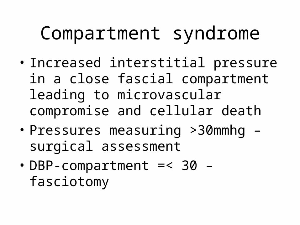
Compartment syndrome
• Increased interstitial pressure in a close fascial compartment leading to microvascular compromise and cellular death
• Pressures measuring >30mmhg –surgical assessment
• DBP-compartment =< 30 –fasciotomy

References• Oda, Jun MD; Tanaka, Hiroshi MD; Analysis of 372 Patients with Crush
Syndrome Caused by the Hanshin-Awaji Earthquake,J of trauma:Volume 42(3), March 1997, pp 470-476
• Gonzalez, Dario MD ,Crush syndrome,J of critical care:Volume 33(1) Supplement, January 2005, pp S34-S41
• Ana L Huerta-Alardín1, Joseph Varon2 and Paul E Marik .Bench-to-bedside review: Rhabdomyolysis – an overview for clinicians; Critical Care 2005, 9:158-169Crush Injury and Crush Syndrome: A Review
• Smith, Jason MD; Greaves, Ian Crush Injury and Crush Syndrome: A Review .J of trauma:Volume 54(5) Supplement, May 2003, pp S226-S230
• Brown,carlos V MD:Rhee,Peter MD ;Preventing Renal Failure in Patients with Rhabdomyolysis: Do Bicarbonate and Mannitol Make a difference . J of Trauma :Vol 56 ,June2004,pp1191-1196

Also Check…
• San Fran crush protocol