Embed Size (px)
Citation preview
COPYRIGHT © 2004 BY THE JOURNAL OF BONE AND JOINT SURGERY, INCORPORATED
Treatment of Lumbar Disc Herniation:
Epidural Steroid Injection Compared with Discectomy
A PROSPECTIVE, RANDOMIZED STUDY
BY GLENN R. BUTTERMANN, MD
Investigation performed at Midwest Spine Institute, Stillwater, Minnesota
Background: Epidural steroid injection is a low-risk alternative to surgical intervention in the treatment of lumbardisc herniation. The objective of this study was to determine the efficacy of epidural steroid injection in the treatmentof patients with a large, symptomatic lumbar herniated nucleus pulposus who are surgical candidates.
Methods: One hundred and sixty-nine patients with a large herniation of the lumbar nucleus pulposus (a herniationof >25% of the cross-sectional area of the spinal canal) were followed over a three-year period. One hundred patientswho had no improvement after a minimum of six weeks of noninvasive treatment were enrolled in a prospective, non-blinded study and were randomly assigned to receive either epidural steroid injection or discectomy. Evaluation wasperformed with the use of outcomes scales and neurological examination.
Results: Patients who had undergone discectomy had the most rapid decrease in symptoms, with 92% to 98% of thepatients reporting that the treatment had been successful over the various follow-up periods. Only 42% to 56% of thefifty patients who had undergone the epidural steroid injection reported that the treatment had been effective. Thosewho did not obtain relief from the injection had a subsequent discectomy, and their outcomes did not appear to havebeen adversely affected by the delay in surgery resulting from the trial of epidural steroid injection.
Conclusions: Epidural steroid injection was not as effective as discectomy with regard to reducing symptoms anddisability associated with a large herniation of the lumbar disc. However, epidural steroid injection did have a role: itwas found to be effective for up to three years by nearly one-half of the patients who had not had improvement withsix or more weeks of noninvasive care.
Level of Evidence: Therapeutic study, Level I-1a (randomized controlled trial [significant difference]). See Instruc-tions to Authors for a complete description of levels of evidence.
he treatment of lumbar disc herniation remains contro-versial. Although, in some studies, lumbar discectomyfor the treatment of large disc herniations accompanied
by severe symptoms was found to produce excellent short-termresults1, other researchers have found less optimal results, a dis-parity that may be related to patient selection2. Epidural steroidinjection is a low-risk alternative to surgical intervention insome patients for whom noninvasive treatment has failed3,4. Ithas been advocated because it modulates the body’s response toinflammatory stimuli, such as those related to a disc herniation5-8.
Epidural steroid injection has been found to be beneficial in an-imal models9, but its clinical efficacy for the treatment of lum-bar disc herniation has not been proven, to my knowledge10,11.Furthermore, as far as I know, it has not been determinedwhether epidural steroids have an effect on various factors re-lated to a disc herniation, such as the duration of pain, weak-ness, and sensory deficits and the size of the herniation. If onecould identify which patients are likely to respond to an epidu-ral steroid injection, the cost associated with the treatmentwould probably be decreased.
The purpose of this prospective study was to comparethe results of epidural steroid injection with those of discec-tomy in patients with a lumbar disc herniation encompassing>25% of the cross-sectional area of the spinal canal and who
T
A commentary is available with the electronic versions of this article,on our web site (www.jbjs.org) and on our quarterly CD-ROM (call oursubscription department, at 781-449-9780, to order the CD-ROM).
TH E JO U R NA L OF BONE & JOINT SURGER Y · JBJS .ORG
VO LU M E 86-A · NU M B E R 4 · APR IL 2004TRE A T M EN T OF LU M B A R DISC HER NIAT ION: EPIDUR A L STEROID INJEC T ION COMPARE D W ITH DISCE C TOMY
had continuous, disabling symptoms after six or more weeksof noninvasive treatment. The criterion for the size of the her-niation was selected because a prior study had suggested thatpatients with disc herniation encompassing <25% of thecross-sectional area of the spinal canal typically could betreated nonoperatively12.
Materials and Methodsne hundred and sixty-nine patients who were referred tome for treatment of a lumbar disc herniation that en-
compassed >25% of the cross-sectional area of the spinal ca-nal (as determined on axial magnetic resonance or computedtomography images) were prospectively followed over a three-year period from September 1995 to September 1998. Thisgroup did not include patients who were younger than eigh-teen years of age or older than seventy years of age; were preg-nant; or had cauda equina syndrome, a pars defect at the levelof the disc herniation, a far-lateral disc herniation, multilevelsymptomatic disc herniations, or a recurrent disc herniation.Also, it did not include sixteen patients who were seen duringthe study period but had an exceptional case or refused to par-ticipate in the study. Two of those sixteen patients had a truecauda equina syndrome and underwent emergent discec-tomy. One had recovery of bowel and bladder function andnearly complete resolution of the lower-extremity neurologi-cal deficit. The other had partial recovery of bowel, bladder,and lower-extremity function. Two other patients had saddleanesthesia in addition to lower-extremity neurological deficits(impending cauda equina syndrome) and underwent urgentdiscectomy. These four patients all had massive disc hernia-tion, and thus, although they met the radiographic criteria forinclusion in the study, they underwent early discectomy andwere not part of the randomized, prospective trial comparingepidural steroid injection with discectomy. Twelve additionalpatients who were eligible on the basis of radiographic criteriaat presentation declined to participate in the study. Theyelected not to wait six weeks before invasive treatment or notto have randomized treatment; eight of the twelve went else-where for treatment.
All other patients met the inclusion criteria and wereasked to participate in this study. Institutional review boardapproval and consent from all patients were obtained. Patientswere prospectively followed and if their condition did not im-prove with noninvasive treatment (physical therapy, chiro-practic treatment, rest, and/or pain medication) after sixweeks, they were randomly assigned (by computer) to one oftwo treatment groups: epidural steroid injection or discec-tomy. Patients were enrolled until there were fifty participantsin each treatment arm. During the study period, sixty-ninepatients who met the criterion for the size of the disc hernia-tion obtained improvement during the first six weeks of non-invasive treatment.
Two patients, after consenting to be part of the group tobe treated with epidural steroid injection, decided to proceedwith a discectomy. In addition, two patients who had been ini-tially assigned to the discectomy group elected to undergo epi-
dural steroid injection and were thus placed in that group. Theresults of the statistical analysis of the fifty patients in eachgroup were not different from those of the analysis of the in-tention-to-treat groups of forty-eight patients each.
The epidural steroid injections were performed by eithera radiologist or an anesthesiologist, who administered as manyas three injections one week apart. If a patient subjectively re-ported a decrease in pain within one week after a single injec-tion, no more injections were administered. If the patient didnot have improvement within a week, a second (or third) injec-tion was performed. All injections were performed at one levelcephalad to the disc herniation, with the needle placed betweenthe laminae; thirty-eight (76%) of the fifty patients were giventhe injection under fluoroscopic guidance. The dose of the cor-ticosteroid (betamethasone) was 10 to 15 mg.
I performed all of the discectomies. The duration of hos-pitalization for the discectomy group averaged less than twenty-four hours (range, less than one to three days). All patients inwhom the epidural steroid injection failed were subsequentlytreated with a discectomy, and those twenty-seven patients con-stituted the crossover group. The decision to proceed with dis-cectomy in the crossover group was made by the patient.
The demographic and radiographic characteristics of theepidural steroid injection, discectomy, and crossover groupswere similar (see Appendix). The mean ages were forty-one,forty, and thirty-nine years old, respectively; the mean dura-tions of symptoms were 3.3, 3.8, and 4.5 months; the percent-ages of smokers were 30%, 36%, and 33%; and the mean sizesof the disc herniations were 42%, 43%, and 41% of the axialcross section of the spinal canal. All but five disc herniations oc-curred at the L4 or L5 level.
All patients in each of the three groups were prospectivelyassessed with an examination and a questionnaire. I performeda neurological examination, which included documentation ofmotor strength on a scale of 0 to 5 (with 5 indicating normalstrength), at each clinical encounter. The self-assessment ques-tionnaire, which has been previously described13, is reliable andvalid. It included a visual analog scale of 0 to 10 for assessmentof current back and lower-extremity pain. A pain drawing wasused to indicate the distribution of the pain (with a high scorerepresenting a greater area of bodily pain), and an OswestryDisability Scale was employed to quantitate the level of function(on a 0 to 100-point scale, in which a higher score representedgreater disability). With the numbers available, the initial ques-tionnaire scores did not differ significantly among the groups,and no significant differences were noted between the grouptreated with epidural steroid injection and the discectomygroup with regard to the initial distribution of the neurologicaldeficits (p > 0.05) for motor, sensory, deep tendon reflex, andnerve root tension sign (Table I). The patients also indicated thetypes of pain medication that they used and the frequency withwhich they used it, provided a self-assessment of the treatmentsuccess, stated whether they would undergo the treatment againunder similar circumstances, and indicated whether they wouldrecommend the treatment to others with a similar condition.
The questionnaire and examination were completed at
O
TH E JO U R NA L OF BONE & JOINT SURGER Y · JBJS .ORG
VO LU M E 86-A · NU M B E R 4 · APR IL 2004TRE A T M EN T OF LU M B A R DISC HER NIAT ION: EPIDUR A L STEROID INJEC T ION COMPARE D W ITH DISCE C TOMY
presentation and at every subsequent clinical visit, and addi-tional surveys were completed by mail. Follow-up was carriedout at one to three months after treatment, at four to sixmonths, at seven to twelve months, at one to two years, and attwo to three years. Only three patients were not assessed at thefinal three-year follow-up interval: two patients in the discec-tomy group and one patient in the crossover group were lostto follow-up at two years. Only one patient, who had been
treated with the epidural steroid injection, did not have a finalfollow-up neurological examination.
The size of the disc herniation was determined in all 169patients (including those who had improvement within the firstsix weeks) on axial images of the affected level: 145 magneticresonance images and thirty computed tomography scans wereassessed, with six patients having both types of scans. The digi-tal images were examined to determine the ratio of the area of
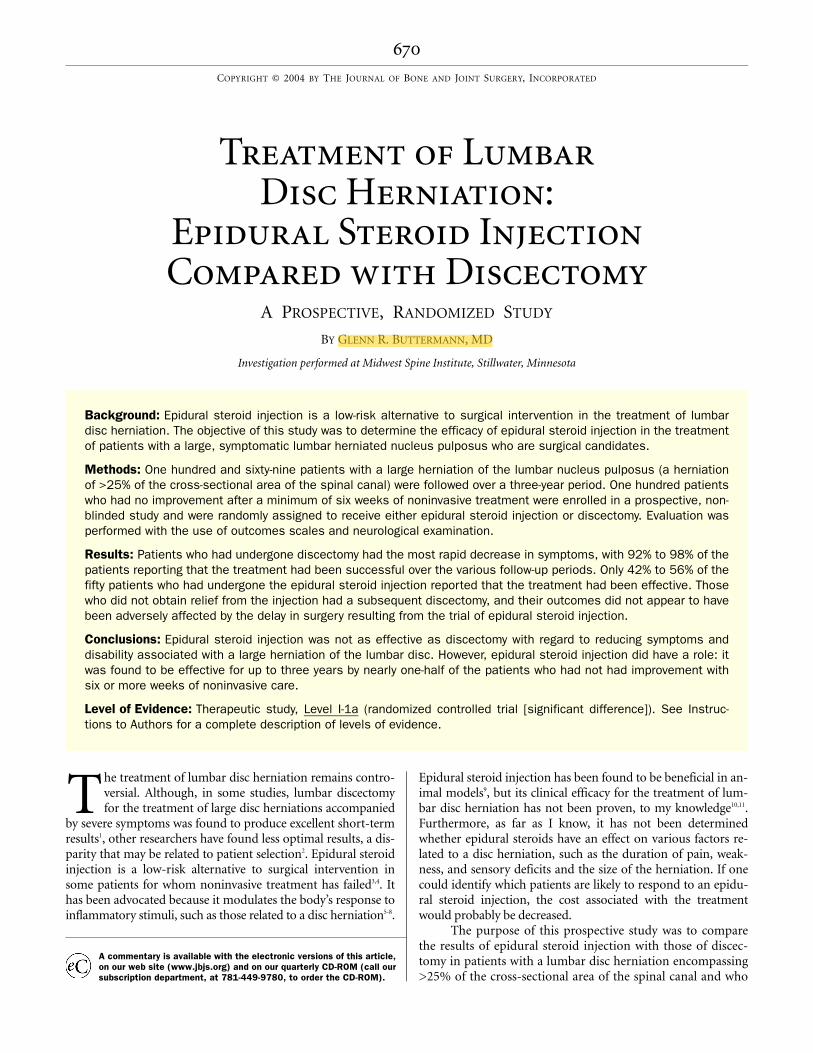
Fig. 1
The rates of neurological motor deficits before
treatment (Pre-Tx) and at the various follow-up
intervals. (The severity of the deficits is not
shown.) EPIDURAL and ESI = group treated
with epidural steroid injection, DECOMP =
discectomy group, and X-OVER = crossover
group. An asterisk indicates a significant dif-
ference between groups (p < 0.05).
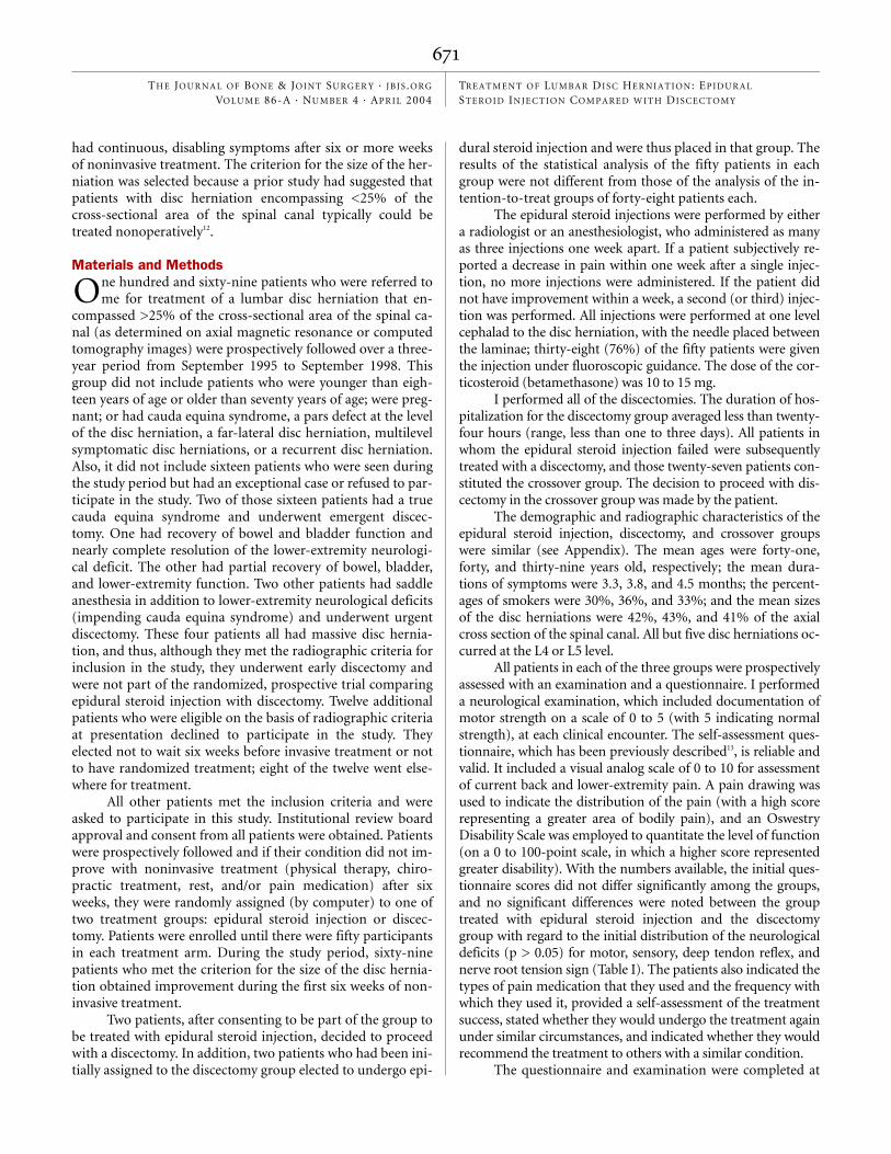
TABLE I Findings of the Neurological Examination at Presentation and the Two to Three-Year Follow-up Interval*
Study Group†
Epidural Steroid Injection Discectomy Crossover
Motor deficit
At presentation 82 88 78
At follow-up 9 4 11
Sensory deficit
At presentation 70 74 59
At follow-up 9 20 11
Reflex deficit
At presentation 54 56 52
At follow-up 14 17 22
Root tension sign
At presentation 82 84 81
At follow-up 0 0 0
Normal neurological findings
At presentation 0 0 0
At follow-up 74 68 63
*The severity of the deficits is not indicated. The severity of the motor deficits is described in text. The subjective severity of the sensory def-icits routinely decreased. †The values are given as the percentage of patients.
TH E JO U R NA L OF BONE & JOINT SURGER Y · JBJS .ORG
VO LU M E 86-A · NU M B E R 4 · APR IL 2004TRE A T M EN T OF LU M B A R DISC HER NIAT ION: EPIDUR A L STEROID INJEC T ION COMPARE D W ITH DISCE C TOMY
the disc herniation to the area of the spinal canal13. Other radio-graphic parameters that were analyzed were the presence of lat-eral recess stenosis (none was severe) at the level of the discherniation, inflammatory end-plate (Modic type-I14) changes,the total number of lumbar levels with disc degeneration, andthe relative degree of hydration of the herniated disc on T2-weighted magnetic resonance images.
Statistical MethodsStatistical analysis was performed with use of univariate chi-square analyses of all relevant pairs of variables, analysis of vari-ance, and the Student t test for the scores on the visual analog,
pain-drawing, and Oswestry Disability scales. The change in theuse of pain medication was analyzed with the Fisher exact test.Comparisons of disc hydration on magnetic resonance imageswere performed with the exact Wilcoxon test. Intention-to-treatanalysis was also performed, and no appreciable differenceswere found. A p value of <0.05 was considered significant.
Resultshe crossover group consisted of twenty-seven patientswho considered the treatment with the epidural steroid
injection a failure and had a subsequent discectomy. Contin-ued pain was the predominant reason mentioned by the pa-
T
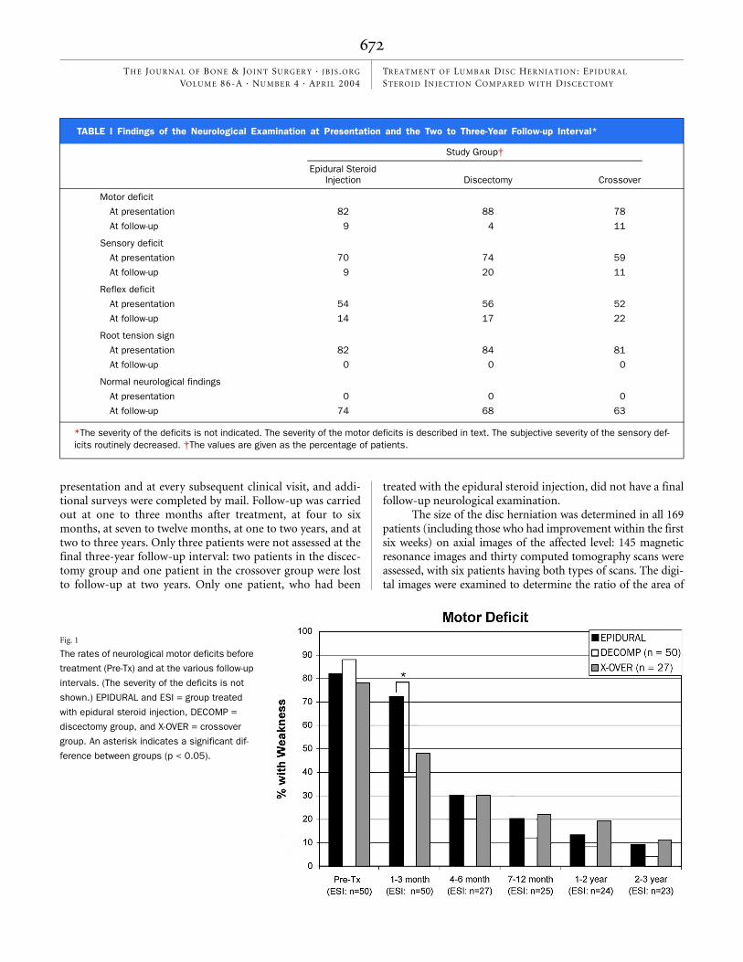
Fig. 2-B
Fig. 2-A
The scores for severity of back pain, as mea-
sured on a visual analog scale (VAS), before
treatment (Pre-Tx) and at the various follow-up
intervals. EPIDURAL and ESI = group treated
with epidural steroid injection, DECOMP = dis-
cectomy group, and X-OVER = crossover group.
The scores for severity of lower-extremity
pain, as measured on a visual analog scale
(VAS), before treatment (Pre-Tx) and at the vari-
ous follow-up intervals. EPIDURAL and ESI =
group treated with epidural steroid injection,
DECOMP = discectomy group, and X-OVER =
crossover group. An asterisk indicates a signifi-
cant difference between groups (p < 0.05).
TH E JO U R NA L OF BONE & JOINT SURGER Y · JBJS .ORG
VO LU M E 86-A · NU M B E R 4 · APR IL 2004TRE A T M EN T OF LU M B A R DISC HER NIAT ION: EPIDUR A L STEROID INJEC T ION COMPARE D W ITH DISCE C TOMY
tients for the failure of the injection; however, six of thetwenty-seven patients stated that a persistent neurological def-icit in the form of either weakness or a sensory deficit was alsoan important factor in their decision to proceed with the dis-cectomy. The average time from the onset of symptoms to theepidural steroid injection in the crossover group was 3.3months, and the average time from the onset of symptoms tothe discectomy was 4.5 months. The time period between thefailure of the epidural steroid injection and the discectomyranged from one to thirteen months.
Neurological function at the time of presentation and atthe final follow-up examination is summarized in Table I. Thediscectomy group had earlier motor recovery than did thegroup treated with the epidural steroid injectioni.e., signifi-cantly fewer patients in the discectomy group still had a motordeficit at one to three months following treatment (p = 0.001;Fig. 1). However, at the two to three-year follow-up point, therewas no significant difference between the two groups with re-gard to the percentage of patients who still had weakness (p =0.201). At the time of presentation, six patientsthree in thediscectomy group and three in the injection groupexhibited aprofound motor weakness (less than grade 3, with 5 being thehighest grade possible). Two of the three patients in the discec-tomy group had full recovery of motor strength and the otherpatient had mild weakness (grade-4 strength) at the time of fi-nal follow-up. In the injection group, two of the three patientswith profound weakness subsequently underwent discectomy(became part of the crossover group); one had the discectomyat two months and the other, at nine months. One of those twopatients had full recovery of grade-5 strength, and the other hadimprovement to grade-4 strength. The third patient in the in-jection group who had profound weakness did not undergo dis-cectomy; that patient also demonstrated a peroneal nerve deficiton electromyography in addition to an L5 radiculopathy and
had only minimal recovery at the time of final follow-up.The responses to the questionnaire demonstrated a sig-
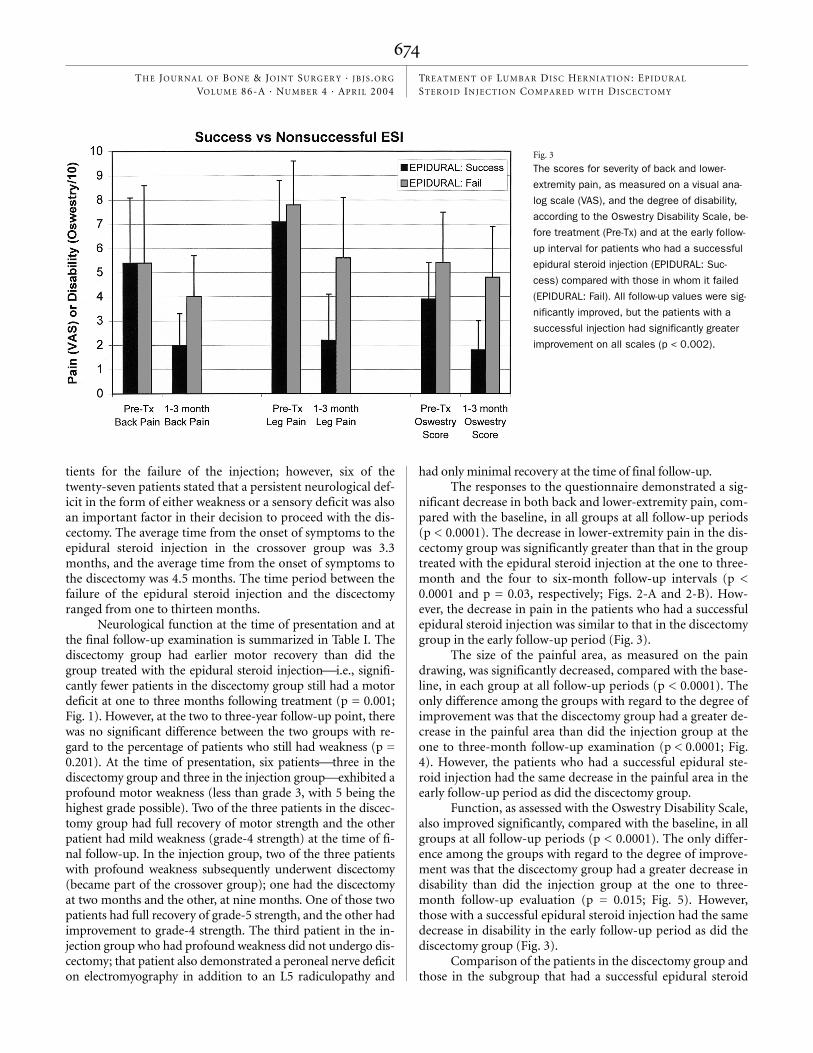
nificant decrease in both back and lower-extremity pain, com-pared with the baseline, in all groups at all follow-up periods(p < 0.0001). The decrease in lower-extremity pain in the dis-cectomy group was significantly greater than that in the grouptreated with the epidural steroid injection at the one to three-month and the four to six-month follow-up intervals (p <0.0001 and p = 0.03, respectively; Figs. 2-A and 2-B). How-ever, the decrease in pain in the patients who had a successfulepidural steroid injection was similar to that in the discectomygroup in the early follow-up period (Fig. 3).
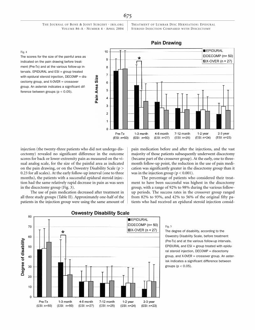
The size of the painful area, as measured on the paindrawing, was significantly decreased, compared with the base-line, in each group at all follow-up periods (p < 0.0001). Theonly difference among the groups with regard to the degree ofimprovement was that the discectomy group had a greater de-crease in the painful area than did the injection group at theone to three-month follow-up examination (p < 0.0001; Fig.4). However, the patients who had a successful epidural ste-roid injection had the same decrease in the painful area in theearly follow-up period as did the discectomy group.
Function, as assessed with the Oswestry Disability Scale,also improved significantly, compared with the baseline, in allgroups at all follow-up periods (p < 0.0001). The only differ-ence among the groups with regard to the degree of improve-ment was that the discectomy group had a greater decrease indisability than did the injection group at the one to three-month follow-up evaluation (p = 0.015; Fig. 5). However,those with a successful epidural steroid injection had the samedecrease in disability in the early follow-up period as did thediscectomy group (Fig. 3).
Comparison of the patients in the discectomy group andthose in the subgroup that had a successful epidural steroid
Fig. 3
The scores for severity of back and lower-
extremity pain, as measured on a visual ana-
log scale (VAS), and the degree of disability,
according to the Oswestry Disability Scale, be-
fore treatment (Pre-Tx) and at the early follow-
up interval for patients who had a successful
epidural steroid injection (EPIDURAL: Suc-
cess) compared with those in whom it failed
(EPIDURAL: Fail). All follow-up values were sig-
nificantly improved, but the patients with a
successful injection had significantly greater
improvement on all scales (p < 0.002).
TH E JO U R NA L OF BONE & JOINT SURGER Y · JBJS .ORG
VO LU M E 86-A · NU M B E R 4 · APR IL 2004TRE A T M EN T OF LU M B A R DISC HER NIAT ION: EPIDUR A L STEROID INJEC T ION COMPARE D W ITH DISCE C TOMY
injection (the twenty-three patients who did not undergo dis-cectomy) revealed no significant difference in the outcomescores for back or lower-extremity pain as measured on the vi-sual analog scale, for the size of the painful area as indicatedon the pain drawing, or on the Oswestry Disability Scale (p >0.23 for all scales). At the early follow-up interval (one to threemonths), the patients with a successful epidural steroid injec-tion had the same relatively rapid decrease in pain as was seenin the discectomy group (Fig. 3).
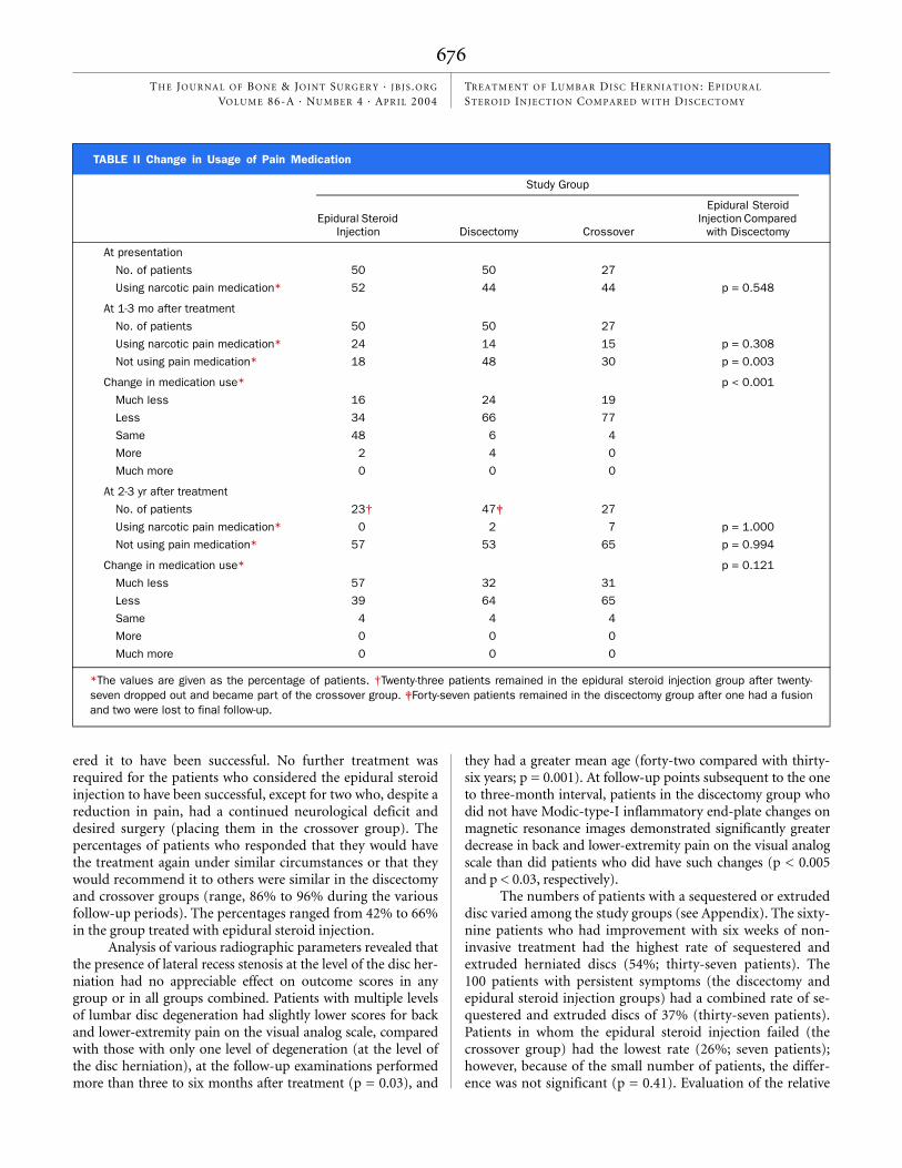
The use of pain medication decreased after treatment inall three study groups (Table II). Approximately one-half of thepatients in the injection group were using the same amount of
pain medication before and after the injections, and the vastmajority of those patients subsequently underwent discectomy(became part of the crossover group). At the early, one to three-month follow-up point, the reduction in the use of pain medi-cation was significantly greater in the discectomy group than itwas in the injection group (p < 0.001).
The percentage of patients who considered their treat-ment to have been successful was highest in the discectomygroup, with a range of 92% to 98% during the various follow-up periods. The success rates in the crossover group rangedfrom 82% to 93%, and 42% to 56% of the original fifty pa-tients who had received an epidural steroid injection consid-
Fig. 4
The scores for the size of the painful area as
indicated on the pain drawing before treat-
ment (Pre-Tx) and at the various follow-up in-
tervals. EPIDURAL and ESI = group treated
with epidural steroid injection, DECOMP = dis-
cectomy group, and X-OVER = crossover
group. An asterisk indicates a significant dif-
ference between groups (p < 0.05).
Fig. 5
The degree of disability, according to the
Oswestry Disability Scale, before treatment
(Pre-Tx) and at the various follow-up intervals.
EPIDURAL and ESI = group treated with epidu-
ral steroid injection, DECOMP = discectomy
group, and X-OVER = crossover group. An aster-
isk indicates a significant difference between
groups (p < 0.05).
TH E JO U R NA L OF BONE & JOINT SURGER Y · JBJS .ORG
VO LU M E 86-A · NU M B E R 4 · APR IL 2004TRE A T M EN T OF LU M B A R DISC HER NIAT ION: EPIDUR A L STEROID INJEC T ION COMPARE D W ITH DISCE C TOMY
ered it to have been successful. No further treatment wasrequired for the patients who considered the epidural steroidinjection to have been successful, except for two who, despite areduction in pain, had a continued neurological deficit anddesired surgery (placing them in the crossover group). Thepercentages of patients who responded that they would havethe treatment again under similar circumstances or that theywould recommend it to others were similar in the discectomyand crossover groups (range, 86% to 96% during the variousfollow-up periods). The percentages ranged from 42% to 66%in the group treated with epidural steroid injection.
Analysis of various radiographic parameters revealed thatthe presence of lateral recess stenosis at the level of the disc her-niation had no appreciable effect on outcome scores in anygroup or in all groups combined. Patients with multiple levelsof lumbar disc degeneration had slightly lower scores for backand lower-extremity pain on the visual analog scale, comparedwith those with only one level of degeneration (at the level ofthe disc herniation), at the follow-up examinations performedmore than three to six months after treatment (p = 0.03), and
they had a greater mean age (forty-two compared with thirty-six years; p = 0.001). At follow-up points subsequent to the oneto three-month interval, patients in the discectomy group whodid not have Modic-type-I inflammatory end-plate changes onmagnetic resonance images demonstrated significantly greaterdecrease in back and lower-extremity pain on the visual analogscale than did patients who did have such changes (p < 0.005and p < 0.03, respectively).
The numbers of patients with a sequestered or extrudeddisc varied among the study groups (see Appendix). The sixty-nine patients who had improvement with six weeks of non-invasive treatment had the highest rate of sequestered andextruded herniated discs (54%; thirty-seven patients). The100 patients with persistent symptoms (the discectomy andepidural steroid injection groups) had a combined rate of se-questered and extruded discs of 37% (thirty-seven patients).Patients in whom the epidural steroid injection failed (thecrossover group) had the lowest rate (26%; seven patients);however, because of the small number of patients, the differ-ence was not significant (p = 0.41). Evaluation of the relative
TABLE II Change in Usage of Pain Medication
Study Group
Epidural Steroid Injection Discectomy Crossover
Epidural Steroid Injection Compared
with Discectomy
At presentation
No. of patients 50 50 27
Using narcotic pain medication* 52 44 44 p = 0.548
At 1-3 mo after treatment
No. of patients 50 50 27
Using narcotic pain medication* 24 14 15 p = 0.308
Not using pain medication* 18 48 30 p = 0.003
Change in medication use* p < 0.001
Much less 16 24 19
Less 34 66 77
Same 48 6 4
More 2 4 0
Much more 0 0 0
At 2-3 yr after treatment
No. of patients 23† 47‡ 27
Using narcotic pain medication* 0 2 7 p = 1.000
Not using pain medication* 57 53 65 p = 0.994
Change in medication use* p = 0.121
Much less 57 32 31
Less 39 64 65
Same 4 4 4
More 0 0 0
Much more 0 0 0
*The values are given as the percentage of patients. †Twenty-three patients remained in the epidural steroid injection group after twenty-seven dropped out and became part of the crossover group. ‡Forty-seven patients remained in the discectomy group after one had a fusionand two were lost to final follow-up.
TH E JO U R NA L OF BONE & JOINT SURGER Y · JBJS .ORG
VO LU M E 86-A · NU M B E R 4 · APR IL 2004TRE A T M EN T OF LU M B A R DISC HER NIAT ION: EPIDUR A L STEROID INJEC T ION COMPARE D W ITH DISCE C TOMY
hydration of the herniated disc, as assessed on the T2-weightedmagnetic resonance images, revealed similar findings. Fifty-two percent (thirty-six patients) who had improvement withsix weeks of noninvasive treatment had a high or moderatelyhigh signal in the herniated disc on T2-weighted magnetic res-onance images; this rate was significantly higher than that inthe other groups (p = 0.001), and the rate was lowest in thecrossover group (7%; two patients).
The patients who had a successful epidural steroid injec-tion tended to be older than those for whom the injectionfailed (mean age, forty-four compared with thirty-nine years;p = 0.12); they were also twice as likely to have an extruded orsequestered disc (57% compared with 26%, p = 0.036), weremore likely to have a hydrated herniated disc (a high signal onthe T2-weighted magnetic resonance image, p = 0.0075), andhad less disability at the time of presentation (Oswestry score,39 compared with 55 points; p = 0.003). With the numbersavailable, there were no differences between the patients whohad a successful and a failed epidural steroid injection with re-gard to the average size of the disc herniation, level of disc her-niation, number of lumbar levels with degeneration, presenceof inflammatory end-plate changes on magnetic resonanceimages, occupation of the patient, duration of symptomsprior to the epidural steroid injection, percentage of smokers,percentage of patients involved in Workers’ Compensationclaims or litigation, back or lower-extremity pain score on thevisual analog scale at presentation, or pain-drawing score atpresentation.
Complications and reoperations were recorded for allgroups. Of the fifty patients treated with the epidural steroid in-jection, two had an incidental dural puncture. Three patientshad recurrent disc herniation at the same level, which was indi-cated by recurrence of symptoms and repeat (third) magneticresonance images demonstrating a larger disc herniation at thesite at which previous follow-up magnetic resonance imageshad shown regression of the original herniation. These repeatherniations occurred at eight, ten, and forty-four months afterthe initial disc herniation. Two of these patients subsequentlyhad a discectomy (became part of the crossover group). Of theseventy-seven patients who underwent discectomy (the discec-tomy and crossover groups), two (3%) had incidental duroto-mies. A seroma developed in one patient and was treated withoral antibiotics. There were no deep infections. There were fourrecurrent disc herniations (5%), all of which were treated withrevision discectomy. Persistent severe low-back pain (a score of>5 on the visual analog scale) was identified in five patients inthe discectomy group and in two patients in the crossovergroup. Two patients in the discectomy group had a spinal ar-throdesis at one and three years after the discectomy, and threeothers were contemplating fusion surgery to relieve disablinglow-back pain. One patient in the crossover group had a newdisc herniation at another level.
Discussionnumber of studies have compared epidural steroid injec-tions with control injections for the nonoperative treat-
ment of lumbar disc herniation. Some investigations, includinga number of randomized, prospective, and blinded studies inwhich patients were followed for periods ranging from weeks toone year15-19, showed epidural steroid injection to be beneficial.However, other comparative randomized and prospective stud-ies of epidural steroid injection demonstrated no substantial ef-fect on the clinical outcome20-24. A common problem with manyof the preceding studies is that the entry criteria usually con-sisted of subjective findings; rarely were findings on imagingstudies used in conjunction with symptoms and signs as entrycriteria. The discrepancies among the preceding studies mayalso be related to the timing of the epidural steroid injection;the injection may have little beneficial effect in the first fewweeks after the onset of symptoms since many patients havespontaneous improvement (along with a decrease in the size ofthe disc herniation)13,25-31. If one can identify the patients whohave not had improvement in the first few weeks, then epiduralsteroid injection may have a better-defined role.
Although there have been studies of the effect of epidu-ral steroid injection on nonoperatively treated patients, to myknowledge no one has previously determined whether epidu-ral steroids can be used as an alternative to surgery. In thepresent prospective, randomized study, I compared the effectsof epidural steroid injection with those of discectomy in pa-tients who fulfilled strict study entry criteria and who were ex-periencing severe symptoms despite an average of more thanthree months of noninvasive treatment. The present study dif-fers from many earlier studies in that the entry criteria in-cluded the morphology of the disc herniation in addition toclinical signs and symptoms. The study did have limitations,particularly because the injections were not completely stan-dardized (the steroid dose and the use of fluoroscopy varied),and the types of noninvasive management varied among thetreating therapists. This reflected restrictions by third-partypayers in this community-based study, which required pa-tients to have the epidural steroid injection and therapy per-formed by different practitioners.
Because most of the patients were referred, this studydoes not truly define the natural history of disc herniation.However, it does support the notion that a minimum of sixweeks of noninvasive treatment is reasonable prior to invasivetreatment. In fact, a study involving follow-up magnetic reso-nance imaging of patients who had improvement within sixweeks demonstrated a substantial decrease in the size of the discherniations13. In the present study, the total number of patientswho did not undergo discectomy was ninety-two (sixty-ninewho had improvement within six weeks without invasive treat-ment and twenty-three who had a successful epidural steroidinjection), and the total number in whom the epidural steroidinjection failed was twenty-seven. Because the treatment withepidural steroid injection or discectomy was randomly as-signed, it can be assumed that the same percentage of patientswould have responded to epidural steroid injection in the dis-cectomy group. Thus, the rate of failure of nonoperative treat-ment during the course of the present study was 31%, which issimilar to the 26% rate predicted in a previous report24.A
TH E JO U R NA L OF BONE & JOINT SURGER Y · JBJS .ORG
VO LU M E 86-A · NU M B E R 4 · APR IL 2004TRE A T M EN T OF LU M B A R DISC HER NIAT ION: EPIDUR A L STEROID INJEC T ION COMPARE D W ITH DISCE C TOMY
The present study supports the use of epidural steroid in-jection in patients with continued severe symptoms after sixweeks of noninvasive treatment because nearly one-half of thepatients who received such an injection had a fairly rapid de-crease in the symptoms. The degree of improvement was simi-lar to that for patients who underwent discectomy. The patientsin whom the epidural steroid injection failed and who subse-quently underwent discectomy (the crossover group) had thesame degree of improvement on all outcome scales as did boththe discectomy group and the patients with a successful epidu-ral steroid injection. On the average, the patients in the cross-over group received their surgical treatment more than onemonth later than did those in the discectomy group; however,this delay did not appear to adversely affect the outcome.
Neurological findings at the final follow-up evaluationwere similar among the three treatment groups (Table I, Fig.1). In the discectomy group, two (4%) of the fifty patients stillhad mild (grade-4) muscle weakness. This result compares fa-vorably with those in prior studies, in which a residual motordeficit was found in approximately 30% of patients (twenty-eight of 116 and twenty-five of seventy-eight) and, as was thecase in the present study, was more common in patients inwhom the weakness was more severe initially32,33. Ten patients(20%) treated with discectomy had mild residual altered sen-sation, which is a somewhat higher rate than that noted inprevious reports34,35. In the present study, 56% (twenty-eight)of the patients in the discectomy group had a loss of deep-tendon reflexes, and it was persistent in eight (16%); however,nerve-root-tension signs resolved in all patients.
A comparison between the epidural steroid injection,discectomy, and crossover groups suggests that delaying surgi-cal treatment after an initial trial of epidural steroid injectionhas no significant effect on the final neurological deficits (p =0.201). In other words, this study failed to show, with thenumber of patients available, that a delay in decompressiondue to an initial trial of epidural steroid injection was detri-mental to neurological recovery at the time of follow-up. Re-cent studies of patients with a nonoperatively treated discherniation also showed neurological improvement with re-gard to motor, sensory, and reflex changes, although up toone-half of the patients who presented with sensory or reflexchanges still had some abnormalities at one year36,37.
In this study, I sought to determine whether epidural
steroid injection in selected patients reduces the need for sur-gical intervention and whether it increases or accelerates painrelief in patients who would eventually have improvementanyway (but in a delayed fashion). Characteristics of the pa-tients who had a successful response to epidural steroid injec-tion were identified. Clinically, these included less severedisability, as demonstrated by the Oswestry Disability Score, atpresentation and a somewhat older age. Findings on magneticresonance images were partially predictive in that successfullytreated patients were more likely to have a sequestered or ex-truded disc herniation (as were patients who had spontaneousimprovement within six weeks), whereas those in whom theepidural steroid injection failed usually had a poorly hydratedherniated disc (a low signal on T2-weighted magnetic reso-nance images). Follow-up magnetic resonance images of thepatients in the epidural steroid injection group, to assesschanges in disc morphology and the possible effects of theinjection13, may be valuable. Such a study could help us to un-derstand whether the success of epidural steroid injections isrelated to resorption of the herniated disc or whether the pa-tients still have a sizable disc herniation and their clinical im-provement is due to modulation of the inflammatory responseto the herniation.
AppendixTables showing the clinical characteristics and radio-graphic characteristics of all three patient groups are
available with the electronic versions of this article, on ourweb site at www.jbjs.org (go to the article citation and click on“Supplementary Material”) and on our quarterly CD-ROM(call our subscription department, at 781-449-9780, to orderthe CD-ROM). �
References
1. Hanley EN Jr, Shapiro DE. The development of low-back pain after excision of a lumbar disc. J Bone Joint Surg Am. 1989;71:719-21.
2. Junge A, Dvorak J, Ahrens S. Predictors of bad and good outcomes of lumbar disc surgery. A prospective clinical study with recommendations for screening to avoid bad outcomes. Spine. 1995;20:460-8.
3. Johnson BA, Schellhas KP, Pollei SR. Epidurography and therapeutic epidural injections: technical considerations and experience with 5334 cases. AJNR Am J Neuroradiol. 1999;20:697-705.
4. Windsor RE, Pinzon EG, Gore HC. Complications of common selective spinal injections: prevention and management. Am J Orthop. 2000;29:759-70.
5. Goupille P, Jayson MI, Valat JP, Freemont AJ. The role of inflammation in disk herniation-associated radiculopathy. Semin Arthritis Rheum. 1998;28:60-71.
6. Gronblad M, Virri J, Seitsalo S, Habtemariam A, Karaharju E. Inflammatory cells, motor weakness, and straight leg raising in transligamentous disc her-niations. Spine. 2000;25:2803-7.
7. Lee HM, Weinstein JN, Meller ST, Hayashi N, Spratt KF, Gebhart GF. The role of steroids and their effects on phospholipase A2. An animal model of radiculopathy. Spine. 1998;23:1191-6.
8. Olmarker K, Byrod G, Cornefjord M, Nordborg C, Rydevik B. Effects of meth-ylprednisolone on nucleus pulposus–induced nerve root injury. Spine. 1994;19:1803-8.
9. Hayashi N, Weinstein JN, Meller ST, Lee HM, Spratt KF, Gebhart GF. The effect of epidural injection of betamethasone or bupivacaine in a rat model of lumbar radiculopathy. Spine. 1998;23:877-85.
10. Spaccarelli KC. Lumbar and caudal epidural corticosteroid injections.
Glenn R. Buttermann, MDMidwest Spine Institute, 1950 Curve Crest Boulevard, Stillwater, MN 55082
The author did not receive grants or outside funding in support of his research or preparation of this manuscript. He did not receive payments or other benefits or a commitment or agreement to provide such benefits from a commercial entity. No commercial entity paid or directed, or agreed to pay or direct, any benefits to any research fund, foundation, educational institution, or other charitable or nonprofit organization with which the author is affiliated or associated.
TH E JO U R NA L OF BONE & JOINT SURGER Y · JBJS .ORG
VO LU M E 86-A · NU M B E R 4 · APR IL 2004TRE A T M EN T OF LU M B A R DISC HER NIAT ION: EPIDUR A L STEROID INJEC T ION COMPARE D W ITH DISCE C TOMY
Mayo Clin Proc. 1996;71:169-78.
11. Weinstein SM, Herring SA, Derby R. Contemporary concepts in spine care. Epidural steroid injections. Spine. 1995;20:1842-6.
12. Carragee EJ, Kim DH. A prospective analysis of magnetic resonance imaging findings in patients with sciatica and lumbar disc herniation. Correlation of outcomes with disc fragment and canal morphology. Spine. 1997;22:1650-60.
13. Buttermann GR. Lumbar disc herniation regression after successful epidural steroid injection. J Spinal Disord Tech. 2002;15:469-76.
14. Modic MT, Steinberg PM, Ross JS, Masaryk TJ, Carter JR. Degenerative disk disease: assessment of changes in vertebral body marrow with MR im-aging. Radiology. 1988;166:193-9.
15. Dilke TF, Burry HC, Grahame R. Extradural corticosteroid injection in man-agement of lumbar nerve root compression. Br Med J. 1973;2:635-7.
16. Yates DW. A comparison of the types of epidural injection commonly used in the treatment of low back pain and sciatica. Rheumatol Rehabil. 1978;17:181-6.
17. Mathews JA, Mills SB, Jenkins VM, Grimes SM, Morkel MJ, Mathews W, Scott CM, Sittampalam Y. Back pain and sciatica: controlled trials of manip-ulation, traction, sclerosant and epidural injections. Br J Rheumatol. 1987;26:416-23.
18. Ridley MG, Kingsley GH, Gibson T, Grahame R. Outpatient lumbar epidural corticosteroid injection in the management of sciatica. Br J Rheumatol. 1988;27:295-9.
19. Helliwell M, Robertson JC, Ellis RM. Outpatient treatment of low back pain and sciatica by a single extradural corticosteroid injection. Br J Clin Pract. 1985;39:228-31.
20. Beliveau P. A comparison between epidural anaesthesia with and without cor-ticosteroid in the treatment of sciatica. Rheumatol Phys Med. 1971;11:40-3.
21. Snoek W, Weber H, Jorgensen B. Double blind evaluation of extradural methyl prednisolone for herniated lumbar discs. Acta Orthop Scand. 1977;48:635-41.
22. Klenerman L, Greenwood R, Davenport HT, White DC, Peskett S. Lumbar epidural injections in the treatment of sciatica. Br J Rheumatol. 1984;23:35-8.
23. Cuckler JM, Bernini PA, Wiesel SW, Booth RE Jr, Rothman RH, Pickens GT. The use of epidural steroids in the treatment of lumbar radicular pain. A prospective, randomized, double-blind study. J Bone Joint Surg Am. 1985;67:63-6.
24. Carette S, Leclaire R, Marcoux S, Morin F, Blaise GA, St-Pierre A, Truchon R, Parent F, Levesque J, Bergeron V, Montminy P, Blanchette C. Epidural
corticosteroid injections for sciatica due to herniated nucleus pulposus. N Engl J Med. 1997;336:1634-40.
25. Ahn SH, Ahn MW, Byun WM. Effect of the transligamentous extension of lumbar disc herniations on their regression and the clinical outcome of sciat-ica. Spine. 2000;25:475-80.
26. Bozzao A, Gallucci M, Masciocchi C, Aprile A, Barile A, Passariello R. Lum-bar disk herniation: MR imaging assessment of natural history in patients treated without surgery. Radiology. 1992;185:135-41.
27. Delauche-Cavallier MC, Budet C, Laredo JD, Debie B, Wybier M, Dorfmann H, Ballner I. Lumbar disc herniation. Computed tomography scan changes after conservative treatment of nerve root compression. Spine. 1992;17:927-33.
28. Komori H, Shinomiya K, Nakai O, Yamaura I, Takeda S, Furuya K. The natu-ral history of herniated nucleus pulposus with radiculopathy. Spine. 1996;21:225-9.
29. Sinel MS, Goldstein TB, Deutsch AL, Mink JH, Lee YP, Jackson KR. Conser-vative management of large lumbar disc extrusions: an MRI imaging study. Read at the Annual Meeting of the American Academy of Orthopaedic Sur-geons; 1999 Feb 4-8; Anaheim, CA.
30. Ellenberg MR, Ross ML, Honet JC, Schwartz M, Chodoroff G, Enochs S. Pro-spective evaluation of the course of disc herniations in patients with proven radiculopathy. Arch Phys Med Rehabil. 1993;74:3-8.
31. Nakamura T. Natural course of lumbar disc herniation—prospective study. Presented at the Annual Meeting of the North American Spine Society; 1998 October 28-31; San Francisco, CA.
32. Weber H. The effect of delayed disc surgery on muscular paresis. Acta Or-thop Scand. 1975;46:631-42.
33. Postacchini F, Giannicola G, Cinotti G. Recovery of motor deficits after micro-discectomy for lumbar disc herniation. J Bone Joint Surg Br. 2002;84:1040-5.
34. Naylor A. Late results of laminectomy for lumbar disc prolapse. A review after ten to twenty-five years. J Bone Joint Surg Br. 1974;56:17-29.
35. Lewis PJ, Weir BK, Broad RW, Grace MG. Long-term prospective study of lumbosacral discectomy. J Neurosurg. 1987;67:49-53.
36. Balague F, Nordin M, Sheikhzadeh A, Echegoyen AC, Brisby H, Hoogewoud HM, Fredman P, Skovron ML. Recovery of severe sciatica. Spine. 1999;24:2516-24.
37. Dubourg G, Rozenberg S, Fautrel B, Valls-Bellec I, Bissery A, Lang T, Faillot T, Duplan B, Briancon D, Levy-Weil F, Morlock G, Crouzet J, Gatfosse M, Bonnet C, Houvenagel E, Hary S, Brocq O, Poiraudeau S, Beaudreuil J, de Sauverzac C, Durieux S, Levade MH, Esposito P, Maitrot D, Goupille P, Valat JP, Bourgeois P. A pilot study on the recovery from paresis after lumbar disc herniation. Spine. 2002;27:1426-31.


![[TMMC Healthcare] Application of advanced techniques in disc herniation surgery](https://img.pdfslide.net/doc/110x75/568ca5b11a28ab186d8e1c71/tmmc-healthcare-application-of-advanced-techniques-in-disc-herniation-surgery-5704601e459ae.jpg)