Embed Size (px)
Citation preview
1
Whitepaper 2011
Treatment of Melasma , Hyperpigmentation ,Rejuvenation and Acne with Revlite By Clinical Professor Niwat Polnikorn Director, Kasemrad Aesthetics Center Kasemrad Hospital Prachacheun Bangkok, Thailand 10800 [email protected]
Melasma is the most common hyperpigment disorders in Asians. Consultation for treatment of this disfiguring condition accounts for more than 50% of aesthetics consulltation in Asian countries. Pathogesis of this condition is complex and relates to genetic, sunexposure , hormonal factor, subclinical vascular reactions, etc. Clinically the lesions occur in middle age women on sunexposure area of face especially on cheek, forhead and upper lips.The common presentation is bimalar and centrofacial types. The lesions starts as a brownish asymptomatic irregular border macules .The lesions slowly spread out forming brownish patches . Later on the color changes to grayish brown with indistinct borders. This disease mainly effect women (female to male ratio =9:1). Eventhough spontaneous remission after menopause happens but in most of the patients the lesions persists for decades. Melasma has been one of the most difficult hyperpigmented lesions to be treated espeicially in colored skin of Fitzpatric’s skin type III-V. Eventhough total clearing upto 60% of cases is possible with long term triple – combination agent formula (hydroquinone+retinoic acid+ topical steroid) . This treatment result in only temporary clearing with possibiltity of complications and common recurrence. Eventhough laser had been introduced for treatment of hyperpigmented lesions for more than thirty years. The early results after ablative laser ,continuous yellow light laser or high fluence nanosecond pulsed laser had been disappointing. Only recently that new high energy nanosecond-pulsed infrared laser with homogenous beam profiles technology has been developed Medlite C 6 laser(Hoya-Conbio, USA).Originally this new technology has been designed for rapid tattoos removal with minimal downtime. The author was the first to report new technique in treatment of refractory melasma with repeatitive subthrehold energy flluence. Recently clinical efficacy of this new technique has been confirmed in larger study of melasma. Studies from other centers in Thailand ,Korea and Japan have been reported confirming effectiveness of this new technique which has been called “Laser toning” or”Minimal photothermolysis” techniques. Most recently, Revlite laser, (Hoya-Conbio, USA with Photothermolysis Pulses Technology mode has been introduced . The author has performed clinical study for application of this newest laser in colored skin for 12 months. Recently the author has started the research project of effectiveness of Revlite in active acne and acne scar. The research is still going on , preliminary result will be presented. Pathogenesis: In melasma genetically predetermined clones of melanocytes on specific facial areas which are stimulated by ultraviolet light, oxidative stress and aging became hyperactive and proliferate. The normal pathway of melanin synthesis is being stimulated leading to hyperpigmentation. Melanin granules are synthesis in epidermal and follicular melanocytes by stimulation with ultraviolet light especially in the range of UVA (320-400 nm). UV stimulation of keratinocytes leads to activation of binding of keratinocytes and melanocytes through specific binding proteins and receptors ( SCF, Stem cell factor and C-kit proteins) stimulation. This lead to releasing of epidermal cytokines (endothelins) . Epidermal cytokines released from keratinocytes stimulate vascular proliferation, and hypermelanization . Endothelins will bind to
2
endothelin receptors on melanocyte cell membrane. These cytokines are potent stimulator of tyrosinase enzymes synthesis. Endothelins stimulation leads to increase in melanin synthesis in melanosomes. The mature melnin granules (Stage IV to V) in melanosones are then transferred to keratinocytes (epidermal melanocyte unit). Melanins granules in keratinocytes are shedded daily. In order that melasma lesions are clinically stable , hyperactive state of melanin synthesis are continuous maintained for long period of time. Vascular dilatation and endothelia cells proliferation results in telangiestasia which is common finding in long standing melasma. Leakage of vascular components lead to activation of arachidonic acid and prostaglandins synthesis. This result in stimulation of melanization and vicious cycles of more vascular proliferation. This visious cycle is the driving force for persistence of melasma. Any treatments that damage basement membrane resulted in dropping of melanins and mlanophage formation. Melanophages persist in the dermis for many years before slowly migrate out from the lesions. In Asian’s skin ( Fitzpatrick’s skin type III to VI) , there is a wide range of melanin synthesis ,aggregation and distribution leading to different shades of brown colour. The darker the colour the higher density of mature and large clumps of melanins .These melanins distribute in both keratinocytes and melanocytes. Only at some specific locations, melanocytes develop greater response to ultraviolet light resulting in epidermal hypermelnization. These hyperactive melanocytes will also proliferatel and populated the areas. Large melanized melanin (stage IV-V) were detected in epidermal melanocytes and keratinocytes. Usually in normal skin these mature melanin granules will be found mainly in keratinocytes. More than two third of the cases of melasma, macrophages containing melanin granules ( melanophages) were detected in upper dermis. These result from dropping of melanin granules through damage basement membranes into dermis. Basement membrane damages were result of treatments with many topical agents or past treatments especially ones that damaged basement membrane e.g. chemical peel, ablative laser, high energy pigment selective laser etc. In some cases telangiectasia of dermal vessels may play important role in chronicity of lesion. Vicious cycle of prostaglandins synthesis through leakage of telangiectatic vessels may be the cause of persistent melanin synthesis. Melasma had been classified into three groups 1) Epidermal type with hyperactive melanocytes only in the epidermis, 2) dermal type with presence of melanophages in dermis and 3) mixed type with presence of both hyperactive epidermal melanocytes and dermal melanophages. Many histologic study from Asians’ melasma lesions had demonstrated that more than two third of cases were mixed type, the remaining were epidermal type. Dermal type melasma had not been found. The presentation of only dermal melanophages could be found in post inflammatory hyperpigmentation. The author believes that melasma should be reclassified into three major groups 1) epidermal melasma 2) mixed melasma and 3) telangiectatic melasma. In order to treat melasma effectively for long lasting result we should be able to control epidermal melanin synthesis , reduce melanins aggregation in both melanocytes and keratinocytes, decrease melanins transportation into keratinocytes , mobilization of melanophages and reduction of telangiectasia. This is not possible with single treatment method. From theoretical point of view combination treatments targeting different steps in pathogenesis of melasma will ensure better, faster and long lasting . For treatment of acne, the inflammatory cytokines released after exposure to laser beam has been believed to promote healing of inflammatory acnes. Long term exposure will result in neocollagenesis and improvement of atrophic scar.
3
Treatments: The treatments of melasma can be grouped as follow: 1.Reduction of melanin synthesis: Tyrosinase enzyme is the most important enzyme in melanization. It changes dopa to dopaquinone and eumelanin. Topical application of tyrosinase inhibitors is the most widely use method. Eventhough there are many new chemical agents claiming to be effective bleaching agent, hydroquinone (2-4%) is still the most widely prescribed topical bleaching agent. Hydroquinone usually result in short term improvement , 30 % has total clearing within three months follow by recurrence .Long term maintainance with even 2% hydroquinone relates to many side effects e.g. skin irritation, hypopigmentation , rebound hyperpigmentation and ochronosis. Other newer agents include, kojic acid, arbutin, licorice , ascorbic acid, fullerine etc are less effective than hydroquinone. In order to enhance effectiveness of hydroquinone , it has been combined with retinoic acid (0.05%) and steroid ( 0.1% dexamethasone) . This formular which is known as Kligman’s formula enhance effectiveness to >60%of cases have total clearing but also increase side effects especially skin irritation, dryness, acneiform eruption , rosacea-like dermatitis ,skin atrophy and telangiectasia. Many patients develop steroid dependent rosacea-like facial dermatitis after prolong application. For mild to moderate epidermal melasma alternative topical treatments are alpha arbutin, licorice extract, paper mulberry extract and phyllanthus emblica extract. Since free radicals or reactive oxygen species are important stimulation of melanin synthesis, application of stable and fat soluble ascorbic acid e.g. ascorbyl phosphate palmitate sodium plus strong free radical scavenger , e.g.fullerene is also effective for both treatment and prevention of repigmentation. Recently topical decapeptides have been introduced, it has been to be as effective as hydroquinone , but safer for prolong usage. 2.Increase melanins transfer and shedding This is possible by superficial peeling which can be done by repetitive peeling of keratinocytes .This interferes with melanin transporation, enhance desquamation and reduction of hyperpigmentation. Retinoic acid (0.1%), glycolic acid (30-50%), and salicylic acid (20%) work on this principles. The clinical effectiveness of superficial peel for melasma is only mild to moderate. Periodic peelings are necessary to sustain the result. This often increase side effects especially skin irritation. Better result is obtained if peeling has been combined with topical bleaching. 4.Skin resurfacing: Eventhough the reported result of treatment of melasma with dermabrasion was good. This treatment did not gain wide acceptance. The prolong post operative downtime and risk of complications had discourage both patients and doctors in performing this operation. Carbondioxide laser resurfacing had been studied in melasma with poor result and unexceptable side effects. Intraepidermal laser resurfacing ended up with severe post treatment hyperpigmentation while deep resurfacing down to mid dermis ended up with persisted hypopigmentation. Erbium Yag laser resurfacing had been studied with similar result. Intense pulsed light works by production of epidermal necrosis form absorbed light that had been converted into heat. The effects are similar to intraepidermal laser resurfacing. Post inflammatory hyperpigmentation and persistent hypopigmentation were common especially in dark skin types. 5.Reduction of telangiecasia: Since vascular dilatations , plasminogen activation and prostaglandins synthesis may be one important factor in prolonging pathogenesis of melasma, reduction of telangiectasia should be one effective treatment of melasma. Oral tranxemic acid which has been used widely in Japan for many years, might
4
work on this principle. Recently a study from Korea has shown effectiveness for intralesion tranxemic acid in melasma. Yellow light laser e.g. copper bromide 577 nm, or Intense pulse light had been shown to be effective in some preliminary studies. Reduction of telangiectasia may be one factor for effectiveness. 6.Pigment selective lasers: High energy pigmented selective laser which destroyed pigmented cells by “Principle of Selective Photothermolysis” e.g. 694 nm, Q-switched ruby laser, 755 nm Q-switched alexandrite laser, 532 nm Frequency –doubled Q-switched Nd:YAG laser and 1064 nm Q-switched Nd:YAG laser had been studied for treatment of melasma with poor results. Balance normal skin colour was rarely achieved. During treatment with high energy fluence almost all epidermal melanins containg cells, e.g. epidermal melanocytes and keratinocytes will be destroyed. Melanocytes with early stage of melanin formation (Stage I-II) and follicular melanocytes will survived. Clinically hypopigmentation followed by hyperpigmentation is normal finding after this treatment. The author had reported had better result after combination of pulsed CO2 laser and Q-switched alexandrite laser when compared to ultrapulse CO2 laser alone. This support the important of follicular melanocytes as source of repigmentation of melasma. Due to complexity of the procedures together with prolonged recovering time and complications this combination laser treatment is still not widely accepted. 7.Fractional laser resurfacing Pigment lightening was a coincidental finding after application of a new type of laser delivering system for collagen stimulation .This laser delivers linear or random scanning of laser beams producing microtermal necrotic zone (MTZ) . Originally laser was diode laser but other lasers have been introduced to produce similar result e.g. pulsed carbondioxide (10,600 nm), pulsed Erbium glass fiber laser (1,550 nm). The microspot size of fractional CO2 laser is 100-300 microns, while fractional erbium glass laser produced smaller spot size of 25 microns. Fractional CO2 laser produce minutes epidermal burns, scabbing , erythma and post inflammatory hyperpigmentation while fractional erbium glass fiber laser produces less erythema and hyperpigmentation .Thus fractional Erbium glass fiber is more appropriate for dark skin color. Generally energy of fractional erbium fiber laser pulse for treatment of melasma is between 5-10 Joules/cm2 delivering depth of MTZ down to 300-500 microns with 1000-2000 dots/cm2.Preliminary data from uncontrolled study has shown fair to moderate result in treatment of epidermal melasma in Fitzpatrick skin type II-III. The mechanism of pigment lightenting may be explained by partial destruction of epidermal melanocytes and transepidermal elimination of dermal melanophages.
7.Minimal Photothermolysis Laser (Medlite C6, Revlite : Hoya –Conbio,USA) Medlite C 6 (Hoya, Conbio, USA) is the new generation of Q-switched Nd:YAG laser systems that has been introduced since 2006. The important properties of this laser that differs from the older or other systems and enable this laser to be applied for treatment of melasma are:
1. High pulse energy of 1.2 Joules 2. Large spot sizes 6 and 8 mm in diameter with maxim energy fluence of 4 and 3 Joules/cm2
respectively. 3. Collimated and hat-top beam profiles 4. Pulse width of 5 nanoSeconds 5. High repetition (10 Hz) 6. Coaxial aiming beam
5
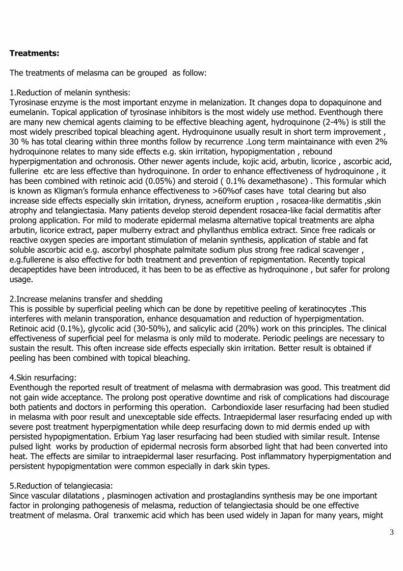
Figure 1: Medlite C 6 Laser ( Hoya-Conbio, USA) Medlite C6 and melasma: Principle of Minimal Photothermolysis Treatment: Recent histologic study of laser toning technique has shown that after exposure to repetitive sub-ablative threshold hat-topped 1064 nm Q-switched Nd:YAG laser beam, melanin granules will be fragmented and dispersed into cellular cytoplasm .Dendrites of epidermal melanocytes also decrease.The smaller size and dispersion of melanins results in pigment lightening. Multiple passes should disperse melanins layer by layer , after 10-20 passes ,the whole thickness of epidermis should receive enough energy for melanin dispersion. The total cumulative dosage of energy should be less than toxic accumulative energy that would lead to irreversible damage of the pigmented cells. Beyond toxic accumulative dosage, melanocytes with dense melanin will die. Subsequent treatment at weekly interval would gradually reduce hyperpigmentation of melasma without damaging to the epidermis. During treatments ,few layers of most superficial keratinocytes will be vaporized. This improves the textures and comlextion of skin similar to superficial chemical peelings . Some dermal melanophages with dense melanin granules should absorbed enough energy to critical level of cellular damage and cell death. By average about 8 to 10 weekly treatments are required to reduce the hyperpigmentation of melasma down close to normal skin colour. Due to the effects of improving complexion and textures some doctors called this technique “ Laser toning”. Then recurrence is prevented by long-term application of topical safe whitening and antioxidant agents e.g. 5% alpha arbutin , Fullerine+Ascorbly phosphate sodium together with UVB+ UVA blocker sunscreen ( SPF>30,PA > +++) , oral Tranexamic acid or topical decapeptides. Laser toning can be repeated if there is recurrence of hyperpigmentation. Prolong continuous treatment is not adviced because of potential side effect . Since the total commulative dosage of laser energy related to destruction of melanocytes and dyschromia, it should be kept to minimum. Revlite ( Hoya-Conbio, USA) is a newest and most powerful Q-switched Nd:YAG laser system available. The high energy 5 nanoseconds, twin pulses (800 mJ) with 140 microsecond interval ,1064 nm wavelength , total 1.6 Joules ( +/- 20%) has been patened as The PhotoacousticTherapy Pulse (PTP) . A This PTP mode , therefore result in both photoacoustic and photothermal effects with breakage of target melanins or tattoos at larger spot size than ordinary Q-switched Nd:YAG laser together with more thermal reaction and less epidermal injuries. Homogenous or hat-topped beam profile deliver deep homogenous energy preventing problems of central hot spot. Large spot size also enables precise overlapping ( 5-10%) .The overall result is more evenly melanin breakages , without wounding and textural changes. Because of high pulse energy, the frequency –doubled mode, produce 532 nm , nanosecond pulse beam
6
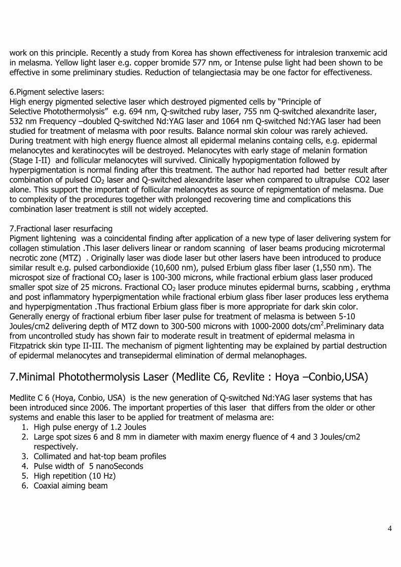
upto 5 joules/cm2 at 2 mm spot size. This is enough for red tattoos removal. The system also provide Optical MultiLite Dye laser handpiece which converted 1064 nm to 585 nm and 650 nm nanosecond pulse laser. With 585 nm,2 mm spot size ,the energy of pulse laser is 10 Joules/cm2 and 6.5 Joules/cm2 for 585 nm wavelength. These are powerful enough for green and blue tattoos removal. The author has studied application of this new laser in many conditions for more than twenty four months. The advantages of this new laser are 1) faster clearing of dermal melanins and multicolor tattoos with reduction of number of treatments required 2) absence of epidermal wounds and recovering time 3)with lower peak energy but wider pulses, there is more thermal effects for dermal collagen stimulation 4) with dye adaptors, this laser has enough energy for removal of both blue and red color tattoos. When PTP mode is turned off, this laser is equivalent to Medlite C6.
Figure 2 : Revlite (Hoya-Conbio, USA)
7
Technique: 1.Treatment area should be cleaned and dried 2.Take standard photographies (front, side views 45 ,90 o, close-up) 3,Measuring of Melanin Index at lesions and normal facial skin 4.Patient should wear cap and protective eye goggles, wanted hair ( eye brows, eyelashes) should be protected with tape and eye goggle. 5.Precooling of treatment area with cool air (-20oC) for few minutes 6.Setting parameters:
Epidermal melasma: Medlite C6, Revlite (PTP off) Fitzpatrick skin type III-IV, 1064 nm, 3-3.6 Joules/cm2, 6-8 mm spot size, 10 Hz, 10-20 passes Fitzpatrick skin type V-VI, 1064 nm, 2.5-3.2 Joules/cm2, 6-8 mm spot size, 10 Hz, 10-20 passes
Dermal/mixed melasma: Medlite C6, Revlite (PTP off) All skin types , 1064 nm, 3.4-4.2 Joules/cm2, 6 mm spot size, 10 Hz, 10-20 passes
Rebound melasma :Medlite C6, Revlite (PTP off) All skin types, 1064 nm, 2.5-3.2 Joules /cm2, 6 mm spot size, 10 Hz, 10-20 passes
Postinflammatory hyperpigmentation: Medlite C6, Revlite (PTP off) All skin types, 1064 nm, 2.8-4.2 Joules /cm2, 6 mm spot size, 10 Hz, 10-20 passes
Rejuvenation /wrinkles /dilated pores: Fitzpatrick skin type II-III,1064 nm, Medlite C6 3.4-4.2 Joules/cm2, 6-8 mm spot size, 10 Hz, 10 passes, Revlite,(PTP on) 6 Joules/cm2, 6 mm spot size 5-10 passes Fitzpatrick skin type IV-VI,Medlite C6,1064 nm, 2.8-3.4 Joules/cm2, 8 mm spot size, 10 Hz, 10 passes Revlite, (PTP off)3.2Joules/cm2, 8 mm spot size 5-10 passes
Ephilides Fitzpatrick skin type II-III,532 nm,Medlite C6 ,Revlite (PTP off)1.8-2.5 Joules/cm2, 3-4 nm spot size, 1-2 Hz, 1 pass Fitzpatrick skin type IV-VI , Medlite C 6, Revlite (PTP off) 1064 nm2.5-3.4Joules/cm2, 6 mm spot size, 10 Hz, 10 passes
Solar lentigoes Fitzpatrick skin type II-III, Medlite C6, Revlite (PTP off) 532 nm, 2-3 Joules/cm2, 3-4 mm spot size, 1-2 Hz, 1 pass Fitzpatrick skin type IV-VI, Medlite C6, Revlite (PTP off) 532 nm, 2-2.5 Joules/cm2, 3-4 mm spot size , 1-2 Hz, 1 pass
Atrophic scars All Fitzpatrick skin type , Medlite C 6,1064 nm, 4-6 Jouls/cm2 ,4-6 mm spot size, 10 HZ, > 20 pulses or Revlite(PTP on) ,1064 nm, 6 Joules/cm2, 6 mm,10 Hz, 10-20 passes (fine petechiaes are observed)
Dermal melanocytic lesions (Nevus of Ota, or Hori’s nevus) All Fitzpatrick skin types ,Medlite C 6, 1064 nm, 6-8 Joules/cm2, 4 mm spot size, 4-10 passes or Revlite (PTP on), 6 Joules/cm2, 6 mm, 10 Hz, 5 passes, repeated
every month until the lesions fade Tattoos removal (Black,blue black )
All Fitzpatrick skin types, Medlite C6,1064 nm, 3-4 mm spot size,6-8 Joules/cm2, 10 Hz, 1-2 passes,or Revlite (PTP on), 6-12 Joules/cm2, 6 or 4 mm spot size, 1-2 passes
Every month until tattoos fade >80% Tattoos removal (Red)
8
All Fitzpatrick skin types,Medlite C 6, 532 nm, 2 mm spot size, 5 Joules/cm2, 1 pass, every month until tattoos fade
Tattoos removal (Blue, Green) All Fitzpatrick’s skin types, Revlite+ Optical Mujltilite Dye laser handpiece For blue tattoos: 585 nm, 10 Joules/cm2, 2 mm spot size For green tattoos: 650 nm, 6.5 Joules/cm2, 2 mm spot size 1-2 passes, every month until clearing
Tattoos (cosmetic, skin color) All Fitzpatrick skin type , Medlite C6 ,1064 nm,3- 4 mm spot size, 6-8 Joules /cm2, 1 pass Revlite (PTP on), 6-12 Joules/cm2, 6 or 4 mm spot size, 1-2 passes ( tattoos darkening is possible, test spot is recommended)
Periorbital darkening Fitzpatrick skin type III-IV, Medlite C6,1064 nm, 4 mm spot size ,4-6 Joules/cm2, 10 passes Revlite (PTP on) 1064 nm, 6 mm spot size 6 Joules/cm2, 10 passes every month
Axillary hyperpigmentation Fitzpatrick skin type III-V, Medlite C6, Revlite (PTP off) 1064 nm, 6 mm spot size, 3-4 Joules/cm2, 10-20 passes every month until the lesions fade (6-10 times)
Hyperpigmented hypertrophic scar Fitzpatrick skin type III –V, Medlite C6,1064 nm, 4 mm spot size, 6-8 Joules/cm2, 10-20 passes every month, Revlite (PTP on), 6 Joules/cm2, 6 mm, 10 Hz, 5 passes
Lips darkening All skin type, Medlite C6, Revlite (PTP off )532 nm, 2-3 Joules/cm2, 3 mm spot size, 1 pass
Lentigines All skin type , Medlite C6, Revlite (PTP off )532 nm, 2-3 Joules/cm2, 2-3 mm spot size, 1 pass
Café au lait, segmental lentigines, Nevus spilus etc All skin types, Medlite C6, Revlite (PTP off ) Medlite C6,1064 nm, 4 mm spot size, 6-8 Joules/cm2, 10-20 passes every month,or Revlite (PTP on), 6 Joules/cm2, 6 mm, 10 Hz, 5 passes follow by 532 nm, 2-3 Joules/cm2, 2-3 mm spot size, 1 pass, every month until hyperpigmented areas improve.
Acne ,post acne redness and acne scar All skin types, Medlite C6, 1064 nm, 4 mm spot size, 6-8 Joules/cm2, 10-20 passes every month,or Revlite (PTP on), 6 Joules/cm2, 6 mm, 10 Hz, 10 passes every two weeks
7.The face should be divided into multiple treatment areas and treat one area at a time. 8.The laser beam should be delivered perpendicular to the surface and move the beam slowly in such away that there is <10 % overlapping between pulses .There are two techniques of treatment
A Painting technique. Laser beam is moved along the linear lines. For treatment of melasma the beam is moved forward and backward along that lines for 10 times before moving to adjacent area. After completion of first pass treatment of that area . The second pass will be performed in similar manner perpendicular to the direction of first pass.
B.Tracing technique, Laser beam is moved following the pattern of lesions. Generally for extensive melasma lesion and rejuvenation, painting technique is recommended. For
telangiectatic or dyschromic lesions tracing technique is better in following the pattern of lesions.
9
9.Clinical end points: Epidermal melasma: immediate pigment lightening, immediate whitening of fine hair and Perilesional erythema Dermal/mixed melasma: immediate darkening of lesions, immediate perilesional erythema
Rebound melasma: immediate pigment lightening, immediate perilesional erythema Dermal melanocytic lesions ( Nevus of Ota, Hori etc): immediate darkening of lesions, immediate perilesional erythema
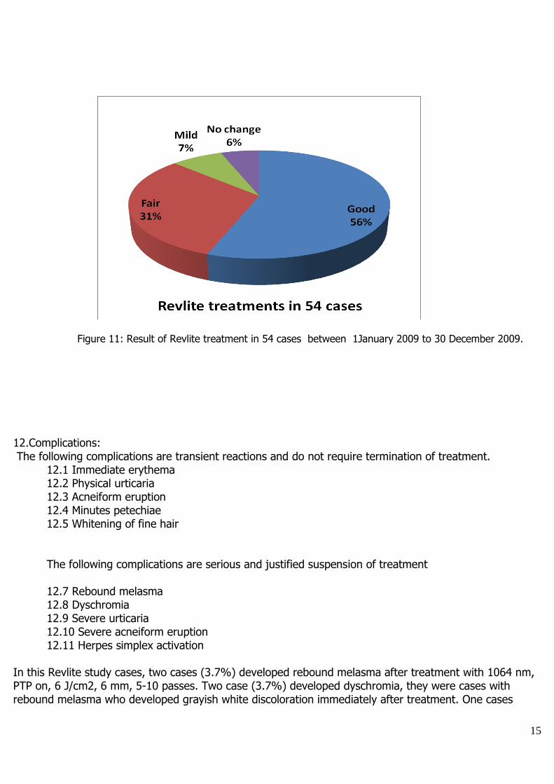
Post inflammatory hyperpigmentation: immediate lightening of lesions, perilesional erythema Periorbital / axillar darkening: immediate lightening of lesions, perilesional erythema Ephilides /solar lentigoes: immediate grayish discolouration (1064 nm)or whitening ( 532 nm) , Rejuvenation: immediate erythema with improvement of fine textures, pore sizes, wrinkles Atrophic scar: immediate erythema +/- minute petechiaes Tattoos: immediate whitening, minutes petechiaes Acne: diffuse erythema, whitening of fine hair 10.Follow-up treatments: For melasma ,the treatment should be performed at 1-2 weeks interval for 5 to 10 times. The number of treatments depends on clinical response. The goal of treatment is to reduce the hyperpigmentation down close to normal skin colour but not to completely treated melasma. Then treatment is then continue with topical whitening agents e.g. 5% Alpha Arbutin+ 2% Kojic acid, Fullerine+ Ascorbyl phosphate palmitate sodium (F+ APPS) or 4% hydroquinone+ 0.05%retinoic acid and broad spectrum sunscreen (PA> +++ and SPF>30) to maintain long term remission. For rejuvenation treatment will be performed every 2 weeks for 4-6 sessions, then every 1-3 months for maintainance. For post inflammatory hyperpigmentation fewer treatments are required. For ephilides and solar lentigoes, few treatments with 532 nm at monthly intervals are required. Most of the cases will develop mild to moderate PIH. Few treatments with 1064 nm similar to treatment for melasma will clear this complication within one month. For acne and acne scar, bi weekly treatments upto three months will be required to treat acne and improved post acne scar. 11.Results: Epidermal melasma response better and faster than dermal/ mixed melasma. Complete clearing of lesions should be expected in more than 60% of cases of epidermal melasma after 10 Revlite treatment. Complete clearing of dermal /mixed melasma will be between 30-50% , while the remaining cases will show moderate improvement. Post inflammatory hyperpigmentation and rebound melasma are sensitive to the treatment. Lower energy and fewer repetition is adequate to produce marked improvement. Ephilides need 1-2 treatments for >80% clearing. Solar lentigoes should be treated with 532 nm wavelength for few times.In Fitzpatrick’s skin type IV-V ,PIH is common complication. PIH should be treated with l1064 nm, laser toning and topical whitening agents. Revlite with higher pulse energy cleared dermal melanocytic lesions faster than Medlite C6.To clear Nevus of Ota, monthly treatment for 4-10 times are required. Usually there will be less dyschromia when compare to older Q-switched NdYAG laser or other Q-switched laser systems.. Hori’s nevus needs 2-6 biweekly treatments for more than 80% clearing Revlite offered advantage for collagen stimulation and gave good result for skin rejuvenation, reduction of wrinkles, dilated pores, telangiectasia and hyper/atrophic scars. For acne ,post acne hyperpigmentation and atrophic scar, the preliminary result by the author has shown
10
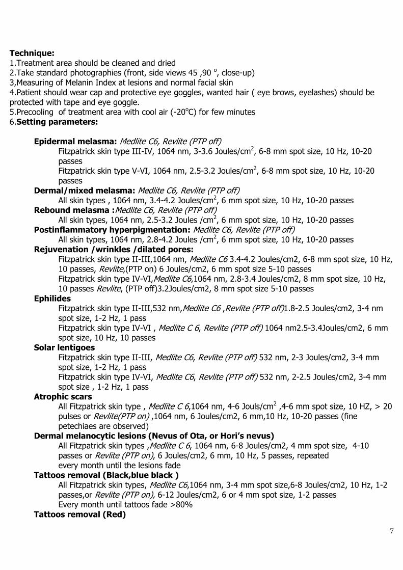
moderate reduction in active acne lesions , good response for post acne hyperpigmentation and fair response for atrophic scar.
Figure 3: Pre and post eight treatment of epidermal melasma with Medlite C 6 laser. Observe Improvement in both melasma and skin textures. ( Clin Prof Niwat Polnikorn )
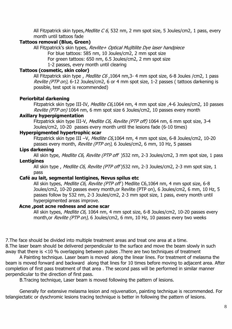
Figure 3: Pre and post 10 treatment of mixed type melasma , PIH and ochronosis with Medlite C6.
Note improvement in melasma, clearing of ochronosis and improvement of textures. (Clin Prof Niwat Polnikorn )
11
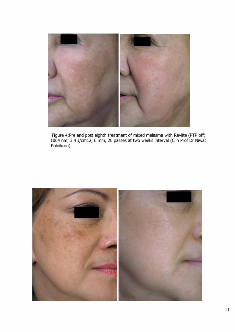
Figure 4:Pre and post eighth treatment of mixed melasma with Revlite (PTP off) 1064 nm, 3.4 J/cm12, 6 mm, 20 passes at two weeks interval (Clin Prof Dr Niwat Polnikorn)
12
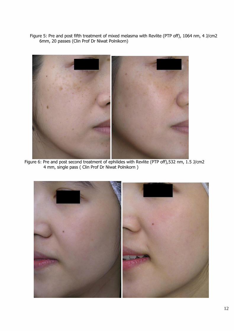
Figure 5: Pre and post fifth treatment of mixed melasma with Revlite (PTP off), 1064 nm, 4 J/cm2 6mm, 20 passes (Clin Prof Dr Niwat Polnikorn)
Figure 6: Pre and post second treatment of ephilides with Revlite (PTP off),532 nm, 1.5 J/cm2 4 mm, single pass ( Clin Prof Dr Niwat Polnikorn )
13
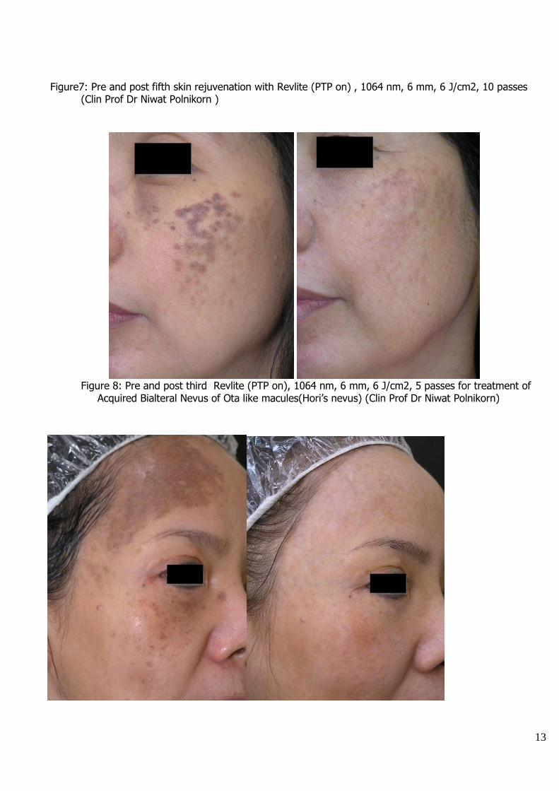
Figure7: Pre and post fifth skin rejuvenation with Revlite (PTP on) , 1064 nm, 6 mm, 6 J/cm2, 10 passes (Clin Prof Dr Niwat Polnikorn )
Figure 8: Pre and post third Revlite (PTP on), 1064 nm, 6 mm, 6 J/cm2, 5 passes for treatment of
Acquired Bialteral Nevus of Ota like macules(Hori’s nevus) (Clin Prof Dr Niwat Polnikorn)
14
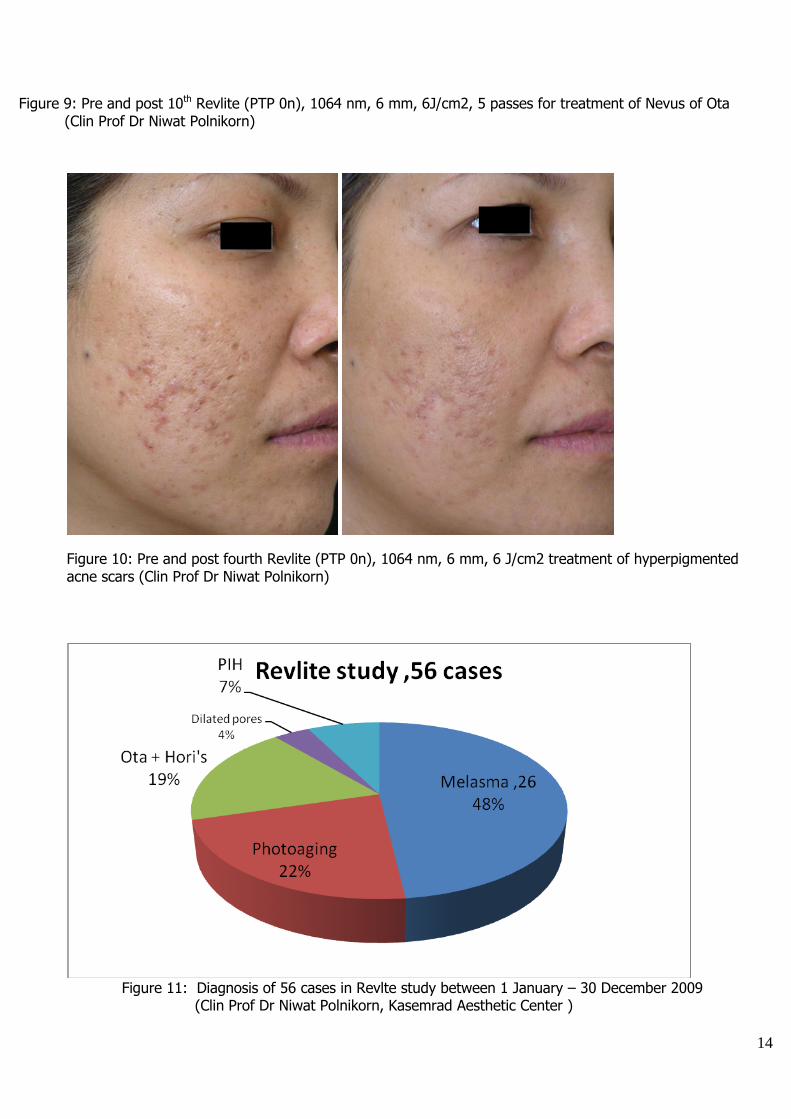
Figure 9: Pre and post 10th Revlite (PTP 0n), 1064 nm, 6 mm, 6J/cm2, 5 passes for treatment of Nevus of Ota (Clin Prof Dr Niwat Polnikorn)
Figure 10: Pre and post fourth Revlite (PTP 0n), 1064 nm, 6 mm, 6 J/cm2 treatment of hyperpigmented acne scars (Clin Prof Dr Niwat Polnikorn)
Figure 11: Diagnosis of 56 cases in Revlte study between 1 January – 30 December 2009 (Clin Prof Dr Niwat Polnikorn, Kasemrad Aesthetic Center )
15
Figure 11: Result of Revlite treatment in 54 cases between 1January 2009 to 30 December 2009.
12.Complications: The following complications are transient reactions and do not require termination of treatment. 12.1 Immediate erythema 12.2 Physical urticaria 12.3 Acneiform eruption 12.4 Minutes petechiae 12.5 Whitening of fine hair The following complications are serious and justified suspension of treatment 12.7 Rebound melasma 12.8 Dyschromia 12.9 Severe urticaria 12.10 Severe acneiform eruption 12.11 Herpes simplex activation In this Revlite study cases, two cases (3.7%) developed rebound melasma after treatment with 1064 nm, PTP on, 6 J/cm2, 6 mm, 5-10 passes. Two case (3.7%) developed dyschromia, they were cases with rebound melasma who developed grayish white discoloration immediately after treatment. One cases
16
developed post inflammatory hyperpigmentation after treatment of Hori’s nevus.Over all complications were 11.11%.
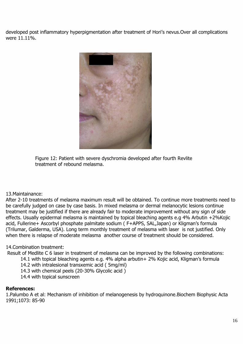
Figure 12: Patient with severe dyschromia developed after fourth Revlite treatment of rebound melasma. 13.Maintainance: After 2-10 treatments of melasma maximum result will be obtained. To continue more treatments need to be carefully judged on case by case basis. In mixed melasma or dermal melanocytic lesions continue treatment may be justified if there are already fair to moderate improvement without any sign of side effects. Usually epidermal melasma is maintained by topical bleaching agents e.g 4% Arbutin +2%Kojic acid, Fullerine+ Ascorbyl phosphate palmitate sodium ( F+APPS, SAL,Japan) or Kligman’s formula (Trilumar, Galderma, USA). Long term monthly treatment of melasma with laser is not justified. Only when there is relapse of moderate melasma another course of treatment should be considered. 14.Combination treatment: Result of Medlite C 6 laser in treatment of melasma can be improved by the following combinations: 14.1 with topical bleaching agents e.g. 4% alpha arbutin+ 2% Kojic acid, Kligman’s formula 14.2 with intralesional transxemic acid ( 5mg/ml) 14.3 with chemical peels (20-30% Glycolic acid ) 14.4 with topical sunscreen References: 1.Palumbo A et al: Mechanism of inhibition of melanogenesis by hydroquinone.Biochem Biophysic Acta 1991;1073: 85-90
17
2.Ennes SBP et al : A double –blind comparative ,placebo-controlled study of efficacy and tolerability of 4% hydroquinone as a depigmenting agent in melasma. J Dermatollog Treat 2000; 11:173-9 3.Haddad AL wt al. A clinical, prospective, randomized, double-blind trial comparing skin whitening complex with hydroquinone vs placebo in treatment of melasma. Int J Dermatol 2003; 42:153-6 4.Balina LM et al. The treatment of melasma. 20% azaleic acid versus 4% hydroquinone cream. Int J Dermatol 1991;30:893-5 5.Lim JT. Treatment of melasma using kojic acid in a gel containing hydroquinone and glycolic acid. Dermatol Surg 1999; 25:282-4 6.Kligmen AM, Willis I. A new formula for depigmentaing human skin. Arch Dermatol 1975; 111: 40-8 7.Taylor SC eta;. Efficacy and safety of a new triple-combination agent for the treatment of facial melasma. Cutis 2003;72:67-72 8.Hurley ME, et al. Efficay of glycolic peels in the treatment of melasma. Arch Dermatol 2002;138:1578-82 9.Lim JTE, Tham SN. Glycolic acid peels in the treatment of melasma among Asian wome. Dermatol Surg 1999;23:177-9 10.Kopora D, Hohenleutner.Ruby laser treatment of melasma and post inflammatory hyperpigmentation.Dermatol Surg 1995;21:990-5 11.Nouri K et al. Combination treatment of melasma with pulsed CO2 laser followed by Q-switched alexandrite laser : a pilot study. Dermatol Surg 1999;25:494-7 12.Angsuwarangsee S, Polnikorn N. Combination ultrapulse CO2 laser and Q-switched alexandrite laser compare with Q-switched alexandrite laser alone for refractory melasma. Dermatol Surg 2003;29:59-64 13.Lee G H et al. The effect of combination treatment of the recalcitrant pigmentary disorders with pigmented laser and chemical peeling agent. Dermatol Surg 2002;28:1120-3 14.Malaloto RM, Alster T. Erbium-YAG laser resurfacing for refractory melasma. Dermatol Surg 1999;25:121-3 15.Operating Manual; Medlite C 6. Hoya-Conbio , USA 2006 16.Physician Guidline for Medlite C 6 . Hoya –Conbio, USA 2006 17.Polnikorn N. Treatment of Refractory Dermal Melasma with Medlite C6 Q-switched Nd:YAG Laser. Report of two cases.J Cos Laser Ther 2008 ;10:167-173 18.Polnikorn N. Treatment of refractory melisma with the Medlite C6 , Q -switched Nd:YAG laser and alpha arbutin.A prospective study. J Cos Laser Ther 2010; 12: 126-131
![Anti-melanogenic effects of black, green, and white tea ... · hyperpigmentation, melasma, postinflammatory melanoderma, and solar lentigo [8,35]. Melanin is one of the most widely](https://img.pdfslide.net/doc/110x75/5ecf5a981e33ba350c72b898/anti-melanogenic-effects-of-black-green-and-white-tea-hyperpigmentation-melasma.jpg)

![Plants used to treat hyperpigmentation in Iranian traditional … · 2020-05-18 · melasma [17,18]. ITM scholars believed that ‘Bahaq’ was similar to ‘Kalaf’ except that](https://img.pdfslide.net/doc/110x75/5ecf5a991e33ba350c72b89c/plants-used-to-treat-hyperpigmentation-in-iranian-traditional-2020-05-18-melasma.jpg)