Embed Size (px)
Citation preview
British Journal of Ophthalmology, 1979, 63, 418-421
Treatment of amoeboid herpetic ulcers withadenine arabinoside or trifluorothymidineDOUGLAS J. COSTER, BARRIE R. JONES, AND JAMES I. McGILL*From the Department of Clinical Ophthalmology, Institute ofOphthalmology, Moorfields Eye Hospital, London
SUMMARY In previous studies adenine arabinoside and trifluorothymidine were found to be equallyeffective treatments for dendritic ulcers of the cornea, but a trend emerged which suggested that inamoeboid ulcers trifluorothymidine was more effective. The collection of additional cases confirmsthe superiority of trifluorothymidine in such cases.
Despite extensive research 5-iodo-2'-deoxyuridine(IDU) has remained until recently the only antiviralagent generally available to clinicians for treatingulcerative herpetic keratitis.Adenine arabinoside (Ara-A) and trifluorothymi-
dine (F3T) (Wellings et al., 1972; Pavan-Langston,1975; Coster et al., 1976) are both highly effective intreating herpetic corneal ulceration. In a largeseries of herpetic ulcers treated with one or other ofthese agents no difference could be found in thedendritic ulcer group (Fig. 1), but a trend emergedwhich suggested that F3T had some advantage overAra-A in treating the less common but more severeamoeboid ulcers (Coster et al., 1976).Not only are amoeboid ulcers less common than
dendritic ulcers, they have often been treated withsteroids before referral to an ophthalmologist, andmany have associated stromal disease and uveitis.They constitute the most difficult therapeutic chal-lenge for antiviral drugs used for ulcerative epithelialherpetic keratitis. Cases of this type are thereforemore likely to show a difference in effectivenessbetween 2 highly active antiviral compounds thanthe less challenging cases of dendritic ulcer.
In the initial trial 102 patients were treated-87with dendritic ulcers, 15 with amoeboid ulcers(Coster et al., 1976). A further 15 amoeboid ulcerswere treated under the same protocol in an effort toclarify the trend which seemed to be emerging in theinitial study. This paper reports the results.
Patients and methods
Thirty unselected patients with typical amoeboid
Address for reprints: Mr D. J. Coster, FRCS, Departmentof Clinical Ophthalmology, Moorfields Eye Hospital, CityRoad, London EClV 2PD*Present address: Southampton Eye Hospital, Wilton Avenue,Southampton S09 4XW
100 -
75 -
w
50 -
(D
F3TN= 13
( ArA-AN =17
251
5 10 15 20Days to heal
Fig. 1 Cumulative frequency graph of times for healingof dendritic ulcers under treatment with 3% adeninearabinoside ointment or 1% trifluorothymidine eye dropsS times daily
ulcers who consented to enter the study were in-cluded. All completed the course of treatment; 17were treated with Ara-A and 13 with F3T.The coded treatments were randomly allocated
by the pharmacist within closely matched strata.The stratification was based on the features con-sidered to be related to poor prognosis. These were:the size of the epithelial defect, whether the patienthad been treated with steroids before coming tohospital. and whether or not the patient was atopic.
All patients were treated on an outpatient basis.The coded preparations consisted of either 3-3%Ara-A ointment or 1% F3T drops, which were
418
I Il
on 16 July 2018 by guest. Protected by copyright.
http://bjo.bmj.com
/B
r J Ophthalm
ol: first published as 10.1136/bjo.63.6.418 on 1 June 1979. Dow
nloaded from
Treatment of amoeboid herpetic ulcers with adenine arabinoside or trifluorothymidine
2a 2b
applied topically 5 times a day. In addition to thecoded antiviral substance the patients receivedatropine drops 1% once daily. If they were alreadyreceiving topical steroids, these were graduallyreduced over the first 3 days of the treatmentprogramme and then stopped. The coded antiviralsubstance was given 5 times a day until the epithelialdefect had healed, as indicated by the absence ofstaining with fluorescein. The frequency of adminis-tration was then reduced to 3 times a day for anadditional 3 days.
Patients were reviewed on alternate days, and at
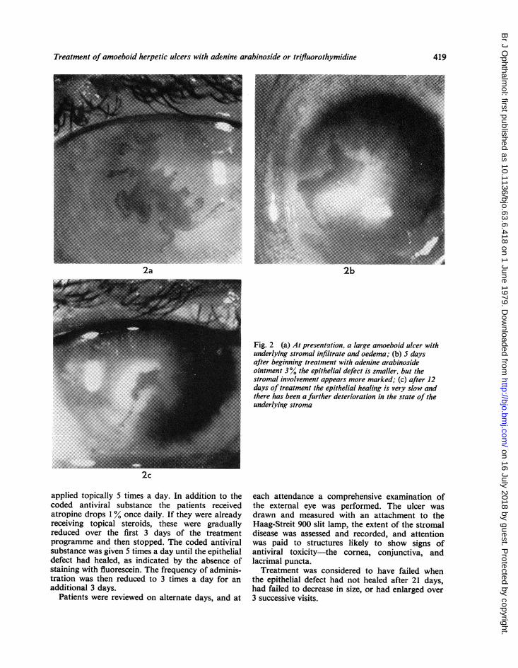
Fig. 2 (a) At presentation, a large amoeboid ulcer withunderlying stromal infiltrate and oedema; (b) S daysafter beginning treatment with adenine arabinosideointment 3% the epithelial defect is smaller, but thestromal involvement appears more marked; (c) after 12days of treatment the epithelial healing is very slow andthere has been a further deterioration in the state of theunderlying stroma
each attendance a comprehensive examination ofthe external eye was performed. The ulcer wasdrawn and measured with an attachment to theHaag-Streit 900 slit lamp, the extent of the stromaldisease was assessed and recorded, and attentionwas paid to structures likely to show signs ofantiviral toxicity-the cornea, conjunctiva, andlacrimal puncta.Treatment was considered to have failed when
the epithelial defect had not healed after 21 days,had failed to decrease in size, or had enlarged over3 successive visits.
419
on 16 July 2018 by guest. Protected by copyright.
http://bjo.bmj.com
/B
r J Ophthalm
ol: first published as 10.1136/bjo.63.6.418 on 1 June 1979. Dow
nloaded from
Douglas J. Coster, Barrie R. Jones, and James L McGill
between groups of patients when the groups aredefined not only by treatment schedule but by any
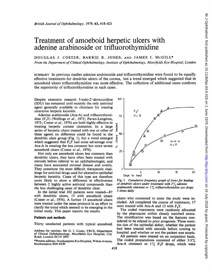
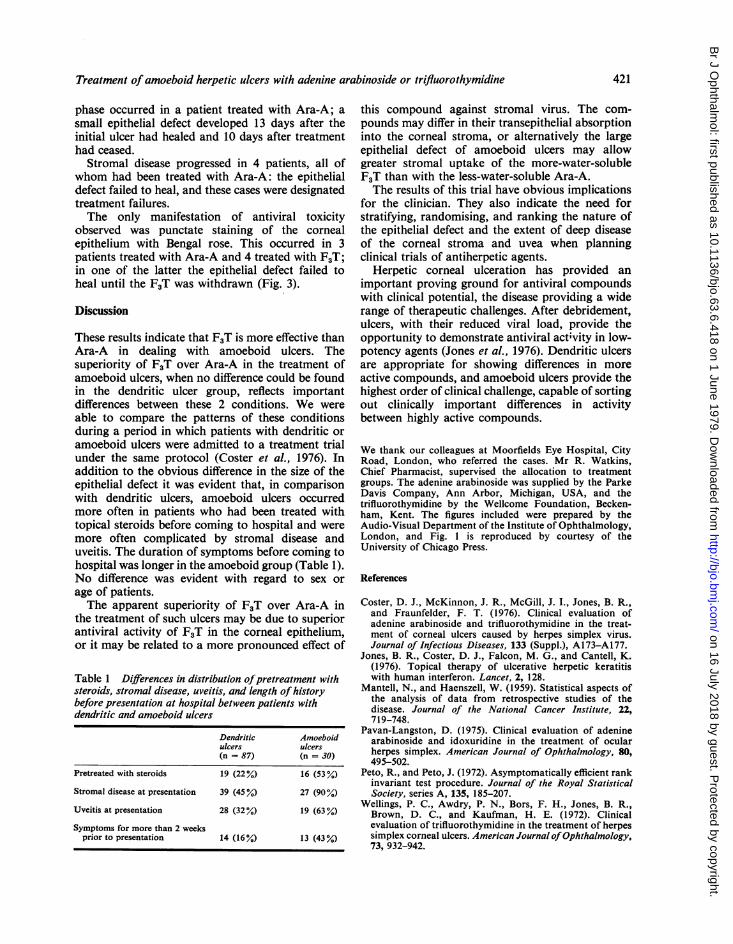
*b ~ factor thought to influence prognosis such as size ofulcer and extent of stromal disease (Mantell andHaenszell, 1959). An additional advantage is thatit is not a parametric test and does not assume thatthe healing times follow a normal distribution. Thedistribution of healing times is set out in Fig. 4,and clearly does not follow such a distribution.A logrank analysis was performed on the data
set out above. It indicates that the difference betweenthe 2 treatment groups is likely to be significant
s|l|||@ (P=0-05). The results are displayed in Fig. 5.
-I ~~~~~~~~~~~~~~~~~~~~~~~~~~~~~~. COMPLICATIONS AND DRUG REACTIONS
Apart from failure to heal, complications were few.The only recurrence in the immediate post-treatment
5o4-
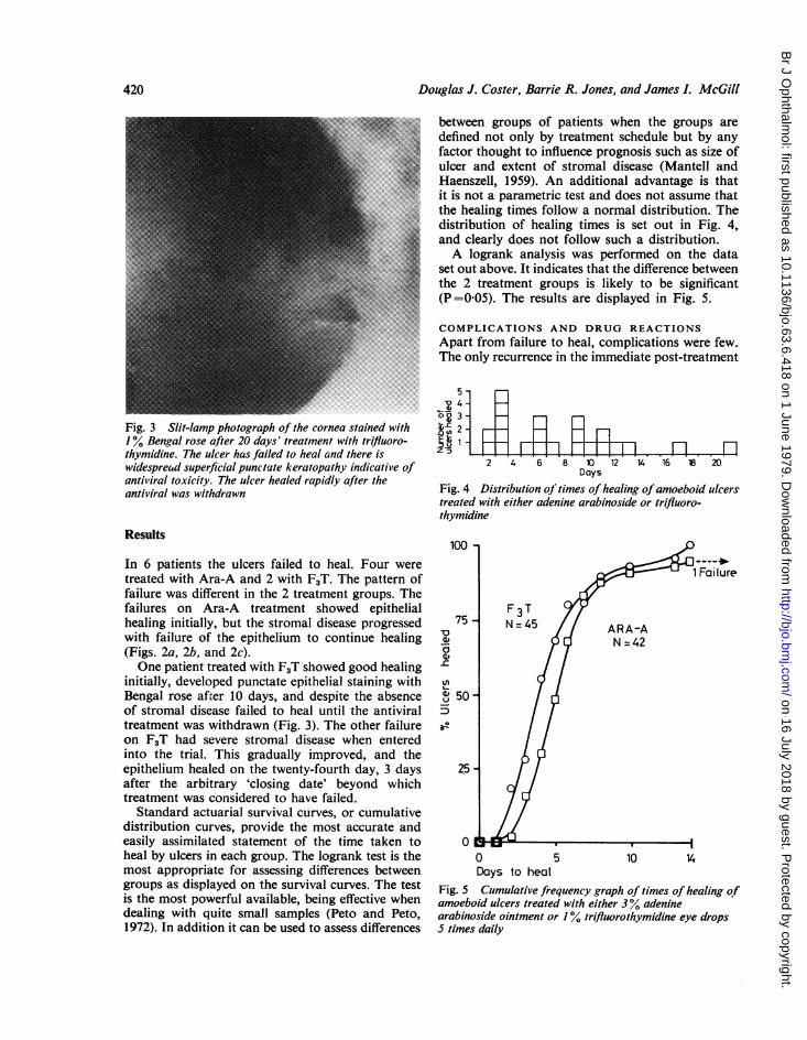
0 3K20Fig. 3 Slit-lamp photograph of the cornea stained with C 21 Ai-I
Bengal rose after 20 days' treatment with trifluoro- Et1 l,lthymidine. The ulcer has failed to heal and there is zlTIwidespread superficial punctate keratopathy indicative of 2 4 6 8 10 12 14 16 18 20antiviral toxicity. The ulcer healed rapidly after the Daysantiviral was withdrawn Fig. 4 Distribution of times of healing ofamoeboid ulcers
treated with either adenine arabinoside or trifluoro-thymidine
Results100
In 6 patients the ulcers failed to heal. Four weretreated with Ara-A and 2 with F3T. The pattern of 1 Failurefailure was different in the 2 treatment groups. Thefailures on Ara-A treatment showed epithelial
7F 3 T
healing initially, but the stromal disease progressed 75 N=45 ARA-Awith failure of the epithelium to continue healing a|, N =42(Figs. 2a, 2b, and 2c).One patient treated with F3T showed good healing l
initially, developed punctate epithelial staining with |/Bengal rose after 10 days, and despite the absence t 50 -
of stromal disease failed to heal until the antiviral 5|treatment was withdrawn (Fig. 3). The other failure a
0
on F3T had severe stromal disease when enteredinto the trial. This gradually improved, and theepithelium healed on the twenty-fourth day, 3 days 25 -
after the arbitrary 'closing date' beyond whichtreatment was considered to have failed.
Standard actuarial survival curves, or cumulativedistribution curves, provide the most accurate andeasily assimilated statement of the time taken to 0heal by ulcers in each group. The logrank test is the 0 5 l0 14most appropriate for assessing differences between Days to healgroups as displayed on the survival curves. The test Fig. 5 Cumulative frequency graph of times of healingofis the most powerful available, being effective when amoeboid ulcers treated with either 3% adeninedealing with quite small samples (Peto and Peto, arabinoside ointment or 1% trifluorothymidine eye drops1972). In addition it can be used to assess differences 5 times daily
420
on 16 July 2018 by guest. Protected by copyright.
http://bjo.bmj.com
/B
r J Ophthalm
ol: first published as 10.1136/bjo.63.6.418 on 1 June 1979. Dow
nloaded from
Treatment ofamoeboid herpetic ulcers with adenine arabinoside or trifluorothymidine
phase occurred in a patient treated with Ara-A; asmall epithelial defect developed 13 days after theinitial ulcer had healed and 10 days after treatmenthad ceased.
Stromal disease progressed in 4 patients, all ofwhom had been treated with Ara-A: the epithelialdefect failed to heal, and these cases were designatedtreatment failures.The only manifestation of antiviral toxicity
observed was punctate staining of the cornealepithelium with Bengal rose. This occurred in 3patients treated with Ara-A and 4 treated with FjT;in one of the latter the epithelial defect failed toheal until the F3T was withdrawn (Fig. 3).
Discussion
These results indicate that F3T is more effective thanAra-A in dealing with amoeboid ulcers. Thesuperiority of F3T over Ara-A in the treatment ofamoeboid ulcers, when no difference could be foundin the dendritic ulcer group, reflects importantdifferences between these 2 conditions. We wereable to compare the patterns of these conditionsduring a period in which patients with dendritic oramoeboid ulcers were admitted to a treatment trialunder the same protocol (Coster et al., 1976). Inaddition to the obvious difference in the size of theepithelial defect it was evident that, in comparisonwith dendritic ulcers, amoeboid ulcers occurredmore often in patients who had been treated withtopical steroids before coming to hospital and weremore often complicated by stromal disease anduveitis. The duration of symptoms before coming tohospital was longer in the amoeboid group (Table 1).No difference was evident with regard to sex orage of patients.The apparent superiority of F3T over Ara-A in
the treatment of such ulcers may be due to superiorantiviral activity of F3T in the corneal epithelium,or it may be related to a more pronounced effect of
Table 1 Differences in distribution of pretreatment withsteroids, stromal disease, uveitis, and length of historybefore presentation at hospital between patients withdendritic and amoeboid ulcers
Dendritic Amoeboidulcers ulcers(n = 87) (n = 30)
Pretreated with steroids 19 (22%) 16 (53 Y7)Stromal disease at presentation 39 (45%) 27 (90%)
Uveitis at presentation 28 (32%) 19 (63%)
Symptoms for more than 2 weeksprior to presentation 14 (16%) 13 (43%)
this compound against stromal virus. The com-pounds may differ in their transepithelial absorptioninto the corneal stroma, or alternatively the largeepithelial defect of amoeboid ulcers may allowgreater stromal uptake of the more-water-solubleF5T than with the less-water-soluble Ara-A.The results of this trial have obvious implications
for the clinician. They also indicate the need forstratifying, randomising, and ranking the nature ofthe epithelial defect and the extent of deep diseaseof the corneal stroma and uvea when planningclinical trials of antiherpetic agents.
Herpetic corneal ulceration has provided animportant proving ground for antiviral compoundswith clinical potential, the disease providing a widerange of therapeutic challenges. After debridement,ulcers, with their reduced viral load, provide theopportunity to demonstrate antiviral activity in low-potency agents (Jones et al., 1976). Dendritic ulcersare appropriate for showing differences in moreactive compounds, and amoeboid ulcers provide thehighest order of clinical challenge, capable of sortingout clinically important differences in activitybetween highly active compounds.
We thank our colleagues at Moorfields Eye Hospital, CityRoad, London, who referred the cases. Mr R. Watkins,Chief Pharmacist, supervised the allocation to treatmentgroups. The adenine arabinoside was supplied by the ParkeDavis Company, Ann Arbor, Michigan, USA, and thetrifluorothymidine by the Wellcome Foundation, Becken-ham, Kent. The figures included were prepared by theAudio-Visual Department of the Institute of Ophthalmology,London, and Fig. 1 is reproduced by courtesy of theUniversity of Chicago Press.
References
Coster, D. J., McKinnon, J. R., McGill, J. I., Jones, B. R.,and Fraunfelder, F. T. (1976). Clinical evaluation ofadenine arabinoside and trifluorothymidine in the treat-ment of corneal ulcers caused by herpes simplex virus.Journal of Infectious Diseases, 133 (Suppl.), A173-A177.
Jones, B. R., Coster, D. J., Falcon, M. G., and Cantell, K.(1976). Topical therapy of ulcerative herpetic keratitiswith human interferon. Lancet, 2, 128.
Mantell, N., and Haenszell, W. (1959). Statistical aspects ofthe analysis of data from retrospective studies of thedisease. Journal of the National Cancer Institute, 22,719-748.
Pavan-Langston, D. (1975). Clinical evaluation of adeninearabinoside and idoxuridine in the treatment of ocularherpes simplex. American Journal of Ophthalmology, 80,495-502.
Peto, R., and Peto, J. (1972). Asymptomatically efficient rankinvariant test procedure. Journal of the Royal StatisticalSociety, series A, 135, 185-207.
Wellings, P. C., Awdry, P. N., Bors, F. H., Jones, B. R.,Brown, D. C., and Kaufman, H. E. (1972). Clinicalevaluation of trifluorothymidine in the treatment of herpessimplex corneal ulcers. American Journal ofOphthalmology,73, 932-942.
421
on 16 July 2018 by guest. Protected by copyright.
http://bjo.bmj.com
/B
r J Ophthalm
ol: first published as 10.1136/bjo.63.6.418 on 1 June 1979. Dow
nloaded from