Embed Size (px)
Citation preview
Tutors
Dra. Maria Sarret Pons Departament de Química Física
Universitat de Barcelona (UB)
Dr. Jordi Esquena Moret Grup de Química de Superfícies
Departament de Nanotecnologia Química i Biomolecular Institut de Química Avançada de Catalunya (IQAC)
Consell Superior d’Investigacions Científiques (CSIC)
Treball Final de Grau
Preparation and characterization of biomimetic materials, by synthesis and templating in structured liquids
Preparación y caracterización de materiales biomiméticos, mediante síntesis en líquidos estructurados, utilizados como plantillas
José Luis Capdevila Echeverria June 2015


Aquesta obra esta subjecta a la llicència de: Reconeixement–NoComercial-SenseObraDerivada
http://creativecommons.org/licenses/by-nc-nd/3.0/es/

“Do you know what the secret to success is?” I of course said no. He then said, “Just keep working.”
Eric Kandel, quoted by (and said to) Eve Marder, interview in Current Biology 12:R5-R7 2007
First of all, I would like to acknowledge Institute of Advanced Chemistry of Catalonia (Institut
de Química Avançada de Catalunya, IQAC) and Spanish National Research Council (Consejo
Superior de Investigaciones Científicas, CSIC) for the research introduction for university
students (INTRO 2014) fellowship. I would like to thank Dr. Jordi Esquena Moret having
welcomed me in his research group for my Final Degree Project (Treball Final de Grau, TFG). It
was a challenging and very profitable opportunity to study in his research group for the
realization of the present project. I am deeply thankful to him, without his support, confidence
and direction; this work would not be possible. I would also like to thank Dr. Maria Sarret Pons
for her guidance, advice and discussion of the present work.
I am also grateful to the Final Degree Project committee for their time, care and
consideration for the finalization of this project.
In addition, I would like to thank all members of the Colloid and Interfacial Chemistry Centre
(formed by Dr. Jordi Esquena Moret and Dr. Conxita Solans Marsà groups) for their very useful
help and support, particularly, Dr. Susana Vílchez Maldonado, Dr. Jonathan Miras Hernández,
Dr. Jérémie Nestor, Dr. Tirso Emmanuel Flores Guia, Míriam Regué Griñó, Yoran Beldengrün,
Montserrat Sindreu Grañe, and Laura Sobrevias Bonells.
Finally, a million thanks to my family and friends, their encouragement and love has gotten
me through all the difficult times and I am grateful to them for all the chats and advice especially
at the end when I was so busy.
Thank you all for everything!


REPORT

Preparation and characterization of biomimetic materials, by synthesis and templating in structured liquids 1
CONTENTS
1. SUMMARY 3
2. RESUMEN 5
3. INTRODUCTION 7
3.1. Bone tissue 7
3.1.1. General description of bone tissue 7
3.1.2. Hydroxyapatite 9
3.1.3. Collagen and gelatin 9
3.2. Emulsions 11
3.2.1. Basic aspects of emulsions 11
3.2.2. Highly concentrated emulsions as reaction media for the formation of
porous materials 13
3.3. Gels 15
3.3.1. Basic aspects of gels 15
3.3.2. Protein polymer hydrogels 15
3.3.3. Cross-linking with genipin 16
3.4. Biomedical application: biomimetic porous composites of protein polymers-
reinforced hydroxyapatite 17
4. OBJECTIVES 18
5. EXPERIMENTAL SECTION 19
5.1. Materials 19
5.1.1. Synthesis of macroporous hydroxyapatite foams 19
5.1.2. Preparation of protein hydrogels 19
5.2. Apparatus and instrumental 20
5.3. Methods and procedures 21
5.3.1. Synthesis of macroporous hydroxyapatite foams 21
5.3.2. Preparation of collagen hydrogels 23
2 Capdevila Echeverria, José Luis
5.3.2.1. Method I: Addition of cross-linker after gelation (gel cross-linking) 23
5.3.2.2. Method II: Addition of cross-linker before gelation (mixing cross-linking) 23
5.3.3. Preparation of gelatin hydrogels 24
5.3.4. Rheological study of hydrogels 24
6. RESULTS AND DISCUSSION 27
6.1. Synthesis of macroporous hydroxyapatite foams 27
6.2. Preparation of protein hydrogels 34
6.2.1. Preparation of collagen hydrogels 34
6.2.1.1. Method I: Addition of cross-linker after gelation (gel cross-linking) 34
6.2.1.2. Method II: Addition of cross-linker before gelation (mixing cross-linking) 36
6.2.2. Preparation of gelatin hydrogels 37
6.3. Feasibility study for obtaining macroporous hydroxyapatite reinforced with
collagen or gelatin 43
7. CONCLUSIONS 45
8. REFERENCES 47
9. ACRONYMS 50
Preparation and characterization of biomimetic materials, by synthesis and templating in structured liquids 3
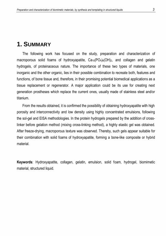
1. SUMMARY
The following work has focused on the study, preparation and characterization of
macroporous solid foams of hydroxyapatite, Ca10(PO4)6(OH)2, and collagen and gelatin
hydrogels, of proteinaceous nature. The importance of these two types of materials, one
inorganic and the other organic, lies in their possible combination to recreate both, features and
functions, of bone tissue and, therefore, in their promising potential biomedical applications as a
tissue replacement or regenerator. A major application could be its use for creating next
generation prostheses which replace the current ones, usually made of stainless steel and/or
titanium.
From the results obtained, it is confirmed the possibility of obtaining hydroxyapatite with high
porosity and interconnectivity and low density using highly concentrated emulsions, following
the sol-gel and EISA methodologies. In the protein hydrogels prepared by the addition of cross-
linker before gelation method (mixing cross-linking method), a highly elastic gel was obtained.
After freeze-drying, macroporous texture was observed. Thereby, such gels appear suitable for
their combination with solid foams of hydroxyapatite, forming a bone-like composite or hybrid
material.
Keywords: Hydroxyapatite, collagen, gelatin, emulsion, solid foam, hydrogel, biomimetic
material, structured liquid.

Preparation and characterization of biomimetic materials, by synthesis and templating in structured liquids 5
2. RESUMEN
El siguiente trabajo se ha focalizado en el estudio, obtención y caracterización de espumas
sólidas macroporosas de hidroxiapatita, Ca10(PO4)6(OH)2, y de hidrogeles de colágeno y
gelatina, de naturaleza peptídica. La importancia de estos dos tipos de materiales, uno
inorgánico y el otro orgánico, recae en su posible combinación para recrear, tanto en
características y funciones, el tejido óseo y, por lo tanto, en sus prometedoras aplicaciones
potenciales en biomedicina como sustituto o regenerador de dicho tejido. Una de las principales
aplicaciones podría ser su utilización para la creación de prótesis de próxima generación que
sustituyeran a las actuales, generalmente de acero inoxidable y/o titanio.
De los resultados obtenidos, se confirma la posibilidad de obtención de hidroxiapatita con
elevada porosidad e interconectividad y de baja densidad mediante emulsiones altamente
concentradas, siguiendo las metodologías sol-gel y EISA. En los hidrogeles de naturaleza
proteica preparados mediante el método de adición de reticulante previa a la gelificación
(método de mezclado-reticulación), se obtuvo un gel altamente elástico. Después de la
liofilización, se observó una textura macroporosa. De este modo, este tipo de geles parecen
adecuados para su combinación con espumas sólidas de hidroxiapatita, formando un material
compuesto o híbrido parecido al hueso.
Palabras clave: Hidroxiapatita, colágeno, gelatina, emulsión, espuma sólida, hidrogel, material
biomimético, líquido estructurado.

Preparation and characterization of biomimetic materials, by synthesis and templating in structured liquids 7
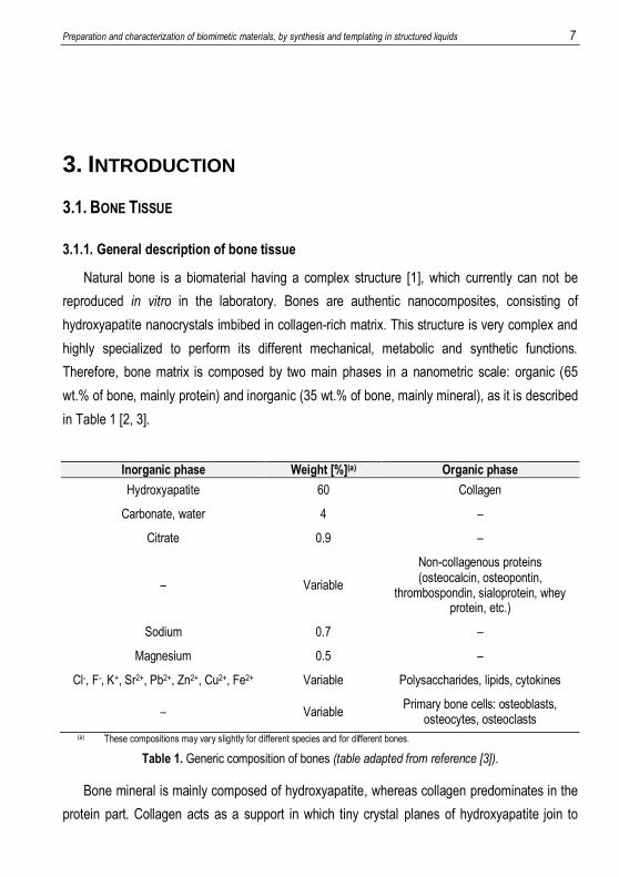
3. INTRODUCTION
3.1. BONE TISSUE
3.1.1. General description of bone tissue
Natural bone is a biomaterial having a complex structure [1], which currently can not be
reproduced in vitro in the laboratory. Bones are authentic nanocomposites, consisting of
hydroxyapatite nanocrystals imbibed in collagen-rich matrix. This structure is very complex and
highly specialized to perform its different mechanical, metabolic and synthetic functions.
Therefore, bone matrix is composed by two main phases in a nanometric scale: organic (65
wt.% of bone, mainly protein) and inorganic (35 wt.% of bone, mainly mineral), as it is described
in Table 1 [2, 3].
Inorganic phase Weight [%](a) Organic phase
Hydroxyapatite 60 Collagen
Carbonate, water 4 –
Citrate 0.9 –
– Variable
Non-collagenous proteins (osteocalcin, osteopontin,
thrombospondin, sialoprotein, whey protein, etc.)
Sodium 0.7 –
Magnesium 0.5 –
Cl-, F-, K+, Sr2+, Pb2+, Zn2+, Cu2+, Fe2+ Variable Polysaccharides, lipids, cytokines
– Variable Primary bone cells: osteoblasts,
osteocytes, osteoclasts (a) These compositions may vary slightly for different species and for different bones.
Table 1. Generic composition of bones (table adapted from reference [3]).
Bone mineral is mainly composed of hydroxyapatite, whereas collagen predominates in the
protein part. Collagen acts as a support in which tiny crystal planes of hydroxyapatite join to
8 Capdevila Echeverria, José Luis
form bone. Bone collagen has a characteristic fibrous structure, whose diameter varies between
100 and 2000 nm. Similarly, the hydroxyapatite in the bone mineral is found in the form of
nanocrystals, with size between 4x50x50 (nm).
The main role of minerals is to provide strength and rigidity to the bone, whereas collagen
imparts resistance and flexibility. It is important to note that inorganic bone crystals are not
directly linked to collagen, but connected through non-collagenous proteins, which represent
between 3 and 5 wt.% of bone composition and provide active sites for biomineralisation and
cell binding.
The amount of water present in bone is an important factor determining its mechanical
behaviour. Lipids, around 2 wt.% of bone, are also required for cellular functions and they play a
crucial role in biomineralisation. It is important to point out that biomineralisation degree is the
most important factor to determine the mechanical capabilities of the bone.
When bone is initially deposited, it is structurally weak and disorganized. But after a few
days the original bone becomes lamellar. In a macroestructural level, mature lamellar bone is
distinguished in compact and spongy types. They differ in density, as their names indicate, and
are organized in pores at multiple levels, from macro to nano, for the establishment of multiple
functions, including the transport of nutrients, oxygen and body fluids.
The spongy or trabecular bone occupies about 20 % of total bone weight. It is lighter, has
higher porosity and concentration of blood vessels than compact bone. The diameter of the
pores can be from micrometres to millimetres. The compact or cortical bone is much denser and
occupies about 80 % of total bone weight. It has lower porosity and concentration of blood
vessels than spongy one. Pores have diameters of 10-20 m and are separated by intervals of
200-300 m [2].
There are five different types of cells associated with bone tissue: osteoprogenitor cells,
osteoblasts, osteocytes, osteoclasts, and bone-lining cells. Nevertheless, the cells responsible
for the formation of new bone are osteoblasts, which produce collagen and cover it with non-
collagenous proteins which can retain minerals from blood flow, especially calcium and
phosphate, creating new bone.
For proper growth and differentiation of osteoblasts, they must be found in a similar
environment of the tissue being regenerated, achieving more favorable environments for their
proliferation with nanometric porosities [2, 4, 5].
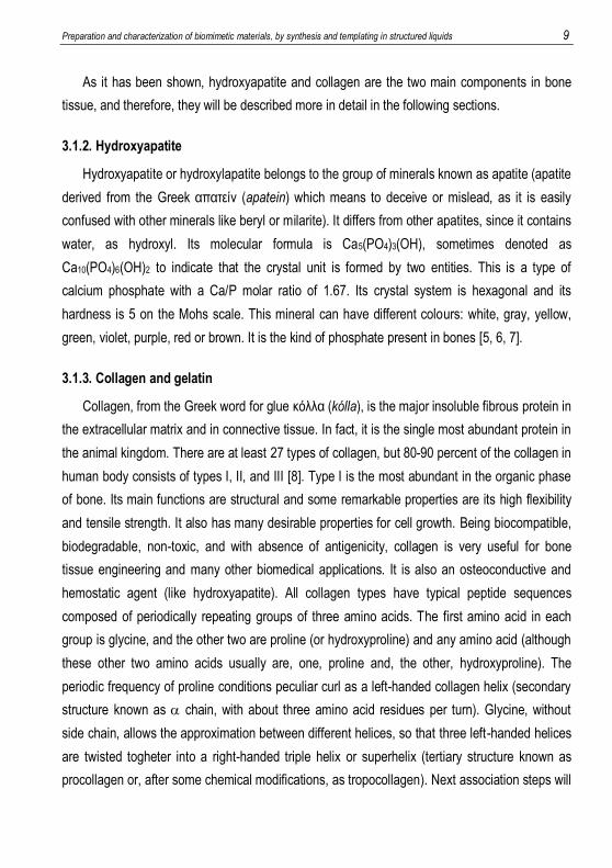
Preparation and characterization of biomimetic materials, by synthesis and templating in structured liquids 9
As it has been shown, hydroxyapatite and collagen are the two main components in bone
tissue, and therefore, they will be described more in detail in the following sections.
3.1.2. Hydroxyapatite
Hydroxyapatite or hydroxylapatite belongs to the group of minerals known as apatite (apatite
derived from the Greek απατείν (apatein) which means to deceive or mislead, as it is easily
confused with other minerals like beryl or milarite). It differs from other apatites, since it contains
water, as hydroxyl. Its molecular formula is Ca5(PO4)3(OH), sometimes denoted as
Ca10(PO4)6(OH)2 to indicate that the crystal unit is formed by two entities. This is a type of
calcium phosphate with a Ca/P molar ratio of 1.67. Its crystal system is hexagonal and its
hardness is 5 on the Mohs scale. This mineral can have different colours: white, gray, yellow,
green, violet, purple, red or brown. It is the kind of phosphate present in bones [5, 6, 7].
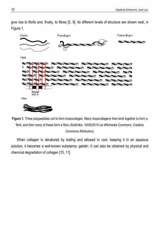
3.1.3. Collagen and gelatin
Collagen, from the Greek word for glue κόλλα (kólla), is the major insoluble fibrous protein in
the extracellular matrix and in connective tissue. In fact, it is the single most abundant protein in
the animal kingdom. There are at least 27 types of collagen, but 80-90 percent of the collagen in
human body consists of types I, II, and III [8]. Type I is the most abundant in the organic phase
of bone. Its main functions are structural and some remarkable properties are its high flexibility
and tensile strength. It also has many desirable properties for cell growth. Being biocompatible,
biodegradable, non-toxic, and with absence of antigenicity, collagen is very useful for bone
tissue engineering and many other biomedical applications. It is also an osteoconductive and
hemostatic agent (like hydroxyapatite). All collagen types have typical peptide sequences
composed of periodically repeating groups of three amino acids. The first amino acid in each
group is glycine, and the other two are proline (or hydroxyproline) and any amino acid (although
these other two amino acids usually are, one, proline and, the other, hydroxyproline). The
periodic frequency of proline conditions peculiar curl as a left-handed collagen helix (secondary
structure known as chain, with about three amino acid residues per turn). Glycine, without
side chain, allows the approximation between different helices, so that three left-handed helices
are twisted togheter into a right-handed triple helix or superhelix (tertiary structure known as
procollagen or, after some chemical modifications, as tropocollagen). Next association steps will
10 Capdevila Echeverria, José Luis
give rise to fibrils and, finally, to fibres [5, 9]. Its different levels of structure are shown next, in
Figure 1.
Figure 1. Three polypeptides coil to form tropocollagen. Many tropocollagens then bind together to form a
fibril, and then many of these form a fibre (Solitchka, 10/05/2015 via Wikimedia Commons, Creative
Commons Attribution).
When collagen is denatured by boiling and allowed to cool, keeping it in an aqueous
solution, it becomes a well-known substance, gelatin. It can also be obtained by physical and
chemical degradation of collagen [10, 11].
Preparation and characterization of biomimetic materials, by synthesis and templating in structured liquids 11
3.2. EMULSIONS
Polymeric, organic or inorganic, foams exhibit specific properties such as high porosity and
low density, which make them suitable candidates for hydroxyapatite recreation. Such materials
can be obtained by using highly concentrated emulsions as templates, by polymerizing or cross-
linking processes in the continuous phase of these emulsions.
3.2.1. Basic aspects of emulsions
Becher [12], in 1965, and Everett [13], in 1972, defined emulsions as thermodynamically
unstable heterogeneous systems composed of at least two immiscible or partially miscible liquid
phases, one of which is dispersed in the other in the form of droplets, with diameter generally
greater than 0.1 m.
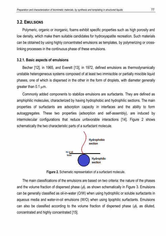
Commonly added components to stabilize emulsions are surfactants. They are defined as
amphiphilic molecules, characterized by having hydrophobic and hydrophilic sections. The main
properties of surfactants are adsorption capacity in interfaces and the ability to form
autoaggregates. These two properties (adsorption and self-assembly), are induced by
intermolecular configurations that reduce unfavorable interactions [14]. Figure 2 shows
schematically the two characteristic parts of a surfactant molecule.
Figure 2. Schematic representation of a surfactant molecule.
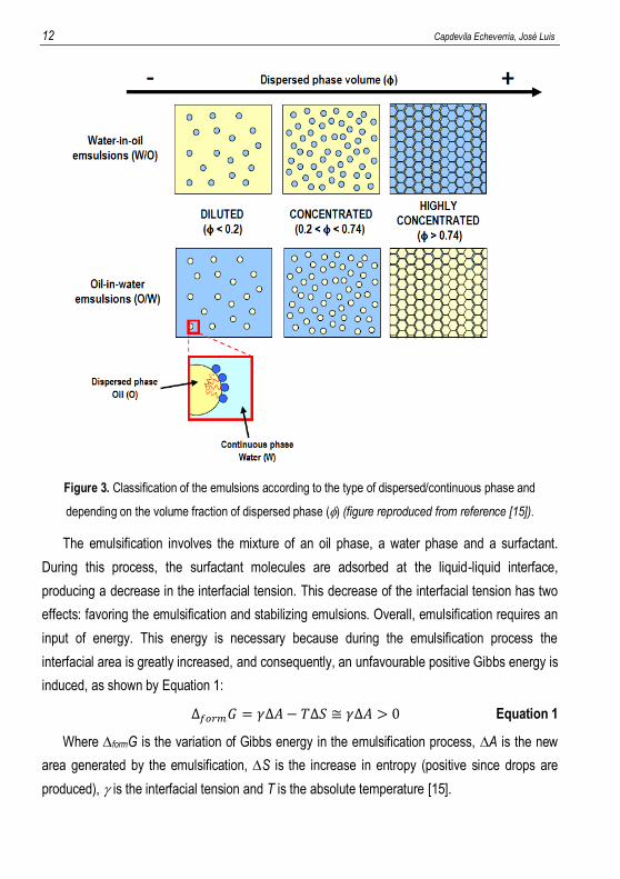
The main classifications of the emulsions are based on two criteria: the nature of the phases
and the volume fraction of dispersed phase (), as shown schematically in Figure 3. Emulsions
can be generally classified as oil-in-water (O/W) when using hydrophilic or soluble surfactants in
aqueous media and water-in-oil emulsions (W/O) when using lipophilic surfactants. Emulsions
can also be classified according to the volume fraction of dispersed phase (), as diluted,
concentrated and highly concentrated [15].
12 Capdevila Echeverria, José Luis
Figure 3. Classification of the emulsions according to the type of dispersed/continuous phase and
depending on the volume fraction of dispersed phase () (figure reproduced from reference [15]).
The emulsification involves the mixture of an oil phase, a water phase and a surfactant.
During this process, the surfactant molecules are adsorbed at the liquid-liquid interface,
producing a decrease in the interfacial tension. This decrease of the interfacial tension has two
effects: favoring the emulsification and stabilizing emulsions. Overall, emulsification requires an
input of energy. This energy is necessary because during the emulsification process the
interfacial area is greatly increased, and consequently, an unfavourable positive Gibbs energy is
induced, as shown by Equation 1:
∆𝑓𝑜𝑟𝑚𝐺 = 𝛾∆𝐴− 𝑇∆𝑆 ≅ 𝛾∆𝐴 > 0 Equation 1
Where formG is the variation of Gibbs energy in the emulsification process, A is the new
area generated by the emulsification, S is the increase in entropy (positive since drops are
produced), is the interfacial tension and T is the absolute temperature [15].
Preparation and characterization of biomimetic materials, by synthesis and templating in structured liquids 13
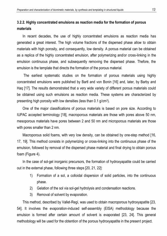
3.2.2. Highly concentrated emulsions as reaction media for the formation of porous
materials
In recent decades, the use of highly concentrated emulsions as reaction media has
generated a great interest. The high volume fractions of the dispersed phase allow to obtain
materials with high porosity, and consequently, low density. A porous material can be obtained
as a replica of the highly concentrated emulsion, after polymerizing and/or cross-linking in the
emulsion continuous phase, and subsequently removing the dispersed phase. Thefore, the
emulsion is the template that directs the formation of the porous material.
The earliest systematic studies on the formation of porous materials using highly
concentrated emulsions were published by Bartl and von Bonin [16] and, later, by Barby and
Haq [17]. The results demonstrated that a very wide variety of different porous materials could
be obtained using such emulsions as reaction media. These systems are characterized by
presenting high porosity with low densities (less than 0.1 g/cm3).
One of the major classifications of porous materials is based on pore size. According to
IUPAC accepted terminology [18], macroporous materials are those with pores above 50 nm,
mesoporous materials have pores between 2 and 50 nm and microporous materials are those
with pores smaller than 2 nm.
Macroporous solid foams, with very low density, can be obtained by one-step method [16,
17, 19]. This method consists in polymerizing or cross-linking into the continuous phase of the
emulsion, followed by removal of the dispersed phase material and final drying to obtain porous
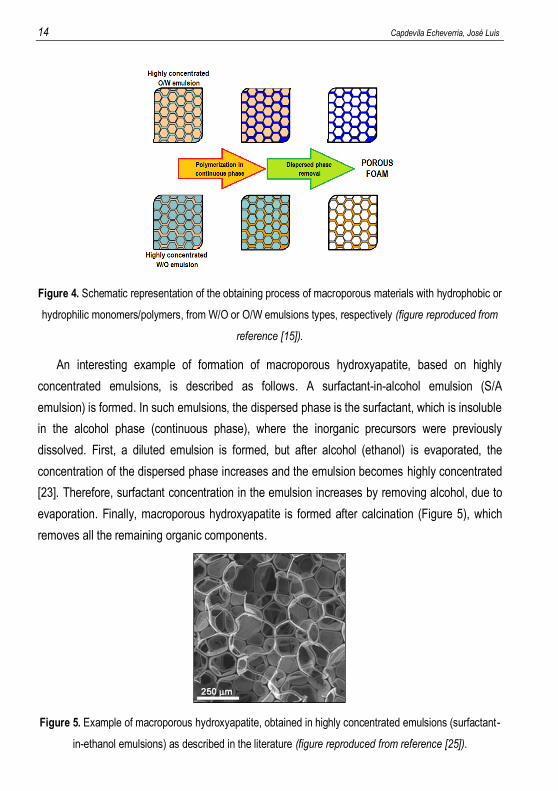
foam (Figure 4).
In the case of sol-gel inorganic precursors, the formation of hydroxyapatite could be carried
out in the external phase, following three steps [20, 21, 22]:
1) Formation of a sol, a colloidal dispersion of solid particles, into the continuous
phase.
2) Gelation of the sol via sol-gel hydrolysis and condensation reactions.
3) Removal of solvent by evaporation.
This method, described by Vallet-Regí, was used to obtain macroporous hydroxyapatite [23,
54]. It involves the evaporation-induced self-assembly (EISA) methodology because the
emulsion is formed after certain amount of solvent is evaporated [23, 24]. This general
methodology will be used for the obtention of the porous hydroxyapatite in the present project.
14 Capdevila Echeverria, José Luis
Figure 4. Schematic representation of the obtaining process of macroporous materials with hydrophobic or
hydrophilic monomers/polymers, from W/O or O/W emulsions types, respectively (figure reproduced from
reference [15]).
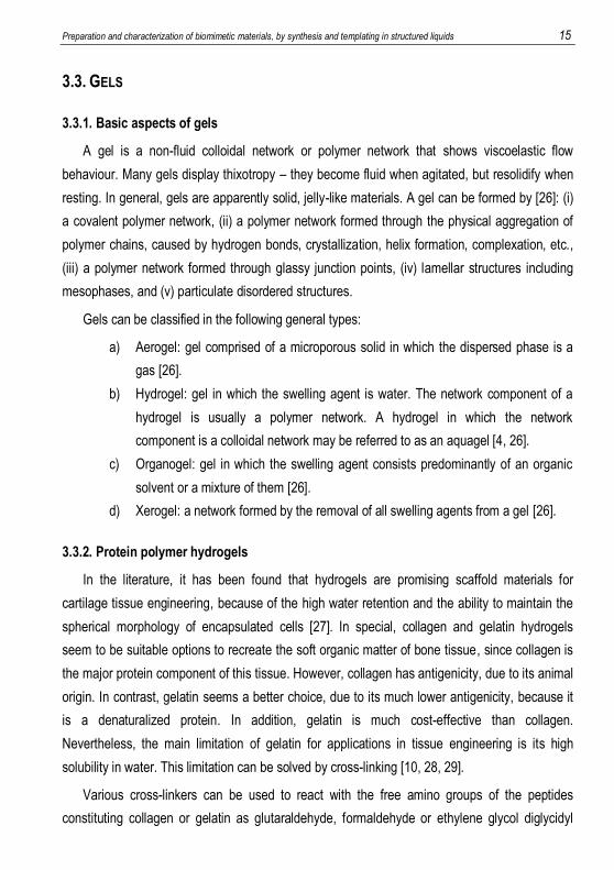
An interesting example of formation of macroporous hydroxyapatite, based on highly
concentrated emulsions, is described as follows. A surfactant-in-alcohol emulsion (S/A
emulsion) is formed. In such emulsions, the dispersed phase is the surfactant, which is insoluble
in the alcohol phase (continuous phase), where the inorganic precursors were previously
dissolved. First, a diluted emulsion is formed, but after alcohol (ethanol) is evaporated, the
concentration of the dispersed phase increases and the emulsion becomes highly concentrated
[23]. Therefore, surfactant concentration in the emulsion increases by removing alcohol, due to
evaporation. Finally, macroporous hydroxyapatite is formed after calcination (Figure 5), which
removes all the remaining organic components.
Figure 5. Example of macroporous hydroxyapatite, obtained in highly concentrated emulsions (surfactant-
in-ethanol emulsions) as described in the literature (figure reproduced from reference [25]).
Preparation and characterization of biomimetic materials, by synthesis and templating in structured liquids 15
3.3. GELS
3.3.1. Basic aspects of gels
A gel is a non-fluid colloidal network or polymer network that shows viscoelastic flow
behaviour. Many gels display thixotropy – they become fluid when agitated, but resolidify when
resting. In general, gels are apparently solid, jelly-like materials. A gel can be formed by [26]: (i)
a covalent polymer network, (ii) a polymer network formed through the physical aggregation of
polymer chains, caused by hydrogen bonds, crystallization, helix formation, complexation, etc.,
(iii) a polymer network formed through glassy junction points, (iv) lamellar structures including
mesophases, and (v) particulate disordered structures.
Gels can be classified in the following general types:
a) Aerogel: gel comprised of a microporous solid in which the dispersed phase is a
gas [26].
b) Hydrogel: gel in which the swelling agent is water. The network component of a
hydrogel is usually a polymer network. A hydrogel in which the network
component is a colloidal network may be referred to as an aquagel [4, 26].
c) Organogel: gel in which the swelling agent consists predominantly of an organic
solvent or a mixture of them [26].
d) Xerogel: a network formed by the removal of all swelling agents from a gel [26].
3.3.2. Protein polymer hydrogels
In the literature, it has been found that hydrogels are promising scaffold materials for
cartilage tissue engineering, because of the high water retention and the ability to maintain the
spherical morphology of encapsulated cells [27]. In special, collagen and gelatin hydrogels
seem to be suitable options to recreate the soft organic matter of bone tissue, since collagen is
the major protein component of this tissue. However, collagen has antigenicity, due to its animal
origin. In contrast, gelatin seems a better choice, due to its much lower antigenicity, because it
is a denaturalized protein. In addition, gelatin is much cost-effective than collagen.
Nevertheless, the main limitation of gelatin for applications in tissue engineering is its high
solubility in water. This limitation can be solved by cross-linking [10, 28, 29].
Various cross-linkers can be used to react with the free amino groups of the peptides
constituting collagen or gelatin as glutaraldehyde, formaldehyde or ethylene glycol diglycidyl
16 Capdevila Echeverria, José Luis
ether [9, 27, 28]. However, these compounds have physiological toxicity. Therefore, in this study
it has been used a naturally occurring cross-linking agent called genipin 1 (Figure 6), obtained
from the fruits of Gardenia jasminoides Ellis, which has toxicity between 5000-10000 times less
than glutaraldehyde [30, 31]. The use of this cross-linker is described in detail in the following
section.
3.3.3. Cross-linking with genipin
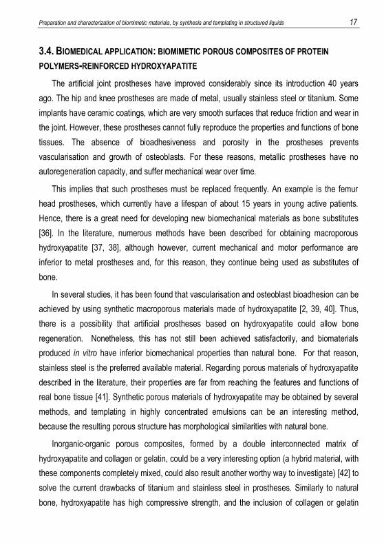
Genipin 1 may react with the primary amino groups of lysine, hydroxylysine or arginine
present in collagen and gelatin, forming new covalent bonds and cross-linking peptide chains, at
ambient conditions and neutral pH. The proposed mechanism [32, 33] involves reacting the
carboxymethyl group of the genipin 1 with the free amino group of lysine, hydroxylysine or
arginine of collagen or gelatin to form a secondary amide and nucleophilic attack of other free
amino groups (but of the same type involved) on an olefinic carbon of genipin 1, producing an
opening of the genipin 1 dihydropyran ring at an intermediate stage, leading to the formation of
a tertiary amide. Several authors [32, 33] have also suggested that dimers and trimers of
genipin 1 are formed during the cross-linking. This process is shown schematically in Figure 6,
in which different bonds between chains of collagen or gelatin, represented as red lines, and the
chemical structure of the cross-linking reaction between genipin 1 and collagen or gelatin are
represented [15, 34, 35]. Dark blue colour is observed in the hydrogel, after cross-linking with
genipin 1.
Figure 6. Schematic representation of cross-linking of collagen, or gelatin, with genipin 1 (figure adapted
from reference [15]).
Preparation and characterization of biomimetic materials, by synthesis and templating in structured liquids 17
3.4. BIOMEDICAL APPLICATION: BIOMIMETIC POROUS COMPOSITES OF PROTEIN
POLYMERS-REINFORCED HYDROXYAPATITE
The artificial joint prostheses have improved considerably since its introduction 40 years
ago. The hip and knee prostheses are made of metal, usually stainless steel or titanium. Some
implants have ceramic coatings, which are very smooth surfaces that reduce friction and wear in
the joint. However, these prostheses cannot fully reproduce the properties and functions of bone
tissues. The absence of bioadhesiveness and porosity in the prostheses prevents
vascularisation and growth of osteoblasts. For these reasons, metallic prostheses have no
autoregeneration capacity, and suffer mechanical wear over time.
This implies that such prostheses must be replaced frequently. An example is the femur
head prostheses, which currently have a lifespan of about 15 years in young active patients.
Hence, there is a great need for developing new biomechanical materials as bone substitutes
[36]. In the literature, numerous methods have been described for obtaining macroporous
hydroxyapatite [37, 38], although however, current mechanical and motor performance are
inferior to metal prostheses and, for this reason, they continue being used as substitutes of
bone.
In several studies, it has been found that vascularisation and osteoblast bioadhesion can be
achieved by using synthetic macroporous materials made of hydroxyapatite [2, 39, 40]. Thus,
there is a possibility that artificial prostheses based on hydroxyapatite could allow bone
regeneration. Nonetheless, this has not still been achieved satisfactorily, and biomaterials
produced in vitro have inferior biomechanical properties than natural bone. For that reason,
stainless steel is the preferred available material. Regarding porous materials of hydroxyapatite
described in the literature, their properties are far from reaching the features and functions of
real bone tissue [41]. Synthetic porous materials of hydroxyapatite may be obtained by several
methods, and templating in highly concentrated emulsions can be an interesting method,
because the resulting porous structure has morphological similarities with natural bone.
Inorganic-organic porous composites, formed by a double interconnected matrix of
hydroxyapatite and collagen or gelatin, could be a very interesting option (a hybrid material, with
these components completely mixed, could also result another worthy way to investigate) [42] to
solve the current drawbacks of titanium and stainless steel in prostheses. Similarly to natural
bone, hydroxyapatite has high compressive strength, and the inclusion of collagen or gelatin
18 Capdevila Echeverria, José Luis
allows to increase fracture strength and toughness. Hence, it could be expected that porous
composites of hydroxyapatite and collagen, or gelatin, possess better biomechanical capabilities
than porous materials consisting solely of hydroxyapatite. For this reason, the present work will
be focused on the study of porous hydroxyapatite and protein hydrogels.
4. OBJECTIVES
Given the importance of finding a suitable material, which biomimics bone tissue and can be
applied in biomedicine to regenerate and replace natural bone, and taking into account the
considerations explained in the introduction, the main objective of this work consisted in the
study, obtention and characterization of macroporous hydroxyapatite and collagen and gelatin
hydrogels.
The work plan is described below:
a) Selection of the most appropriate components: hydroxyapatite precursors, protein
precursors (collagen and gelatin) and components of the emulsions (surfactant
and continuous phase solvent).
b) Preparation and characterization of hydroxyapatite by infrared spectroscopy (IR),
X-ray diffraction (XRD) and scanning electron microscopy (SEM).
c) Preparation of hydrogels of proteinaceous nature (collagen and gelatin) and study
of their rheological properties, as function of the protein precursor, cross-linking
agent, their concentrations, and temperature. As well as qualitative monitoring of
the hydrogels formation and observation of their structure by scanning electron
microscopy (SEM).
d) Feasibility study for obtaining macroporous hydroxyapatite, reinforced with
collagen or gelatin.
Preparation and characterization of biomimetic materials, by synthesis and templating in structured liquids 19
5. EXPERIMENTAL SECTION
5.1. MATERIALS
5.1.1. Synthesis of macroporous hydroxyapatite foams
For the synthesis and cross-linking of hydroxyapatite, the following reagents have been
selected:
a) Triethyl phosphite: 2, P(OCH2CH3)3, 98 wt.%, with a molecular weight of 166.16
g/mol. Supplied by Sigma-Aldrich. See molecule in Figure 7A, 2.
b) Calcium nitrate tetrahydrate: Ca(NO3)2·4H2O, puriss. p.a., ACS reagent, 99-103
wt.%. It has a molecular weight of 236.15 g/mol. Supplied by Sigma-Aldrich.
c) Filtered deionized water: H2O, Milli-Q® water (ultra-pure Millipore water system,
Milli-Qplus 185 filter). Supplied by Merck Millipore.
d) Pluronic® F 127: 3, Poloxamer 407 (prill). It is a non-ionic surfactant (HLB = 18-23)
consisting of a triblock structure composed of ethylene glycol and propylene glycol
units, HO(C2H4O)101(C3H6O)56(C2H4O)101H. It has an average molecular weight of
12600 Da (12600 g/mol). Supplied by BASF. See molecule in Figure 7B, 3.
e) Ethanol: CH3CH2OH, absolute for analysis EMSURE® ACS, ISO, Reag. Ph Eur.
Supplied by Merck Millipore.
Figure 7. Formulas of A) triethyl phosphite 2 and B) Pluronic® F 127 3.
5.1.2. Preparation of protein hydrogels
For the preparation of collagen and gelatin hydrogels, the following reagents have been
selected:
a) Peptan® P 2000 LD: porcine collagen peptides or hydrolyzed collagen with low
density, with a content of type I collagen ≥ 90 wt.%. It has an average molecular
weight of 2000 Da (2000 g/mol). Supplied by Rousselot.
20 Capdevila Echeverria, José Luis
b) Gelatin: from bovine skin, Type B (derived from lime-cured tissue). It is a
heterogeneous mixture of water-soluble proteins of high average molecular
weights, present in collagen (proteins are extracted by boiling skin, tendons,
ligaments, bones, etc). It has an average molecular weight between 40000-50000
Da (40000-50000 g/mol) which correlates with a Bloom number (which is an
indication of the strength of a gel formed from a solution of known concentration
and this is directly proportional to molecular weight) between 175-225 (medium
Bloom). Supplied by Sigma-Aldrich.
c) Genipin: 1, 98 wt.%, with a molecular weight of 226.226 g/mol. It is a natural
cross-linker for protein, collagen, gelatin and chitosan. Supplied by Challenge
Bioproducts Co., Ltd. (Taiwan).
d) Phosphate buffered saline: PBS, in tablets. It provides a pH = 7.4 (physiological
pH), at 25 ºC. It consists of a mixture of Na2HPO4·2H2O (11.88 g/L) and KH2PO4
(9.08 g/L). Supplied by Sigma-Aldrich.
e) Glacial acetic acid: CH3COOH, 100 wt.%, Ph Eur, BP, JP, USP, E 260. It has a
molecular weight of 60.05 g/mol. Supplied by Merck Millipore.
f) Sodium hydroxide: NaOH, BioXtra, ≥ 98 wt.%, pellets (anhydrous). It has a
molecular weight of 40.00 g/mol. Supplied by Sigma-Aldrich.
g) Filtered deionized water: H2O, Milli-Q® water (ultra-pure Millipore water system,
Milli-Qplus 185 filter). Supplied by Merck Millipore.
5.2. APPARATUS AND INSTRUMENTAL
The equipment and instruments are listed below:
- Mettler Toledo AB204-S/FACT analytical balance with a precision of ±10-4 g
(maximum capacity: 220 g).
- Sartorius CPA3202-S top-loading balance with a precision of ±10-2 g (maximum
capacity: 3000 g).
- Heidolph MR Hei-Standard magnetic stirrer hot plate, with temperature controlled
by means of Heidolph EKT3001 probe (maximum stirring capacity: 2500 rpm).
- IKA VORTEX 3 (vortex shaker).
Preparation and characterization of biomimetic materials, by synthesis and templating in structured liquids 21
- Vibromatric vibratory shaker, JP SELECTA, S.A.
- Memmert Waterbath WNB 7-45 (shaking bath with temperature control).
- Burdinola V21 Space ST 1500 fume hood.
- MBRAUN MB GB-2202-S glove box.
- Mettler Toledo Seven Easy pH-meter.
- HERAEUS T6 heating and drying oven (maximum temperature: 250 ºC).
- KOTTERMANN 2712 heating and drying oven (maximum temperature: 250 ºC).
- GHA 12/450 CARBOLITE horizontal tube furnace with compressed air flow
(maximum temperature: 1200 ºC).
- Liebherr GX823 freezer (minimum temperature: -32 ºC).
- Christ Alpha 2-4 LD Plus lyophiliser, with pressure and temperature of ~0.03 mbar
and -85 ºC, respectively.
- AR-G2 strain-controlled rheometer (TA Instruments).
- Hitachi TM-1000 Tabletop Microscope (Scanning Electron Microscopy, SEM).
- Nicolet Avatar 360 FT-IR spectrophotometer.
- Bruker D8 A25 Advance X-ray diffractometer with monochromatic Cu-K1 and
Cu-K2 radiation (=1.5405 Å), primary parallel X-ray beam generated by a
Göbbel mirror and the scattered beam analyzed by a lineal detector with 90-
position sample loader.
5.3. METHODS AND PROCEDURES
5.3.1. Synthesis of macroporous hydroxyapatite foams
The 3D-macroporous HAp foams have been synthesized by the sol-gel technique [20, 21,
22, 37, 43] including a non-ionic surfactant, Pluronic® F 127 3, as macropore inducer in the
accelerated evaporation-induced self-assembly (EISA) method.
Aqueous sols were prepared hydrolysing triethyl phosphite (TIP) 2 with Milli-Q® water for 24
h with magnetic stirring and adding it onto an ethanol mixture of non-ionic surfactant Pluronic® F
127 3. It is important to respect the hydrolysis reaction time to avoid uncompleted hydrolysed
intermediates, i.e., diethyl phosphite or monoethyl phosphite, since this reduces further cross-
linking (see Figure 8 for hydrolysis and polycondensation processes). Molar ratios of 0.06, 0.11,
0.44, and 11.0 of F127/TIP have been tested in order to obtain different degrees of
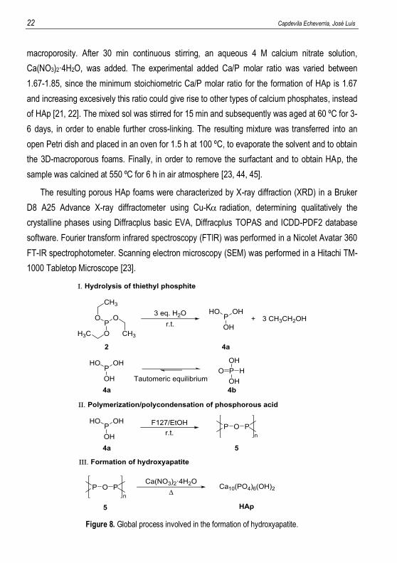
22 Capdevila Echeverria, José Luis
macroporosity. After 30 min continuous stirring, an aqueous 4 M calcium nitrate solution,
Ca(NO3)2·4H2O, was added. The experimental added Ca/P molar ratio was varied between
1.67-1.85, since the minimum stoichiometric Ca/P molar ratio for the formation of HAp is 1.67
and increasing excesively this ratio could give rise to other types of calcium phosphates, instead
of HAp [21, 22]. The mixed sol was stirred for 15 min and subsequently was aged at 60 ºC for 3-
6 days, in order to enable further cross-linking. The resulting mixture was transferred into an
open Petri dish and placed in an oven for 1.5 h at 100 ºC, to evaporate the solvent and to obtain
the 3D-macroporous foams. Finally, in order to remove the surfactant and to obtain HAp, the
sample was calcined at 550 ºC for 6 h in air atmosphere [23, 44, 45].
The resulting porous HAp foams were characterized by X-ray diffraction (XRD) in a Bruker
D8 A25 Advance X-ray diffractometer using Cu-Kradiation, determining qualitatively the
crystalline phases using Diffracplus basic EVA, Diffracplus TOPAS and ICDD-PDF2 database
software. Fourier transform infrared spectroscopy (FTIR) was performed in a Nicolet Avatar 360
FT-IR spectrophotometer. Scanning electron microscopy (SEM) was performed in a Hitachi TM-
1000 Tabletop Microscope [23].
Figure 8. Global process involved in the formation of hydroxyapatite.
Preparation and characterization of biomimetic materials, by synthesis and templating in structured liquids 23
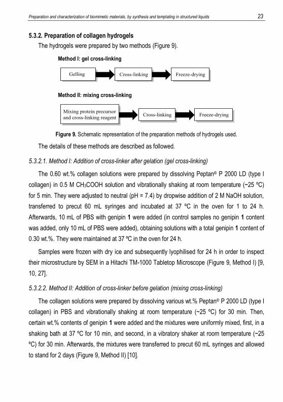
5.3.2. Preparation of collagen hydrogels
The hydrogels were prepared by two methods (Figure 9).
Figure 9. Schematic representation of the preparation methods of hydrogels used.
The details of these methods are described as followed.
5.3.2.1. Method I: Addition of cross-linker after gelation (gel cross-linking)
The 0.60 wt.% collagen solutions were prepared by dissolving Peptan® P 2000 LD (type I
collagen) in 0.5 M CH3COOH solution and vibrationally shaking at room temperature (~25 ºC)
for 5 min. They were adjusted to neutral (pH = 7.4) by dropwise addition of 2 M NaOH solution,
transferred to precut 60 mL syringes and incubated at 37 ºC in the oven for 1 to 24 h.
Afterwards, 10 mL of PBS with genipin 1 were added (in control samples no genipin 1 content
was added, only 10 mL of PBS were added), obtaining solutions with a total genipin 1 content of
0.30 wt.%. They were maintained at 37 ºC in the oven for 24 h.
Samples were frozen with dry ice and subsequently lyophilised for 24 h in order to inspect
their microstructure by SEM in a Hitachi TM-1000 Tabletop Microscope (Figure 9, Method I) [9,
10, 27].
5.3.2.2. Method II: Addition of cross-linker before gelation (mixing cross-linking)
The collagen solutions were prepared by dissolving various wt.% Peptan® P 2000 LD (type I
collagen) in PBS and vibrationally shaking at room temperature (~25 ºC) for 30 min. Then,
certain wt.% contents of genipin 1 were added and the mixtures were uniformly mixed, first, in a
shaking bath at 37 ºC for 10 min, and second, in a vibratory shaker at room temperature (~25
ºC) for 30 min. Afterwards, the mixtures were transferred to precut 60 mL syringes and allowed
to stand for 2 days (Figure 9, Method II) [10].
24 Capdevila Echeverria, José Luis
5.3.3. Preparation of gelatin hydrogels
The gelatin stock solutions were prepared by dissolving gelatin (from bovine skin, type B,
175-225 Bloom) in PBS and magnetically stirring at 50 ºC for 24 h. The gelatin contents were 2,
6, and 10 wt.%. For these final gelatin concentrations, 4, 12, and 20 wt.% gelatin stock solutions
were required, respectively. Genipin 1 stock solutions were prepared at different concentrations
(1 and 0.25 wt.% in order to obtain final genipin 1 concentrations of 0.5 and 0.125 wt.%,
respectively) by dissolving genipin 1 in phosphate buffer, at pH 7.4, by magnetically stirring for 1
h at room temperature (~25 ºC). The reaction of gelatin and genipin 1 occurred after mixing both
solutions at a ratio of 1:1 (w/w) inside the precut 60 mL syringes [10, 11]. Mixed solutions were
allowed to gel for 24 h at 25 ºC [28, 29, 30].
The rheological characterization was performed on freshly synthesized hydrogels using an
AR-G2 strain-controlled rheometer (TA Instruments) at controlled temperature of 37 ºC. The
geometry was plate-plate (standard steel parallel plates) of 40 mm diameter and the gap used
was 2000 m. Fresh samples were frozen at -32 ºC (Liebherr GX823 freezer) for 24 h and
subsequently lyophilised (Christ Alpha 2-4 LD Plus lyophiliser) for 24 h (freeze-drying step) in
order to observe their microstructure by means of a scanning electron microscopy (Hitachi TM-
1000 Tabletop Microscope) (Figure 9, Method II) [28, 29, 35, 42].
5.3.4. Rheological study of hydrogels
The rheological study of the hydrogels was performed using dynamic oscillatory tests. In
these tests, a sinusoidal stress () with amplitude is applied and a strain () with amplitude
is measured simultaneously (Figure 10). Thus, a time shift (t) between the amplitudes of
stress and strain, at a certain frequency (), occurs. The phase angle () is calculated from the
time shift (t) and frequency (), following the relationship shown in Equation 2.
𝛿 = 𝜔∆𝑡 Equation 2
The phase angle indicates the nature of the system: = 0º (elastic solid), = 90º (viscous
liquid), and 0º < < 90º (viscoelastic system).
Preparation and characterization of biomimetic materials, by synthesis and templating in structured liquids 25
Figure 10. Sinusoidal curves for an applied oscillatory stress and its response in the form of a strain in a viscoelastic system (figure reproduced from reference [15]).
From , , and , the following rheological parameters are obtained: complex modulus,
|G*| (it measures the resistance of the material to be deformed, see Equation 3), elastic or
storage modulus, G’ (it measures the elasticity of the material, which means the ability of the
material to store energy, see Equation 4), and viscous or loss modulus, G’’ (it represents the
ability of the material to dissipate energy as heat, see Equation 5). The relation between elastic
and viscous moduli is called tangent (it measures the energy dissipated in relation to the
stored one, see Equation 6). This parameter indicates the viscoelastic behaviour of a material.
The greater the elasticity, the smaller the tangent [30].
|𝐺∗| =𝜎0
𝛾0⁄ Equation 3
𝐺′ = |𝐺∗| cos 𝛿 Equation 4
𝐺′′ = |𝐺∗| sin𝛿 Equation 5
tan 𝛿 = 𝐺′′
𝐺′⁄ Equation 6
Such oscillatory tests can be divided into two categories: strain or frequency oscillatory
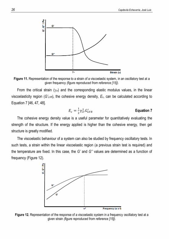
tests. In the strain tests, the frequency (, rad/s or f, Hz being = f) and temperature are
fixed and a strain sweep is performed whilst G’ and G’’ values are recorded. Generally, two
distinct regions are detected based on the values of G’ and G’’ (Figure 11). The first region,
known as linear viscoelastic region (LVR), is where G’ remains virtually constant and
independent of the applied strain. The second region is observed above a certain critical strain
value, cr, in which the G’ and G’’ values show a nonlinear behaviour and G’ decreases with
increasing strain. This critical value indicates the strain value where the structure of the material
begins to be irreversibly fractured.
26 Capdevila Echeverria, José Luis
Figure 11. Representation of the response to a strain of a viscoelastic system, in an oscillatory test at a given frequency (figure reproduced from reference [15]).
From the critical strain (cr) and the corresponding elastic modulus values, in the linear
viscoelasticity region (G’LVR), the cohesive energy density, Ec, can be calculated according to
Equation 7 [46, 47, 48].
𝐸𝑐 =1
2𝛾𝑐𝑟2 𝐺𝐿𝑉𝑅
′ Equation 7
The cohesive energy density value is a useful parameter for quantitatively evaluating the
strength of the structure. If the energy applied is higher than the cohesive energy, then gel
structure is greatly modified.
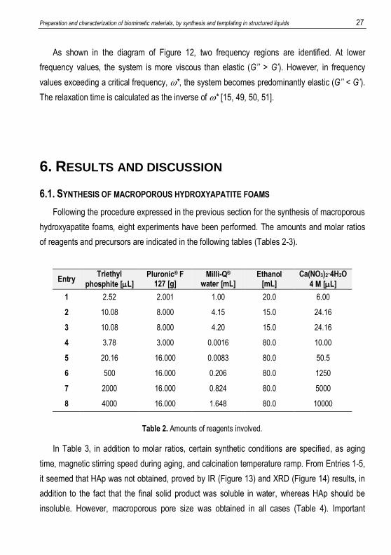
The viscoelastic behaviour of a system can also be studied by frequency oscillatory tests. In
such tests, a strain within the linear viscoelastic region (a previous strain test is required) and
the temperature are fixed. In this case, the G’ and G’’ values are determined as a function of
frequency (Figure 12).
Figure 12. Representation of the response of a viscoelastic system in a frequency oscillatory test at a given strain (figure reproduced from reference [15]).
Preparation and characterization of biomimetic materials, by synthesis and templating in structured liquids 27
As shown in the diagram of Figure 12, two frequency regions are identified. At lower
frequency values, the system is more viscous than elastic (G’’ > G’). However, in frequency
values exceeding a critical frequency, *, the system becomes predominantly elastic (G’’ < G’).
The relaxation time is calculated as the inverse of * [15, 49, 50, 51].
6. RESULTS AND DISCUSSION
6.1. SYNTHESIS OF MACROPOROUS HYDROXYAPATITE FOAMS
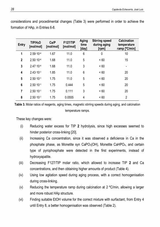
Following the procedure expressed in the previous section for the synthesis of macroporous
hydroxyapatite foams, eight experiments have been performed. The amounts and molar ratios
of reagents and precursors are indicated in the following tables (Tables 2-3).
Entry Triethyl
phosphite [L] Pluronic® F
127 [g] Milli-Q®
water [mL] Ethanol
[mL] Ca(NO3)2·4H2O
4 M [L]
1 2.52 2.001 1.00 20.0 6.00
2 10.08 8.000 4.15 15.0 24.16
3 10.08 8.000 4.20 15.0 24.16
4 3.78 3.000 0.0016 80.0 10.00
5 20.16 16.000 0.0083 80.0 50.5
6 500 16.000 0.206 80.0 1250
7 2000 16.000 0.824 80.0 5000
8 4000 16.000 1.648 80.0 10000
Table 2. Amounts of reagents involved.
In Table 3, in addition to molar ratios, certain synthetic conditions are specified, as aging
time, magnetic stirring speed during aging, and calcination temperature ramp. From Entries 1-5,
it seemed that HAp was not obtained, proved by IR (Figure 13) and XRD (Figure 14) results, in
addition to the fact that the final solid product was soluble in water, whereas HAp should be
insoluble. However, macroporous pore size was obtained in all cases (Table 4). Important
28 Capdevila Echeverria, José Luis
considerations and procedimental changes (Table 3) were performed in order to achieve the
formation of HAp, in Entries 6-8.
Entry TIP/H2O
[mol/mol] Ca/P
[mol/mol] F127/TIP [mol/mol]
Aging time [day]
Stirring speed during aging
[rpm]
Calcination temperature
ramp [ºC/min]
1 2.59·10-4 1.67 11.0 6 0 10
2 2.50·10-4 1.68 11.0 5 < 60 15
3 2.47·10-4 1.68 11.0 3 < 60 -
4 2.43·10-1 1.85 11.0 6 < 60 20
5 2.50·10-1 1.75 11.0 5 < 60 20
6 2.50·10-1 1.75 0.444 5 < 60 20
7 2.50·10-1 1.75 0.111 3 < 60 20
8 2.50·10-1 1.75 0.0555 4 < 60 2
Table 3. Molar ratios of reagents, aging times, magnetic stirring speeds during aging, and calcination
temperature ramps.
These key changes were:
(i) Reducing water excess for TIP 2 hydrolysis, since high excesses seemed to
hinder posterior cross-linking [20].
(ii) Increasing Ca concentration, since it was observed a deficience in Ca in the
phosphate phase, as Monetite syn CaPO3(OH), Monetite CaHPO4, and certain
type of pyrophosphate were detected in the first experiments, instead of
hydroxyapatite.
(iii) Decreasing F127/TIP molar ratio, which allowed to increase TIP 2 and Ca
concentrations, and then obtaining higher amounts of product (Table 4).
(iv) Using low agitation speed during aging process, with a correct homogenisation
during cross-linking.
(v) Reducing the temperature ramp during calcination at 2 ºC/min, allowing a larger
and more robust HAp structure.
(vi) Finding suitable EtOH volume for the correct mixture with surfactant, from Entry 4
until Entry 8, a better homogenisation was observed (Table 2).
Preparation and characterization of biomimetic materials, by synthesis and templating in structured liquids 29
Among these changes, point (iii) was remarkably important and the results were the
opposite as mentioned in certain literature [23], which recommended a F127/TIP molar ratio of
11 in order to obtain suitable macroporosity. As observed in Table 4, macroporous pore sizes
were formed with a F127/TIP molar ratio less than 1. An aging time between 3-6 days was
found to be suitable for cross-linking.
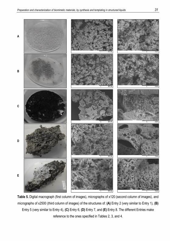
Complementing the information described in Table 4, different types of macroscopic and
microscopic appearances of the products are illustrated in Table 5. Among experiments of
Entries 6-8, which resulted in HAp formation, Entries 7 and 8 had more uniform pores, with
shapes approximately circular, and also with good interconnectivities.
Entry Product
weight [mg] Macroscopic appearance
Soluble in water?
Pore size (SEM) [nm](a)
1 4.6 Porous light brown powder Yes 130-2400,
macroporous(b)
2 27.7 Porous gray powder Yes 200-4000,
macroporous(b)
3 - - Yes (intermediate
raw product) -
4 10.1 Porous brown and gray particles Yes 220-3800,
macroporous(c)
5 41.8 Porous dark gray particles Yes 400-5700,
macroporous(c)
6 311.4 Porous dark gray structures No 100-3600,
macroporous(d)
7 1613.6 Porous gray-white structures No 130-7100,
macroporous(e)
8 3888.0 Porous gray-white structures No 250-7100,
macroporous(e)
(a) Pore size measured as pore diameter by scanning electron microscopy (SEM). (b) Microscopic appearance: amorphous. (c) Microscopic appearance: particulated. (d) Microscopic appearance: cracked. (e) Microscopic appearance: approximately circular.
Table 4. Weight, appearance, water solubility, and pore size (measured as pore diameter).
30 Capdevila Echeverria, José Luis
Among Entries 6-8, it seemed that lower F127/TIP molar ratio produced better macroporous
HAp. Besides, the lowest calcination temperature ramp allowed to obtain the largest and the
most robust HAp structures (Table 3). Lower temperature ramp seemed to produce weaker
mechanical tensions between pores during calcination, in such a way that macrostructure was
less affected and less fractured. Trace amounts of retained solvents, EtOH or H2O, could also
participate in the fracture of the structure when they were rapidly removed during calcination.
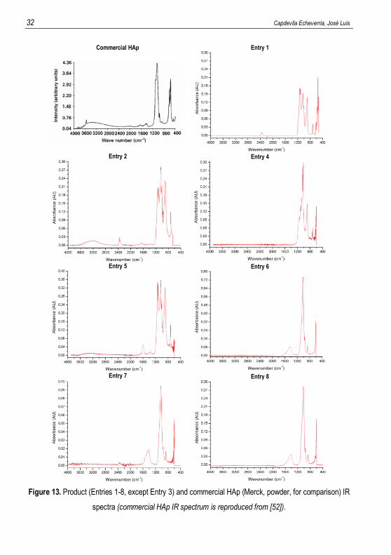
In Figure 13, it is observed that only IR spectra of Entries 6-8 correspond to HAp, when
comparing to IR spectrum of the commercial HAp, Entry 1. Absorption bands located at 460 and
960 cm-1 are stretching modes of PO43-, and the bands located at 560-600 and 1020 cm-1
correspond to bending modes of PO43-. A characteristical OH- stretch is observed at 3569 cm-1
in commercial HAp, but in synthetic HAp is not observed due to CO32- substitution (CO32- can
also substitute PO43-), which is difficult to eliminate at the calcination step. In this regard, it is
observed a growth in carbonate bands (870, 1400-1450, and 1600-1650 cm-1) in detrimental to
hydroxyl or phosphate ones in the synthesized HAp. This kind of HAp is called as B-type HAp.
In some spectra, some adsorbed water was detected, 2600-3600 cm-1 (absorption band
becomes narrower under influence of thermal treatment) [52, 53, 54].
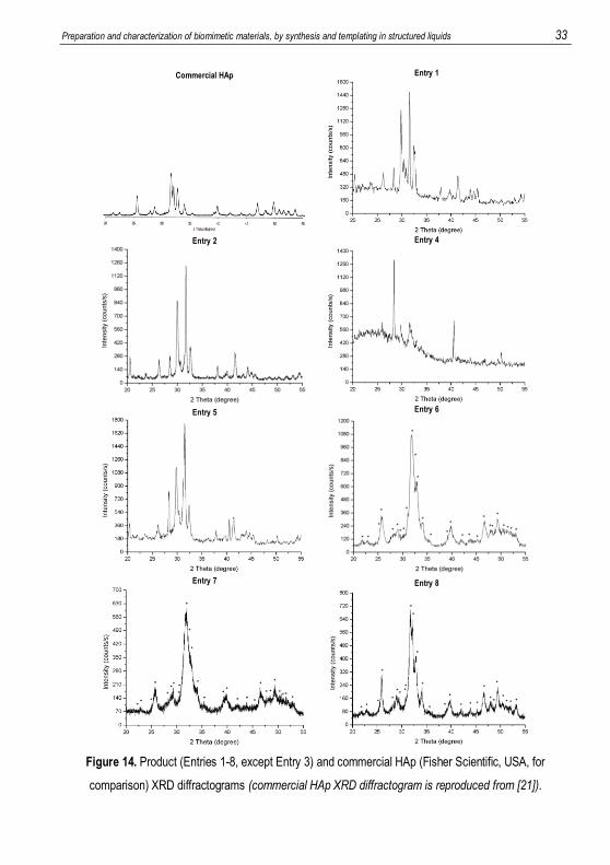
By means of XRD, the HAp identification was clearer than by IR for Entries 6-8, as it can be
observed in Figure 14, if they are compared with the commercial HAp diffractogram. The other
diffractograms were found not to be HAp, but other crystalline phosphate phases, as Monetite
syn, CaPO3(OH); Monetite, CaHPO4; and an unidentified pyrophosphate, which were soluble in
water.
Preparation and characterization of biomimetic materials, by synthesis and templating in structured liquids 31
A
B
C
D
E
Table 5. Digital macrograph (first column of images), micrographs of x120 (second column of images), and
micrographs of x2500 (third column of images) of the structures of: (A) Entry 2 (very similar to Entry 1), (B)
Entry 5 (very similar to Entry 4), (C) Entry 6, (D) Entry 7, and (E) Entry 8. The different Entries make
reference to the ones specified in Tables 2, 3, and 4.
32 Capdevila Echeverria, José Luis
Commercial HAp
Entry 1
Entry 2
Entry 4
Entry 5
Entry 6
Entry 7
Entry 8
Figure 13. Product (Entries 1-8, except Entry 3) and commercial HAp (Merck, powder, for comparison) IR
spectra (commercial HAp IR spectrum is reproduced from [52]).
Preparation and characterization of biomimetic materials, by synthesis and templating in structured liquids 33
Commercial HAp
Entry 1
Entry 2
Entry 4
Entry 5
Entry 6
Entry 7
Entry 8
Figure 14. Product (Entries 1-8, except Entry 3) and commercial HAp (Fisher Scientific, USA, for
comparison) XRD diffractograms (commercial HAp XRD diffractogram is reproduced from [21]).
34 Capdevila Echeverria, José Luis
6.2. PREPARATION OF PROTEIN HYDROGELS
6.2.1. Preparation of collagen hydrogels
6.2.1.1. Method I: Addition of cross-linker after gelation (gel cross-linking)
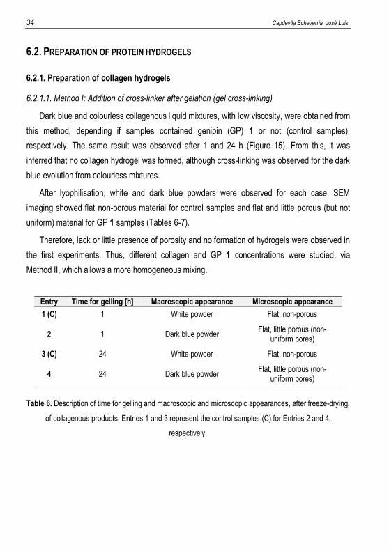
Dark blue and colourless collagenous liquid mixtures, with low viscosity, were obtained from
this method, depending if samples contained genipin (GP) 1 or not (control samples),
respectively. The same result was observed after 1 and 24 h (Figure 15). From this, it was
inferred that no collagen hydrogel was formed, although cross-linking was observed for the dark
blue evolution from colourless mixtures.
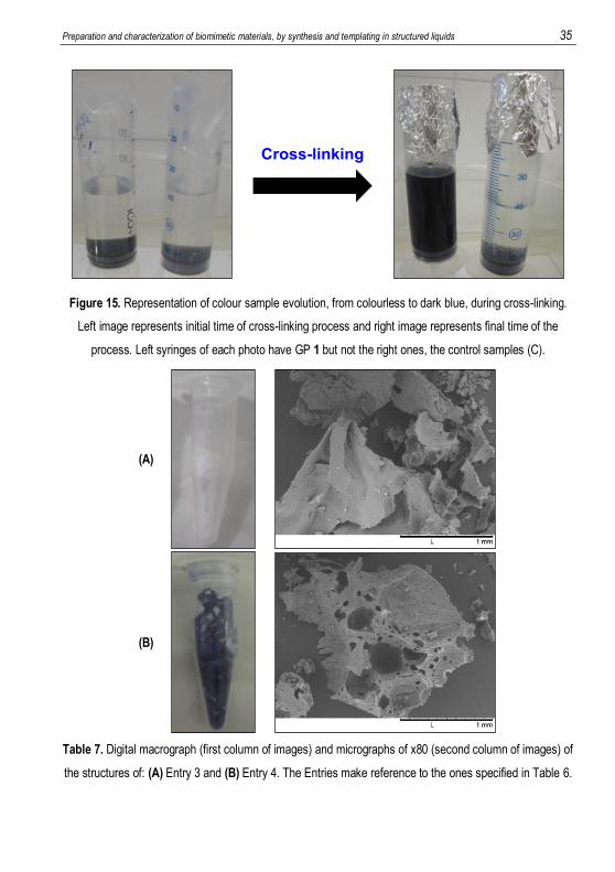
After lyophilisation, white and dark blue powders were observed for each case. SEM
imaging showed flat non-porous material for control samples and flat and little porous (but not
uniform) material for GP 1 samples (Tables 6-7).
Therefore, lack or little presence of porosity and no formation of hydrogels were observed in
the first experiments. Thus, different collagen and GP 1 concentrations were studied, via
Method II, which allows a more homogeneous mixing.
Entry Time for gelling [h] Macroscopic appearance Microscopic appearance
1 (C) 1 White powder Flat, non-porous
2 1 Dark blue powder Flat, little porous (non-
uniform pores)
3 (C) 24 White powder Flat, non-porous
4 24 Dark blue powder Flat, little porous (non-
uniform pores)
Table 6. Description of time for gelling and macroscopic and microscopic appearances, after freeze-drying,
of collagenous products. Entries 1 and 3 represent the control samples (C) for Entries 2 and 4,
respectively.
Preparation and characterization of biomimetic materials, by synthesis and templating in structured liquids 35
Figure 15. Representation of colour sample evolution, from colourless to dark blue, during cross-linking.
Left image represents initial time of cross-linking process and right image represents final time of the
process. Left syringes of each photo have GP 1 but not the right ones, the control samples (C).
(A)
(B)
Table 7. Digital macrograph (first column of images) and micrographs of x80 (second column of images) of
the structures of: (A) Entry 3 and (B) Entry 4. The Entries make reference to the ones specified in Table 6.
36 Capdevila Echeverria, José Luis
6.2.1.2. Method II: Addition of cross-linker before gelation (mixing cross-linking)
Firstly, Peptan® (collagen) concentration dependence was studied with GP 1 concentration
fixed to 0.5 wt.% (Table 8). From this study, no hydrogel formation was observed. An increase
in viscosity was observed with increasing Peptan® concentration, but gelation did not occur.
Entry Peptan® [wt.%] Genipin [wt.%] Entry Peptan® [wt.%] Genipin [wt.%]
1 (C) 8 0 10 40 0.5
2 4 0.5 11 45 0.5
3 8 0.5 12 50 0.5
4 12 0.5 13 52 0.5
5 16 0.5 14 54 0.5
6 20 0.5 15 56 0.5
7 25 0.5 16 58 0.5
8 30 0.5 17 60 0.5
9 35 0.5 18 62 0.5
Table 8. Peptan® (not fixed) and GP 1 (fixed) concentrations tested in Method II with collagen. (C) means
control sample, which did not contain GP 1.
Secondly, GP 1 concentration dependence was studied with Peptan® (collagen)
concentration fixed constant at 30 wt.%. This Peptan® concentration presented enough viscosity
in the previous study and it was considered that higher cross-linker concentration would give
rise to hydrogel formation (Table 9). However, hydrogel formation was unsuccessful again,
despite an increase in viscosity induced by higher GP 1 concentration.
Entry Peptan® [wt.%] Genipin [wt.%]
1 (C) 30 0
2 30 3
3 30 6
4 30 9
5 30 12
6 30 15
Table 9. Peptan® (fixed) and GP 1 (not fixed) concentrations tested in Method II with collagen. (C) means
control sample, which did not contain GP 1.
Preparation and characterization of biomimetic materials, by synthesis and templating in structured liquids 37
In all results, up to this point, gelation was not observed, in spite of the cross-linking
reaction, which was indicated by the appearance of dark blue colour. This absence of gelation
was attributed to the low molecular weight of the collagen (2000 Da). It could be presumed that
short collagen chains could not form the polymer network required for hydrogel formation.
Therefore, the next experiments were performed with gelatin with a much higher molecular
weight (40000-50000 Da). It should be mentioned that gelatin and collagen are both similar
proteins, but the gelatin tested posessed a much higher molecular weight than the available
collagen.
6.2.2. Preparation of gelatin hydrogels
The formation of gelatin hydrogels was studied, and successful results were obtained. Table
10 shows the colour development and the gelling effect. After 24 h, hydrogels were observed in
all cases, although little gelation was detected in Entry 1. From Table 10, it is important to
remark that control hydrogel samples (Entries 1-3), which did not contain GP 1, took longer to
gel than samples with GP 1 (Entries 4-9). In absence of GP 1, only intermolecular ionic forces
and/or hydrogen bonding and Van der Waals interactions are present, in non cross-linked
structures. However, cross-liking occurs in presence of GP 1, and therefore covalent bonding
between gelatin chains is produced, giving rise to a more elastic structure. Reference samples
did not change the initial orange colour of gelatin, whereas cross-linked samples with GP 1
turned dark blue after 24 h, because of the reaction with GP 1 (Figure 16). Moreover, it was also
observed that the higher the GP 1 concentration, the faster the gelation. This was related to
higher cross-linking with higher GP 1 contents. In all cases, a quicker gelation was observed
and more resistant hydrogels were formed with higher concentrations of gelatin; this seemed to
be caused by an increase of intermolecular interactions between peptide chains.
Macroscopic aspect after 24 h can be observed in Figure 17. Control samples were orange
and gelatin-GP samples were dark blue. Entry 1 was so little gelled that it was more like a liquid
than a solid. This type of sample was used for the rheological study, which will be described at
the end of this section.
38 Capdevila Echeverria, José Luis
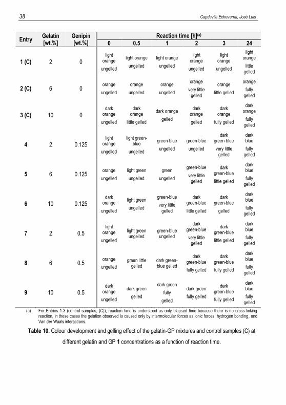
Entry Gelatin [wt.%]
Genipin [wt.%]
Reaction time [h](a)
0 0.5 1 2 3 24
1 (C) 2 0 light
orange
ungelled
light orange
ungelled
light orange
ungelled
light orange
ungelled
light orange
ungelled
light orange
little gelled
2 (C) 6 0 orange
ungelled
orange
ungelled
orange
ungelled
orange
very little gelled
orange
little gelled
orange
fully gelled
3 (C) 10 0 dark
orange
ungelled
dark orange
little gelled
dark orange
gelled
dark orange
gelled
dark orange
fully gelled
dark orange
fully gelled
4 2 0.125 light
orange
ungelled
light green-blue
ungelled
green-blue
ungelled
green-blue
ungelled
dark green-blue
very little gelled
dark blue
fully gelled
5 6 0.125 orange
ungelled
light green
ungelled
green
ungelled
green-blue
very little gelled
dark green-blue
little gelled
dark blue
fully gelled
6 10 0.125 dark
orange
ungelled
light green
ungelled
green-blue
very little gelled
dark green-blue
little gelled
dark green-blue
gelled
dark blue
fully gelled
7 2 0.5 light
orange
ungelled
light green ungelled
green-blue ungelled
dark green-blue
very little gelled
dark green-blue
little gelled
dark blue
fully gelled
8 6 0.5 orange
ungelled
green little gelled
dark green-blue gelled
dark green-blue
fully gelled
dark green-blue
fully gelled
dark blue
fully gelled
9 10 0.5 dark
orange
ungelled
dark green
gelled
dark green
fully
gelled
dark green
fully gelled
dark green-blue
fully gelled
dark blue
fully gelled
(a) For Entries 1-3 (control samples, (C)), reaction time is understood as only elapsed time because there is no cross-linking reaction, in these cases the gelation observed is caused only by intermolecular forces as ionic forces, hydrogen bonding, and Van der Waals interactions.
Table 10. Colour development and gelling effect of the gelatin-GP mixtures and control samples (C) at
different gelatin and GP 1 concentrations as a function of reaction time.
Preparation and characterization of biomimetic materials, by synthesis and templating in structured liquids 39
0 h
0.5 h
1 h
2 h
3 h
24 h
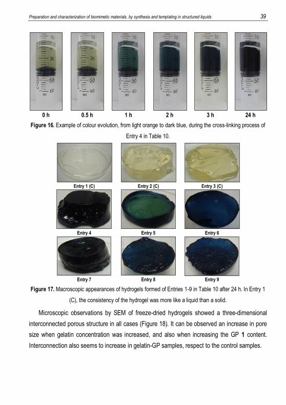
Figure 16. Example of colour evolution, from light orange to dark blue, during the cross-linking process of
Entry 4 in Table 10.
Entry 1 (C)
Entry 2 (C)
Entry 3 (C)
Entry 4
Entry 5
Entry 6
Entry 7
Entry 8
Entry 9
Figure 17. Macroscopic appearances of hydrogels formed of Entries 1-9 in Table 10 after 24 h. In Entry 1
(C), the consistency of the hydrogel was more like a liquid than a solid.
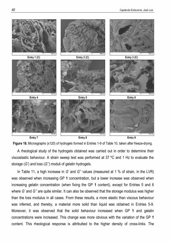
Microscopic observations by SEM of freeze-dried hydrogels showed a three-dimensional
interconnected porous structure in all cases (Figure 18). It can be observed an increase in pore
size when gelatin concentration was increased, and also when increasing the GP 1 content.
Interconnection also seems to increase in gelatin-GP samples, respect to the control samples.
40 Capdevila Echeverria, José Luis
Entry 1 (C)
Entry 2 (C)
Entry 3 (C)
Entry 4
Entry 5
Entry 6
Entry 7
Entry 8
Entry 9
Figure 18. Microsgraphs (x120) of hydrogels formed in Entries 1-9 of Table 10, taken after freeze-drying.
A rheological study of the hydrogels obtained was carried out in order to determine their
viscoelastic behaviour. A strain sweep test was performed at 37 ºC and 1 Hz to evaluate the
storage (G’) and loss (G’’) moduli of gelatin hydrogels.
In Table 11, a high increase in G’ and G’’ values (measured at 1 % of strain, in the LVR)
was observed when increasing GP 1 concentration, but a lower increase was observed when
increasing gelatin concentration (when fixing the GP 1 content), except for Entries 5 and 6
where G’ and G’’ are quite similar. It can also be observed that the storage modulus was higher
than the loss modulus in all cases. From these results, a more elastic than viscous behaviour
was inferred, and thereby, a material more solid than liquid was obtained in Entries 5-9.
Moreover, it was observed that the solid behaviour increased when GP 1 and gelatin
concentrations were increased. This change was more obvious with the variation of the GP 1
content. This rheological response is attributted to the higher density of cross-links. The
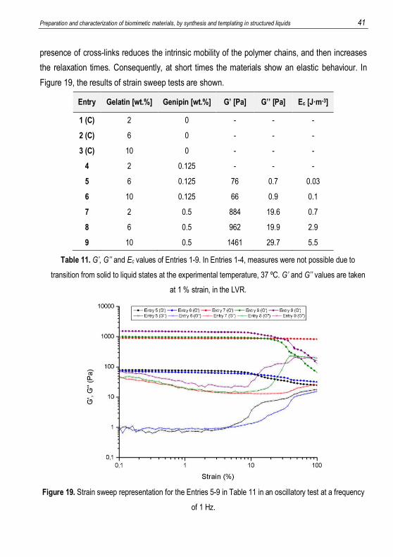
Preparation and characterization of biomimetic materials, by synthesis and templating in structured liquids 41
presence of cross-links reduces the intrinsic mobility of the polymer chains, and then increases
the relaxation times. Consequently, at short times the materials show an elastic behaviour. In
Figure 19, the results of strain sweep tests are shown.
Entry Gelatin [wt.%] Genipin [wt.%] G’ [Pa] G’’ [Pa] Ec [J·m-3]
1 (C) 2 0 - - -
2 (C) 6 0 - - -
3 (C) 10 0 - - -
4 2 0.125 - - -
5 6 0.125 76 0.7 0.03
6 10 0.125 66 0.9 0.1
7 2 0.5 884 19.6 0.7
8 6 0.5 962 19.9 2.9
9 10 0.5 1461 29.7 5.5
Table 11. G’, G’’ and Ec values of Entries 1-9. In Entries 1-4, measures were not possible due to
transition from solid to liquid states at the experimental temperature, 37 ºC. G’ and G’’ values are taken
at 1 % strain, in the LVR.
Figure 19. Strain sweep representation for the Entries 5-9 in Table 11 in an oscillatory test at a frequency
of 1 Hz.
42 Capdevila Echeverria, José Luis
For Entries 1-4, rheological determinations could not be performed due to the transition from
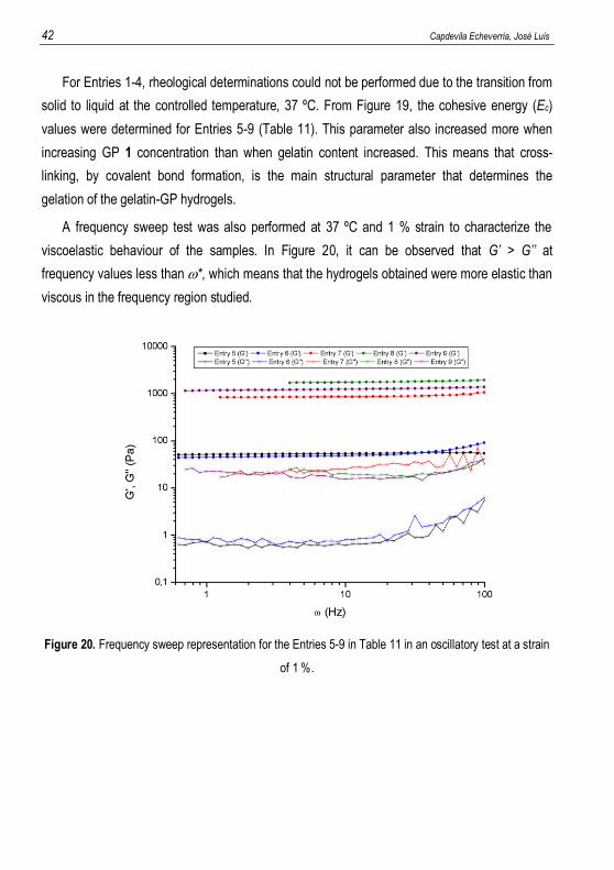
solid to liquid at the controlled temperature, 37 ºC. From Figure 19, the cohesive energy (Ec)
values were determined for Entries 5-9 (Table 11). This parameter also increased more when
increasing GP 1 concentration than when gelatin content increased. This means that cross-
linking, by covalent bond formation, is the main structural parameter that determines the
gelation of the gelatin-GP hydrogels.
A frequency sweep test was also performed at 37 ºC and 1 % strain to characterize the
viscoelastic behaviour of the samples. In Figure 20, it can be observed that G’ > G’’ at
frequency values less than *, which means that the hydrogels obtained were more elastic than
viscous in the frequency region studied.
Figure 20. Frequency sweep representation for the Entries 5-9 in Table 11 in an oscillatory test at a strain
of 1 %.
Preparation and characterization of biomimetic materials, by synthesis and templating in structured liquids 43
6.3. FEASIBILITY STUDY FOR OBTAINING MACROPOROUS HYDROXYAPATITE
REINFORCED WITH COLLAGEN OR GELATIN
The results obtained in this research allow to formulate the hypothesis that artificial bone
tissue could be simulated in vitro, by obtaining macroporous hydroxyapatite and reinforcing it
with imbibed cross-linking gelatin hydrogels.
A novel hydroxyapatite/gelatin reinforced porous material could be obtained, by imbibing
macroporous hydroxyapatite with gelatin, previous to its gelation. This seems also possible for
collagen if done with a higher average molecular weight, since, then, gelation seems that could
occur, as with gelatin.
The formation of these novel biomaterials could be studied in the future. This subject could
be a very interesting and promising continuation of the present work.

Preparation and characterization of biomimetic materials, by synthesis and templating in structured liquids 45
7. CONCLUSIONS
The interpretation of the results has allowed to reach the following conclusions:
I. Synthesis of macroporous hydroxyapatite foams
a) Macroporous hydroxyapatite has been synthesized, by a method based on previous
publications [23, 25, 37, 45]. The synthesis has been optimized, by studying the
influence of the different parameters.
b) Water concentration in stoichiometric excess was required for TIP 2 hydrolysis. Ca
concentration, in excess respect to the stoichiometric molar ratio of Ca/P = 1.67, was
also required to obtain hydroxyapatite.
c) The volume of ethanol, used as solvent, was optimized in order to obtain a good
homogeneity during the whole process.
d) Low agitation speed, < 60 rpm, was enough for good homogenisation during the sol-
gel reactions, and an aging time between 3-6 days was suitable for cross-linking.
e) Hydroxyapatite was identified by XRD and IR spectra, and also by its water
insolubility. The results confirmed the synthesis of hydroxyapatite.
f) A F127/TIP molar ratio smaller than 1 was suitable for the formation of a
macroporous structure.
g) Slow temperature ramps during calcination, 2 ºC/min, leaded to less fractured
macroporous hydroxyapatite.
II. Preparation of collagen hydrogels
a) Formation of collagen hydrogels was not observed after the addition of genipin 1.
b) An increase in viscosity was observed when increasing both collagen and genipin 1
concentrations. However, elastic gels were not observed by rheology, even at very
high concentrations of both collagen and genipin 1. Consequently, it was concluded
that gelation had not occurred.
c) The absence of gelation was attributed to low average molecular weight of the
collagen molecule (2000 Da). The chains could be too short for producing a polymer
network that would increase the elastic modulus (G’).
46 Capdevila Echeverria, José Luis
III. Preparation of gelatin hydrogels
a) Gelatin hydrogels were obtained by adding genipin 1. The rheological determinations
showed that the elastic modulus (G’) was much higher than the viscous modulus
(G’’), which is typical of hydrogels. The formation of gelatin/genipin hydrogels was
achieved thanks to the high molecular weight of gelatin, which is much higher than
the molecular weight of the collagen, previously used.
b) Control samples, in absence of genipin 1, remained orange whereas gelatin/genipin
samples became dark blue. This change was attributed to cross-linking reactions.
c) Samples in presence of genipin 1 gelled much faster than samples in absence of
genipin 1, demonstrating the formation of cross-links between gelatin chains.
d) Gelation velocity increased with genipin 1 concentration. This was attributed to a
higher degree of cross-linking. Moreover, the elastic modulus (G’) also increased with
genipin 1 concentration. These results clearly confirmed the formation of gelatin-
genipin-gelatin covalent bridges, demonstrating cross-linkage.
e) The increase in gelatin concentration also increased both gelation velocity and gel
elastic modulus (G’).
f) The cohesive energy (Ec), calculated from the rheological properties, greatly
increased with the increase in genipin 1, but increased slightly with gelatin content.
This is consistent to a gelatin polymer network, which is cross-linked with genipin 1.
A method for the synthesis of macroporus hydroxyapatite has been optimized. The
macropores are formed by the templating effect of surfactant droplets dispersed in the
ethanol/precursors mixture, as described in a previous publication [23]. The method allows
formation of large porous monoliths, and it could be scaled-up to obtain large porous blocks,
resembling natural bone. The formation of cross-linked gelatin materials has also been
achieved. Therefore, the synthesis of porous hydroxyapatite/cross-linked gelatin composite
materials can be possible. This material, consisting of hydroxyapatite reinforced with gelatin,
could be more similar to natural bone, and thus, it could be considered a biomimetic material.
The hydroxyapatite/gelatin composite could simulate the features and functions of bone tissue.
Therefore, the synthesis of hydroxyapatite/gelatin composites can be a very interesting subject
for research, and possible studies as bone substitute could be carried out. This subject can be
the scope of a future research.
Preparation and characterization of biomimetic materials, by synthesis and templating in structured liquids 47
8. REFERENCES 1. Steele, D. G.; Bramblett, C. A. The Anatomy and Biology of the Human Skeleton, Revised ed.; Texas
A&M Univ. Pr.: Austin, 1988. 2. Fundación MAPFRE. Martínez, E. B.; Muñoz, F. L.; Pellejero, A. L.; Gil, A. T.; Gordo, M. C.; Escobar,
M. M. Bone composition study for an appropriate regeneration with implanted materials http://www.mapfre.com/fundacion/html/revistas/patologia/v4n3/pag02_01_res.html (accessed Apr 19, 2015).
3. Murugan, R.; Ramakrishna S. Development of nanocomposites for bone grafting. Compos. Sci. Technol. 2005, 65, 2385-2406.
4. Escuela de Medicina. Pontificia Universidad Católica de Chile. Tejido conectivo http://escuela.med.puc.cl/publ/Histologia/paginas/co26277.html# (accessed Apr 19, 2015).
5. II Congreso Virtual Hispanoamericano de Anatomía Patológica. Serrano, S. Estructura y función del hueso normal http://www.conganat.org/iicongreso/conf/018/matriz.htm (accessed Apr 19, 2015).
6. Rangel, N. A.; de Alva, H. E.; Romero, J.; Rivera, J. L.; Álvarez, A.; García, E. Síntesis y caracterización de materiales reforzados (“composites”) de poliuretano poroso/hidroxiapatita. Rev. Iberoam. Polim. 2007, 8, 2, 99-111.
7. Mineralogical database. Hydroxylapatite http://www.mindat.org/min-1992.html (accessed May 9, 2015).
8. Lodish, H.; Berk, A.; Zipursky, S. L.; Matsudaira, P.; Baltimore, D.; Darnell, J. Molecular Cell Biology, 4th ed.; W. H. Freeman: New York, 2000.
9. Zhang, X.; Chen, X.; Yang, T.; Zhang, N.; Dong, L.; Ma, S.; Liu, X.; Zhou, M.; Li, B. The effects of different crossing-linking conditions of genipin on type I collagen scaffolds: an in vitro evaluation. Cell Tissue Bank 2014, 15, 531-541.
10. Lien, S.; Li, W.; Huang, T. Genipin-crosslinked gelatin scaffolds for articular cartilage tissue engineering with a novel crosslinking method. Mater. Sci. Eng., C 2008, 28, 36-43.
11. Zandi, M. (2008). Studies on the Gelation of Gelatin Solutions and on the Use of Resulting Gels for Medical Scaffols. Ph. D. Thesis. University of Duisburg-Essen. Germany.
12. Becher, P. Emulsions: Theory and practice, 2nd ed.; Reinhold Publishing Corp.: New York, 1965. 13. Everett, D. H. Definitions, Terminology and Symbols in Colloid and Surface Chemistry. Pure Appl.
Chem. 1972, 31, 579-638. 14. Rosen, M. J.; Kunjappu, J. T. Characteristic Features of Surfactants (Chapter 1), in: Surfactants and
Interfacial Phenomena, 4th ed.; John Wiley & Sons, Inc.: USA, 1978. 15. Miras, J. (2015). Formación y propiedades de espumas macroporosas de quitosano obtenidas a partir
de emulsions altamente concentradas. Ph. D. Thesis. University of Barcelona. Spain. 16. Bartl, V. H.; Prof, H.; Ing, E. Uber die polymerisation in umgekehrter emulsion. Macromol. Chem.
Phys. 1962, 57, 74-95. 17. Barby, D.; Haq, Z. Low density porous cross-linked polymeric materials and their preparation.
European patent 0060138 B1, September 3, 1986. 18. Sing, K. S. W.; Everett, D. H., Haul, R. A. W.; Moscou, L.; Pierotti, R. A.; Rouquérol, J.;
Siemieniewska, T. Reporting physisorption data for gas/solid systems with Special Reference to the Determination of Surface Area and Porosity. Pure & Appl. Chem. 1985, 57, 603-619.
48 Capdevila Echeverria, José Luis
19. Esquena, J.; Solans, C. Highly Concentrated Emulsions as Templates for Solid Foams (Chapter 6), in:
Emulsions Stability, 2nd ed.; Boca Raton, FL, Taylor & Francis (Surfactant Science Series, Volume 132): USA, 2006.
20. López-Goerne, T. M. Nanotecnología y nanomedicina: la ciencia del futuro… hoy, 1st ed.; Arkhé: México, 2011.
21. Dean-Mo, L.; Troczynski, T.; Wenjea J. T. Water-based sol-gel synthesis of hydroxyapatite: process development. Biomater. 2001, 22, 1721-1730.
22. Vila, M.; Izquierdo-Barba, I.; Bourgeois, A.; Vallet-Regí, M. Bimodal meso/macro porous hydroxyapatite coatings. J. Sol-Gel Sci. Technol. 2011, 57, 109-113.
23. Sánchez-Salcedo, S.; Vila, M.; Izquierdo-Barba, I.; Cicuéndez, M.; Vallet-Regí, M. Biopolymer-coated hydroxyapatite foams: a new antidote for heavy metals intoxication. J. Mater. Chem. 2010, 20, 6956-6961.
24. Brezesinski T.; Groenewolt, M.; Gibaud, A.; Pinna, N.; Antonietti, M.; Smarsly, B. M. Evaporation-Induced Self-Assembly (EISA) at Its Limit: Ultrathin, Crystalline by Templating of Micellar Monolayers. 2006, 18, 2260-2263.
25. Vila, M.; Sánchez-Salcedo, S.; Cicuéndez, M.; Izquierdo-Barba, I.; Vallet-Regí, M. Novel biopolymer-coated hydroxyapatite foams for removing heavy-metals from polluted water. J. Hazard. Mater. 2011, 192, 71-77.
26. Jones, R. G.; Wilks, E. S.; Val Metanomski, W.; Kahovec, J.; Hess, M.; Stepto, R.; Kitayama, T. Compendium of Polymer Terminology and Nomenclature (IUPAC Recommendations 2008), 2nd ed.; RSC Publishing: Cambridge, 2009.
27. Yang, X.; Guo, L.; Fan, Y.; Zhang, X. Preparation and characterization of macromolecule cross-linked collagen hydrogels for chondrocyte delivery. Int. J. Biol. Macromol. 2013, 61, 487-493.
28. Shyamkuwar, A. L.; Chokashi, K. P.; Waje, S. S.; Thorat, B. N. Synthesis, Characterization, and drying of absorbible Gelatin. Drying Technol. 2010, 28, 659-668.
29. Bigi, A.; Cojazzi, G.; Panzzvolta, S.; Roveri, N.; Rubini, K. Stabilization of gelatin films by crosslinking with genipin. Biomater. 2002, 23, 4827-4832.
30. Vílchez, S.; Samitier, V.; Porras, M.; Esquena, J.; Erra, P. Chitosan Hydrogels Covalently crosslinked with a Natural Reagent. Tenside Surf. Det. 2009, 46, 1-5.
31. Nishi, C.; Nakajima, N.; Ikada, Y. In vitro evaluation of cytotoxicity of diepoxy compounds used for biomaterial modification. J. Biomed. Mater. Res. 1995, 29, 829-834.
32. Mi, F.; Sung, H.; Shyu, S. Synthesis and characterization of a novel chitosan-based network prepared using naturally occurring crosslinker. J. Polym. Sci., Part A: Polym. Chem. 2000, 38, 2804-2814.
33. Butler, M. F.; Ng, Y.; Pudney, P. D. A.; Mechanism and kinetics of the crosslinking reaction between biopolymers containing primary amine groups and genipin. J. Polym. Sci., Part A: Polym. Chem. 2003, 41, 3941-3953.
34. Sundararaghavan, H. G.; Monteiro, G. A.; Lapin, N. A.; Chabal, Y. J.; Miksan, J. R.; Shreiber, D. I. Genipin-induced changes in collagen gels: correlation of mechanical properties to fluorescence. J. Biomed. Mater. Res. A 2008, 87, 2, 308-320.
35. Liang, H.; Chang, H.; Liang, H.; Lee, M.; Sung, H. Crosslinking Structures of Gelatin Hydrogels Crosslinked with Genipin or a Water-Soluble Carbodiimide. J. Appl. Polym. Sci. 2004, 91, 4017-4026.
36. Hall, S. J. Basic Biomechanics, 5th ed.; McGraw-Hill Higher Education: New York, 2007. 37. Cicuéndez, M.; Izquierdo-Barba, I.; Sánchez-Salcedo, S.; Vila, M.; Vallet-Regí, M. Biological
performance of hydroxyapatite-biopolymer foams: In vitro cell response. Acta Biomater. 2012, 8, 802-810.
38. Colilla, M.; Manzano, M.; Vallet-Regí, M. Recent advances in ceramic implants as drug delivery Systems for biomedical Applications. Int. J. Nanomedicine 2008, 3, 4, 403-414.
39. Salinas, A. J.; Vallet-Regí, M. Bioactive ceramics: from bone grafts to tissue engineering. RSC Adv. 2013, 3, 11116-11131.
Preparation and characterization of biomimetic materials, by synthesis and templating in structured liquids 49
40. García-Garduño, M. V.; Reyes-Gasga, J. La hidroxiapatita, su importancia en los tejidos
mineralizados y su aplicación biomédica. TIP Rev. Esp. Cienc. Quím. Biol. 2006, 9, 2, 90-95. 41. Turner, C. H.; Wang, T.; Burr, D. B. Shear strength and fatigue properties of human cortical bone
determined from pure shear tests. Calcif. Tissue Int. 2001, 69, 373-378. 42. Yao, C.; Liu, B.; Chen, Y. Calvarial bone response to a tricalcium phosphate-genipin crosslinked
gelatin composite. Biomater. 2005, 26, 3065-3074. 43. Hijón, N.; Cabañas, M. V.; Izquierdo-Barba, I.; Vallet-Regí, M. Bioactive Carbonate-Hydroxyapatite
Coatings Deposited onto Ti6Al4V Substrate. Chem. Mater. 2004, 16, 1451-1455. 44. Rodríguez-Lorenzo, L. M.; Vallet-Regí, M.; Ferreira, J. M. F. Colloidal processing of hydroxyapatite.
Biomater. 2001, 22, 1847-1852. 45. Padilla, S.; Román, J.; Vallet-Regí, M. Synthesis of porous hydroxyapatites by combination of
gelcasting and foams burn out methods. J. Mater. Sci. - Mater. Med. 2002, 13, 1193-1197. 46. Ramsay, J. D. F. Colloidal properties of synthetic hectorite clay dispersions. I. Rheology. J. Colloid
Interface Sci. 1986, 109, 441-447. 47. Sohm, R.; Tadros, T. F. Viscoelastic properties of sodium montmorillonite (Gelwhite H) suspensions.
J. Colloid Interface Sci. 1989, 132, 62-71. 48. Tadros, T. F. Use of viscoelastic measurements in studying interactions in concentrated dispersions.
Langmuir 1990, 6, 28-35. 49. Tadros, T. F.; Rheology of dispersions: Principles ans Applications, 1st ed.; Wiley-VCH: Weinheim,
2010. 50. Barnes, H. A.; Hutton, J. F.; Walters, K. An introduction to Rheology (Rheology Series 3), 1st ed.;
Elsevier Science: Amsterdam, 1989. 51. Brummer, R. Rheology essentials of cosmetic and food emulsions, 1st ed.; Springer-Verlag: Berlin,
Heidelberg, 2006. 52. Rehman I.; Bonfield, W. Characterization of hydroxyapatite and carbonated apatite by photo acoustic
FTIR spectroscopy. J. Mater. Sci. - Mater. Med. 1997, 8, 1-4. 53. Berzina-Cimdina, L.; Borodajenko, N. Research of Calcium Phosphates Using Fourier Transform
Infrared Spectroscopy, Infrared Spectroscopy – Materials Science, Engeneering and Technology, 1st ed.; Prof. Theophanides Theophile (Ed.), InTech: Rijeka, 2012.
54. Agrawal, K.; Singh, G.; Puri, D.; Prakash, S. Synthesis and Characterization of Hydroxyapatite Powder by Sol-Gel Method for Biomedical Application. J. Mater. Miner. Charact. Eng. 2011, 10, 727-734.
50 Capdevila Echeverria, José Luis
8. ACRONYMS
EISA – Evaporation-induced self-assembly
EtOH – Ethanol
F127 – Pluronic® F 127 NF
FTIR or FT-IR – Fourier transform infrared spectroscopy
GP – Genipin
HAp – Hydroxyapatite
HLB – Hydrophilic-lipophilic balance
LVR – Linear viscoelastic region or linear viscoelasticity region
O/W (emulsion) – Oil-in-water (emulsion)
PBS – Phosphate buffered saline
rpm – Revolutions per minute
r. t. – Room temperature
S/A (emulsion) – Surfactant-in-alcohol (emulsion)
SEM – Scanning electron microscopy
TIP – Triethyl phosphite
W/O (emulsion) – Water-in-oil (emulsion)
XRD – X-ray diffraction