Embed Size (px)
Citation preview
RESEARCH Open Access
TRIB2 functions as novel oncogene incolorectal cancer by blocking cellularsenescence through AP4/p21 signalingZhenlin Hou1, Kaixuan Guo1, Xuling Sun1, Fuqing Hu1, Qianzhi Chen1, Xuelai Luo1, Guihua Wang1, Junbo Hu1
and Li Sun2*
Abstract
Background: Cellular senescence is a state of irreversible cell growth arrest and senescence cells permanently loseproliferation potential. Induction of cellular senescence might be a novel therapy for cancer cells. TRIB2 has beenreported to participate in regulating proliferation and drug resistance of various cancer cells. However, the role ofTRIB2 in cellular senescence of colorectal cancer (CRC) and its molecular mechanism remains unclear.
Methods: The expression of TRIB2 in colorectal cancer tissues and adjacent tissues was detected byimmunohistochemistry and RT-PCR. The growth, cell cycle distribution and cellular senescence of colorectalcancer cells were evaluated by Cell Counting Kit-8 (CCK8) assay, flow cytometry detection and senescence-associated β-galactosidase staining, respectively. Western blot, RT-PCR and luciferase assay were performed todetermine how TRIB2 regulates p21. Immunoprecipitation (IP) and chromatin-immunoprecipitation (ChIP)were used to investigate the molecular mechanisms.
Results: We found that TRIB2 expression was elevated in CRC tissues compared to normal adjacent tissuesand high TRIB2 expression indicated poor prognosis of CRC patients. Functionally, depletion of TRIB2 inhibited cancercells proliferation, induced cell cycle arrest and promoted cellular senescence, whereas overexpression of TRIB2accelerated cell growth, cell cycle progression and blocked cellular senescence. Further studies showed thatTRIB2 physically interacted with AP4 and inhibited p21 expression through enhancing transcription activities ofAP4. The rescue experiments indicated that silencing of AP4 abrogated the inhibition of cellular senescenceinduced by TRIB2 overexpression.
Conclusion: These data demonstrate that TRIB2 suppresses cellular senescence through interaction with AP4 todown-regulate p21 expression. Therefore, TRIB2 could be a potential target for CRC treatment.
Keywords: Colorectal cancer, Cellular senescence, TRIB2, AP4, p53, p21
BackgroundTribbles homolog 2, a member of the tribbles family(TRIB1, TRIB2, TRIB3), is first identified in Drosophilaas mitosis blocker that regulates embryo and germ celldevelopment [1]. It comprises an N-terminal domain, aC-terminal domain, and a central pseudokinase domainthat contains a Ser/Thr protein kinase-like domain but
lacks ATP affinity and catalytic activity [2]. In the ab-sence of kinase activity, TRIB2 functions as a scaffoldprotein to regulate different signaling pathway in fun-damental biological processes as well as in pathologicalconditions, including cancer [3]. TRIB2 plays a crucialrole in regulating various cellular processes in cancer,such as proliferation, apoptosis and drug resistance[4–6]. Currently, the role of TRIB2 in cancer remainscontroversial. TRIB2 is overexpressed in human acutemyeloid leukemia (AML) and accelerates AMLprogression via the inactivity of C/EBPα [7]. In livercancer, TRIB2 functions as an adaptor protein and
* Correspondence: [email protected] of Oncology, Tongji Hospital, Huazhong University of Scienceand Technology, 1095 Jiefang Av, Wuhan, Hubei 430030, People’s Republicof ChinaFull list of author information is available at the end of the article
© The Author(s). 2018 Open Access This article is distributed under the terms of the Creative Commons Attribution 4.0International License (http://creativecommons.org/licenses/by/4.0/), which permits unrestricted use, distribution, andreproduction in any medium, provided you give appropriate credit to the original author(s) and the source, provide a link tothe Creative Commons license, and indicate if changes were made. The Creative Commons Public Domain Dedication waiver(http://creativecommons.org/publicdomain/zero/1.0/) applies to the data made available in this article, unless otherwise stated.
Hou et al. Molecular Cancer (2018) 17:172 https://doi.org/10.1186/s12943-018-0922-x
promotes YAP protein stabilization through the E3 ubiqui-tin ligase βTrCP, contributing to cancer cell proliferationand transformation [8]. In contrast, Mara et al. reportedthat TRIB2 might counteract the chemotherapy resistanceand propagation in myeloid leukemia via activation of p38;in liver cancer, TRIB2 inhibits Wnt-signaling by regulatingthe degradation of key factors, such as βTrCP, COP1 andSmurf1 [6, 9]. Interestingly, recent literature has reportedthat high-TRIB2 expression correlated with a worse clinicaloutcome of colorectal cancer (CRC) [10]. However, thebiological role of TRIB2 and its underlying mechanism inCRC are not fully understood.Cellular senescence is a state of growth arrest and
characterized as some phenotypic alterations, such as re-modeled chromatin, reprogrammed metabolism, morph-ology changes and up-regulated senescence-associatedβ-galactosidase (SA-β-gal) activity [11, 12]. Various intrin-sic and extrinsic insults could trigger cellular senescence,including oxidative stress, mitochondrial dysfunction,DNA damage and therapeutic drugs or radiation [13].Substantial evidence has shown that disruption of senes-cence accelerates and induction of senescence inhibitscancer development [14]. Therefore, senescence might bea promising target for tumor therapy.The cyclin-dependent kinase inhibitor p21 (CDKN1A
or p21WAF1/Cip1), a member of the Cip/Kip family, is acritical regulator of cell cycle exit and cellular senes-cence through blocking the activities of cyclin-dependent kinases (CDK), including CDK1 and CDK2[15–17]. Microarray-based studies indicate that p21 ispositively correlated with genes involved in cellular sen-escence [18]. Currently, induction of p21 expression bya variety of stimuli is thought to be the driver of senes-cence initiation [19]. The tumor suppressor protein p53is the major transcription regulator for p21 and mul-tiple proteins involved in regulating cellular senescencework through p53/p21 pathway. Besides, many othertranscription factors like Smad3, BRCA1, CHK2 andtranscription factor activating enhancer-binding protein4 (AP4), have been reported to control p21 expression[20, 21]. As a member of the basic helix-loop-helixtranscription factors superfamily, AP4 activates or re-presses a series of genes by recognizing and binding tothe E-box sequence CAGCTG in the promoter [22]. Ithas been reported that AP4 occupies the four CAGCTG motifs in the promoter of p21 and subsequentlyrepressing its transcription activity to contribute tocancer cell proliferation and cell cycle arrest [21, 23].In the present study, we found that TRIB2 was over-
expressed in colorectal cancer and inversely correlatedwith survival rate of CRC patients. Down-regulation ofTRIB2 inhibited cancer cells proliferation, induced cellcycle arrest and promoted senescence in CRC cells.Moreover, TRIB2 physically interacted with AP4 and
the TRIB2-AP4 interaction enhanced AP4-mediatedtranscriptional activity. Using rescue experiments, wedemonstrated TRIB2 negatively regulated cellular sen-escence through cooperating with AP4 to repress p21expression. Thus, our study identifies a novel mechan-ism mediated by TRIB2/AP4/P21 axis in regulating cel-lular senescence, and suggests that TRIB2 might be anew target in clinical practice for CRC treatment.
Materials and methodsColorectal cancer samplesPrimary tumor samples and the corresponding adjacentnormal tissues were obtained from CRC patients whoreceived surgical resection at Tongji Hospital (Wuhan,China), between January 2017 and January 2018, aftertheir written informed consent. None of the patientsreceived chemotherapy or radiotherapy before surgery.This study was approved by the Huazhong Universityof Science and Technology Ethics Committee.
Cell lines, antibodies and reagentsThe cell lines HEK 293 T, SW48 and LoVo were obtainedfrom the American Type Culture Collection (ATCC, Ma-nassas, VA, USA). These cells were cultured in Dulbecco’smodified Eagle’s medium (DMEM) plus 10% fetal bovineserum (FBS) at 5% CO2 and 37 °C. The antibodies againstp53 (sc-47698) and p21 (sc-397) were purchased fromSanta Cruz Company (Santa Cruz, CA, USA), the anti-body against GAPDH (A00227) were purchased fromBoster Company (Boster, Wuhan, China), the antibodyto TRIB2 (A11661) were purchased from AbclonalCompany (Abclonal, Wuhan, China) and the antibodyto AP4 (ab28512) were purchased from Abcam Com-pany (Abcam, MA, USA). Doxorubicin (Dox), used toestablish the cellular senescent model, was obtainedfrom Calbiochem (La Jolla, CA, USA).
Cell viability assayCell viability was determined by CCK8 assays. Briefly, coloncancer cells were seeded in 96-well plates (5 × 10³ cells/well) and treated with corresponding processes. CCK8 wasadded into the wells for 3 h at indicated times. The absorb-ance in each well at wavelength of 450 nm (A450) was mea-sured with a Thermomax microplate reader.
Cell cycle analysisCells were trypsinized, washed with cold phosphate-buf-fered saline (PBS) and fixed in 80% ethanol overnight at −20 °C. Cells were then washed twice with PBS, and stainedwith PI at room temperature for 1 h. Cell cycle distribu-tion was measured by the Becton-Dickinson FACScanSystem (Franklin Lakes, NJ, USA).
Hou et al. Molecular Cancer (2018) 17:172 Page 2 of 15
Apoptosis assayCells were trypsinized, washed with cold phosphate-buf-fered saline (PBS), stained with Annexin V-FITC and pro-pidiumiodide (PI) using an Annexin V-FITC/PI-stainingkit (BD Pharmingen, San Diego, CA, USA) and placed atroom temperature for 30min. The apoptosis of cells wasmeasured by flow cytometry.
Senescence-associated β-galactosidase stainingTumor cells were transfected with or without siRNA orplasmid, treated with doxorubicin and cultured for 48h. Cells were washed with PBS for 3 times and stainedwith freshly prepared SA-β-Gal staining solution fol-lowing the protocol provided by the manufacturer(Beyotime Biotechnology Ltd., Shanghai, China). Thestained cells were detected with a microscope and thepercentage of senescence cells was quantified by calcu-lating the percentage of SA-β-Gal-positive cells in ran-domly selected fields (n = 3).
Western blot analysis and Immunoprecipitation (IP)Cancer cells were collected, washed twice with cold PBSand lysed in NP-40 lysis buffer for 30 min at 4 °C. Pro-tein concentration was measured using bicinchoninicacid assay kit (Thermo). Protein extracts were separatedby electrophoresis in an 8~12% premade sodium dodecylsulfate-polyacrylamide minigel (Tris-HCL SDS-PAGE)and transferred to a PVDF membrane. The membranewas incubated with indicated antibodies and detected byusing a chemiluminescence method. For immunoprecip-itation, total cell lysates were incubated with appropriateantibodies overnight and subsequently rotated with pro-tein A/G beads for 2~4 h at 4 °C. Beads were washed threetimes using NP-40 lysis buffer, mixed with 2 × SDS samplebuffer and boiled for 5~10min. The co-precipitates wereanalyzed by western blot analysis.
Immunohistochemistry (IHC)The procedures followed standard manufacturer’s pro-tocols as described previously. Two pathologistsreviewed and scored IHC staining for each sample in-dependently. IRS system was used to quantify IHCstaining. The percentage of positively stained tumorcells was scored as follows: 1 (< 10%), 2 (10–50%), 3(50–75%) and 4 (> 75%). Staining intensity was scored0–3: 0, no staining; 1, weak staining (light yellow); 2,moderate staining (yellow brown); 3, strong staining(brown). The staining index ranged from 0 to 12, whichwas calculated by multiplying the score of the percent-age of positive tumor cells and the staining intensity.
GST pull-down assayGST-TRIB2 fusion proteins and His-AP4 proteins wereexpressed in BL21 and purified using Glutathione
Sepharose 4B beads (Amersham Pharmacia, Piscataway,NJ, USA) or Ni beads (GE Healthcare, CA, USA), re-spectively. Purified His-AP4 protein was incubated withGST or GST-TRIB2 fusion proteins bound to GlutathioneSepharose beads at 4 °C overnight. Beads-associated pro-teins were detected by western blot. Expression of GST fu-sion proteins was confirmed by Coomassie Blue staining.
Luciferase activity assayCancer cells were seeded in 12-well plates (2 × 10⁵ cells/well) and co-transfected with luciferase reporter constructscontained p21 promoter (p21-Luc), TK-Renilla expressionplasmids, along with indicated expression plasmids orsiRNA by lipofectamine 2000 reagent. After 48 h of trans-fection, luciferase activity assay was performed using thedual luciferase assay kit according to the manufacturer’s in-struction (Promega, Madison, WI, USA). Firefly luciferaseactivity was normalized to Renilla luciferase activity. Allthe experiments were carried out three times.
ChIP assayChIP assay was carried out using the ChIP assay kit ac-cording to the protocol. Briefly, 1 × 10⁷ cancer cells wereharvested and treated with 1% formaldehyde for 10min at37 °C to cross-link. To stop the reaction, glycine wasadded to the cell suspension at a final concentration of0.125M. Chromatin was sheared on ice by sonication togenerate DNA fragments with a bulk size of 200~1000 bp.After centrifugation, the cell lysates were incubated withindicated antibody overnight and subsequently with pro-tein G-agarose beads for 2~4 h at 4 °C with agitation.Beads were washed and eluted, and the cross links werereversed by incubation at 65 °C for 4 h. Purified DNA wasused to analyze the binding of Flag-AP4 or His-TRIB2 top21 promoter locus by PCR reactions. The sequences ofoligonucleotides used as qChIP primers: p21 promoterforward 5’-TGTGTCCTCCTGGAGAGTGC-3′ and p21promoter reverse 5’-CAGTCCCTCGCCTGCGTTG-3′.
RNA interferenceShort interfering RNA (siRNA) oligonucleotide duplexestargeting TRIB2, AP4 and p53 used in this study weresynthesized and purified by RiboBio (Ribobio Co.,Guangzhou, China). The sequences of TRIB2 (#1 and#2), AP4 and p53 are as follows.siTRIB2 #1: 5′- CGTGGACTCTAGTATGTAAAT -3′,siTRIB2 #2: 5′- GCGTTTCTTGTATCGGGAAAT -3’.siAP4#1: 5′- GTGATAGGAGGGCTCTGTAG -3’.siAP4#2: 5’-GCAGAGCATCAACGCGGGATT -3′.sip53: 5′- GACTCCAGTGGTAATCTAC -3′.A nonsense siRNA with no homology to the known
genes in human cells was used as negative control.siRNA transfections of cancer cells were performed byusing Lipofectamine 2000 (Invitrogen, Carlsbad, CA,
Hou et al. Molecular Cancer (2018) 17:172 Page 3 of 15
USA) according to the manufacturer’s instructions, andthe knockdown efficiency was verified 48 h after trans-fection. All the siRNAs were used at a final concentra-tion of 100 nM.
Quantitative real-time PCRTotal RNA was extracted using the TRIzol (Invitrogen,Carlsbad, CA, USA) method in accordance with themanufacture’s protocol. Reverse transcription of totalRNA was performed to generate cDNA. Real-time PCRwas carried out using the Multi-color Real-Time PCRDetection System (Bio-Rad, Hercules, CA, USA) andSYBR Green Real-time PCR Master Mix (TOYOBO,Shanghai, China). The primer sequences for real-timePCR analysis are listed as follows.
Gene Forward primer (5′- to 3′) Reverse primer (5′- to 3′)
TRIB2 ATGAACATACACAGGTCTACCCC GGGCTGAAACTCTGGCTGG
p21 CGATGGAACTTCGACTTTGTCA GCACAAGGGTACAAGACAGTG
GAPDH GGAGCGAGATCCCTCCAAAAT GGCTGTTGTCATACTTCTCATGG
p53 CAGCACATGACGGAGGTTGT TCATCCAAATACTCCACACGC
AP4 GAGGGCTCTGTAGCCTTGC GAATCCCGCGTTGATGCTCT
Animal studyCancer cells were collected, washed with PBS,resuspended in culture medium and mixed withMatrigel (BD Biosciences) at the ratio of 1:1. The nudemice were randomly divided into two groups andsubcutaneously injected with the prepared cells above(1 × 10⁵ cells/mouse). After a week of injection, tumorsize was measured with calipers every 4 days. At theend of the experiment, all mice were sacrificed and thetumors were isolated and weighed.
Statistical analysisAll statistical analyses were performed using SPSS 24.0(SPSS Inc.). All data were quantified as mean ± SD.Two-tailed Student’s t-test was used to evaluate the dif-ferences between two groups. Survival curves were plot-ted using Kaplan-Meier methodology with log-rank teston univariate analysis. On multivariate analysis, Coxproportional hazards model was used for analyzing prog-nostic factors. The cut-offs for the expression levels wereidentified by reference to the Human Protein Atlas(www.proteinatlas.org). Values of p < 0.05 were consid-ered as statistically significant in all cases.
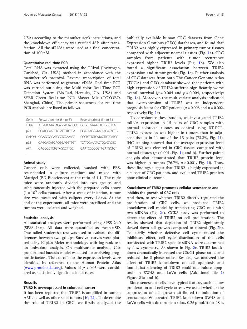
ResultsTRIB2 is overexpressed in colorectal cancerIt has been reported that TRIB2 is amplified in humanAML as well as other solid tumors [10, 24]. To determinethe role of TRIB2 in CRC, we firstly analyzed the
publically available human CRC datasets from GeneExpression Omnibus (GEO) databases, and found thatTRIB2 was highly expressed in primary tumor tissuescompared with adjacent normal tissues (Fig. 1a). CRCsamples from patients with tumor recurrenceexpressed higher TRIB2 levels (Fig. 1b). We alsofound a significant association between TRIB2expression and tumor grade (Fig. 1c). Further analysisof CRC datasets from both The Cancer Genome Atlas(TCGA) and GEO database showed that patients withhigh expression of TRIB2 suffered significantly worseoverall survival (p = 0.004 and p = 0.004, respectively;Fig. 1d). Moreover, the multivariate analysis indicatedthat overexpression of TRIB2 was an independentprognosis factor for CRC patients (p = 0.006 and p = 0.002,respectively; Fig. 1e).To corroborate these studies, we investigated TRIB2
mRNA expression in 15 pairs of CRC samples withnormal colorectal tissues as control using RT-PCR.TRIB2 expression was higher in tumors than in adja-cent tissues in 11 out of the 15 pairs (73.3%, Fig. 1f ).IHC staining showed that the average expression levelof TRIB2 was elevated in CRC tissues compared withnormal tissues (p < 0.001, Fig. 1g and h). Further pairedanalysis also demonstrated that TRIB2 protein levelwas higher in tumors (76.7%, p < 0.001, Fig. 1i). Thus,these findings suggest that TRIB2 is highly expressed ina subset of CRC patients, and evaluated TRIB2 predictspoor clinical outcome.
Knockdown of TRIB2 promotes cellular senescence andinhibits the growth of CRC cellsAnd then, to test whether TRIB2 directly regulated theproliferation of CRC cells, we produced TRIB2knockdown cell model by transfecting CRC cells withtwo siRNAs (Fig. 2a). CCK8 assay was performed todetect the effect of TRIB2 on cell proliferation. Theresults showed that depletion of TRIB2 significantlyslowed down cell growth compared to control (Fig. 2b).To clarify whether defective cell cycle caused theinhibitory effect, cell cycle distribution of the cellstransfected with TRIB2-specific siRNA were determinedby flow cytometry. As shown in Fig. 2c, TRIB2 knock-down dramatically increased the G0/G1-phase ratios andreduced the S-phase ratios. Besides, we analyzed theeffect of TRIB2 knockdown on cell apoptosis andfound that silencing of TRIB2 could not induce apop-tosis in SW48 and LoVo cells (Additional file 1:Figure S1a and b).Since senescent cells have typical feature, such as low
proliferation and cell cycle arrest, we asked whether thesuppression of cell growth attributed to induction ofsenescence. We treated TRIB2-knockdown SW48 andLoVo cells with doxorubicin (dox, 0.25 μmol/l) for 48 h,
Hou et al. Molecular Cancer (2018) 17:172 Page 4 of 15
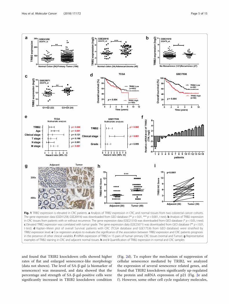
and found that TRIB2 knockdown cells showed higherrates of flat and enlarged senescence-like morphology(data not shown). The level of SA-β-gal (a biomarker ofsenescence) was measured, and data showed that thepercentage and strength of SA-β-gal-positive cells weresignificantly increased in TRIB2 knockdown condition
(Fig. 2d). To explore the mechanism of suppression ofcellular senescence mediated by TRIB2, we analyzedthe expression of several senescence related genes, andfound that TRIB2 knockdown significantly up-regulatedthe protein and mRNA expression of p21 (Fig. 2e andf ). However, some other cell cycle regulatory molecules,
Fig. 1 TRIB2 expression is elevated in CRC patients. a Analysis of TRIB2 expression in CRC and normal tissues from two colorectal cancer cohorts.The gene expression data (GSE41258, GSE20916) was downloaded from GEO database (** p < 0.01, *** p < 0.001, t-test). b Analysis of TRIB2 expressionin CRC tissues from patients with or without recurrence. The gene expression data (GSE21510) was downloaded from GEO database (* p < 0.05, t-test).c Elevated TRIB2 expression was correlated with tumor grade. The gene expression data (GSE25071) was downloaded from GEO database (** p < 0.01,t-test). d Kaplan–Meier plot of overall Survival: patients with CRC (TCGA database and GSE17536 from GEO database) were stratified byTRIB2 expression level. e Cox regression analysis to evaluate the significance of the association between TRIB2 expression and CRC patients prognosisin the presence of other clinical variables. f mRNA expression of TRIB2 in 15 pairs of human primary CRC tissues (normal and Tumor). g Representativeexamples of TRIB2 staining in CRC and adjacent normal tissues. h and i Quantification of TRIB2 expression in normal and CRC samples
Hou et al. Molecular Cancer (2018) 17:172 Page 5 of 15
such as cyclin D1 and p16, could not be regulated by si-lencing of TRIB2 (Additional file 2: Figure S2a). Ac-cording to the results above, we speculated that TRIB2regulated the expression of p21 at transcription level.
We further performed luciferase assays using pGl3 con-taining p21 promoter (~ 2.0 kb) (p21-Luc). The lucifer-ase activity of p21 promoter was stimulated by TRIB2knockdown (Fig. 2g). On the contrary, overexpression
Fig. 2 Knockdown of TRIB2 in CRC cells inhibits tumor cell growth and enhances cellular senescence. a Western blot of TRIB2 in SW48 and LoVocells transfected with TRIB2-specific siRNA (siTRIB2#1 and siTRIB2#2) or control siRNA. b CCK8 analysis of cell viability in TRIB2-knockdown SW48and LoVo cells at 0, 24, 48 and 72 h, respectively. c Cell cycle distribution measured by flow cytometry in TRIB2-knockdown SW48 and LoVo cells.d SA-β-gal staining analysis in TRIB2-knockdown SW48 and LoVo cells treated with dox (0.25 μmol/l, 48 h, left panel, representative images of SA-β-gal staining). e Western blot of TRIB2, p53 and p21 in SW48 and LoVo cells transfected with TRIB2-specific or control siRNA. f mRNA expressionof p53 and p21 in SW48 and LoVo cells transfected with TRIB2-specific or control siRNA. g Luciferase activity of p21 promoter in SW48 and LoVocells transiently transfected with p21-Luc plus TRIB2-specific or control siRNA. Results are presented as mean ± SD from three independent assays,* p < 0.05, ** p < 0.01, *** p < 0.001, t-test
Hou et al. Molecular Cancer (2018) 17:172 Page 6 of 15
of TRIB2 inhibited the mRNA and protein levels ofp21, facilitated proliferation and cell cycle, and blockedcellular senescence in SW48 and LoVo cells. Inaddition, the luciferase activity of p21 promoter wasmarkedly weakened in TRIB2 overexpressed SW48 andLoVo cells (Additional file 3: Figure S3). Our resultsshow that TRIB2 is crucial for pro-proliferation andsuppressing cellular senescence in CRC cells.
TRIB2 regulates the expression of p21 in a p53-independent mannerp53 is an important transcription factor involved in cellcycle inhibition and directly target the p21 promoter.We then examined levels of p53 in both TRIB2knockdown/overexpressed and control cells, and foundno significant changes in p53 expression at both proteinand mRNA levels (Fig. 2e and f; Additional file 3: FigureS3e and f). To confirm TRIB2 regulated p21 expressionin a p53-independent manner, we transfected p53-spe-cific siRNA in TRIB2 knockdown SW48 cells to observethe p21 expression changes. As shown in Fig. 3 a and b,TRIB2 knockdown elevated the expression of p21 atprotein and mRNA levels irrespective of p53 knock-down. The luciferase assays showed consistent resultsthat the inhibitory effect of p21-luciferase activity byp53 knockdown was alleviated by silencing TRIB2(Fig. 3 c). Moreover, silencing TRIB2 still inhibitedcell growth and induced cellular senescence in p53knockdown SW48 cells (Fig. 3 d and e). In parallel,we chose HCT116p53−/− cells, a p53-null originatingfrom HCT116 wt cells, and explored whether TRIB2regulated p21 expression in these cells. As expected,no matter what the status of p53 was, silencingTRIB2 dramatically up-regulated p21 expression,inhibited cell growth, induced cell cycle arrest andenhanced cellular senescence. Conversely, overexpres-sion of TRIB2 down-regulated p21 expression, accel-erated proliferation and cell cycle, and blockedsenescence (Fig. 3 f-j). These findings indicate thatTRIB2 regulates proliferation and cellular senescencein a p53-independent manner.
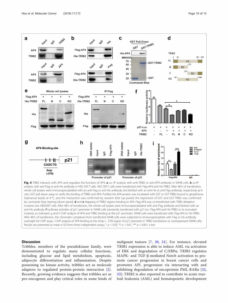
TRIB2 interacts with AP4 and enhances AP4transcriptional activitiesThe transcription factor AP4 belongs to bHLZ family,and has an important role in cell growth anddevelopment. It forms homodimers and binds to theE-box motif CAGCTG to repress viral and cellular genes[25, 26]. The expression of AP4 was elevated in CRCtumor tissues compared with their corresponding nor-mal tissues (9/15; Additional file 4: Figure S4a). AP4 hasbeen shown to induce cell cycle arrest and repress p21
expression via occupying four CAGCTG motifs in thep21 promoter [21]. Deletion of AP4 by AP4-specificsiRNA (siAP4#1 and siAP4#2) significantly increasedmRNA and protein levels of p21, inhibited cell prolifera-tion and promoted cellular senescence in SW48 andLoVo cells (Additional file 4: Figure S4b, c, d and e).Data from mass spectrometry (BioGRID) identify a num-ber of proteins including TRIB2 as partners of AP4. Toconfirm the TRIB2/AP4 interaction, we carried outco-IP assay using SW48 cells or HEK 293 T cellsco-transfected with Flag-AP4 and His-TRIB2. AP4 andTRIB2 were found to precipitate with each other (Fig. 4a and b). Besides, pull down assay verified that AP4 bandto GST-TRIB2 in vitro but not GST protein alone (Fig. 4c). Next we constructed a series of different deletionmutants of His-TRIB2, and transfected them withFlag-AP4 into HEK 293 T cells. co-IP results indicatedthat it was kinase-like domain (KD) of TRIB2 interactedwith AP4 (Fig. 4 d and e).TRIBs are members of the pseudokinase family, and
position protein ‘substrates’ and control their E3ligase-dependent ubiquitination [27]. Thus we hypothe-sized that TRIB2 regulated p21 expression and senes-cence predominantly through regulating AP4 expression.However, as shown in Additional file 2: Figure S2a andb, TRIB2 could not directly regulate the expression ofAP4. Recent studies have shown that TRIB3 binds tosome transcription factor, such as ATF4 and PPARγ, andregulates their transcription activities [28, 29]. So wesupposed that TRIB2 might regulate AP4 mediated tran-scription and performed luciferase assay using p21-Luc.The results showed that overexpression of AP4 sup-pressed and co-transfection of TRIB2 further suppressedthe activities of p21 promoter (Fig. 4f ), which indicatedTRIB2 enhanced the function of AP4. Further studyconfirmed that it was kinase-like domain (KD) of TRIB2,but not the N- or C-terminus, that promoted the inhibi-tory effect of AP4 on p21 promoter activities (Fig. 4f ).To further address the mechanism of how TRIB2
regulated the function of AP4, we conducted ChIPassays on SW48 cells that ectopically expressedFlag-AP4. The results showed that Flag-AP4 wasenriched in the proximal region (− 218 to − 324) of thep21 gene promoter (Fig. 4h, left panel). Next, we per-formed ChIP assays on SW48 cells ectopically express-ing His-TRIB2, and found that His-TRIB2 wasenriched in the same region with Flag-AP4 (Fig. 4h,right panel). To elucidate how TRIB2 affected thebinding of AP4 on p21 promoter, we performed ChIPexperiments in TRIB2 knockdown or overexpressedSW48 cells. The results indicated that knockdown ofTRIB2 decreased and overexpression of TRIB2 in-creased AP4-binding on p21 promoter (Fig. 4i). Takentogether, TRIB2 physically interacts with AP4 and
Hou et al. Molecular Cancer (2018) 17:172 Page 7 of 15
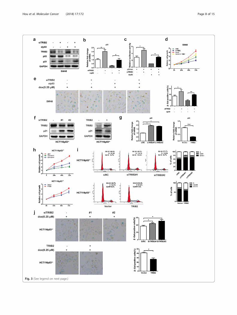
Fig. 3 (See legend on next page.)
Hou et al. Molecular Cancer (2018) 17:172 Page 8 of 15
enhances binding of AP4 on p21 promoter to nega-tively regulate p21 expression.
Enhanced transcription activities of AP4 by TRIB2accelerates CRC cells proliferation and hinders cellularsenescenceIn view of results above, we investigated the role ofTRIB2/AP4 interaction in tumorigenicity. Todemonstrate whether AP4 was critical in TRIB2-inducedsuppression of p21 expression, we overexpressed TRIB2with or without AP4 knockdown and detected the proteinand mRNA levels of p21. Elevated TRIB2 markedly re-pressed p21 expression in SW48 and LoVo cells, and thiscould be reversed by AP4 knockdown (Fig. 5a and b). Theluciferase assay indicated TRIB2 inhibited p21 promoteractivities in an AP4-dependent manner (Fig. 5c). We thenperformed CCK8 and SA-β-gal staining analysis to exam-ine the effects of AP4 on TRIB2-mediated functions, andfound that silencing AP4 could significantly block TRIB2overexpression-promoted SW48 and LoVo cells growthand enhance TRIB2 overexpression-inhibited cellularsenescence (Fig. 5g and h).Next, we investigated the importance of TRIB2 in
modulating AP4 functions, and co-transfected AP4 ex-pressing plasmid with or without TRIB2-specific siRNAin SW48 and LoVo cells. As shown in Fig. 5d, the de-crease of p21 protein levels caused by elevated AP4 wasabrogated by TRIB2 knockdown. The RT-PCR and lu-ciferase assay indicated that the transcription activitiesof AP4 were dependent on TRIB2/AP4 interaction (Fig.5e and f ). We then examined the role of TRIB2/AP4interaction in AP4-mediated tumorigenesis. Data fromCCK8 and SA-β-gal staining analysis showed that thefunctions of AP4 in cell proliferation were markedlyimpaired and repressed cellular senescence by AP4 wasenhanced in the absence of TRIB2 (Fig. 5i and j). Takentogether, these results reveal that TRIB2 promotes can-cer cells growth and suppresses cellular senescence viaenhancing AP4 transcription activities.
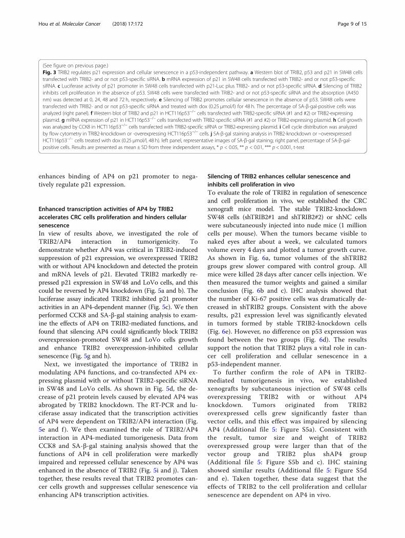
Silencing of TRIB2 enhances cellular senescence andinhibits cell proliferation in vivoTo evaluate the role of TRIB2 in regulation of senescenceand cell proliferation in vivo, we established the CRCxenograft mice model. The stable TRIB2-knockdownSW48 cells (shTRIB2#1 and shTRIB2#2) or shNC cellswere subcutaneously injected into nude mice (1 millioncells per mouse). When the tumors became visible tonaked eyes after about a week, we calculated tumorsvolume every 4 days and plotted a tumor growth curve.As shown in Fig. 6a, tumor volumes of the shTRIB2groups grew slower compared with control group. Allmice were killed 28 days after cancer cells injection. Wethen measured the tumor weights and gained a similarconclusion (Fig. 6b and c). IHC analysis showed thatthe number of Ki-67 positive cells was dramatically de-creased in shTRIB2 groups. Consistent with the aboveresults, p21 expression level was significantly elevatedin tumors formed by stable TRIB2-knockdown cells(Fig. 6e). However, no difference on p53 expression wasfound between the two groups (Fig. 6d). The resultssupport the notion that TRIB2 plays a vital role in can-cer cell proliferation and cellular senescence in ap53-independent manner.To further confirm the role of AP4 in TRIB2-
mediated tumorigenesis in vivo, we establishedxenografts by subcutaneous injection of SW48 cellsoverexpressing TRIB2 with or without AP4knockdown. Tumors originated from TRIB2overexpressed cells grew significantly faster thanvector cells, and this effect was impaired by silencingAP4 (Additional file 5: Figure S5a). Consistent withthe result, tumor size and weight of TRIB2overexpressed group were larger than that of thevector group and TRIB2 plus shAP4 group(Additional file 5: Figure S5b and c). IHC stainingshowed similar results (Additional file 5: Figure S5dand e). Taken together, these data suggest that theeffects of TRIB2 to the cell proliferation and cellularsenescence are dependent on AP4 in vivo.
(See figure on previous page.)Fig. 3 TRIB2 regulates p21 expression and cellular senescence in a p53-independent pathway. a Western blot of TRIB2, p53 and p21 in SW48 cellstransfected with TRIB2- and or not p53-specific siRNA. b mRNA expression of p21 in SW48 cells transfected with TRIB2- and or not p53-specificsiRNA. c Luciferase activity of p21 promoter in SW48 cells transfected with p21-Luc plus TRIB2- and or not p53-specific siRNA. d Silencing of TRIB2inhibits cell proliferation in the absence of p53. SW48 cells were transfected with TRIB2- and or not p53-specific siRNA and the absorption (A450nm) was detected at 0, 24, 48 and 72 h, respectively. e Silencing of TRIB2 promotes cellular senescence in the absence of p53. SW48 cells weretransfected with TRIB2- and or not p53-specific siRNA and treated with dox (0.25 μmol/l) for 48 h. The percentage of SA-β-gal-positive cells wasanalyzed (right panel). f Western blot of TRIB2 and p21 in HCT116p53−/− cells transfected with TRIB2-specific siRNA (#1 and #2) or TRIB2-expressingplasmid. g mRNA expression of p21 in HCT116p53−/− cells transfected with TRIB2-specific siRNA (#1 and #2) or TRIB2-expressing plasmid. h Cell growthwas analyzed by CCK8 in HCT116p53−/− cells transfected with TRIB2-specific siRNA or TRIB2-expressing plasmid. i Cell cycle distribution was analyzedby flow cytometry in TRIB2-knockdown or -overexpressing HCT116p53−/− cells. j SA-β-gal staining analysis in TRIB2-knockdown or –overexpressedHCT116p53−/− cells treated with dox (0.25 μmol/l, 48 h). left panel, representative images of SA-β-gal staining; right panel, percentage of SA-β-gal-positive cells. Results are presented as mean ± SD from three independent assays, * p < 0.05, ** p < 0.01, *** p < 0.001, t-test
Hou et al. Molecular Cancer (2018) 17:172 Page 9 of 15
DiscussionTribbles, members of the pseudokinase family, weredemonstrated to regulate many cellular functions,including glucose and lipid metabolism, apoptosis,adipocyte differentiation and inflammation. Despitepossessing no kinase activity, tribbles act as moleculeadaptors to regulated protein-protein interaction [2].Recently, growing evidence suggests that tribbles act aspro-oncogenes and play critical roles in some kinds of
malignant tumors [7, 30, 31]. For instance, elevatedTRIB1 expression is able to induce AML via activationof ERK and degradation of C/EBPα; TRIB3 regulatesMAPK- and TGF-β-mediated Notch activation to pro-mote cancer progression in breast cancer cells andpromotes APL progression via interacting with andinhibiting degradation of oncoprotein PML-RARα [32,33]; TRIB2 is also reported to contribute to acute mye-loid leukemia (AML) and hematopoietic development
Fig. 4 TRIB2 interacts with AP4 and regulates the function of AP4. a co--IP analysis with anti-TRIB2 or anti-AP4 antibody in SW48 cells. b co-IPanalysis with anti-Flag or anti-His antibody in HEK 293 T cells. HEK 293 T cells were transfected with Flag-AP4 and His-TRIB2. After 48 h of transfection,whole cell lysates were immunoprecipitated with an anti-Flag or anti-His antibody and blotted with an anti-His or anti-Flag antibody, respectively. c Invitro GST pull down assay to verify the binding of TRIB2 and AP4. Purified His-AP4 protein was incubated with GST or GST-TRIB2 bound to glutathione-Sepharose beads at 4 °C, and the interaction was confirmed by western blot (up panel); the expression of GST and GST-TRIB2 was confirmedby coomassie blue staining (down panel). d and e Mapping of TRIB2 regions binding to AP4. Flag-AP4 was co-transfected with TRIB2-deleptionmutants into HEK293T cells. After 48 h of transfection, the whole cell lysates were immunoprecipitated with anti-Flag antibody and blotted with ananti-His antibody. f luciferase activities of p21 promoter in SW48 cells transiently transfected with p21-luc, Flag-AP4 and His-TRIB2 or its truncatedmutants, as indicated. g and h ChIP analysis of AP4 and TRIB2 binding at the p21 promoter. SW48 cells were transfected with Flag-AP4 or His-TRIB2.After 48 h of transfection, the chromatin complexes from transfected SW48 cells were subjected to immunoprecipitate with Flag or His antibodyovernight for ChIP assay. i ChIP analysis of AP4 binding at the Amp (− 270) region of p21 promoter in TRIB2 knockdown or overexpressed SW48 cells.Results are presented as mean ± SD from three independent assays, * p < 0.05, ** p < 0.01, *** p < 0.001, t-test
Hou et al. Molecular Cancer (2018) 17:172 Page 10 of 15
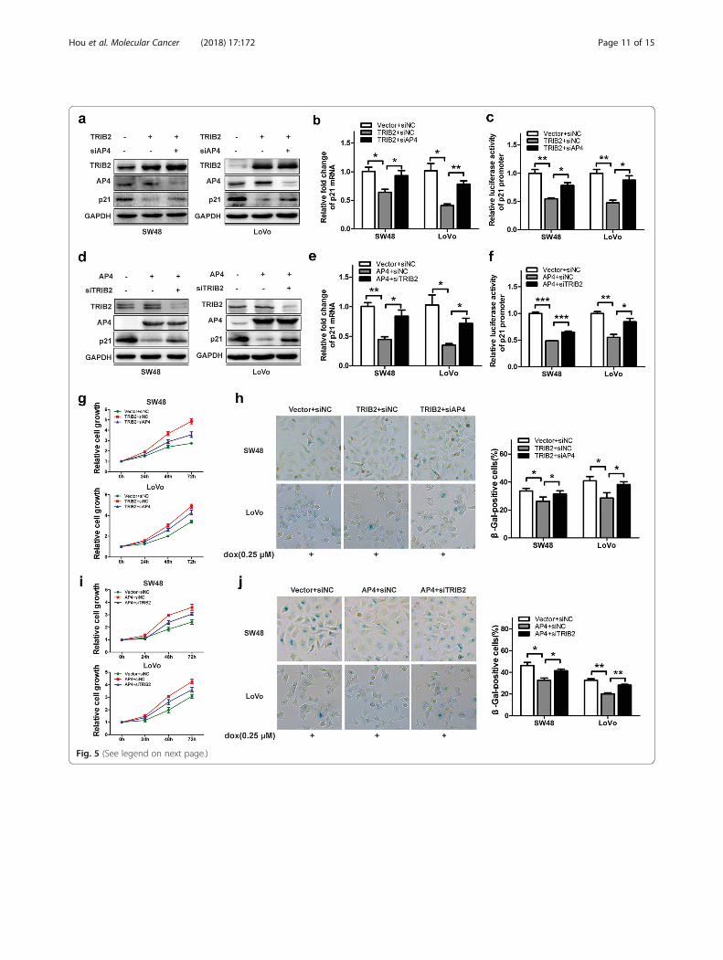
Fig. 5 (See legend on next page.)
Hou et al. Molecular Cancer (2018) 17:172 Page 11 of 15
[34]. In the present study, we demonstrated that TRIB2expression in human CRC tissues was much higher thanthat in adjacent tissues and negatively correlated withprognosis of CRC patients, which is consistent with theresults reported by Hill, R. et al. [10]. Moreover, we identi-fied TRIB2 as an independent prognostic factor affectingthe survival of CRC patients through multivariate analysis,further illustrating its important clinical role in CRC pa-tients. These results indicated that TRIB2 might be anavailable prognostic marker for CRC patients.Studies on the mechanism of regulating the
expression of TRIB2 have been widely conducted, andtranscriptional regulation is likely to be pivotal inTRIB2 expression. The aberrant expression of someoncogenes has been reported to account for elevatedTRIB2. Wang et al. showed that the TRIB2 is a directtarget of Wnt/TCF pathway in liver cancer [8]. InAML cells, dysregulated C/EBPalpha and E2F1contributes to up-regulation of TRIB2 [35]. In humanT cell acute lymphoblastic leukemia (T-ALL), NOTCH,PITX and TAL1 are also found to up-regulate TRIB2[36]. However, it is unclear why TRIB2 is overex-pressed in CRC patients. It would be interesting to fur-ther investigate the mechanism of TRIB2 up-regulationin CRC.To further explore the oncogenic role of TRIB2 in
tumorigenesis, we performed a series of experiments inCRC cell lines and found that ectopic expression ofTRIB2 dramatically blocked cellular senescence.Cellular senescence is identified as a state ofirreversible cell-cycle arrest induced by a series of in-sults, and induction of senescence is considered as apotent strategy for suppressing cancer progression withminor side effects [37]. As the critical marker for cellu-lar senescence, p21 has been proved to drive senes-cence initiation. Various stimuli including ROS,mitochondrial dysfunction, irradiation and chemothera-peutic drugs induce cellular senescence through DNAdamage response by activating ATM and eventually
activating p53/p21 pathway [38]. Although p21 ismainly regulated by p53 at the transcription level, mul-tiple transcription factors (BRCA1, Smad3, AP4 andc-myc) have also been reported to control the tran-scriptional activation or repression of p21 [39–41].Inour study, TRIB2 could negatively regulate p21 pro-moter activities and expression, and inhibit cellular sen-escence in CRC cells. Earlier reports of Richard et al.have showed that in melanoma cells TRIB2 inhibits p53and p21 expression in an AKT-dependent manner [4].However, in the current study we did not detect signifi-cant changes of p53 expression in SW48 cells whenTRIB2 was silenced. Experiments in CRC cells, whichwere transfected with p53- and or not TRIB2-specificsiRNA, suggested that TRIB2 knockdown could stilltranscriptionally increase p21 expression even in theabsence of p53. These results revealed that TRIB2 regu-lated p21 expression in a p53-independent manner,which is opposite with previous study. We speculatedthat TRIB2 may have distinct functions depending onetiologic background of different cancer types.To extensively study the mechanisms responsible for
TRIB2-mediated regulation of cellular senescence, weanalyzed the BioGRID database and found TRIB2 dir-ectly interacted with transcription factor AP4. AP4, be-longing to the basic helix-loop-helix transcriptionfactors (bHLH-LZ) superfamily, is identified as anoncogene and accelerates development and progressionin a variety of human cancers [23, 42–45]. Moreover,AP4 is frequently up-regulated in these malignanciesand positively correlates with poor prognosis. The func-tions of AP4 in modulating tumorigenic properties havebeen extensively studied. It plays a critical role in regulat-ing stem-like phenotypes and epithelial-mesenchymaltransition (EMT) through directly targeting genes suchas CD44, LGR5, VIM and E-cadherin [45]. Mechanic-ally, AP4 recognizes and binds to consensus E-boxmotif CAGCTG in promoters of downstream targetgenes [22]. There is evidence that AP4 directly binds to
(See figure on previous page.)Fig. 5 TRIB2 regulates cell proliferation and cellular senescence dependent on AP4. a Western blot analysis of TRIB2, AP4 and p21 expression inSW48 and LoVo cells transfected with TRIB2-expressing plasmid and or not AP4-specific siRNA. b mRNA expression of p21 in SW48 and LoVo cellstransfected with TRIB2-expressing plasmid and or not AP4-specific siRNA. c p21-Luc was co-transfected with TRIB2-expressing plasmid and or notAP4-specific siRNA into SW48 and LoVo cells. After 48 h of transfection, the cells were harvest and luciferase activities were measured. d Westernblot of TRIB2, AP4 and p21 expression in SW48 and LoVo cells transfected with AP4-expressing plasmid and or not TRIB2-specific siRNA. e mRNAexpression of p21 in SW48 and LoVo cells transfected with AP4-expressing plasmid and or not TRIB2-specific siRNA, respectively. f p21-Luc wasco-transfected with AP4-expressing plasmid and or not TRIB2-specific siRNA into SW48 and LoVo cells. After 48 h of transfection, the cells wereharvest and luciferase activities were measured. g Cell proliferation analysis of TRIB2-overexpressed SW48 and LoVo cells transfected with orwithout AP4-specific siRNA. h SA-β-gal staining analysis in TRIB2 overexpressed SW48 and LoVo cells transfected with or without AP4-specificsiRNA (dox, 0.25 μmol/l, 48 h). The percentage of SA-β-gal-positive cells was analyzed (right panel). i Cell proliferation analysis of AP4-overexpressed SW48 and LoVo cells transfected with or without TRIB2-specific siRNA. j SA-β-gal staining analysis in AP4-overexpressed SW48 andLoVo cells transfected with or without TRIB2-specific siRNA (dox, 0.25 μmol/l, 48 h). The percentage of SA-β-gal-positive cells was analyzed (rightpanel). Results are presented as mean ± SD from three independent assays, * p < 0.05, ** p < 0.01, *** p < 0.001, t-test
Hou et al. Molecular Cancer (2018) 17:172 Page 12 of 15

p21 promoter and transcriptionally represses p21 ex-pression [21]. Our results suggest that TRIB2 regulatesp21 expression by enhancing the functions of AP4,through protein-protein interaction. Similar to our re-sults, Nobumichi et al. showed that TRIB3, anothermember of the tribbles family, interacts with transcrip-tion factor ATF4 and then regulates transcriptional ac-tivity of ATF4 [29]. In addition, TRIB3 alters PPARγtranscriptional activity through directly binding toPPARγ during adipocyte differentiation [28]. The aboveevidence indicates that it is a common phenomenon forthe tribble family members to influence the activity oftranscription factors through protein-protein inter-action. However, the exact mechanism of how TRIB2regulate the transcription activities of AP4 remains to
be clarified. It is likely that TRIB2 acts as scaffold to re-cruit some corepressor (s) or trigger histone modifica-tion. Future studies are needed to determine thespecific mechanism.
ConclusionsIn summary, we noted the oncogenic effect of TRIB2 inhuman colorectal cancer. The increased level of TRIB2promotes proliferation and induces cell cycle arrest byinhibiting cellular senescence, and this is mediated byTRIB2/AP4 interaction which leads to enhanced AP4transcription activity. The present study expands ourunderstanding of TRIB2 in cellular senescence, andsuggests that TRIB2/AP4/p21 pathway is a potentialtarget for tumor therapeutic intervention.
Fig. 6 TRIB2 knockdown inhibits cell proliferation and enhances cellular senescence in vivo. a Tumor volumes of the TRIB2-knockdownand control groups were calculated every 4 days. b and c Tumor size and weight of the TRIB2-knockdown and control groups. d IHCstaining of TRIB2, p53, p21 and Ki-67 in the indicated xenograft tumors. e Quantification of p21 expression, and proliferation index in theindicated xenograft tumors, proliferation index was determined using the percentage of Ki-67 positive cells. The results were presentedas mean ± SD, n = 6, * p < 0.05, ** p < 0.01, *** p < 0.001, t-test. f Proposed model: TRIB2/AP4 interaction enhances binding of AP4 onp21 promoter to negatively regulate p21 expression, which consequently leads to increased cell growth and inhibited cellularsenescence in CRC
Hou et al. Molecular Cancer (2018) 17:172 Page 13 of 15
Additional files
Additional file 1: Figure S1. The functions of TRIB2 on cell growth arenot due to cell apoptosis in CRC. a and b Flow cytometry analysis of thepercentage of apoptotic cells for SW48 and LoVo cells transfected withsiTRIB2 (#1 and #2) or siNC for 72 h. (TIF 1224 kb)
Additional file 2: Figure S2. TRIB2 could not affect cyclin D1 and p16expression.a Western blot analysis of Cyclin D1 and p16 expression inSW48 and LoVo cells transfected with siTRIB2 (#1 and #2) or siNC. bWestern blot analysis of Cyclin D1 and p16 expression in SW48 and LoVocells transfected with TRIB2 expressing plasmid or vector. (TIF 1118 kb)
Additional file 3: Figure S3. Overexpression of TRIB2 in CRC cellspromotes tumor cell growth and inhibits cellular senescence. a Westernblot analysis of TRIB2 in SW48 and LoVo cells transfected with TRIB2-expressing plasmid or vector. b Cell viability of TRIB2-overexpressed orcontrol SW48 and LoVo cells at 0, 24, 48, 72 h, respectively. c Cell cycledistribution by flow cytometry detection in TRIB2-overexpressed orcontrol SW48 and LoVo cells; d SA-β-gal staining analysis of TRIB2-overexpressed or control SW48 and LoVo cells treated with dox(0.25 μmol/l, 48 h, left panel, representative images of SA-β-gal staining). eWestern blot analysis of TRIB2, p53 and p21 in SW48 and LoVo cellstransfected with TRIB2-expressing plasmid or vector. f RT-PCR analysis ofp53 and p21 expression in SW48 and LoVo cells transfected with TRIB2-expressing plasmid or vector. g Relative luciferase activity of p21 in SW48and LoVo cells transiently transfected with p21-Luc plus TRIB2-expressingplasmid or vector. Results are presented as mean ± SD from threeindependent assays, * p < 0.05, ** p < 0.01, *** p < 0.001, t-test. (TIF 2282 kb)
Additional file 4: Figure S4. AP4 promotes tumorigenesis in colorectalcancer. a mRNA expression of AP4 in 15 pairs of human primary CRCtissues (normal and Tumor). b Western blot analysis of AP4 and p21 inSW48 and LoVo cells transfected with AP4 specific siRNA (#1 and #2) orsiNC. c RT-PCR analysis of AP4 and p21 in SW48 and LoVo cells transfected withAP4 specific siRNA (#1 and #2) or siNC. d CCK8 analysis of cell proliferationcapacity in AP4-knockdown or control SW48 and LoVo cells. e SA-β-gal staininganalysis of SW48 and LoVo cells transfected with AP4 specific siRNA (#1 and #2)or siNC (dox, 0.25 μmol/l, 48 h). left panel, representative images ofSA-β-gal staining; right panel, percentage of SA-β-gal-positive cells.Results are presented as mean ± SD from three independent assays,* p < 0.05, ** p < 0.01, *** p < 0.001, t-test. (TIF 2707 kb)
Additional file 5: Figure S5 The functions that TRIB2 mediated in vivoare dependent on AP4. a Tumor volumes of TRIB2-overexpressed or TRIB2-overexpressed plus AP4 knockdown or control groups were calculated every4 days. b and c Tumor size and weight of tumors generated from TRIB2-overexpressed SW48 cells transfected with or without AP4-specific siRNAand control cells. d IHC staining of TRIB2, p53, p21 and Ki-67 in the indicatedxenograft tumors. e Quantification of p21 expression, and proliferation indexin the indicated xenograft tumors, proliferation index was determined usingthe percentage of Ki-67 positive cells. The results were presented as mean± SD, n = 6, * p < 0.05, ** p < 0.01, *** p < 0.001, t-test. (TIF 4363 kb)
AbbreviationsAML: Acute myeloid leukemia; AP4 CDK: Cyclin-dependent kinases;AP4: Transcription factor; CDKN1A: Cyclin dependent kinase inhibitor 1A;ChIP: Chromatin immunoprecipitation; co-IP: Co-immunoprecipitation;CRC: Colorectal cancer; DDR: DNA damage response; FBS: Fetal bovineserum; GEO: Gene Expression Omnibus; IHC: Immunohistochemistry; RNATCGA: The Cancer Genome Atlas; SA-β-gal: Senescence-associated β-galactosidase; siRNA: Short interfering; TRIB2: Tribbles pseudokinase 2
AcknowledgementsNot applicable.
FundingThis work was supported by grants from the National Natural Science Foundationof China (No. 81572725, No. 81570525, and No. 81773113) and the ‘973’programme (No. 2015CB553903–1).
Availability of data and materialsAll the dataset and materials generated and/or analyzed during the currentstudy were available.
Authors’ contributionsWGH and HZL designed the study. HZL and GKX performed most of theexperiments. SXL and HFQ performed animal experiments and constructedthe expression plasmids. GKX, SXL and HFQ collected biological samples,analyzed the data and made the figures. HZL, SXL and GKX analyzed theGEO and TCGA data and participated in writing the manuscript. All authorsread and approved the final manuscript.
Ethics approval and consent to participateThis study was approved by the Huazhong University of Science andTechnology Ethics Committee. Signed informed consents were obtainedfrom all the patients. The animal experiments conducted strictly inaccordance with the Animal Study Guidelines of Huazhong University ofScience and Technology.
Consent for publicationNot applicable.
Competing interestsThe authors declare that they have no competing interests.
Publisher’s NoteSpringer Nature remains neutral with regard to jurisdictional claims in publishedmaps and institutional affiliations.
Author details1Cancer Research Institute, Tongji Hospital, Huazhong University of Scienceand Technology, Wuhan, China. 2Department of Oncology, Tongji Hospital,Huazhong University of Science and Technology, 1095 Jiefang Av, Wuhan,Hubei 430030, People’s Republic of China.
Received: 9 August 2018 Accepted: 21 November 2018
References1. Grosshans J, Wieschaus E. A genetic link between morphogenesis and cell
division during formation of the ventral furrow in Drosophila. Cell. 2000;101(5):523–31.
2. Hua F, Mu R, Liu J, Xue J, Wang Z, Lin H, et al. TRB3 interacts withSMAD3 promoting tumor cell migration and invasion. J Cell Sci. 2011;124(Pt 19):3235–46.
3. Do EK, Park JK, Cheon HC, Kwon YW, Heo SC, Choi EJ, et al. Trib2 regulatesthe pluripotency of embryonic stem cells and enhances reprogrammingefficiency. Exp Mol Med. 2017;49(11):e401.
4. Sakai S, Miyajima C, Uchida C, Itoh Y, Hayashi H, Inoue Y. Tribbles-RelatedProtein Family Members as Regulators or Substrates of the Ubiquitin-Proteasome System in Cancer Development. Curr Cancer Drug Targets.2016;16(2):147–56.
5. Kritsch D, Hoffmann F, Steinbach D, Jansen L, Mary Photini S, Gajda M, et al.Tribbles 2 mediates cisplatin sensitivity and DNA damage response inepithelial ovarian cancer. Int J Cancer. 2017;141(8):1600–14.
6. Salome M, Magee A, Yalla K, Chaudhury S, Sarrou E, Carmody RJ, et al. ATrib2-p38 axis controls myeloid leukaemia cell cycle and stress responsesignalling. Cell Death Dis. 2018;9(5):443.
7. Keeshan K, He Y, Wouters BJ, Shestova O, Xu L, Sai H, et al. Tribbleshomolog 2 inactivates C/EBPalpha and causes acute myelogenousleukemia. Cancer Cell. 2006;10(5):401–11.
8. Wang J, Park JS, Wei Y, Rajurkar M, Cotton JL, Fan Q, et al. TRIB2 actsdownstream of Wnt/TCF in liver cancer cells to regulate YAP and C/EBPalpha function. Mol Cell. 2013;51(2):211–25.
9. Xu S, Tong M, Huang J, Zhang Y, Qiao Y, Weng W, et al. TRIB2 inhibits Wnt/β-catenin/TCF4 signaling through its associated ubiquitin E3 ligases, β-TrCP,COP1 and Smurf1, in liver cancer cells. FEBS Lett. 2014;588(23):4334–41.
10. Hill R, Madureira PA, Ferreira B, Baptista I, Machado S, Colaço L, et al. TRIB2confers resistance to anti-cancer therapy by activating the serine/threonineprotein kinase AKT. Nat Commun. 2017;8:14687.
Hou et al. Molecular Cancer (2018) 17:172 Page 14 of 15
11. Collado M, Blasco MA, Serrano M. Cellular senescence in cancer and aging.Cell. 2007;130(2):223–33.
12. Campisi J. Aging, cellular senescence, and cancer. Annu Rev Physiol.2013;75:685–705.
13. Kuilman T, Michaloglou C, Mooi WJ, Peeper DS. The essence of senescence.Genes Dev. 2010;24(22):2463–79.
14. Sun P, Yoshizuka N, New L, Moser BA, Li Y, Liao R, et al. PRAK is essential forras-induced senescence and tumor suppression. Cell. 2007;128(2):295–308.
15. Wu HL, Li SM, Hu J, Yu X, Xu H, Chen Z, et al. Demystifying the mechanisticand functional aspects of p21 gene activation with double-stranded RNAsin human cancer cells. J Exp Clin Cancer Res. 2016;35(1):145.
16. Stein GH, Drullinger LF, Soulard A, Dulic V. Differential roles for cyclin-dependent kinase inhibitors p21 and p16 in the mechanisms of senescenceand differentiation in human fibroblasts. Mol Cell Biol. 1999;19(3):2109–17.
17. Malumbres M, Barbacid M. Mammalian cyclin-dependent kinases. TrendsBiochem Sci. 2005;30(11):630–41.
18. Abbas T, Dutta A. p21 in cancer: intricate networks and multiple activities.Nat Rev Cancer. 2009;9(6):400–14.
19. Muñoz-Espín D, Cañamero M, Maraver A, Gómez-López G, Contreras J,Murillo-Cuesta S, et al. Programmed cell senescence during mammalianembryonic development. Cell. 2013;155(5):1104–18.
20. Dulic V, Kaufmann WK, Wilson SJ, Tlsty TD, Lees E, Harper JW, et al. p53-dependent inhibition of cyclin-dependent kinase activities in humanfibroblasts during radiation-induced G1 arrest. Cell. 1994;76(6):1013–23.
21. Jung P, Menssen A, Mayr D, Hermeking H. AP4 encodes a c-MYC-induciblerepressor of p21. Proc Natl Acad Sci U S A. 2008;105(39):15046–51.
22. Hu YF, Luscher B, Admon A, Mermod N, Tjian R. Transcription factor AP-4contains multiple dimerization domains that regulate dimer specificity.Genes Dev. 1990;4(10):1741–52.
23. Liu X, Zhang B, Guo Y, Liang Q, Wu C, Wu L, et al. Down-regulation of AP-4inhibits proliferation, induces cell cycle arrest and promotes apoptosis inhuman gastric cancer cells. PLoS One. 2012;7(5):e37096.
24. Grandinetti KB, Stevens TA, Ha S, Salamone RJ, Walker JR, Zhang J, et al.Overexpression of TRIB2 in human lung cancers contributes totumorigenesis through downregulation of C/EBPalpha. Oncogene. 2011;30(30):3328–35.
25. Tsujimoto K, Ono T, Sato M, Nishida T, Oguma T, Tadakuma T. Regulation ofthe expression of caspase-9 by the transcription factor activator protein-4 inglucocorticoid-induced apoptosis. J Biol Chem. 2005;280(30):27638–44.
26. Imai K, Okamoto T. Transcriptional repression of human immunodeficiencyvirus type 1 by AP-4. J Biol Chem. 2006;281(18):12495–505.
27. Eyers PA, Keeshan K, Kannan N. Tribbles in the 21st century: the evolvingroles of tribbles Pseudokinases in biology and disease. Trends Cell Biol.2017;27(4):284–98.
28. Takahashi Y, Ohoka N, Hayashi H, Sato R. TRB3 suppresses adipocytedifferentiation by negatively regulating PPARgamma transcriptional activity.J Lipid Res. 2008;49(4):880–92.
29. Ohoka N, Yoshii S, Hattori T, Onozaki K, Hayashi H. TRB3, a novel ER stress-inducible gene, is induced via ATF4-CHOP pathway and is involved in celldeath. EMBO J. 2005;24(6):1243–55.
30. Zanella F, Renner O, Garcia B, Callejas S, Dopazo A, Peregrina S, et al.Human TRIB2 is a repressor of FOXO that contributes to the malignantphenotype of melanoma cells. Oncogene. 2010;29(20):2973–82.
31. Hill R, Kalathur RK, Colaco L, Brandao R, Ugurel S, Futschik M, et al. TRIB2 asa biomarker for diagnosis and progression of melanoma. Carcinogenesis.2015;36(4):469–77.
32. Izrailit J, Berman HK, Datti A, Wrana JL, Reedijk M. High throughput kinaseinhibitor screens reveal TRB3 and MAPK-ERK/TGFbeta pathways asfundamental Notch regulators in breast cancer. Proc Natl Acad Sci U S A.2013;110(5):1714–9.
33. Li K, Wang F, Cao WB, Lv XX, Hua F, Cui B, et al. TRIB3 promotes APLprogression through stabilization of the Oncoprotein PML-RARalpha andinhibition of p53-mediated senescence. Cancer Cell. 2017;31(5):697–710 e7.
34. Stein SJ, Mack EA, Rome KS, Pear WS. Tribbles in normal and malignanthaematopoiesis. Biochem Soc Trans. 2015;43(5):1112–5.
35. Rishi L, Hannon M, Salomè M, Hasemann M, Frank AK, Campos J, et al.Regulation of Trib2 by an E2F1-C/EBPα feedback loop in AML cellproliferation. Blood. 2014;123(15):2389–400.
36. Sanda T, Lawton LN, Barrasa MI, Fan ZP, Kohlhammer H, Gutierrez A, et al.Core transcriptional regulatory circuit controlled by the TAL1 complex inhuman T cell acute lymphoblastic leukemia. Cancer Cell. 2012;22(2):209–21.
37. Lleonart ME, Artero-Castro A, Kondoh H. Senescence induction; a possiblecancer therapy. Mol Cancer. 2009;8:3.
38. Herranz N, Gil J. Mechanisms and functions of cellular senescence. J ClinInvest. 2018;128(4):1238–46.
39. Elbendary A, Berchuck A, Davis P, Havrilesky L, Bast RC Jr, Iglehart JD, et al.Transforming growth factor beta 1 can induce CIP1/WAF1 expressionindependent of the p53 pathway in ovarian cancer cells. Cell growth &differentiation : the molecular biology journal of the American Associationfor Cancer Research. 1994;5(12):1301–7.
40. Somasundaram K, El-Deiry WS. Inhibition of p53-mediated transactivationand cell cycle arrest by E1A through its p300/CBP-interacting region.Oncogene. 1997;14(9):1047–57.
41. Wu S, Cetinkaya C, Munoz-Alonso MJ, von der Lehr N, Bahram F,Beuger V, et al. Myc represses differentiation-induced p21CIP1expression via Miz-1-dependent interaction with the p21 core promoter.Oncogene. 2003;22(3):351–60.
42. Cao J, Tang M, Li WL, Xie J, Du H, Tang WB, et al. Upregulation of activatorprotein-4 in human colorectal cancer with metastasis. Int J Surg Pathol.2009;17(1):16–21.
43. Xinghua L, Bo Z, Yan G, Lei W, Changyao W, Qi L, et al. The overexpressionof AP-4 as a prognostic indicator for gastric carcinoma. Med Oncol. 2012;29(2):871–7.
44. Hu BS, Zhao G, Yu HF, Chen K, Dong JH, Tan JW. High expression of AP-4predicts poor prognosis for hepatocellular carcinoma after curativehepatectomy. Tumour biology : the journal of the International Society forOncodevelopmental Biology and Medicine. 2013;34(1):271–6.
45. Jackstadt R, Roh S, Neumann J, Jung P, Hoffmann R, Horst D, et al. AP4 is amediator of epithelial-mesenchymal transition and metastasis in colorectalcancer. J Exp Med. 2013;210(7):1331–50.
Hou et al. Molecular Cancer (2018) 17:172 Page 15 of 15

![Presentazione standard di PowerPointmedia.aiom.it/userfiles/files/doc/AIOM-Servizi/slide/...paronychia, dermatitis, stomatitis,..) 63% (vs 41%) Oncogene Addicted [ARCHER 1050] Oncogene](https://img.pdfslide.net/doc/110x75/5e8e913ff7852e421e584c5b/presentazione-standard-di-paronychia-dermatitis-stomatitis-63-vs-41.jpg)