Embed Size (px)
Citation preview

Phyton (Horn, Austria) Vol. 43 Fasc. 1 109-133 21. 7. 2003
Trichomes of Origanum dictamnus L. (Labiatae)
By
Theodoros VRACHNAKIS*)
With 5 figures
Accepted October 1, 2002
Key words : Origanum dictamnus L., Labiatae, Lamiaceae, development, tri-chomes, glandular, non-glandular, morphology, functions.
S u m m a r y
VRACHNAKIS Th. 2003. Trichomes of Origanum dictamnus L. (Labiatae). - Phyton(Horn, Austria) 43 (1): 109-133, with 5 figures. - English with German summary.
The morphology and distribution of the trichomes on the aerial parts of Or-iganum dictamnus were studied by light and scanning electron microscopy. The leafystem is covered with a thick indumentum as a result of the branched non-glandularhairs unique within the genus. The glands are distinguished to two main types: thebig peltate hairs, characteristic within Labiatae, as main site of the lipophilic secre-tions and the diverse forms of capitate hairs as the most numerous type of glandulartrichomes. In the early stage of the plant and in the young expanding tissues a dis-tinct type of capitate hair, the trichome-hydathodes are abundant. Their name, ac-cording to their function, is due to their unique hydrophilic secretion. On the re-productive organs, the "fragile" floral glandular trichomes, as another type of capi-tate hairs with alluring function are distinguished. The stalked glandular hairs, areconsidered as a third type of capitate hairs, are the most common glands, present onall the plant parts. The complex hairiness of the plant is depicted in the floral ele-ments where glandular, non glandular, integrated glandular/eglandular and peculiarhairs are present. The trichome versatility and the variegation of the papillate-cell(transformed ordinary cell) in flowers are attributed to their role as (scent/visual/tactile) attractants and make obscure the division of the epidermal elements in Or-iganum dictamnus.
*) Dr. Theodoros VRACHNAKIS, Institut für Pflanzenphysiologie, KFUNIGraz, Austria, present address: School of Agricultural Technology, TEI of Crete,Stavromenos, P.O. Box 140, GR-71500 Heraklion-Crete, Greece, e-mail: [email protected]
©Verlag Ferdinand Berger & Söhne Ges.m.b.H., Horn, Austria, download unter www.biologiezentrum.at

110
Zusammenfassung
VRACHNAKIS Th. 2003. Haartypen von Origanum dictamnus L. {Labiatae). -Phyton (Horn, Austria) 43 (1): 109-133, 5 Abbildungen. - Englisch mit deutscherZusammenfassung.
Die Morphologie und Verteilung der verschiedenen Trichome auf den ober-irdischen Teilen von Origanum dictamnus wurden licht- und rasterelektronen-mikroskopisch untersucht. Der Stengel und die Laubblätter sind von einem dichtenFilz von verzweigten nicht drüsigen Haaren bedeckt, die charakteristisch für dieGattung Origanum sind. Zwei Haupt-Typen von Drüsenhaaren sind anzutreffen: diefür Lamiaceen charakteristischen großen sitzenden Drüsenschuppen, die am meistenlipophile Substanzen produzieren und verschieden gestaltete Köpfchenhaare als dieam häufigsten vorkommenden Drüsehaare. In frühen Entwicklungsstadien derPflanze, auf jungen noch wachsenden Geweben, finden sich zahlreiche Trichom-hydathoden, die ein wässriges Sekret abgeben. Auf allen Teilen der Blüte finden sichfragile Drüsenhaare mit einem großen einzelligen Drüsenköpfchen, die eventuell imDienste der Anlockung stehen. Eine dritte Type von gestielten Köpfchenhaaren fin-det sich häufig an allen Pflanzenteilen. Die meisten Haartypen finden sich auf denTeilen der Blüte, auf denen Drüsenhaare und nicht drüsige Haare anzutreffen sindund eine Reihe von sonderbaren Haarausprägungen, die sich nicht sicher einer Typezuordnen lassen. Diese Vielfalt der Haare im Blütenbereich, zu denen noch einfachePapillen als Auswüchse normaler Epidermen dazukommen, hängt eventuell mit de-ren Rolle bei der Attraktion von Blütenbesuchern zusammen, wobei teils Geruchs-stoffe, teils visuelle Reize, teils Berührung eine Rolle spielen könnten.
Introduction
Origanum dictamnus L. [syn: O. pseudodictamnus SIEBER, O. dictam-nifolium SAINT-LAGER, O. saxatile SALISBURY, Majorana dictamnus (L.)KOSTELETZKY, M. tomentosa STOKES, Amaracus dictamnus (L.) BENTH., A.tomentosus MOENCH, Dictamnus creticus HILL (cit. IETSWAART 1980)] is anaromatic subshrub of Labiatae (Lamiaceae), endemic of the island of Crete.Recorded in error from East Peloponnese by HALACSY 1902, ZAGANIARIS
1940, and as a casual from Malta (TURLAND & al. 1993), O. dictamnus is theonly Origanum species with branched hairs. Belonging to the sectionAmaracus it is closely related to O. calcaratum JUSS. (SE. Aegean, Creteendemic); it resembles other endemics in the area: O. vettert BRIQUET &BARBEY on Karpathos (Cretan area), O. symes A. CARLSTRÖM on Symi(SE. Aegean), O. scabrum; Boiss. & HELDR. on Euboea and Peloponnese,O. cordifolium (MONTBRET ET AUCHER EX BENTHAM) VOGEL on Cyprus andhas also similarities with other endemics of the continental East Medi-terranean (IETSWAART 1980, CARLSTRÖM 1984, PATON 1994, KOKKINI 1997).
O. dictamnus is mentioned from the Minoan era (FAURE 1987), throughthe centuries from physicians and philosophers like Asklepios, Euripides,Aristotle, Hippocrates, Theophrastus, Virgil, Galen, Dioscorides, to manyin our time, due to the healing properties attributed to it. The ability toheal wounds of arrowed wild-goats (Fig. lb), to stop bleeding, to stimulate
©Verlag Ferdinand Berger & Söhne Ges.m.b.H., Horn, Austria, download unter www.biologiezentrum.at

I l l
the nervous system and cure skin diseases, have made it synonymous to apanacea in the folk medicine of Crete, where it is widely used as a tea. Ithas been known under many names such as: "dictamnos" deriving eitherfrom shrub (thamnos) of mount Dicti (locality where it grows) or from theCretan goddess Dictinna, who helped women during childbirth; "mal-liarohorto" (woolly herb), referring to its hairy leaves, and 'erontas" (Eros),indicating that like true love which is hard to find so is this plant hard tofind and collect from the rather inaccessible sites where it thrives (FRAGAKI
1969, PLATAKIS 1975, HAVAKIS 1980).The plant occurs on several places on Crete (Fig. la), in calcareous
cliffs, crevices, gorge-beds, alt. 0-2000 m, as an obligate chasmophyte (KY-PRIOTAKIS 1998). It is commercially cultivated at the SW. edge of the mountDicti (Fig.la4), and occasionally seen in gardens elsewhere. It can befound, as "Dittany of Crete", in rock gardens, treated as an alpine-houseplant in the old and new world where a mediterranean climate prevails(ELLIOTT 1966, INGWERSEN 1981, TUCKEE & ROLLINS 1989, LEADLEY 1997).Several artificial hybrids, all of garden origin, are known. The expectednatural hybridisation with O. calcaratum in the NE. Crete, where the dis-tribution of the two species just overlaps (IETSWAART 1980, PATON 1994), isnot confirmed (KYPRIOTAKIS 1998, VRACHNAKIS 2002).
As one of the oldest pharmaceutical plants with commercial interest(OHLOFF 1992, LANGE & SCHIPPMANN 1997, SKOULA & KAMENOPOULOS 1997)
it has been widely studied. These studies are focused on the composition ofits essential oils (SKRUBIS 1979, KATSIOTIS & OIKONOMOU 1986, HARVALA &al. 1987, ECONOMAKIS & al. 1999, SKOULA & al. 1999) and on the peltateglandular trichomes, characteristic within the Lamiaceae genera as the(main) site of the essential oils. BOSABALIDIS & TSEKOS 1982 and BOSABALI-
DIS 1987, 1990, investigated the glandular scale (peltate glandular tri-chome) formation on the leaf epidermis of O. dictamnus. HUSAIN& al. 1990studied by SEM its leaf and nutlet surface. VALENTINI & al. 1991, comparedit with O. cordifolium and MATTERN & VOGEL 1994 compared the glands ofthe calyx with that of the leaf of Amaracus dictamnus (O. dictamnus).
In the present work a study of the aerial surface of O. dictamnus fromseed germination until seed formation takes place. The morphology anddistribution of all the types of trichomes on the different organs were in-vestigated and compared with those of the related species and especiallywith O. calcaratum studied by VRACHNAKIS 2002.
Materials and Methods
Plant material
Flowering branches of O. dictamnus were collected from characteristic sites ofits distribution: mount Giouchtas, alt. 750 m; coastal-rock at sea level in SW. Crete;mount Ida, alt. 450 m; SW. edge of the mount Dicti, alt. 400 m; gorge Ha, SE. Crete,
©Verlag Ferdinand Berger & Söhne Ges.m.b.H., Horn, Austria, download unter www.biologiezentrum.at

112
alt. 320 m (Fig. l a l & Fig. lc; Fig. Ia2; Fig. Ia3; Fig. Ia4; Fig. Ia5 & Fig. Id respec-tively) and from specific local gardens and local nurseries, during the summer 1998and 1999. Rooted cuttings from the nursery Mauroskotis (Athens) were cultivated inthe greenhouse of the Institut für Pflanzenphysiologie, KFUNI Graz, Austria androoted plants (garden origin) were transplanted to the Botanical Garden of Graz.Seeds (nutlets) from different samples were germinated in containers under 12hlight/dark, 20°/15° C, 70% RH, 180 (.imol nT2 s"1 PAR in growth chamber (HeraeusVötsch) and in greenhouse conditions starting on 1 December 1999. The conditions inthe growth chamber were shifted to: 17h light/7h dark, 23°/17° C, 55% RH on30 March 2000. Containers from growth chamber and greenhouse, as well as con-tainers with germinated seeds and growing plants from cuttings, were placed outsideof the greenhouse area on 14 April 2000. A potting mix based on sand was used andirrigation was the only agriculture practice to the growing plants. For a comprehen-sive study, samples were collected from the Botanical Garden of Innsbruck, Austria,the Botanical Garden of Wageningen, Holland and herbarium material was providedby Dr. Kypriotakis Z. (Technological Education Institute of Crete). Voucher speci-mens are deposited in Institut für Pflanzenphysiologie, Graz and plants can be foundin Hortus Botanicus Graecensis.
Light microscopy (LM)
Free-hand sections of all plant's parts were observed under a Zeiss AxioplanPhotomicroscope. Paraffin-oil was used for hydrophilic secretions (HEINRICH 1973a)and Oil Red for the localisation of lipophilic substances (GREEN 1991).
Scanning electron microscopy (SEM) •
Plant parts were fixed with 2.5% glutaraldehyde in 0.05 M cacodylate buffer atpH 7.2 for 4 h. and dehydrated in a graded acetone series, critical point dried withCO2 and the specimens sputter-coated with a thin layer of gold (Agar Sputter CoaterB7340). Observations were carried out on a Philips XL 30 ESEM, operating at 20 kV.
Measurements
Measurements took place during the life-cycle of the plants. The number of tri-chomes was counted on SEM images, and directly on LM at 400 x magnification. Thesize of cells and trichomes was measured and the number of trichome cells was de-termined on LM (1000 x magnification). Micromeasurements were confirmed using animage analysing system (Optimas 5.2 for Microsoft Windows, Optimas Corporation).Measurements (per mm2 surface area) took place at cotyledons, leaves and bracts.
Fig. 1. Origanum dictamnus.a. distribution on Crete: after KYPRIOTAKIS Z. 1998 and the author; 1,2,3,4,5: samples
capitalb. arrowed wild-goat eating "dittany" by Cornelius G. Decker (1643-1678)c. growing on calcareous rock, mount Giouchtas, alt. 750 m.d. growing on calcareous rock, gorge Ha, alt. 320 m.e. flowering with exposed flower-organsf. axillary flowering in early stage
©Verlag Ferdinand Berger & Söhne Ges.m.b.H., Horn, Austria, download unter www.biologiezentrum.at

113
©Verlag Ferdinand Berger & Söhne Ges.m.b.H., Horn, Austria, download unter www.biologiezentrum.at

114
Data represent mean values of randomly chosen plant material of different origin.Observations took place in vivo and under a Zeiss SV8 Stereomicroscope.
Results
Trichome formation during growth and development
The life cycle of O. dictamnus is divided in seedling, vegetative andreproductive stage.
In the early stage when the seedling emerges on germination, the hy-pocotyl and cotyledons, although looking glabrous, are pubescent (Figs. 2a,b). In hypocotyl capitate hairs (stalked glandular trichomes and trichome-hydathodes); 1-celled non-glandular trichomes and occasionally peltateglandular trichomes at its top can be found. In the abaxial (lower) side ofthe cotyledon capitate hairs are present, while peltate hairs and non-glandular hairs are absent. In the adaxial (upper) side, all types of glan-dular hairs are present and lor 2-celled non glandular ones (Figs. 2a, cand Table 1). Trichomes are present with developing ordinary cells anddeveloping stomata (of the abaxial cotyledon). At the tip of the cotyledonepithem-hydathodes can be found. Hypocotyl and the cotyledons have apink-red appearance.
Table 1. Trichomes on a mature cotyledon, a fully-expanded leaf and floral ele-ments of O. dictamnus (per mm2 area for flat tissues, n = + 60).
Plant parts
Abaxial side of cotyledonAdaxial side of cotyledonAbaxial side of leafAdaxial side of leafAbaxial side of bractAdaxial side of bractOuter calyxInner calyxOuter corollaInner corolla
Peltateglandular
-3.1-3.53.2-3.92.8-3.60.1-0.40.8-1.1+-+
-
Capitateglandular
68-10380-11048-9562-106155-320135-295+-+
-
Non- Otherglandular types
-+++
+ / -
+ / -+
+ ++
Variation in the values is partly caused by the non homogenous distribution of theepidermal elements on the plant surfaces (see also text).
In the vegetative stage the decussate phyllotaxy is consisting fromleaves of increasing size up to the middle of the stem and then decreasingupwards to the top (Figs, lb, c). When the first leaf primordia appears,glandular and non-glandular hairs with undeveloped ordinary cells consisttheir epidermis (Fig. 5a). With the leaf expansion, in the abaxial side ca-pitate hairs are concentrated across the raised veins and on the petiole.Peltate hairs are located at the periphery of the lamina and on the sides of
©Verlag Ferdinand Berger & Söhne Ges.m.b.H., Horn, Austria, download unter www.biologiezentrum.at
115
the veins. The non-glandular hairs protrude from the veins, leaf-marginand petiole. Stomata are in abundance on the leaf-blade (Figs. 2d, e andTable 1). On the adaxial leaf side, all types of hairs and few stomata areuniformly distributed (Figs. 2f, g and Table 1). In young leaves the mostupper part is devoid of peltate hairs while different stages of these hairsco-exist at the leaf-base (Figs. 2d, f). Epithem-hydathodes can be found asending of the main vein and anthocyanin is present at the leaf-margin. Thenon-glandular hairs are branched, resulting in an obscured epidermalsurface due to their "woolly" cover (Figs. Id, f and 3a, b, d). The leaf sizedepends on its age and position on the stem, reaching its maximum at themiddle stem (±6th node). In these fully-expanded leaves all the epidermalelements are differentiated and their density on both leaf sides is almostequal (Table 1). The indumentum of the internodes is similar to that of theleaves.
The reproductive stage: After six months of cultivation, the main andaxillary stems terminate into inflorescence, and flowering branches ap-peared from the upper stems. The spike like inflorescence comprises suc-cessive pairs of bracts subtending two flowers per verticillaster, attachedwith pedicel (stalk of individual flower) to the peduncle (main axis of theinflorescence). The conversion from the vegetative to the reproductivestage results in intermediate leaves-bracts. Within this stage, the char-acteristic white woolly of the "leafy" stage is replaced by the rather glab-rous and remarkably variegation of the floral elements (Figs, lb, c, e, f).
The bract although looking "naked", bears glandular and eglandulartrichomes. Few peltate hairs are concentrated at the base of the abaxial(outer) side, while at the adaxial (inner) bract blade a greater number ofthese trichomes is uniformly-distributed (Figs, le, f). Occasionally, shortnon-glandular hairs (branched or unbranched) are present at the bractbase or along the bract-margin. The capitate hairs are in abundance onboth sides, densely at the base (Figs. 4a, g, h and Table 1). Their char-acteristic big cuticular-head, within this stage, allows the name: floralglandular trichomes (Fig. 5e). The tip of bracts possesses epithem-hy-dathodes and stomata as in the leaves, but in reduced number. The wholebract has a membranous appearance with a brilliant red-purple colour in adecreasing gradient from bract-apex to bract-base (Figs, le, f and 4a). Theleaf/bract indumentum is intermediate to that of a most upper leaf and abract (Fig. 3h).
In the outer calyx peltate hairs on the sides of the raised veins andmany floral trichomes densely downwards to its base and pedicel are pre-sent. Small non-glandular trichomes can be found along the calyx marginand towards its base (Figs. 4b, d and Table 1). The inner calyx lacks peltatehairs; but possesses floral hairs and in some ecotypes is characterised by ahairy throat of long 4-8 celled unbranched non-glandular hairs (Figs. 4c, d;
©Verlag Ferdinand Berger & Söhne Ges.m.b.H., Horn, Austria, download unter www.biologiezentrum.at
116
5d, m and Table 1). The tip of calyx lobe possesses epithem-hydathodes.Stomata and colour of the calyx are similar to that of bracts. The peducle'sindumentum is similar to that of the pedicel, dominated by the floralglandular trichomes.
When the corolla comes out of the calyx and opens, it reveals a pub-escent epidermis (Fig. le). On the outer corolla, (from the tubular part upto the bilabiate mouth) peltate hairs and several types of stalked hairs arepresent (Figs. 4d, e, i; 51 and Table 1). Their distinction between glandularand non-glandular is not clear since branched and unbranched hairs withacute to variously "headed" terminations can be found (Figs. 4i and 51, n).The inner corolla lacks peltate hairs while peculiar hairs are present nearthe insertions of the filaments (Figs. 4n, o; 5o and Table 1). The colour ofthe opened corolla is pinkish to purple, intense in the inner corolla and thecorolla's lobes where the typical puzzle-like ordinary cells are transformedto papillae-cells (Figs. 4j, k, o). The didynamous stamens ascends under theupper lip of the corolla (Fig. le). The 2-lobed anther, joined by the ex-panding connective with the hairless filament presents a variegated pa-pillosous epidermis. Distinct glandular hairs on the underside of the con-nective and occasionally few trichomes protruding from the pollen sacs arethe only trichomes to be found on the anthers. Papillate-cells of differentforms and the white colour of the aggregated pollen grains (visible withnaked-eye) in contrast with the deep-purple of the anther are the char-acteristic features of the stamen (Figs. 4f, 1, p and 5p). The style lacks tri-chomes, while in the bifid stigma the conical papillate-cells are trans-formed to smaller receptive papillae on the inner stigmatic lobes. Like theother floral tissues the papillosous style and stigma exhibit a pinkish topurple coloration. Trichomes on the ovary and on the seeds were not ob-served.
Types of Trichomes
The aerial parts of O. dictamnus possess non-glandular (eglandular)and glandular trichomes. The glandular trichomes are of two kinds, peltateand capitate ones.
Fig. 2. The early vegetative stage.(SEM micrographs)a. adaxial side of a cotyledon, bar: 1 mmb. capitate hairs on hypocotyl, trichome-hydathodes in bending position, bar: 20 |.imc. adaxial side of a cotyledon, peltate hair surrounded by 1-celled eglandular hairs,
bar: 100 (.imd. abaxial side of a first leaf, bar: 500 |ime. trichome-hydathodes and stomata on abaxial side of a first leaf, bar: 20 |amf. adaxial side of a first leaf, bar: 500 (.img. peltate and non-glandular hairs on adaxial side of a first leaf, bar: 100 \xm
©Verlag Ferdinand Berger & Söhne Ges.m.b.H., Horn, Austria, download unter www.biologiezentrum.at

117
©Verlag Ferdinand Berger & Söhne Ges.m.b.H., Horn, Austria, download unter www.biologiezentrum.at

118
A peltate glandular trichome consists of a basal cell, a very short stalkcell and a large round head of up to 16 secretory cells arranged in twoconcentric circles. The diameter of a 12-16 celled head is: 75 um-95 |im(+ 10) (Figs. 3g and 5h, i, j , k). In SEM micrographs, the heads show asmooth or wrinkled surface, revealing the presence of a large subcuticularspace or emphasises the head-cells outlines, due to close attachment of thecuticle to the upper cell walls (Figs. 2c, g; 3c, e, f and 4g, h). Under LM, thecuticular head is opaque to hyaline, the subcuticular space is eitherhomogeneously filled with material, or emulsified by numerous coloureddroplets (Figs. 5g, h, i). With Oil Red, reddish droplets can be seen in longtime immersed material (Figs. 5k, 1). When using paraffin oil, hydrophilicmaterial was secreted out of the head (Fig. 5j). Under the stereoscope orwith unaided-eye (Fig. If), the colour varies from opalescent/pale yellow toorange/red-brown (the latter common to old material). The peltate hairsare often sunken to various degrees into epidermis, forming a ring ofperibasal cells (Figs. 2c and 3c, e, f). On the leaf blade, peltate hairs aresunken, "forming" more peribasal cells than on non-flattened tissues(vein sides, internodes, calyx, corolla), where the peltate hairs are notsunken (Figs. 4d, h, i and 51). Nevertheless, on a flat tissue a 12-16 celled-head peltate hair can be surrounded by equal in number or more peribasalcells. Peltate hairs can be found at sheltered locations: grooves formed byinfolds of the abaxial leaf margins, sides of raised veins, inner bract.They are rare on protruding or hairless tissues: veins; leaf, bract, calyxtop; abaxial cotyledon; inner corolla; stamen and pistil (Table 1). Generallythey are present under the protective "canopy" of the eglandular hairs(Figs. 3a, b, d, h). The long life span of the peltate hair is evident,since, even in old (herbarium) samples the spherical head, due to accu-mulated material, is similar to that of a young tissue. Evidence of secre-tion of lipophilic material out of the cuticle or pores on it was notobserved. The occasionally rupture of the cuticular layer was caused byartificial damage. Undamaged peltate hairs can be found after a heavyrain or snow.
Fig. 3. The leafy stage. (SEM micrographs)a. small leaf covered by non-glandular hairs, bar: 500 (imb. cross section of mature leaf (sample: SW. Crete), bar: 500 (imc. abaxial side of leaf, epidermal elements and hollow scars of the peltate hairs, bar:
50 (imd. cross section of mature leaf (sample: Botanical Garden Innsbruck, Austria) bar:
500 (ime. trichomes on abaxial epidermal surface, bar: 100 |imf. sunken, swollen and collapsed peltate hairs in opposed leaf surfaces, bar: 50 \\mg. peltate hair with detached cuticle and secreted cells in turgor, bar: 20 |imh. trichomes on the base of a leaf/bract, bar: 500 (im
©Verlag Ferdinand Berger & Söhne Ges.m.b.H., Horn, Austria, download unter www.biologiezentrum.at
119©Verlag Ferdinand Berger & Söhne Ges.m.b.H., Horn, Austria, download unter www.biologiezentrum.at

120
The capitate glandular trichomes are trichome-hydathodes, stalkedglandular trichomes and floral glandular trichomes.
Trichome-hydathodes consist of an almost invisible basal cell and ashort stalk bearing a pear-like head cell of 15 \\m (+ 5), characterised bytheir early presence on growing parts of vegetative tissues. They do notprotrude from the plant surface, are almost in a bending position, and donot form a subcuticular space (Figs. 2b, e). Under LM, the head is opaqueto hyaline. Using Oil Red, colouration of the head was not observed. Itscharacter is shown using paraffin oil, with distinct droplets of aqueous/hydrophilic material secreted out of the globose head, obviously in freshmaterial of well watered plants (Fig. 5b). The life span of the trichome-hydathodes is short and their detection in dry or old material is not easy.
Under the name stalked glandular trichomes, several forms of gland-ular trichomes with distinct stalk are included (Figs. 2b; 3c and 5c, 1). Theform and size of the basal cell varies, as well as the number and size of thestalk cell(s). The variation in shape and size of the head-cell did not allowto predict the maturity of a stalked hair. A developing stalked hair does notdiffer from a developing trichome-hydathode, and the difference of a ca-pitate stalked hair from a "floral" one is just the head size. At the samelocation of the plant tissue different forms of stalked hairs can be found. Inthe reproductive organs, their variation is even greater and branchedstalked glandular trichomes are observed in the outer corolla (Fig. 5n). Thestalked hairs are the most common trichomes (not always the most nu-merous), and protrude from all plant surfaces. Under LM, no specific col-ouration was detected within the "head", which was either opaque tohyaline, or filled with an emulsion of droplets. The reaction with Oil Redand paraffin oil is similar to that of the peltate hairs (Figs. 5c, 1). The shortlife span is evident due to occasionally collapsed stalked hairs in dry ma-terial.
"Floral" glandular trichomes are characterised by the big "cuticular"sphere of the head-cell, which can reach 35 \im in diameter and theirabundance on to reproductive organs (Figs. 4d, g, h and 5m). In surfaceview, a floral hair resembles a small peltate one, due to a short, narrowstalk cell and a short basal cell (Fig. 5f). In many cases due to the optical"emptiness" of the stalk cell, floral hairs look suspended from the tissue(Fig. 5e). SEM micrographs of successive stages reveal their "pump-like"nature. The material secreted in the subcuticular space, seems to be suckedand transported to the head cell via the stalk cell, at the expense of thebasal cell. Floral trichomes can often be seen collapsed or with the cuticleunstuck, revealing the temporariness of the accumulated secretion. Cuticledetachment occurs only at the upper region of the glandular head, whiletowards the base the cuticle adheres closely to the cell wall (Fig. 4m).Under LM, the head is filled with a homogenous opalescence to pale-yel-
©Verlag Ferdinand Berger & Söhne Ges.m.b.H., Horn, Austria, download unter www.biologiezentrum.at

121
low secretion. Observations were not always clear due to their "fragile"character and the interference of the secretion with solutes of other origin(nectar). The use of Oil Red intensified the peripheral "cuticular" line andvarious coloured (in the red tone) droplets can be seen (Fig. 5f). Usingparaffin oil, solutes in the size of the head-sphere were secreted and inter-fered with it (Fig. 5e, m). Their short life span is evident, whilst the sphereis "shining" in carefully dried material (visible even with unaided eye).
The non-glandular trichomes (eglandular hairs), at least in the vege-tative stage, are considered to be of one type. In cotyledons 1-celled hairsappear as conical papillae, due to their form and warty cuticle (Fig. 2c).Branching of the hairs occurs generally at the 2-leaves stage of the plant.The non-glandular trichomes of O. dictamnus are unbranched or brancheduniseriate, unicellular to multicellular, pointed, distinctly articulated be-tween cells, with acute to obtuse apex, erect or leaning from the plant or-gans and can reach 3mm length (Figs. 2d, f, g; 3e, h; 4g and 5c, g). Theirsurfaces are cuticular ornamented or smooth. The hair base is embedded inthe epidermis, surrounded by a collar of upwardly inclined epidermal cells(pedestal cells), (Figs. 2g and 3c, e, f). The number and the degree of ele-vation of pedestal cells follows the rules of the peribasal cells of the peltatehair. In the reproductive stage the hairs (only in some ecotypes) of the ca-lyx-throat are unbranched and uniform (Fig. 5d), but those at the corollaare out of rules. Non-glandular hairs are in abundance on the leafy stem,covering entirely the leaves; they are present in specific places on re-productive organs and their presence is related with that of the glandularhairs (Figs. Id; 3a, b, d, h and Tablel). In young tissues, the vacuole of theirbasal-cell is often coloured by anthocyanins. As the eglandular hairs donot terminate in a gland, predictions of their maturation are relative. Theirunclear life span seems to be not important for the plant itself, since evendead (air filled), they can be functional as a protection.
Unusual hairs of the reproductive stage: in the outer corolla branchedglandular hairs (Fig. 5n) and integrated (glandular/eglandular) hairs canbe found (Figs. 4i and 51). In the inner corolla unicellular, narrow hairs,with blunt end and warty cuticle, 25 um length (Fig. 4n), can be found andunicellular, club forming hairs, with several knobs, 15-35 |im length(Figs. 4o and 5o), are occasionally present. The use of Oil Red shows a pinkcolouration. The glandular hairs on the underside of the anther's con-nective (Figs. 41, p and 5p) look like peltate hairs, but consist of a 1-celledhead (30-50 \xm lenght) instead of the multicellular head of the peltate hair.
Although papillae can be classified as trichomes, they are treated aspapillate-cells, a differentiation of ordinary cells (pavement cells). The or-dinary cell is the most frequently occurring cell type in the epidermis ofO. dictamnus. In surface view a developed pavement cell is either puzzle-like for flatly surfaces, or rectangular elongated for elongated tissues
©Verlag Ferdinand Berger & Söhne Ges.m.b.H., Horn, Austria, download unter www.biologiezentrum.at

122
(Figs. 2b, e; 3c and 5b). This is the rule in the vegetative stage, while in thefloral organs, pavement cells with protruding walls and cuticular stria-tions (papillate-cells) are apparent (Figs. 4f, k, o and 5o). This change inthe cell shape is to be seen also as change in colouration and was observedto be massive (as a group of cells). Papillate-cells are uncommon on theouter tubular part of corolla, while in the region of the bilabiate mouth andin the entire inner corolla, papillate-cells are the rule. The uniformity ofthe "papillosous" pollen sacks can be Interrupted" from the distinctglands under the connective. Papillate-cells are able to secrete, as the re-ceptive papillae of the stigmatic lobes and the papillate cells of the tape-turn.
Epithem-hydathodes (passive-hydathodes) were observed in most ofthe plant's organs as vein terminations. Under LM, 3-5 big open stomataobviously on young tissues can be seen. When using paraffin oil guttationor any secretion was not observed. The use of Oil Red coloured the open-ings with intense red.
Discussion
All the examined ecotypes of O. dictamnus showed a stability in theirgross and trichome phenology, at least in the vegetative stage, with thebranched non-glandular hairs, as a taxonomic marker, unique within Or-iganum (IETSWAART 1980). The early presence of trichomes on leaf pri-mordia is the rule in Lamiaceae (BRUN& al. 1991, WERKER 1993, ASCENSAO
& al. 1995, 1999) and in other families as early pointed out by HOFMEISTER
1868, PFITZER 1872. The glandular hair types of the leaf epidermis in
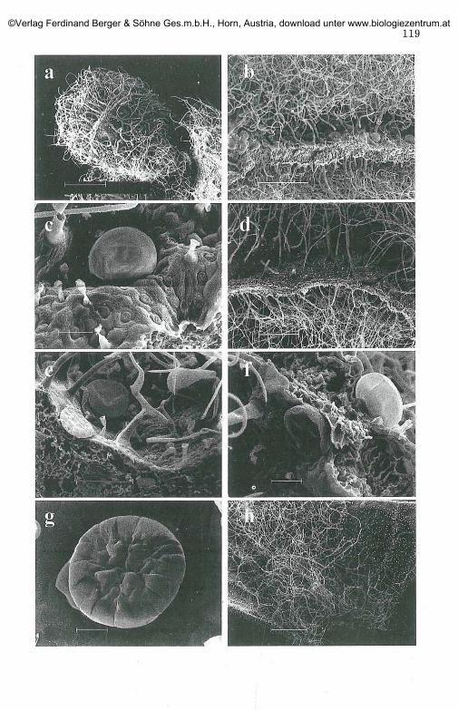
Fig. 4. The reproductive stage.(SEM micrographs)a. successive bracts and enclosed calyx, bar: lmmb. outer calyx with enclosed part of the corolla's tube, bar: lmmc. inner calyx, bar: 500 urnd. closed corolla covered by trichomes inside partly destroyed calyx, bar: 500 (ime. corolla's tube and part of bilabiate mouth, bar: 1 mmf. protruded connective connects the papillosous pollen sacs, bar: 200 [img. "hairy" base of the outer bract, bar: 100 (amh. peltate hair surrounding by floral hairs on outer bract, bar: 50 |ami. glandular and non-glandular hairs on the outer tubular part of the corolla,
bar:100 |imj . lobes of the bilabiate corolla's mouth, bar: 500 (amk. papillate-cells on inner corolla, bar: 20 (im1. glands on the underside of the anther's connective, bar: 100 \imm. cuticle's detachment of floral glandular trichomes, bar: 20 jamn. unicellular, cuticular ornamented hairs on inner corolla, bar: 20 jamo. "tuberculate" hair on inner corolla, bar: 20 (imp. glands and papillate-cells on the underside of the anther's connective, bar: 20 (im
©Verlag Ferdinand Berger & Söhne Ges.m.b.H., Horn, Austria, download unter www.biologiezentrum.at

123©Verlag Ferdinand Berger & Söhne Ges.m.b.H., Horn, Austria, download unter www.biologiezentrum.at

124
0. dictamnus are identical to those reported for Mona.7'da fistulosa (HEIN-RICH 1973), O. syriacum (DUDAI & al. 1988), O. vulgäre (WERKER & al.
1985a) and O. calcaratum (VRACHNAKIS 2002).The head of the peltate hairs (characteristic for Labiatae: "Labiatae-
Drüsen" KLUG 1926) in O. dictamnus was found to consist of up to 16 cells,while BOSABALIDIS & TSEKOS 1982 reported a 12-celled head. The density ofthe peltate hairs on developed leaves agrees with the findings of IETSWAART1980, BOSABALIDIS 1990. The appearance of the head content as emulsioncorrelated with the reaction of paraffin oil and Oil Red indicates thedifferent nature of the secretion (hydrophilic and lipophilic). HUSAIN & al.1982 report too a hydrophilic nature, whilst BOSABALIDIS 1990 consider thatthe subcuticular space is filled entirely with essential oil. Both natures forthe enclosed material are reported for O. x intercedens (BOSABALIDIS & al.1998) and O. calcaratum (VRACHNAKIS 2002). The role of the peltate hairs,to be the "first line of defence" against herbivores (KELSEY & al. 1984),seems to be not the case with O. dictamnus. Due to their hidden charac-ter and the absence of evident secretion of the phytotoxic (BROWN &al. 1987) components out of the cuticle, the peltate hairs are rather pro-tected than providing protection to the plant (WERKER 2000, VRACHNAKIS2002). For these trichomes DUKE 1994 points out that there is no evidencefor a direct function as a physical impedance to insects, and this isconfirmed in O. dictamnus due to the impermeable "barrier" of theeglandular hairs. The undamaged, reddish, peltate hairs of this plant arealso reported from herbarium material by BOSABALIDIS & TSEKOS 1982,HUSAIN & al. 1990.
The appearance of trichome-hydathodes (salt glands: WERKER 2000) inthe early stage of O. dictamnus correlated with their bending position and
Fig. 5. LM micrographs: a.-o.; Stereomicroscope: p.a. trichomes on first leaf primordia, bar: 100 |jmb. trichome-hydathodes secreting hydrophilic droplets, with paraffin-oil, bar: 30 |imc. stalked glandular (right) and 1-celled eglandular hair on a young leaf, bar: 30 |imd. hairy calyx-throat from sample 1 (mount Giouchtas), with Oil Red, bar: 100 jame. "fragile" floral glandular trichomes on bract, with paraffin-oil, bar: 30 (imf. floral glandular trichomes on bract reacting with Oil Red, bar: 25 \\mg. branched eglandular and 8-celled peltate hair on a young leaf, bar: 25 (.imh. peltate hair on bract, bar: 25 (im1. peltate hair on leaf emulsified of different osmiophilic droplets, bar: 25 |imj . peltate hair on bract, with paraffin-oil, bar: 25 jamk. 12-celled peltate hair on flower primordia, with Oil Red , bar: 30 urn1. capitate and peltate hair on outer corolla, with Oil Red, bar: 20 |imm. floral hairs on calyx primordium, with paraffin-oil, bar: 100 (imn. branched glandular hair on the outer corolla's tube, bar: 15 |imo. "tuberculate" hair on inner papillosous corolla, with Oil Red, bar: 5 (imp. glands and pollen-grains contrasting in colour the anther's lobes, bar: 400 (im
©Verlag Ferdinand Berger & Söhne Ges.m.b.H., Horn, Austria, download unter www.biologiezentrum.at

121
©Verlag Ferdinand Berger & Söhne Ges.m.b.H., Horn, Austria, download unter www.biologiezentrum.at

126
their unique hydrophilic secretion confirm their function as active hy-dathodes. RENNER 1909 relates the basiscopic orientation (bending posi-tion) of these trichomes, as opposed to the original acroscopic one of theglandular trichomes, with a special function and NESTLER 1893 points outthat the capitula of small trichome-hydathodes may be appressed. Theearly forming and functioning of the trichome hydathodes in immatureorgans as well their short-life is reported by MIELKE 1891, UPHOF 1962 andFAHN 1979. ZIEGENSPECK 1949 reported Trichomhydathoden in all the59 species of Labiatae investigated. Their role as active hydathodes is re-ported for Monarda fistulosa, Mentha piperita (HEINRICH 1973a, 1977);Teucrium (BINI-MALECI & SERVETTAZ 1991); Scutellaria altissima (THALER &al. 1992); Melissa officinalis (SCHULTZE & al. 1992); Nepeta racemosa(BOURETT & al. 1994); Salvia officinalis (CORSI & BOTTEGA 1999) and O. cal-caratum (VRACHNAKIS 2002).
The variability of the other capitate glandular hairs concerning theshape and the nature of secreted material is common for all Labiatae so farinvestigated (KELSEY & al. 1984, WERKER 1993, 2000). In regard their life-span, UPHOF 1962 stated for Labiatae that in old leaves these hairs areusually shed, but the scars remain visible, while in Hyssopus officinalis the1 to 4-cellular capitate hairs persist for a long time (TUNMANN 1906).
The branched eglandular hairs, that envelop the leaves of O. dictam-nus with a mat, thicker than the leaf itself, are unique in Origanum but notrare within Labiatae (MEFCALFE & CHALK 1950, EL-GAZZAR & WATSON
1970). A thick indumentum is a common feature of plants from medi-terranean climate and steppes, deserts, and alpine habitats (HABERLANDT1884, WARMING 1909, EHLERINGER 1984). The first appearance of theeglandular hairs as conical papillae is also reported for Lamium album(RAUTER 1871), Satureja thymbra (WERKER 2000), O. calcaratum (VRACHNA-
KIS 2002), while in the case of Coridothymus capitatus they are consideredas another type of non-glandular trichomes (ECONOMOU-AMILII & al. 1982).The occasionally presence of long unbranched eglandular hairs in the ca-lyx throat, without taxonomic value, is not rare within other "hairy" Or-iganum (IETSWAART 1980, CARLSTRÖM 1984). Besides the variability of theeglandular hairs in O. dictamnus their defence against various externalfactors is evident (LEVIN 1973, JOHNSON 1975, HOLMES & KEILLER 2002).Their potential function to protect the glandular hairs and their utilityeven when dead is reported for many plant species (UPHOF 1962, WERKER
2000).
The reduced pubescence (as compared to leaves) and the remarkablecolouration of the bracts is also reported for O. vulgäre occurring inGreece (KOKKINI & al. 1994) and for O. calcaratum (VRACHNAKIS 2002). Theouter calyx of O. dictamnus, densely covered by glands in contrast to theglabrous inner one, is common within Labiatae and is associated with the
©Verlag Ferdinand Berger & Söhne Ges.m.b.H., Horn, Austria, download unter www.biologiezentrum.at

127
leaf hairiness (EL-GAZZAR & WATSON 1970, CANTINO & SANDERS 1986,MATTERN & VOGEL 1994). The role of the "hairy" outer calyx is rather theproduction of floral fragrances (MEEUSE 1992, MATTERN & VOGEL 1994)than the protection of the enclosed parts of the corolla (WERKER 2000),due to the "fragileness" of the numerous floral hairs which are "ready"to release the secretion even with a light wind. The proposed alluringfunction of the floral hairs is in agreement with the interference of theirsecretion with the nectar and with their "pump-like" character, sug-gested for O. calcaratum (VRACHNAKIS 2002). The "pump-like" characterof similar anatomical floral attractants is reported by FAHN 1979; whileMATILE & ALTENBURGER 1998 point out the rhythmicity of fragranceemission in flowers. Peltate hairs on the outer corolla of O. dictamnus,in agreement with finding of MATTERN & VOGEL 1994, are also reportedfor O. syriacum, O. vulgäre, Salvia sp. and Leonotis leonorus (WERKER &al. 1985a,b, DUDAI & al. 1988, ASCENSAO & al. 1995). The branchedglandular hairs are also reported (but with one glandular branch) forMeriandra (BOKHARI & HEDGE 1971), Phlomis (AZIZIAN & CUTLER 1982),Hyptis (RUDALL 1980), Rosmarinus officinalis (WERKER & al. 1985c).Integrated glandular/eglandular hairs are reported on both corolla'ssides of O. calcaratum (VRACHNAKIS 2002), while UPHOF 1962 points outthat in Labiatae a confusion between the two kinds of hairs cannot beexcluded. The highly differentiated hairs inside the corolla are commonin Lamiaceae (WERKER 2000) and according to UPHOF 1962, abnormalhairs are not rare in strongly pubescent plants. Trichomes with bluntends and warty cuticle were also reported for the inner corolla ofMelissa officinalis (SCHULTZE & al. 1992) and in the inner flower spur ofTropaeolum majus (RACHMILEVITZ & FAHN 1975). The unicellular clubforming hairs with several knobs, called "tuberculate" by SOLEREDER &MEYER 1928, are reported on petals in Viola tricolor and on flower partsof Vinga minor, Capparis, Digitalis, Mentha, Veronica and Verbascum(KURER 1917). Similar trichomes reported on the inside of the corollatube of Lavandula officinalis (WERKER 1993). Glandular hairs on theunderside of the connective and trichomes protruding from the antherswere reported in O. calcaratum (VRACHNAKIS 2002). Peltate hairs wereobserved on the underside of the anther lobes in O. syriacum (DUDAI &al. 1988), Leonotis leonorus (ASCENSAO & al. 1995) and in Cannabis sa-tiva (MAHLBERG & al. 1984). The peculiarities of the reproductive organsand in particular of the trichomes can be related with the pollinationecology of O. dictamnus, as in the Lamiaceae generally (HUCK 1992,PETANIDOU & VOKOÜ 1993). Hairs inside the corolla can act as osmopho-res (FAHN 1979), or serve as a guiding arrangement for nectar-searchinginsects (SCHULTZE & al. 1992). LUNAU 2000 suggests a signalling function"by an expansion of connectives", in species other than of Labiatae.
©Verlag Ferdinand Berger & Söhne Ges.m.b.H., Horn, Austria, download unter www.biologiezentrum.at

128
The remarkable floral colouration (red to violet-purple) of O. dic-tamnus, like most Labiatae, is caused by anthocyanins (HARBORNE 1992).Although bees (main pollinators for phryganic Labiatae: PETANIDOU 1996)are insensitive to red colours, they can receive a visual signal by con-trasting colours (HARBORNE 1993). The distinct white pollen-grains andthe glands contrasting with the deep purple anther, as well as the dis-tinct venation of the membranous bracts of O. dictamnus, which canalso be found in O. calcaratum (VRACHNAKIS 2002), can act as visualsignal and honey guides for the pollinators (HARBORNE 1993, LUNAU2000). The association of the colouration (anthocyanins) with papillate-cells and "hairless" epidermis is identical for O. calcaratum (VRACHNAKIS2002). Ordinary epidermal cells that undergo an increase in surfaceextension with cuticular striations (papillate-cells) are the rule inreproductive organs (GLOVER & MARTIN 2000) and the ability of theircuticular striations to enhance light absorption and focusing/reflectingthe light into pigment-containing areas is pointed out by GORTON &VOGELMANN 1996. Presence of anthocyanins in young non-floral tissues isreported for other "hairy" plants (NTEFIDOU & MANETAS 1996, NEIL &
GOULD 1999). The defensive role of anthocyanins against herbivory andpathogen attack in the glabrous early stage (contrary to their alluringfunction on flowers) is suggested for O. calcaratum (VRACHNAKIS 2002),in agreement with LAMBERS & al. 1998, NEILL & al. 2002. The ability ofthe papillate-cells for secretion (as those on stigma and tapetum), andthe confusion of glandular/eglandular hairs, which start their develop-ment as papillae, makes the division between trichomes in O. dictamnusa difficult task, or according to UPHOF 1962, "the decision to whichgroup the hairs are to be referred, will always remain more or less sub-jective".
Concluding Remarks
As a plant which thrives in arid environments from sea level up to thealpine zone O. dictamnus needs a plasticity in the behaviour, necessary foradaptation in diverse abiotic and biotic factors. To cope with the adverseconditions the epidermis is equipped with specific structures which mayact also as receptor for beneficial signals. The results of these needs are themultiforming trichomes. The trichome types are genetically linked, buttheir development and distribution is forced by environmental demands.In the reproductive stage the ephemeron makes the more easy (in term oftime) changes or peculiarities of hairs. Besides the proposals of the ecolo-gical significance of plant trichomes, comparative studies from differentdisciplines will allow us to understand better such hairy aromatic plantswhich are confined in isolation (endemic) but thrive in a wide range ofenvironments.
©Verlag Ferdinand Berger & Söhne Ges.m.b.H., Horn, Austria, download unter www.biologiezentrum.at

129
Acknowledgemen t s
Prof. Dr. G. HEINRICH for corrections and comments on the manuscript, Ass.-Prof. Dr. E. STABENTHEINER for guidance on SEM, Dr A. PERKTOLD for technical sup-port on figures and Mr. V. MAUROSKOTIS for providing material of O. dictamnus.
References
ASCENAO L., MARQUES N. & PAIS M. S. 1995. Glandular trichomes on vegetative and
reproductive organs of Leonotis leonurus (Lamiaceae). - Ann. Bot. 75: 619-626.
•— , MOTA L. & CASTRO M. DE M. 1999. Glandular trichomes on the leaves andflowers of Plectranthus ornatus: morphology, distribution and histochemistry.- Ann. Bot. 84: 437-447.
AZIZIAND. & CUTLER D. F. 1982. Anatomical, cytological and phytochemical studieson Phlomis L. and Eremostachys Bunge (Labiatae). - Bot. J. Linn. Soc. 85:249-281.
BINI-MALECI L. & SERVETTAZ O. 1991. Morphology and distribution of trichomes inItalian species of Teucrium sect. Chamaedrys (Labiatae)-a taxonomical eva-luation. - PL Syst. Evol. 174: 83-91.
BOKHARI M. H. & HEDGE I. C. 1971. Observations on the tribe Meriandreae of theLabiatae. - Notes Roy. Bot. Gard. Edinburgh 31: 53-67.
BOSABALIDIS A. M. 1987. Morphometric evaluation of inclusion body-containing leu-coplasts in leaf epidermal cells of Origanum dictamnus L. - Bot. Helvetica 97/2: 315-321.
— 1990. Quantitave aspects of Origanum dictamnus L. glandular scales. - Bot.Helvetica 100/2: 199-206.
— & TSEKOS I. 1982. Glandular scale development and essential oil secretion inOriganum dictamnus L. - Planta 156: 496-504.
— , GABRIELI C. & NIOPAS I. 1998. Flavone aglycones in glandular hairs of Or-iganum x intercedens. - Phytochemistry 49(6): 1549-1553.
BROWN J. T., HEGARTY P. K. & CHARLWOOD B. V. 1987. The toxicity of monoterpenes to
plant cell cultures. - Plant Sei. 48: 195-201.
BRUNN., COLSONM., PERRIN A. & VOIRINB. 1991. Chemical and morphological studies
of the effects of ageing on monoterpene composition in Menta x piperitaleaves. - Can. J. Bot. 69: 2271-2278.
BOURETT T. M., HOWARD R. J., O'KEEFE D. P. & HALLAHAN D. L. 1994. Gland develop-
ment on leaf surfaces of Nepeta racemosa. - Int. J. Plant Sei. 155(6): 623-632
CANTINO P. D. & SANDERS R. W. 1986. Subfamilial classification of Labiatae. - Syst.-Bot. 11(1): 163-185.
CARLSTRÖM A. 1984. New species of Alyssum, Consolida, Origanum & Umbilicus fromthe SE Aegean Sea. - Wildenowia 14: 15-26.
CORSI G. & BOTTEGA S. 1999. Glandular hairs of Salvia officinalis: new data on mor-phology, localization and histochemistry in relation to function. - Ann. Bot. 84:657-664.
DUDAI N., WERKER E., PUTIEVSKY E., RAVID U., PALEVITCH D. & HALEVY H. 1988.
Glandular hairs and essential oils in the leaves and flowers of Majorana syr-iaca. - Isr. J. Bot. 37: 11-18.
©Verlag Ferdinand Berger & Söhne Ges.m.b.H., Horn, Austria, download unter www.biologiezentrum.at
130
DUKE S. O. 1994. Glandular trichomes - a focal point of chemical and structural in-teractions. - Int. J. Plant Sei. 155(6): 617-620.
ECONOMAKIS C, DEMETZOS C, ANASTASSAKIT., PAPAZOGLOU V., GAZOULIM., LOUKIS A.,
THANOS C. & HAEVALA C. 1999. Volatile constituents of bracts and leaves ofwild and cultivated Origanum dictamnus. -Planta Med. 65: 189-191.
ECONOMOU-AMILII A., VOKOUD., ANAGNOSTIDIS K., & MARGARIS N. S. 1982. Leaf mor-
phology of Thymus capitatus (Labiatae) by scanning electron microscopy. - In:MARGARIS N. S., KOEDAM A. & VOKOU D. (Eds.), Aromatic plants: basic andapplied aspects, pp. 13-24. - Martinus Nijhoff, The Hague:
EHLERINGER J. 1984. Ecology and ecophysiology of leaf pubescence in North Amer-ican desert plants. - In: EODRIGUEZE., HEALEYP. L. & METHAI. (Eds.), Biologyand chemistry of plant trichomes. - Plenum Press, pp. 113-133.
ELLIOTT R. 1966. Of marjorams and dittanies. - Bull. Alpine Gard. Soc. Gr. Brit. 34:198-205.
EL-GAZZAR A. & WATSON L. 1970. A taxonomic study of Labiatae and related genera.- New Phytol. 69: 451-486.
FAHN A. 1979. Secretory tissues in plants. - Academic Press.FAURE P. 1987. Parfüms et aromates de l'Antiquite'. - Editions A. Fayard, Paris.FRAGAKI E. 1969. Contribution in common naming of native, naturalised pharma-
ceutical, dye, ornamental and edible plants of Crete. - Athens (in Greek).GLOVER B. J. & MARTIN C. 2000. Specification of epidermal cell morphology. - In:
HALLAHAND. L. & GRAY J. C. (Eds.), Plant trichomes. - Advances in BotanicalResearch Vol. 31, pp. 193-217., Academic Press.
GORTON H. L. & VOGELMANN T. C. 1996. Effects of epidermal cell shape and pigmen-tation on optical properties of Antirrhinum petals at visible and ultravioletwavelengths. - Plant Physiol. 112: 879-888.
GREEN F. J. 1991. The Sigma-Aldrich handbook of stains, dyes and indicators,pp.656-657. - Aldrich Chemical Company, Inc., Milwaukee, Wisconsin.
HABERLANDT G. 1884. Physiologische Pflanzenanatomie. - Engelmann, Leipzig.HALACSYE. VON 1902. Conspectus florae graecae 2: 552-557. - Engelmann, Leipzig.HARBORNE J. B. 1992. Chemistry of flower colour in the Lamiales. - In: HARLEY R. M.
& REYNOLDS T. (Eds.), Advances in labiate science. - Royal Botanic Gardens,Kew. pp. 307-314.
•— 1993. Introduction to ecological biochemistry. - 4th edition. Academic Press.318 pp.
HARVALA C, MENOUNOS P. & ARGYRIADOU N. 1987. Essential oil from Origanum dic-tamnus. - Planta Medica 53(1): 107-109.
HAVAKIS I. E. 1980. Plants and herbs of Crete. - ZHTA Press, Athens, (in Greek).HEINRICH G. 1973. Entwicklung, Feinbau und Ölgehalt der Drüsenschuppen von
Monarda fistulosa. - Planta Med. 23: 154-166— 1973a. Die Feinstruktur der Trichom-Hydathoden von Monarda fistulosa. -
Protoplasma 77: 271-278.— 1977. Die Feinstruktur und das ätherische Öl eines Drüsenhaares von Mon-
arda fistulosa. - Biochem. Physiol. Pflanzen 77: 17-24.HOFMEISTER W. 1868. Allgemeine Morphologie. - Leipzig.HOLMES M.G. & KEILLER D.R. 2002. Effects of pubescence and waxes on the re-
flectance of leaves in the ultraviolet and photosynthetic wavebands: a com-parison of a range of species. - Plant Cell Environ. 25: 85-93.
©Verlag Ferdinand Berger & Söhne Ges.m.b.H., Horn, Austria, download unter www.biologiezentrum.at

131
HUCKR. B. 1992. Overview of pollination biology in the Lamiaceae. - In: HARLEY R. M.& REYNOLDS T. (Eds.), Advances in labiate science, pp. 167-181. - Royal Bota-nical Gardens, Kew.
HUSAINS. Z., HEYWOOD V. H. & MARKHAMK. R. 1982. Distribution of flavonoids of thegenus Origanum L. and related genera in Labiatae. - In: MARGARIS N. S.,KOEDAM A. & VOKOU D. (Eds.), Aromatic plants: basic and applied aspects,pp. 141-152. - Martinus Nijhoff, The Hague.
— , MARIN P. D., SILIC C, QAISER M. & PETROVIC B. 1990. A micromorphological
study of some representative genera in the tribe Saturejeae (Lamiaceae). - Bot.J. Linn. Soc. 103: 59-80.
IETSWAART J. H. 1980. Leiden botanical Series 4: A taxonomic revision of the genusOriganum (Labiatae). - Leiden University Press, Leiden.
INGWERSEN W. 1981. Some origanums for the garden. - Plantsman 3: 128-132.JOHNSONH. B. 1975. Plant pubescence: an ecological perspective. -Bot. Rev. 41: 233-
258.KATSIOTIS S. & OIKONOMOU G. N. 1986. Vergleichende Untersuchung verschiedener
wildwachsender und in Kreta angebauter Muster von Origanum dictamnus L.- Sei. Pharm. 54: 49-52.
KELSEYR., REYNOLDS G. W. & RODRIGUEZ E. 1984. The chemistry of biologically active
constituents secreted and stored in plant glandular trichomes. - In: RODRIGUEZE., HEALEY P. L. & MEHTA I. (Eds), Biology and chemistry of plant trichomes.pp. 187-241. - Plenum Press, New York.
KLUG J. 1926. Über die Sekretdrüsen bei den Labiaten und Compositen. - Diss.Frankfurt a.M.
KOKKINI S. 1997. Taxonomy, diversity and distribution of Origanum species. - In:PADULOSI S. (Ed.), Proceedings of the IPGRI International Workshop onOregano, 8-12 May 1996 CIHEAM. - Valenrano, Bari, Italy, pp: 2-13.
— , KAROUSOUR. & VOKOU D. 1994. Pattern of geographic variation of Origanumvulgäre trichomes and essential oils in Greece. - Biochem. Syst. Ecol. 22(5):517-528.
KURER G. A. 1917. Kutikularfalten und Protuberanzen an Haaren und Epidermen. -Diss. Zürich.
KYPRIOTAKIS Z. 1998. Contribution to the study of the chasmophytic flora of Crete,pp. 142-147. - Ph.D Thesis, University of Patras (in Greek with English sum-mary).
LAMBERS H., CHAPIN III F. S. & PONS T L. 1998. Enviromental effects on the produc-tion of secondary plant metabolites. - In: Plant Physiological Ecology, pp. 427-47. Springer.
LANGE D. & SCHIPPMANN U. 1997. Trade survey of medicinal plants in Germany (acontribution to international plant species conservation), 128 p. - Bundesamtfür Naturschutz 1997, Münster.
LEADLEYP. 1997. Conservation of Origanum spp. in botanical gardens. - In: PADULOSIS. (Ed.), Proceedings of the IPGRI International Workshop on Oregano, 8-12Mayl996 CIHEAM. - Valenrano, Bari, Italy, pp. 24-26.
LEVIN D. A. 1973. The role of trichomes in plant defence. - Quart. Rev. Biol. 48: 3-15.LUNAUK. 2000. The ecology and evolution of visual pollen signals. - Plant Syst. Evol.
222(1-4): 89-111.
©Verlag Ferdinand Berger & Söhne Ges.m.b.H., Horn, Austria, download unter www.biologiezentrum.at

132
MAHLBERG P. G., HAMMOND C. T., TURNER J. C. & HEMPHILL J. K. 1984. Structure, de-
velopment and composition of glandular trichomes of Cannabis sativa L. - In:RODRIGUEZ E., HEALEY P. L. & METHA I. (Eds.), Biology and chemistry of planttrichomes. - Plenum Press, pp. 23-53.
MATILE P. H. & ALTENBURGER R. 1998. Floral fragrance and its rhythmic emission inHoya carnosa and Stephanotis floribunda. - Asklepios 8-13.
MATTERN VON G. & VOGEL S. 1994. Lamiaceen-Blüten duften mit dem Kelch - Prüfungeiner Hypothese. I: Anatomische Untersuchungen: Vergleich der Laub- undKelchdrüsen. - Beitr. Biol. Pflanzen 68: 125-156.
MEEUSE A. D. J. 1992. Anthecology of the Labiatae: An armchair approach. - In:HARLEYR. M. & REYNOLDST. (Eds.), Advances in labiate science, pp. 183-191. -Royal Botanical Gardens, Kew.
MEFCALFE C. R. & CHALK L. (Eds.) 1950. Labiatae. Anatomy of the DicotyledonsVol. II. - Oxford University Press, Oxford, pp. 1041-1053.
MIELKE G. 1891. Anatomische und physiologische Beobachtungen an den Blätterneiniger Eucalyptus-Arten. - Diss. Jena.
NEILL S. O. & GOULD K. S. 1999. Optical properties of leaves in relation to antho-cyanin concentration and distribution. - Can. J. Bot. 77: 1777-1782.
— , GOULD K. S., KILMARTIN P. A., MITCCHELL K. A. & MARKHAM K. R. 2002. Anti-
oxidant activities of red versus green leaves in Elatostema rugosum. - PlantCell Environ. 25: 539-547.
NESTLER A. 1893. Die Perldrüsen von Artante cordifolia. - Österr. bot. Zeitschr. 43:333.
NTEFIDOU M. & MANETAS Y. 1996. Optical properties of hairs during the early stages ofleaf development in Platanus orientalis. - Aust. J. Plant Physiol. 23(4): *535-538.
OHLOFF G. 1992. Auf der Duftspur zum Abendland: Kreta und Mykene /Der Kräu-terdurft des Minos. In: Irdische Düfte - himmlische Lust. Eine Kultur-geschichte der Duftstoffe, pp: 71-96. - Birkhäuser Verlag.
PATON A. 1994. Three membranous-bracted species of Origanum. - Kew Mag. 11(3):109-117.
PETANIDOU T. 1996. Labiatae: A key family for wild bees and the pollination ecologyin mediterranean phryganic communities. - Lamiales Newsl. 4: 4-6.
— & VOKOU D. 1993. Pollination ecology of Labiatae in a phryganic (East Medi-terranean ) ecosystem. - Am. J. Bot. 80(8): 892-899.
PFITZER E. 1872. Über die mehrschichtige Epidermis und das Hypoderma. - Jahrb. f.wiss. Bot. 8: 2.
PLATAKIS E. 1975. O Diktamos tis Kritis {Origanum dictamnus L.). - 2nd edit. AlexiouVer. Iraklion, Kreta (in Greek).
RACHMILEVITZ T. & FAHN A. 1975. The floral nectary of Tropaeolum majus L. Thenature of the secretory cells and the manner of nectar secretion. - Ann. Bot. 37:1-9.
RAUTER J. 1871. Zur Entwicklungsgeschichte einiger Trichomgebilde. - Denkschr.Akad. Wiss. Wien 31: 2.
RENNER O. 1909. Morphologie und Ökologie der pflanzlichen Behaarung. - Flora 99.RUDALLP. J. 1980. Leaf anatomy of the subtribe Hyptidinae (Labiatae). -Bot. J. Linn.
Soc. 80: 319-340.
©Verlag Ferdinand Berger & Söhne Ges.m.b.H., Horn, Austria, download unter www.biologiezentrum.at

133
SCHULTZE W., ZÄNGLEIN A., HOSE S., KUBECZKAK. H. & CZYGANF. C. 1992. Volatiles in
flowers of balm (Melissa officinalis L.). - In: HARLEY R. M. & REYNOLDS T.(Eds.) Advances in labiate science. - Royal Botanic Gardens, Kew. pp. 357-367.
SKRUBISB. 1979. Origanum dictamnus L., a Greek native plant. - J. Ethnopharmacol.1: 411-415.
SKOULAM. & KAMENOPOULOS C. 1997. Origanum dictamnus L. and Origanum vulgäreL. subsp. hirtum (Link) Ietswaart: traditional uses and production in Greece. -In: PADULOSI S. (Ed.), Proceedings of the IPGRI International Workshop onOregano, 8-12 Mayl996. - CIHEAM, Valenrano, Bari, Italy, pp: 26-33.
— , GOTSIOU P., NAXAKIS G. & JOHNSON C. B. 1999. A chemosystematic investiga-tion on the mono- and sesquiterpenoids in the genus Origanum (Labiatae). -Phytochemistry 52: 649-657.
SOLEREDER H. & MEYER F. J. 1928. Systematische Anatomie der Monokotyledonen. -Berlin.
THALER I., GAILHOFER M. & PFEIFHOFER H. W. 1992. Proteinkörper in Drüsenhaarenvon Scutellaria altissima (Lamiaceae). - Phyton 31: 263-280.
TUCKER A. O. & ROLLINS E. D. 1989. The species, hybrids, and cultivars of Origanum(Lamiaceae) cultivated in the United States. - Baileya 23(1): 14-27.
TUNMANN O. 1906. Beiträge zur Kenntnis der Hautdrüsen.- Ber. deut. Pharm. Ges. 18.TURLAND N. J., CHILTON L. & PRESS J. R. 1993. Flora of the Cretan area. Annotated
checklist & atlas, pp. 97-98. - The Natural History Museum, LondonUPHOF J. C. T. 1962. Plant hairs. Enncyclopedia of plant anatomy IV; Vol. 5, pp. 1-206.
- Gebrüder Borntraeger, Berlin, Nikolassee.VALENTINI G., ARNOLD N., BELLOMARIAB. & ARNOLD H. J. 1991. Study of the anatomy
and of the essential oil of Origanum cordifolium, an endemic of Cyprus. -J. Ethnopharmacol. 35: 115-122.
VRACHNAKIS T 2002. On the epidermal elements of Origanum calcaratum Juss. (La-biatae). - Phyton 42 (1): 39-68.
WARMING E. 1909. Oecology of plants: an introduction to the study of plant commu-nities. - Oxford University Press, London.
WERKER E. 1993. Function of essential oil-secreting glandular hairs in aromaticplants of the Lamiaceae - A Review. - Flavour Fragr. 8: 249-255.
— 2000. Trichome diversity and development. - In: HALLAHAN D. L. & GRAY J. C.(Eds.), Advances in botanical research-plant trichomes, Vol. 3, pp. 1-35. -Academic Press.
— , PUTIEVSKYE. & RAVID U. 1985a. The essential oils and glandular hairs in dif-ferent chemotypes of Origanum vulgäre L. - Ann. Bot 55: 793-801.
— , RAVID U. & PUTIEVSKYE. 1985b. Glandular hairs and their secretions in thevegetative and reproductive organs of Salvia sclarea and S. dominica. - Isr. J.Bot. 34: 239-252.
— , — •& 1985c. Structure of glandular hairs and identification of the maincomponents of their secreted materials in some species of the Labiatae. - Isr. J.Bot 34: 31-45.
ZAGANIARIS D. 1940. La Flore de Porös. - In: POLITIS J. (Ed.), Actes de l'Institut Bo-tanique del'Universite' d'Athenes. - Tome I., Pyrsos S.A., Athenes, pp. 235-251.
ZIEGENSPECK H. 1949. Zur Phylogenie der Hydathoden. - Phyton 1: 302-317.
©Verlag Ferdinand Berger & Söhne Ges.m.b.H., Horn, Austria, download unter www.biologiezentrum.at