Embed Size (px)
Citation preview

T.R.N.C.
NEAR EAST UNIVERSITY
INSTITUTE OF HEALTH SCIENCES
THE EFFECT OF AMITRIPTYLINE ON EQUINE SERUM
BUTYRYLCHOLINESTERASE: KINETIC STUDIES AND DOCKING
CALCULATIONS
Sani Muhammad UZAIRU
MEDICAL BIOCHEMISTRY PROGRAM
MASTER OF SCIENCE THESIS
NICOSIA
2017
T.R.N.C.
NEAR EAST UNIVERSITY
INSTITUTE OF HEALTH SCIENCES
THE EFFECT OF AMITRIPTYLINE ON EQUINE SERUM
BUTYRYLCHOLINESTERASE: KINETIC STUDIES AND DOCKING
CALCULATIONS
Sani Muhammad UZAIRU
MEDICAL BIOCHEMISTRY PROGRAM
MASTER OF SCIENCE THESIS
SUPERVISOR
Asst. Prof. Dr. Kerem TERALI, PhD
NICOSIA
2017

iii
The Directorate of Health Sciences Institute,
This study has been accepted by the Thesis Committee in Medical Biochemistry Program
as a Master of Science Thesis.
Thesis committee:
Chair: Professor Nazmi ӦZER, PhD
Near East University
Supervisor: Assistant Professor Kerem TERALI, PhD
Near East University
Member: Professor Naciye Leyla AҪAN, PhD
Hacettepe University
Approval:
According to the relevant articles of the Near East University Postgraduate study-
Education and Examination Regulations, this thesis has been approved by the above
mentioned members of the thesis committee and the decision of the Board of Directors of
the institute.
Professor K. Hünsü Can BAŞER, PhD
Director of Graduate School of Health Sciences

iv
DECLARATION
I hereby declare that the work in this thesis entitled “THE EFFECT OF
AMITRIPTYLINE ON EQUINE SERUM BUTYRYLCHOLINESTERASE:
KINETIC STUDIES AND DOCKING CALCULATIONS” is the product of my own
research efforts undertaken under the supervision of Assistant Professor Kerem Terali. No
part of this thesis was previously presented for another degree or diploma in any university
elsewhere, and all information in this document has been obtained and presented in
accordance with academic ethical conduct and rules. All materials and results that are not
original to this work have been duly acknowledged, fully cited and referenced.
Name, Last Name:
Signature:
Date:

v
ACKNOWLEDGEMENT
Utmost gratitude goes to Almighty Allah. And without further ado, some special mention
has to be made of Prof. (Dr.) Nazmi Ӧzer (Chair: Department of Medical Biochemistry),
Prof. (Dr.) Hamdi Ogus, Assoc. Prof. (Dr.) Ӧzlem Dalmizrak and ultimately, my
supervisor, Asst. Prof. (Dr.) Kerem Terali for their generous contributions towards the
success of this work.
Asst. Prof. (Dr.) Kerem Terali particularly has been of tremendous resourcefulness and
support.
Equally deserving of gratitude is Dr. Abubakar Labaran Yusuf, BMBch, FCMP, as well
as the duo of Professor Muhammad Atiku Kano and Professor Alhassan Muhammad
Wudil who groomed me whilst at Bayero University, Kano as an undergraduate student.
Worthy of note also is my single mother who defied financial odds to support my
education up to the secondary school level, from where a string of scholarships brought
me this far.
Finally, I wish to express my indebtedness to my laboratory colleague, Qendresa Hotti,
and everyone that bought into and supported my vision to pursue M.Sc.

vi
ABSTRACT
Uzairu S.M. The Effect of Amitriptyline on Equine Serum Butyrylcholinesterase:
Kinetic Studies and Docking Calculations. Near East University, Institute of Health
Sciences, Medical Biochemistry Program, M.Sc. Thesis, Nicosia, 2017.
Butyrylcholinesterase, a serine hydrolase, is an important detoxification enzyme with
unique roles in cholinergic function. In this study, the effect of amitriptyline on the
hydrolysis of butyrylthiocholine by butyrycholinesterase, purified from equine serum, was
investigated using kinetic and molecular docking procedures. Amitriptyline (0.244 µM –
125 µM) inhibited the activity of butyrylcholinesterase in a dose dependent fashion with
an IC50 of 10 µM. Lineweaver‒Burk plot and the secondary replots of Dixon revealed a
linear mixed-type inhibition with a predominantly competitive nature. The inhibitory rate
constant, Ki, was 2.25 ± 0.66 µM whilst the Vm and α were found to be 1070 ± 28 U mg-1
protein, and 7.34 ± 1 µM, respectively. Analysis of kinetic constants showed that
amitriptyline triggered more than 3-fold decline in butyrylcholinesterase’s affinity for
butyrylthiocholine. This is even as the Ks was discovered to have rose from 0.169 ±0.019
to 0.551±0.028 mM. Amino acid sequence alignment of equine and human serum
butyrylcholinesterase indicated a 90.4% sequence identity. Docking study revealed that
the Phe329 and Trp231 of the lowest energy cluster established an effective ᴫ‒ᴫ stacking
with the aryl moiety of amitriptyline. The subtle noncompetitive component was
discovered to have resulted from an electrostatic interaction (salt bridge) between
amitriptyline and Asp70 of the peripheral anionic residue. Amyloid beta peptides are
presumed to deposit at the peripheral anionic site, hence a target in anti-AD therapy. Taken
together, amitriptyline exerted strong inhibitory potency against butyrylcholinesterase
slightly above therapeutic doses; and hence, could be optimized for therapeutic utility in
Alzheimer disease.
Keywords: amitriptyline, butyrylcholinesterase, linear mixed type inhibition, Alzheimer
disease

vii
ÖZET
Uzairu S.M. At Serum Butirilkolinesteraz Amitriptilinin Etkisi: Kinetik
Araştırmalar ve Doklama Hesaplamaları. Yakın Doğu Üniversitesi, Sağlık Bilimleri
Enstitüsü, Tıbbi Biyokimya Programı, Yüksek Lisans Tez, Lefkoşa, 2017.
Butilklolinesteraz, serin hidrolaz, kolinerjik fonksiyonda eşsiz rolü olan önemli bir
detoksifikasyon enzimidir. Bu çalışmada, at serumundan arıtılmış butirilkolinesteraz ile
butiriltiokolin hidrolizi üzerine amitriptilinin kinetik ve moleküler yerleştirme yöntemleri
üzerindeki etkisi araştırılmıştır. Amitriptilin (0.244 μM - 125 uM), butirilkolinesteraz'ın
aktivitesini 10 uM IC50 ile doz bağımlı bir tarzda inhibe etti. Lineweaver-Burk ve Dixon'ın
ikincil replotları ağırlıklı olarak rekabetçi nitelikte doğrusal bir karışık tip inhibisyonu
ortaya koydu. Engel hız sabiti Ki, 2.25 ± 0.66 μM iken, Vm ve α'nın sırasıyla 1070 ± 28 U
mg-1 protein ve 7.34 ± 1 μM olduğu bulunmuştur. Kinetik sabitlerin analizi amitriptilinin
butirilkolinesterazın bütiriltiokolin için olan afinitesinde 3 kattan fazla bir düşüşe neden
olduğunu gösterdi. Bu, Ks'nin 0.169 ± 0.019'dan 0.551 ± 0.028 mM'ye yükselmesi ile
sonuçlanmiştir. At ve insan serum butirilkolinesterazının amino asit dizilimleri,% 90.4'lük
bir dizi benzerliği gösterdi. Takma çalışması, en düşük enerji kümelenmesinin Phe329 ve
Trp231'inin amitriptilinin aril kısmı ile etkili bir ᴫ-ᴫ istifleme oluşturduğunu ortaya
koymuştur. Hafif rekabetçi olmayan bileşenin, periferik anyonik kalıntıya ait amitriptilin
ve Asp70 arasındaki bir elektrostatik etkileşimden (tuz köprüsü) kaynaklandığı
keşfedilmiştir. Amiloid beta peptitlerin periferik anyonik bölgede çökeldiği ve dolayısıyla
anti-AD terapisinde bir hedef olduğu varsayılmaktadır. Bu bulgular birlikte ele
alındığında, amitriptilin, butirilkolinesteraz'a karşı terapötik dozların birazcık üstünde
güçlü inhibisyon potensiyeli sergilediği gözlendi; Ve dolayısıyla, Alzheimer hastalığında
terapötik kullanım için optimize edilebileceği düşülebilir.
Anahtar Kelimeler: amitriptilin, bütirilkolinesteraz, lineer karışık tip inhibisyonu,
Alzheimer hastalığı

viii
TABLE OF CONTENTS
Page No
APPROVAL iii
DECLARATION iv
ACKNOWLEDGEMENT v
ABSTRACT vi
ӦZET vii
TABLE OF CONTENTS viii
SYMBOLS AND ABBREVIATIONS xi
LIST OF FIGURES xiv
LIST OF TABLES xvi
1. INTRODUCTION 1
2. GENERAL INFORMATION 4
2.1. Cholinesterase 4
2.2. Acetylcholinesterase 4
2.3. Butyrylcholinesterase 6
2.3.1. Molecular Structure of Butyrylcholinesterase 7
2.3.2. Differences in the Active sites of Cholinesterase 8
2.4. Genetic Variants of Butyrylcholinesterase 10
2.4.1. Atypical 10
2.4.2. K Variant 10
2.4.3. Flouride 10
2.4.4. Silent 11
2.5. Distribution of Butyrylcholinesterase in Human Tissues 11
2.6. Function of Butyrylcholinesterase 13
2.6.1. Detoxification 13
2.6.2. Acetylcholine Hydrolysis 13
2.6.3. Fat Metabolism 14
2.6.4. Scavenger of Polyproline-Rich Pepetides 14

ix
2.7. Neurodegeneration 14
2.7.1. The Cholinergic Hypothesis and The Role of Butyrylcholinesterase in 15
Alzheimer’s
2.7.2. The Amyloid Hypothesis 16
2.8. Amitriptyline 16
2.8.1. Pharmacokinetics of Amitriptyline 17
2.8.2. Pharmacodynamics of Amitriptyline 18
2.9. Molecular Docking 18
3.0 MATERIALS AND METHODS 20
2.10. Enzyme and Chemicals 20
2.11. Protein Concentration Determination 20
2.12. Reagents Preparation 20
3.3.1. 200 mM MOPS/KOH, pH 7.5, Volume 250 mL 20
3.3.2. 20 mM MOPS/KOH, pH 7.5, Volume 200 mL 20
3.3.3. Substrate (Butyrylthiocholine) and DTNB 21
3.3.4. Inhibitor (Amitriptyline) 21
3.3.5. Enzyme Dilution 21
3.4. Butyrylcholinesterase Activity Assay 21
3.5. Butyrylcholinesterase Inhibition Assay 22
3.6. Kinetic Studies of Butyrylcholinesterase Inhibition 23
3.7. Statistical Analysis 23
3.8. Homology Modeling 23
3.9. Molecular Docking 24
3.10. Protein‒Ligand Interaction Profiling 24
4. RESULTS 25
4.1. Substrate Kinetics 26
4.2. Effect of Amitriptyline on Butyrylcholinesterase Activity 29
4.3. Determination of the Reversibility or otherwise of Amitriptyline 32
Induced Inhibition of Butyrylcholinesterase
4.4. Effect of Amitriptyline on the Steady-State Kinetic 33

x
Behaviour of Butyrylcholinesterase
4.5. Homolgy Modeling 40
4.6. Binding of Amitriptyline to Butyrylcholinesterase 43
4.7. Superimposition of Amitriptyline and Butyrylcholinesterase 45
4.8. Binding of Amitriptyline and Butyrylthiocholine inside the Active Site 46
Site gorge of Butyrylcholinesterase
5. DISCUSSION 50
6. CONCLUSION 55
REFERENCES 56

xi
SYMBOLS AND ABBREVIATIONS
A: Adenine
Aβ: Beta Amyloid
ABCBAI: P-glycoprotein
ABP: Acyl Binding Pocket
ACh: Acetylcholine
AChE: Acetylcholinesterase
AD: Alzheimer Disease
AMI: Amitriptyline
APP: Amyloid Precursor Protein
AS: Acylation Site
Asp: Aspartate
BChE: Butyrylcholinesterase
BTCh: Butyrylthiocholine
C: Cytosine
CBS: Choline Binding Site
Ch: Choline
ChAT: Choline Acetyltransferase
ChE: Cholinesterase
CNS: Central Nervous System
CYP3A4: Cytochrome P450 34
CYP2C19: Cytochrome P450 2C19
CYP2D6: Cytochrome P450 2D6
dH20: Distilled Water
DTNB: 5,5I dithiobis (2-nitrobenzoic Acid)
E: Glutamine
EqBChE: Equine Butyrylcholinesterase
G: Guanine
F: Phenylalanine
GABAA: γ- aminobutyric Acid type A

xii
Glu: Glutamine
huAChE: Human Acetylcholinesterase
huBChE: Human Butyrylcholinesterase
His: Histidine
IC50: Half maximal Inhibitory Concentration
K+: Potassium
Kcat: Turnover Number
kDa: Kilo Dalton
Ki: Inhibition Constant
Km: Substrate Concentration at Half maximal Velocity
Ks: Dissociation Constant for Enzyme-Substrate Complex
KOH: Potassium Hydroxide
Leu: Leucine
MOPS: 3-(N-morpholino) Propane Sulfonic Acid
mRNA: Messenger RNA
Na+: Sodium
NaF: Sodium Flouride
NOR: Nortriptyline
PAS: Peripheral Anionic Site
PDB: Protein Data Bank
Phe: Phenylalanine
PLIP: Protein Ligand Interaction Profiler
sAPP: Soluble Amyloid Precursor Protein
Ser: Serine
SSRI: Selective Serotonergic
T: Thymine
TCA: Tricyclic Antidepressant
TcAChE: Torpedo carlifornica Acetylcholinesterase
TCh: Thiocholine
TNFα: Glutamate NMDA Receptor
Trp: Tryptophan
xiii
Tyr: Tyrosine
Val: Valine
Vm: Maximum Velocity
W: Tryptophan
Y: Tyrosine
5-HT2: 5 hydroxytryptamine receptors

xiv
LIST OF FIGURES
PAGE No
Figure 2.1. Hydrolysis of Acetylcholine 5
Figure 2.2. Human Acetylcholinesterase Tetramer 5
Figure 2.3. Human Butyrylcholinesterase Tetramer 6
Figure 2.4. Binding sites of Butyrylcholinesterase Monomer 7
Figure 2.5. Butyrylcholinesterase Gene Organization 8
Figure 2.6. The Active Sites of human acetylcholinesterase and 9
Human Butyrylcholinesterase
Figure 2.7. Chemical Structure of Amitriptyline 17
Figure 4.1. Michaelis‒Menten Plot of Butyrylthiocholine Hydrolysis 27
Figure 4.2. Double Reciprocal Plot of Butyrylthiocholine Hydrolysis 28
Figure 4.3. Dose Dependent Inhibition of Equine Butyrylcholinesterase 30
Figure 4.4. Hill Plot of Butyrylcholinesterase 31
Figure 4.5. Plot of Specific Activity versus Enzyme Concentrations 32
Figure 4.6. Michaelis‒Menten plot for the Inhibition of Equine 34
Butyrylcholinesterase by Amitriptyline
Figure 4.7. Lineweaver‒Burk Plot for the Inhibition of Equine 35
Butyrylcholinesterase by amitriptyline
Figure 4.8. Lineweaver‒Burk Secondary Plot of Intercept and Slope 36
against Amitriptyline Concentration
Figure 4.9. Dixon Plot of Butyrylcholinesterase Inhibition 37
Figure 4.10. Secondary Plot of Dixon Plot: Slope versus 1/[BTCh] 38
Figure 4.11. Secondary Plot of Dixon plot: 1/slope versus 1/[AMI] 39
Figure 4.12. Structural Model of Equine Butyrylcholinesterase 42
Figure 4.13. Close-up View of key Butyrylcholinesterase Active Site Residues 42
Figure 4.14. The Active Site Gorge of Butyrylcholinesterase with bound 43
Amitriptyline and its Interaction Profile
Figure 4.15. Interaction Profile of Amitriptyline inside the Active Site 44
Gorge of Butyrylcholinesterase
xv
Figure 4.16. Superimposition of Amitriptyline and Butyrylthiocholine inside 46
the Active Site gorge of Butyrylcholinesterase
Figure 4.17. The Active Site Gorge of Butyrylcholinesterase with bound 47
Amitriptyline and Butyrylthiocholine
Figure 4.18. Interaction Profile of Amitriptyline inside the Peripheral Anionic 48
Site of Butyrylcholinesterase Active Site Gorge.

xvi
LIST OF TABLES
PAGE No
TABLE 1. Summary of Butyrylcholinesterase Distribution in 12
Adult Human Body
TABLE 2. Kinetic Parameters for the Inhibition of 40
Butyrylcholinesterase by Amitriptyline
TABLE 3. Amino Acid Sequence of Equine Serum Butyrylcholinesterase 40
TABLE 4. Amino Acid Sequence Alignment of Equine and Human 41
Butyrylcholinesterase
TABLE 5. Relatively Strong Competitive Binding Interaction 45
Profile for Amitriptyline
TABLE 6. Binding Interaction Profile for Butyrylthiocholine 48
TABLE 7. Noncompetitive Binding Interaction Profile for Amitriptyline 49

1
1. INTRODUCTION
The World Health Organization (WHO) 2016 estimates put the prevalence of dementia at
47.5 million people worldwide. Fifty eight percent of these live in low and middle income
countries and the projections are that the proportion will shoot up to 71% by 2050. Overall,
135 million people would have suffered dementia by 2050, the report said; and, further
stated that 60%‒70% of these cases would have been caused by Alzheimer disease (AD),
the most common neurodegenerative disease.
Alzheimer disease is triggered by a progressive depletion of the neurotransmitter
acetylcholine (ACh) as a result of impaired activity of choline acetyltransferase (ChAT)
due to the significant loss of cholinergic neurons culminating in cognitive impairment and
memory loss (Mucke, 2009). Therefore, the cholinergic hypothesis, which propounded
that there is a material attenuation of cholinergic function in the brain of AD patients,
forms the nucleus upon which AD therapy is based. The approach is to restore the pool of
the cholinergic neurotransmitter acetylcholine through the use of reversible cholinesterase
inhibitors (Hitzeman, 2006).
Cholinesterase is broadly divided into acetylcholinesterase (AChE; EC: 3.1.1.7) and
butyrylcholinesterase (BChE; EC: 3.1.1.8). AChE is responsible for hydrolytic breakdown
of acetylcholine, and its inhibition has been validated as an efficient way to lessen some
behavioral and cognitive system disorders of AD (Citron, 2010). Presently, AChE
inhibitors like donepezil, galantamine, tacrine have been utilized for clinical treatment of
AD even though their effect is essentially palliative than curative (Shaikh et al., 2014).
Besides, their clinical effectiveness is limited by their deleterious side effect on the liver
and the peripheral nervous system in addition to their poor bioavailability and selectivity
(Toda et al., 2010). These, more than anything, underscore the need to beam the
searchlight on butyrylcholinesterase.
Butyrylcholinesterase is a tetrameric enzyme. Its monomers are arranged as a dimer of
dimers. Each dimer contains identical monomers linked by inter-chain disulfide bridge
(Lockridge et al., 1979). Each of the catalytic subunits of this homo tetramer has 574
amino acid residues. It consists of a catalytic triad made up of serine 198, glutamic acid

2
325 and histidine 438 (Lockridge et al., 1987). Aside the catalytic triad, the active site
gorge has an anionic site which can bond to the positively charged quaternary nitrogen of
the choline. The anionic site contains tryptophan 82. Three crucial aromatic amino acids
found in the peripheral anionic site (PAS) of AChE (Tyr 70, Tyr 121 and Trp 279) are
absent at the entrance of the active site gorge of BChE. For this reason, it is at times
speculated that BChE has no PAS, at least not one identical to AChE PAS (Masson et al.,
2009). Cationic substrates are guided into the active site gorge by communication with
tyrosine 332 and aspartic acid 70 which are found at the entrance of the gorge (Velom et
al., 1993; Soreq and Seidman, 2001). In BChE, lysine 286 and valine 288 line the acyl
pocket within the gorge and this is where the acyl groups are held during catalysis (Velom
et al., 1993).
BChE catalyzes the hydrolytic cleavage of butyrylcholine, acetylcholine, succinylcholine
etc. It also degrades esters of choline such as cocaine, heroin as well as scavenges
anticholinesterase (Lockridge et al., 1980; Galey, 1991; Masson, 1998). Curiously, whilst
the biochemical features of BChE are altered in AD, a significant amplification of its
activity has been noted. Research has revealed that the surge in the activity of BChE plays
a major role in amyloid beta (Aβ) aggregation in the early phases of senile plaque
formation (Anand and Singh, 2013). Given that BChE can also hydrolyze acetylcholine,
its inhibition might result in further advancement of cholinergic transmission.
Consequently, therapeutic candidates that could inhibit BChE could provide a novel
benefit and add value in AD treatment (Sim, 1999; Giacobini, 2000).
And this serves the bases for screening the inhibitory activity of the tricyclic
antidepressant, amitriptyline (AMI) against purified BChE. AMI is a selective
serotonergic re-uptake inhibitor (SSRI). It is degraded into nortriptyline which might
complement its effectiveness on norepinephrine re-uptake (Breyer-Pfaff, 2004). Merry
(1997) observed that AMI raised the expression of brain derived neurotrophic factor and
B-cell lymphoma 2 both of which possess neuroprotective effect. And in the central
nervous system of mammals, it enhanced neuronal regeneration (Chen et al., 1997).
According to Hu et al. (2003), the level of copper, zinc superoxide dismutase is increased
by AMI, and this serves a neuroprotective effect in the brain hippocampus.

3
There are reports in the literature suggesting that antidepressants inhibit cholinesterase
(Ahmed et al., 2008; Muller et al., 2002). Whilst Barcellos et al. (1998) epitomized that
imipramine and diazepam but not AMI inhibited AChE from the cerebral cortex of adult
rats in vitro, Nunes-Tavares et al. (2002) observed that AMI inhibits purified AChE from
Electrophorous electricus (the electric eel) through an interaction at its peripheral anionic
site. But most significantly, Cokugras and Tezcan (1997) speculated that AMI inhibits
serum BChE by either binding to its putative anionic site or possibly through an interaction
with the hydrophobic pocket near its active site; yet fell short of unravelling how the
inhibitor docks into either of those sites; and consequently, the veracity of such a
proposition requires validation.
Besides the fact that the kinetic behavior of purified BChE in the presence of AMI remains
unclear, their molecular mechanism of interaction is also unknown; and even yet, how this
interaction equally impacts on the cholinergic system exist only in the realms of
conjecture. And by virtue of the forgoing, the kinetic and molecular docking studies of
purified BChE with AMI become a fascinating subject to investigate, in the interest of
identifying viable therapeutic alternatives for AD treatment. Given the aforementioned
prerogatives therefore, the objectives of this study are to:
a. screen and evaluate the inhibitory action of AMI against BChE
b. identify the kinetic mechanism of the inhibition and compute kinetic parameters
c. elucidate the structural features and molecular bases for their binding as
well as interaction
d. relate the molecular interaction of AMI and BChE with the cholinergic system

4
2. GENERAL INFORMATION
2.1. Cholinesterases
Cholinesterases are classified under the protein family called serine hydrolases (P family).
This is due to their ability to utilize their catalytic site’s serine residue, a nucleophile, to
hydrolyze substrates. Cholinesterases have been distinguished into two basic groups, viz.:
acetylcholinesterase (AChE; EC: 3.1.1.7) and butyrylcholinesterase (BChE; EC: 3.1.1.8).
The former catalyzes the hydrolytic breakdown of its primary substrate, acetylcholine into
choline and acetate; whilst the latter, with its broad specificity catalyzes the hydrolytic
cleavage of acylcholines to choline and a weak acid. AChE also catalyzes the
transacetylation of variety of acetic esters (Silman and Sussman, 2005). The grounds for
this division hinges primarily on their specificities for their natural substrates, inhibitors,
tissue distribution and kinetic properties (Mesulam et al., 2002). The serine hydrolase
super family consists of myriad proteins involved in diverse physiological actions,
including blood clotting (Flemmig and Melzig, 2012), digestion (Whitcomb and Lowe,
2007) and most significantly neurotransmission (Pohanka, 2011). Even strikingly, many
of these cholinesterases have been implicated in pathological conditions, ranging from
pancreatitis, thrombosis to AD (Pohanka, 2011; Flemmig and Melzig, 2012). And in view
of the aforesaid, the focus has therefore shifted to cholinesterases as valid and prime
targets in drug discovery. It is therefore not surprising that the inhibitors of these enzymes
have been used in the therapy of myansthenia gravis (Mehndiratta et al., 2011) and AD
(Birks, 2006). Today, drugs like Exelon® (Novartis) for AD; Xarelto® (Bayer) for
thrombosis and Onglyza® (Bristol-Myers Squibb) for type 2 diabetes have been developed
from the inhibitors of this enzyme superfamily (Bachovchin and Cravatt, 2012).
2.2. Acetylcholinesterase
AChE is viewed as the key enzyme in the cholinesterase family. A serine hydrolase, AChE
is affiliated to the α/β hydrolase family (Nardini and Dijkstra, 1999). Getman et al. detailed
that AChE is encoded by a single gene located on the long arm of chromosome 7 at
position 7q22. AChE has narrow substrate specificity and acts classically on its
physiological substrate, acetylcholine (ACh) (Getman et al., 1992).

5
Figure 2.1. Acetylcholinesterase hydrolyzes acetylcholine into acetic acid and choline.
Adapted from Xu et al. (2008).
The crucial role AChE plays in nerve impulse by hydrolyzing the neurotransmitter
acetylcholine and terminating its actions in cholinergic system is well documented. This
notwithstanding, suggestions abound of its role as an adhesion protein in synaptic
development and maintenance (Munoz-Muriedas et al., 2004); and in addition, evidence
of its involvement in neurite growth are beginning to surface (Sharma et al., 2001). AChE
has also been figured in the promotion of pathological cluster of amyloid peptide into
amyloid fibrils in vitro (Bartolini et al., 2003) and in vivo (Rees et al., 2003) with
complexes of AChE and beta amyloid (Aβ) displaying acute neurotoxicity in relation to
fibrils formed by Aβ alone (Reyes et al., 2004).
Figure 2.2. Crystallographic assembly of Electrophorous electricus acetylcholinesterase
tetramer (AChE; PDB ID: 1C2O). Modified from Bourne et al. (1999).

6
2.3. Butyrylcholinesterase
BChE is a serine esterase that is predominant in the plasma and tissues of higher
vertebrates where it hydrolyzes a wide range of substrates. These substrates include but
not limited to acetylcholine, butyrylcholine and succinylcholine (Ringvold, 2005). BChE
has been severally referred to as non-specific choline esterase, pseudocholinesterase,
acylcholine acylhydrolase and choline esterase II (Asojo et al., 2011). Liston et al.
reported that BChE in contrast to AChE is highly active in the peripheral tissues as against
the brain (Liston et al., 2004). Also found in the neurons, it is however prevalent in serum
and glial cells (Darvesh and Hopkins, 2003). BChE has been linked to various
physiological processes like the hydrolysis of choline and non-choline esters such as
succinylcholine (Kaufman et al., 2011), acetylcholine (Mesulam et al., 2002), aspirin
(Masson et al., 1998) and cocaine (Xue et al., 2011), hence playing an important role in
anesthesia, neurotransmission and drug abuse.
More so, BChE has huge pharmacological and toxicological significance. This is informed
not only by its ability to hydrolytically cleave ester containing drugs as hinted in the
preceding paragraph; but also, by its capacity to serve as scavenger for ChE inhibitors like
potent organophosphorous nerve agents, just before they arrive at their synaptic targets
(Raveh et al., 1997).
Figure 2.3. Human butyrylcholinesterase tetramer. Adapted from Lockridge O. (2015).

7
2.3.1. Molecular Structure of Butylrylcholinesterase
A tetramer of four identical subunits, human BChE is a siaglycoprotein of 340 kDa
molecular weight. Each monomer has 574 amino acids and 9 linked asparagine complexed
glycans. The 198th residue from the amino terminal end is the active site serine. The
substrate binding sites of this enzyme protein consist of a peripheral anionic site and a
catalytic active site located deep inside a 20 Å gorge. Eight half-cystine is contained in
each subunit of the 574 amino acids of human BChE. Three internal disulphide bridges
are formed by six of these half-cystine viz.: cys65_cys92; cys252_cys263 and
cys400_cys519 (Lockridge et al., 1987).
Figure 2.4. Binding sites of BChE monomer. Adapted from Cokugras (2003).
This sialoglycoprotein has a sugar content of 24% by weight. The polypeptide chain has
a molecular weight of 65.1 kDa, however as a result of glycosylation it reaches to about
85 kDa on deactivating polyacrylamide gel electrophoresis. The higher amount of
glycosylation of BChE is responsible for its longer half-life (Nachon et al., 2002).
Human BChE is encoded by an autosomal gene located on the long arm of chromosome
3 at position 3q26.1_q26.2 (Allderdice et al., 1991). The BCHE gene is approximately 73
kb in length, and consists of 1,722 base pairs of coding sequence and four exons.

8
Figure 2.5. BCHE gene organization: Chromosome Location: chr3: 165,491,169-
165,548,821; the genome is the ruler that shows the nucleotide graphic coding for each
nucleotide. The gene represents the gene-structure on the genome, white boxes represent
the UTRs (untranslated regions) whilst the black lines connecting boxes represent introns.
Repeats show various repeat regions that have been annotated along the genome (A: green,
T: red, G: yellow, C: blue). Cytogenetic location: 3q26.1; Length coding sequence: 1806
nucleotides. Retrieved from http://www.rcsb.org/pdb/gene/BCHE, accessed 17-04-2017.
Exon 1 has untranslated sequences and two possible translation initiation domains at
codons 69 and 47. Exon 2 (1,525 base pairs) comprises 83% of the coding sequence for
mature protein and a third potential translation initiation at codon 28. Exon 3 is about 167
nucleotides in length, whilst exon 4 consisting of 604 base pairs codes for the carboxyl
terminal end of the protein and the 3I untranslated segment where two polyadenylation
signals have been discovered. Whilst intron 1 is about 6.5 kb long, the estimated minimum
sizes of introns 2 and 3 are about 32 kb each (Arpagaus, 1990).
2.3.2. Differences in the Active Sites of Human Cholinesterases
Even though AChE and BChE are products of distinct genes located on chromosomes 7
and 3 in human, they exhibit 65% homology in their amino acid sequence (Giancobini,
2004). AChE is considered to be the ancestral ChE in vertebrates (Chatonnet and
Lockridge, 1989). Additionally, Chatonnet and Lockridge (1989) posited that an early
gene duplication incident and its concomitant divergent structural and functional evolution
engendered the acetylcholinesterase and butyrylcholinesterase of higher vertebrates.
AChE contains a catalytic triad that consist of amino acids serine, histidine and glutamic
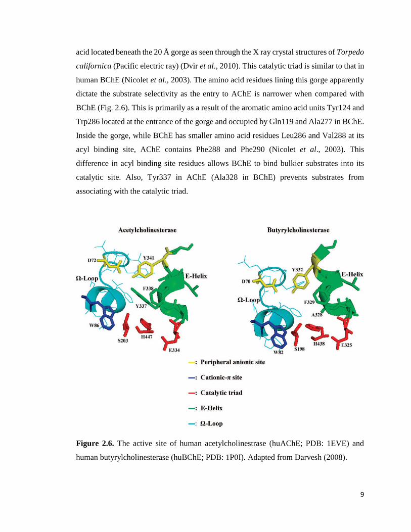
9
acid located beneath the 20 Å gorge as seen through the X ray crystal structures of Torpedo
californica (Pacific electric ray) (Dvir et al., 2010). This catalytic triad is similar to that in
human BChE (Nicolet et al., 2003). The amino acid residues lining this gorge apparently
dictate the substrate selectivity as the entry to AChE is narrower when compared with
BChE (Fig. 2.6). This is primarily as a result of the aromatic amino acid units Tyr124 and
Trp286 located at the entrance of the gorge and occupied by Gln119 and Ala277 in BChE.
Inside the gorge, while BChE has smaller amino acid residues Leu286 and Val288 at its
acyl binding site, AChE contains Phe288 and Phe290 (Nicolet et al., 2003). This
difference in acyl binding site residues allows BChE to bind bulkier substrates into its
catalytic site. Also, Tyr337 in AChE (Ala328 in BChE) prevents substrates from
associating with the catalytic triad.
Figure 2.6. The active site of human acetylcholinestrase (huAChE; PDB: 1EVE) and
human butyrylcholinesterase (huBChE; PDB: 1P0I). Adapted from Darvesh (2008).

10
2.4. Genetic Variants of Butyrylcholinesterase
The genetic polymorphism of BChE dictates: (i) its level of serum activity; (ii) its
functional properties such as its specificity and sensitivity for substrates. Different variants
exhibit different ability to hydrolyze certain xenobiotics; and conversely, are suppressed
at different rates when exposed to certain chemical nerve agents (Lockridge et al., 2016).
Thus far, succinylcholine is the only drug which obviously induces clinical defects in
persons with rare BChE genetic variant (Tunek and Svensson, 1988). Some of the
common genetic variants of BChE include: atypical, K variant, fluoride, and silent (La Du
et al., 1990).
2.4.1. Atypical
All atypicals that have been sequenced so far have a substitution at nucleotide 209, where
A replaces G and subsequently alters codon 70 from aspartic acid to glycine (McGuire et
al., 1989). Atypical BChE possess the kinetic features of an enzyme with deficient anionic
site (La Du et al., 1990). Lockridge and La Du (1978) reported that atypical BChE has a
low affinity for neutral substrates but a normal turnover number, Kcat. Apparently, it could
be safe to speculate that Asp 70 is an essential component of the anionic site.
2.4.2. K Variant
This variant is associated with 33% decrease in serum BChE activity (Rubinstein et al.,
1978). It is a result of linkage disequilibrium as it occurs on the same BChE chromosomal
strand carrying the atypical phenotype. The mutation leading to this genetic variant was
discovered at nucleotide 1615 where G was substituted with A, and the codon 539 altered
from alanine to threonine. Although the most common BChE mutation, the clinical
significance of this variant is only pronounced when it occurs in synchrony with atypical
BChE (Bartels et al., 1989).
2.4.3. Fluoride
The fluoride phenotype resists the inhibitor NaF. Two mutations that engender the fluoride
phenotype have been discovered, namely: Flouride-1 and Flouride-2. Flouride-1 occurs at
11
the nucleotide 728 where C is substituted with G, thereby altering codon 243 from
Threonine to Methionine. The other (Flouride-2) occurs at nucleotide 1169 with G
replaced by T, resulting in alteration of codon 390 from Glycine to Valine (La Du et al.,
1990). Fluoride-1 is the first fluoride mutation discovered through DNA sequencing and
the second being fluoride-2 (Nogueira et al., 1992).
2.4.4. Silent
The silent phenotype is marked by little or no BChE activity, or activity far below 2% of
normal (Rubinstein et al., 1970). Nogueira et al. discovered a type of silent mutation
referred to as silent-1 mutation; and it is a complicated mutation in which the T at
nucleotide 351 is substituted by two nucleotides, AG. Protein synthesis is prematurely
abated because a frame shift caused a stop codon to emerge at codon 129. A truncated
(22% the length of typical enzyme) and inactive BChE that has lost its active serine at
position 198 is therefore synthesized. The serum from this phenotype has no activity with
benzoylcholine and alpha-naphthylacetate; and besides, does not react with antibody
against human serum BChE (Nogueira et al., 1990). The likelihood of silent-2 has been
mooted; given that a different serum with the silent phenotype exhibited zero activity and
absence of cross-reactive material, yet did not contain the above mutation (Aspagaus et
al., 1990).
2.5. Distribution of Butyrylcholinesterase in Human Tissue
The plasma, liver, skin and leg muscle respectively are considered the best source of BChE
enzyme (Lockridge, 2014). Northern blot analysis (Jbilo et al., 1994) has revealed that the
most predominant human BChE mRNA was in the lung, liver, brain and heart. This
revelation is in sync with the relative BChE enzyme content in lung, liver and brain.
Curiously, the heart seemingly has more BChE mRNA than would be imagined from the
little enzyme content captured below.

12
Table 1. Summary of Butyrylcholinesterase enzyme distribution in adult human
body. Modified from Lockridge (2014).
Tissue Weight of tissue, g BChE, mg
Plasma 3,500 16
Liver 1,400‒1,500 13
Skin 4,000‒5,000 7
Leg muscle 3,500 6
Small intestine 800‒900
Lungs 400
Cerebral cortex 1200 3
Stomach 300 1.8
Spleen 150‒200 1
Kidney 130‒160 1
Cerebellum 150 0.8
Heart 300 0.6
Medulla oblongata 20‒25 0.085
Thyroid 20 0.0085

13
2.6. Functions of Butyrylcholinestrase
The existence of healthy individuals with ‘silent’ BChE aroused curiosity about the
physiological significance of the BChE enzyme. Girard et al. (2007) observed that in
AChE knock-out mice, BChE may act as surrogate of AChE in the central nervous system
and at neuromuscular junctions. Myriad functions, both enzymatic and non-enzymatic,
have been attributed to BChE.
2.6.1. Detoxification
Butyrylcholinesterase executes a very important role in the detoxification of poisons that
have been ingested or inhaled. It hydrolyzes naturally occurring toxicants like
physostigmine (eserine) in the calabar bean and cocaine in the leaves of the coca plant to
inactive by-products. Also, BChE scavenges and destroys organophosphorus esters,
including pesticides, nerve agents and a neurotoxic and anticholinesterase called anatoxin-
a(s) formed by the blue-green alga Anabaena flosaque (Mahmood and Carmichael, 1987).
More so, aspirin is hydrolyzed to sialic acid by BChE (Lockridge, 2014). The action of
BChE converts heroin (a diester) to morphine (Lockridge, 2014). BChE again converts a
precursor drug, bambuterol to active antiasthma drug, Terbutaline (Barricklow and
Blatnik, 2013).
2.6.2. Acetylcholine Hydrolysis
Lockridge et al. indicated that BChE hydrolyzes the neurotransmitter acetylcholine in their
studies with AChE and BChE knock-out mice (Lockridge et al., 2011). Before this,
Boudinot et al. (2005) and Duysen et al. (2007) had stated that huperzine A and donepezil,
both Alzheimer drugs, inhibit AChE but not BChE. Indeed, the toxic effects of these drugs
have not been observed in AChE knock-out mice with normal levels of BChE. Duysen et
al. (2007) further added that BChE deficient mice with normal levels of AChE die from a
symptom of excess acetylcholine called tonic convulsion after been treated
subcutaneously with 1.5 mg kg-1 huperzine and 10 mg kg-1 donepezil. From the foregoing,
it could therefore be rationalized that BChE acts in crises situation to dispose of the
physiological role of AChE (that is hydrolysis of acetylcholine).

14
2.6.3. Fat Metabolism
BChE hydrolyzes ghrelin even beyond nanomolar peptide concentrations to desacyl
ghrelin and octanoic acid (De vriese et al., 2004). Octanoyl ghrelin is a 28 amino acid
containing peptide that has been esterified on Ser3 to octanoic acid (De vriese et al., 2004).
BChE knock-out mice turn obese when placed on a high fat diet, not the wild-type though.
BChE deficiency is believed to contribute to decreased fat breakdown (Lockridge, 2015).
2.6.4. Scavenger of Polyproline-Rich Peptides
Biberoglu et al. (2013) reported that a mass spectrometric study of plasma BChE suggests
a putative function for BChE in scavenging peptides rich in polyproline. The authors
further stated that the four homologous subunits that constitute the BChE tetramer are
wound around one polyproline-rich peptide. Polyprotein-rich regions in proteins partake
in protein-protein interaction with effects on cell motility, signaling, transcription,
elasticity, immune respond and self-assembly (Adzhubei et al., 2013). By sequestering
polyproline-rich peptides into the structure of BChE, they are cut off from interacting with
potential proteins, thus protecting cells from unregulated signaling.
2.7. Neurodegeneration
Neurodegeneration has been defined as the progressive attenuation/loss of structure and
function or even death of neuronal cells (Mann, 1996). Dementia is a consequence of
neurodegeneration, and its most common form is the Alzheimer disease.
AD progressively and significantly ruins brain structure and function. It is characterized
by the continuous depletion of cortical neurons that mediate advanced cognitive activities
(Norfray and Provenzale, 2004). AD disrupts synaptic function early in the disease
process, impeding communication in the neural circuits remarkable for memory and yet
other cognitive activities. AD linked degeneration starts within the medial temporal lobe,
mainly in the hippo campus and entorhinal cortex (Jack et al., 1997). Injury to these brain
structures leads to learning deficit and memory loss. The deterioration then spreads over
the temporal association cortex, then to parietal areas (Holtzman et al., 2011).

15
Norfray and Provenzale hypothesized that the neuronal destruction observed in AD is
associated with the settling and build-up of abnormal proteins within and beyond the
neurons. And this represents the hallmark of plaques and tangles. The abnormal proteins
settle in the cerebral cortex due to the archetypal manner of expansion along neural
pathways responsible for mediation of memory and myriad cognitive functions (Norfray
and Provenzale, 2004). Senile plaques are extracellular build-up of amyloid proteins and
are made up of insoluble amyloid-beta protein (Aβ). Querfurth submitted that cells
throughout life usually liberate soluble Aβ after breakdown of the amyloid precursor
protein (APP). AD results from abnormal cleavage of APP that engenders the settling of
Aβ into dense beta sheets and leads to the development of senile plaques (Querfurth,
2010).
2.7.1. The Cholinergic Hypothesis and Role of Butyrylcholinesterase in Alzheimer’s
Cholinergic neurotransmission in the central nervous system (CNS) of mammals is
controlled largely by AChE which catalyzes the hydrolytic cleavage of acetylcholine, the
cholinergic neurotransmitter (Silver, 1974). The indication that there is a substantial loss
of cholinergic neurons in the brain of AD patients coupled with the decreased activity of
choline acetyltransferase (ChAT); and ultimately, diminished neurotransmission and
cognitive dysfunctions lead to the cholinergic hypothesis (Gauthier, 2002). Besides, a
decrease in nicotinic and muscarinic receptors has been reported (Francis et al., 2010).
Liston et al. (2004) observed that by utilizing cholinesterase inhibitors, the amount of
acetylcholine which has a vital role in cognitive functions can be restored. Presently,
research has been directed on the quest for BChE inhibitors (Carolan et al., 2010; Nawaz
et al., 2011). In AChE knock-out mice especially, BChE ensure the perpetual control of
cholinergic neurotransmission by compensating for the deficiency of AChE. This
reinforces the significance of BChE in cholinergic neurotransmission (Li et al., 2000).
According to Giacobini (2004), BChE is seemingly unaffected by changes taking place in
the AD brain while the activity of AChE obviously plummets. Therefore, in AD brain
where there is little or no acetylcholine, BChE assumes a pivotal role in cholinergic
transmission thus promoting further cognitive decline. Lane et al. (2006) reported that the
amount of free acetylcholine available to interact with neuronal receptor is enhanced with

16
the inhibition of both AChE and BChE. In an experiment using adult rats, Greig et al.
(2005) demonstrated that selective inhibition of BChE augmented acetylcholine level,
decreased amyloid deposit and raised cognitive function. And essentially, Podoly et al.
(2009) advanced that researchers have discovered a connection between the K variant of
BChE and low vulnerability to develop AD.
2.7.2. The Amyloid Hypothesis
This hypothesis holds that rise in the beta amyloid (Aβ) protein in the brain induces a
sequence of events that results in Alzheimer disease, and that targeting Aβ could bring
about a reduction, or even terminate, the stream of AD progression (Giordano et al., 2005).
Amyloid plaques comprises amyloid beta (Aβ) peptides that aggregate to form
extracellular clusters hindering neuronal function and triggering neurotoxicity (Lorenzo
and Yankner, 1994). Turner et al. contends that the proteolytic breakdown of amyloid
precursor protein (APP) engenders Aβ1-40/42 peptides which have been identified as the
neurotoxic species of Aβ. As an integral membrane protein, APP is ubiquitous in the
biological system and is presumed to control myriad processes such as neural plasticity
and synapse formation (Turner et al., 2003) in addition to dendritic spine formation as
well as maintenance (Lee et al., 2010). There are two ways of processing APP. One is via
the amyloidogenic pathway which generates Aβ as a result of its cleavage by β- and γ-
secretase; and the second is through the non-amyloidogenic pathway where α- and γ-
secretase cleaves APP which leads to the production of the non-toxic component called α-
sAPP. It has been demonstrated that a link exist between BChE and the evolution of Aβ
fibrils (Lockridge, 2011) and that the excitement of muscarinic acetylcholine receptors
enhances the non-amyloidogenic pathway and ultimately connects the cholinergic and
amyloid hypothesis (Giordano et al., 2005)
2.8. Amitriptyline
Tricyclic antidepressants (TCAs) are predominantly used in the therapy of major
depression disorders. Amitriptyline (AMI) is a clinical TCA extensively used for the
treatment of chronic pain, depressive disorders and prophylactic therapy of migraine
(Schmider et al., 1995). Chemically, AMI is 3-(10, 11- dihydro-5H-dibenzo [a,d]

17
cycloheptane-5-ylidene)-N,N-dimethyl-1-propanamine hydrochloride with C20H23N,HCL
as its molecular formula and 313.9 as its formula weight. AMI essentially exudes its action
as a serotonin-norepinephrine re-uptake inhibitor. Aaltonen et al. (1985) and Cokugras
(2003) stated that some of its anticholinergic side effects manifest in symptoms such as
dry mouth, cardiovascular dysfunction, constipation, delirium, sinus tachycardia, memory
impairment, and blurred vision. It has been reported that these side effects are triggered
by the inhibition of cholinesterase (Perkinson et al., 1969) or are end result of the
antagonizing reactions of TCAs on the cholinergic receptors in the brain (Schein and
Smith, 1978) or both. Documented evidence abounds on the inhibitory potency of AMI
against AChE from erythrocyte membrane (Muller, 2002) and from cerebral cortex
(Barcellos, 1998).
Figure 2.7. Chemical structure of amitriptyline. 3D structure generated using Corina
(accessed via: https://www.mn-am.com/online_demos/corina_demo) and visualized with
PyMOL (The PyMOL Molecular Graphics System, Schrödinger LCC, open source v.1.2x.
http://www.pymol.org).
2.8.1. Pharmacokinetics of Amitriptyline
Amitriptyline is an exceedingly lipophilic drug that goes through extensive metabolism.
After it is administered orally, its bioavailability is 47±11%. This is a result of first pass
effect (Schulz et al., 1983). AMI is a putative substrate for P-glycoprotein (ABCBAI)
transporter at hepato-biliary and intestinal level (Abaut, 2007; Abaut, 2009). Hence, this
might possibly account for its low bioavailability. Upon intravenous administration, AMI
terminal half-life varies between 15 to 19h (Jorgensen and Hansen, 1976); and when

18
administered orally, it ranges from 17 to 26h (Burch and Hullin, 1981). It undergoes
extensive metabolism basically by N-demethylation, producing nortriptyline (NOR) its
active metabolite, and to a lesser degree by N-oxidation and hydroxylation (Kruger et al.,
1986). There is a huge individual variation in the formation of nortriptyline from
amitriptyline (Rollins et al., 1980). Different cytochromes partake in amitriptyline
metabolism. AMI and NOR hydrolysis is catalyzed by CYP2D6 whereas the
demethylation of AMI and NOR is mediated by CYP2C19 and CYP3A4. There is also the
likelihood of the clinical significance of AMI metabolic pathway given that genetic
polymorphism may highlight the higher or lower susceptibilities of adverse reactions
(Steimer et al., 2005) and possibly life threatening drug-drug reaction (Newberry et al.,
1997; Castberg et al., 2005).
2.8.2. Pharmacodynamics of Amitriptyline
AMI has multiple and varied pharmacological targets and this perhaps explains why it is
a rather unselective drug when placed in comparison with new antidepressants. Though
this nonselectivity is responsible for its toxicity, it could also possibly account for its
effectiveness in chronic pain treatment. There is no gainsaying that the antidepressant and
antinociceptive action of AMI and NOR are essentially but not solely due to its ability to
bind serotonin and noradrenaline transporters at central sites (Mico et al., 2006; Verdu et
al., 2008). It has been hypothesized that the antinociceptive efficiency of AMI is regulated
by other peripheral and central mechanisms of action namely: α2 adrenergic receptor
agonism, GABAB receptor potentiation, 5-HT2 receptor antagonism, blockade of Na+ and
K+ channel activation and ca2+ channels action, activation of the endogenous opioid
system, decrease in prostaglandin E2 and TNFα production, and glutamate NMDA
receptor antagonism (Mico et al., 2006; Verdu et al., 2008).
2.9. Molecular Docking
Interactions between proteins and their ligands are fundamental to life. Living organisms
by virtue of these interactions maintain a system of regulatory and metabolic
communication networks that make up the processes of life. Comprehending the nexus of
metabolic pathways and the communication between a protein and its ligand has become

19
necessary and molecular docking has served this need. Molecular docking may be defined
as a concept of structural bioinformatics which serve solutions to unravel the mechanism
behind protein-ligand interactions and some other kind of biomolecular interactions. It
enables the interacting molecule to bind together by way of their topography. The goal of
docking is to find out the best viable conformation of protein–ligand; or possibly, other
kind of interactions using minimal energy (Mukesh and Rakesh, 2011; Guedes, 2014).
This docking approach is an effective way to model the interaction between protein and
small molecule at the molecular level. It helps in profiling the behavior of these molecules
at their sites of binding (Ferreira et al., 2015). The two main approaches used in docking
involves firstly acquiring a stable ligand conformation and secondly evaluating its binding
strength, and in many cases, binding sites are estimated before carrying out docking.
Generally, binding sites are identified through comparison of the target of interest with
other proteins sharing similar function and from same family (Gschwend et al., 1996). The
molecular docking mechanism, which started with the “lock and key” model, has since
evolved a great deal. “Induced-fit” model was considered as a logical transformation of
the primitive lock and key model, where a conformational change is induced in the active
site depending on the binding ligand (Lamb and Jorgensen, 1997). The applications of
molecular docking are widespread whilst the information gleaned is profound. And also,
it has open a new vista into the arena of research which focuses on target discovery, lead
molecule designing, and analyzing application possibility of compounds (Shoichet et al.,
2002).
20
3. MATERIALS AND METHODS
3.1. Enzyme and Chemicals
Butyrylcholinesterase (BChE) from equine serum, S-butyrylthiocholine (BTCh) iodide,
5,51 dithiobis[2-nitrobenzoic acid] (DTNB), Methanol, Potassium hydroxide, 3-(N-
morpholino) propane sulfonic acid (MOPS) and amitriptyline hydrochloride were
obtained from sigma Aldrich (St. Louis, MO, USA). All other reagents utilized were of
analytical grade.
3.2. Protein Concentration Determination
A NanoDrop spectrophotometer was used to quantify the protein. The concentration of
protein was calculated using Warburg–Christian and Kalb–Bernlohr Methods.
3.3. Reagents Preparation
3.3.1. 200 mM MOPS/KOH, pH 7.5, volume 250 mL
a. 10.46 g of MOPS was weighed using electronic weighing balance. This was dissolved
in 200 mL of distilled water. A clear solution was attained with the aid of the magnetic
stirrer.
b. 28 g of KOH was also weighed using electronic weighing balance. This was also
dissolved in 50 mL of distilled water. A clear solution was eventually made with the aid
of the magnetic stirrer. The concentration of the KOH solution was 10 M.
c. The pH of the MOPS buffer was adjusted to 7.5 by adding 10 M KOH in drops while
simultaneous stirred using magnetic stirrer. The final volume was then made up to 250
mL with distilled water. Thus, 200 mM MOPS/KOH buffer at pH 7.5 was obtained.
3.3.2. 20 mM MOPS/KOH, pH 7.5, volume 200 mL
200 mM MOPS/KOH, pH 7.5 buffer was diluted 10 times using distilled water. Thus, 20
mM MOPS/KOH at pH 7.5 buffer was prepared.

21
3.3.3. Substrate (Butyrylthiocholine) and 5,5I – Dithio-Bis (2-Nitro Benzoic acid)
a. 19.03 mg butyrylthiocholine iodide was weighed and dissolved in 1200 µL of 20 mM
MOPS/KOH of pH 7.5. This made the stock BTCh concentration 50 mM.
b. 5.94 mg 5,51 dithio-bis (2-nitro benzoic acid) was weighed and dissolve in 6 mL of 200
mM MOPS/KOH of pH 7.5 to attain a stock DTNB concentration of 2.5 mM
3.3.4. Inhibitor (Amitriptyline)
15.7 mg of AMI was dissolved in 1,000 µl of methanol to make 50 mM stock solution;
and was serially diluted to 25 mM, 12.5 mM, 6.25 mM, 3.125 mM, 1.56 mM, 0.7815 mM,
0.391 mM, 0.195 mM, 0.0977 mM, 0.0488 mM and 0.0244 mM respectively. These were
the working solutions used to screen for the inhibitory activity of AMI against BChE.
Given that only 5 µl of the foregoing stock was added to the reaction mixture to make a
total volume of 500 µl (1:100 dilution) during the assay, the final concentrations in the
reaction mixture corresponded to 250 µM, 125 µM, 62.5 µM, 31.25 µM, 15.625 µM,
7.8125 µM, 3.906 µM, 1.953 µM, 0.977 µM, 0.488 µM and 0.244 µM respectively.
3.3.5. Enzyme Dilution
BChE was diluted 1:1000 with 20 mM MOPS/KOH, pH 7.5 buffer.
3.4. Butyrylcholinesterase Activity Assay
Kinetic studies were performed to find out Michaelis-Menten constants (Km and Vm).
Using Perkin Elmer Lambda 25 UV/VIS, enzyme activity was measured
spectrophotometrically according to the method of Ellman et al. (1961). BChE activity
was measured at 370C under the following conditions: 250 µl of 200 mM MOPS/KOH
buffer (pH 7.5), 165 µl of distilled water, 10 µl of BTCh iodide at increasing
concentrations from 0.05 mM, 0.1 mM, 0.25 mM, 0.50 mM, 1 mM to 2 mM, 50 µl of 2.5
mM DTNB and 25 µl of BChE in the absence of amitriptyline. The concentration of BChE
in the assay medium was 5 µg ml-1. The reaction was started in each case with the addition
of BChE. Assay was conducted in triplicates and the readings were taken after 20 s in each
case. The reaction was observed to be linear within this period. During the experiment, a

22
blank tube which contains all compounds and additional 25 µl of 20 mM MOPS/KOH,
pH 7.5 buffer (instead of BChE) was made use of in other to eliminate the spontaneous
hydrolysis of butyrylthiocholine.
Specific activity was calculated from the average activity readings and with this
Michaelis-Menten, Lineweaver-Burk, Dixon and other plots were generated (Segel,
1975). Specific Activity (Units/mg Protein) = ∆𝐴𝑏𝑠412 ∗ 𝑉𝑡
13.6 ∗ 𝑉𝑠 ∗ [𝑃𝑟𝑜𝑡𝑒𝑖𝑛]
Δ Abs 412 / min: Absorbance change per minute at 412 nm
Vt: Total volume of assay medium (500 μl)
Vs: Sample volume (25 μl)
13.6: Extinction coefficient of 5-thio-2-nitrobenzoic acid (mM)
[Protein]: concentration of protein in mg ml-1
3.5. Butyrylcholinesterase Inhibition Assay
The ability of AMI to inhibit BChE purified from equine serum was tested. AMI was
prepared as explained in section 3.3.4. The activity of BChE without either AMI or
methanol was measured in accordance with Ellman et al. (1961) procedure at 412 nm for
20 s. As a reference, the activity of BChE in the reaction mixture containing 5 µl of
methanol instead of the inhibitor was measured. This served as a baseline (control) for
subsequent measurements in the presence of AMI (test sample). The concentration of
methanol in the assay mixture was 1%. Methanol at this concentration exhibited
infinitesimal inhibitory effect on BChE. The reaction medium contained 250 µl of 200
mM MOPS/KOH pH 7.5, 160 µl of dH2O, 10 µl of 50 mM BTCh, 50 µl of 2.5 mM DTNB,
5 µl of AMI prepared at varied concentrations and 25 µl of BChE. The final concentrations
of AMI in the assay medium were: 0.244 µM, 0.488 µM, 0.977 µM, 1.953 µM, 3.906 µM,
7.8125 µM, 15.625 µM, 31.25 µM, 62.5 µM and 125 µM. The reaction was initiated with
the addition of BChE after rapid and immediate mixing and the absorbance was measured
at 412 nm for 20 s. BChE had the concentration of 5 µg ml-1 in the reaction mixture. Assay
was performed in triplicates. A blank tube consisting of all compounds except BChE was
utilized during the assay. The half maximal inhibitory concentration (IC50) of AMI against

23
BChE was estimated from the eventual plot of percentage remaining activity against
inhibitor concentration. Percent remaining activity was computed as below:
Percent remaining activity (%) = 𝐴𝑏𝑠 𝑐𝑜𝑛𝑡𝑟𝑜𝑙−(𝐴𝑏𝑠 𝑐𝑜𝑛𝑡𝑟𝑜𝑙 – 𝐴𝑏𝑠 𝑡𝑒𝑠𝑡 𝑠𝑎𝑚𝑝𝑙𝑒)
𝐴𝑏𝑠𝑜𝑟𝑏𝑎𝑛𝑐𝑒 𝑜𝑓 𝑐𝑜𝑛𝑡𝑟𝑜𝑙 x100
3.6. Kinetic Studies of Butyrylcholinesterase Inhibition
The essence of performing the inhibitory kinetic experiment was to find out the Km, Vm
and Ki of BChE under the influence of varying AMI and BTCh concentrations. Six
different concentrations of AMI (0.25 µM, 0.5 µM, 1 µM, 2 µM, 4 µM, 8 µM) and six
different BTCh concentrations (0.05 mM, 0.1 mM, 0.25 mM, 0.5 mM, 1 mM, 2 mM) were
made use of in this experiment. For any fixed AMI concentration, the activity of BChE at
different substrate concentrations was determined in triplicates at each stage. The assay
mixture was made up of 250 µL of 200 mM MOPS/KOH, pH 7.5, 160 µL of dH2O, 10
µL of BTCh, 50 µL of 2.5 mM DTNB, 5 µL of AMI and 25 µL of BChE. The
concentration of BChE in the reaction mixture was determined as 5 µg ml-1. The reaction
was at each time started after the addition of enzyme and with rapid and immediate mixing.
Increased in absorbance was monitored at 412 nm according to Ellman et al. (196l) method
for 20 s. A blank tube consisting of all compounds except BChE was made use of during
the assay. It is pertinent to state here that AMI and BTCh were prepared as stock solutions
with different concentrations and their final concentrations stated above were only
obtained after their addition to the reaction medium. Besides, the concentrations of the
inhibitor were varied between stages, whilst that of the substrate were varied within stages.
3.7. Statistical Analysis
For the computation of the kinetic parameters and the determination of the inhibition type,
STATISTICA ’99 Edition (Tulsa, OK, USA) was used.
3.8.1. Homology Modeling
The 3D structure of eqBChE was built by aligning a target eqBChE sequence with a
template huBChE sequence. Foremost, the eqBChE (accession number P81908) was
retrieved from www.uniprot.org and its protein sequence downloaded in the FASTA

24
format. Sequence alignment was run using CPH model (Nielsen et al., 2010) and the
percentage identity between the target and template sequences was observed to be about
90.4%. This indicates that it was in the safe zone (> 30%) and accuracy of the model can
be compared to crystallography. Model was built based on the target-template alignment.
Coordinates which were conserved between the target and the template were copied from
the template to the model. Finally, the geometry of the resulting model was regularized by
using a force field and the accuracy of the modeled protein evaluated using VERIFY3D
(Luthy et. al., 1992) web server.
3.9. Molecular Docking
Molecular Docking was conducted using SwissDock. A web service that is based on the
docking software EADock DSS (Grosdidier et al., 2011a). SwissDock was chosen because
it had a user friendly interface with a facility for input of protein and ligand structures
straight from databases, alter docking parameters, and ultimately visualize the best
favorable clusters on the web. More so, results obtained can be downloaded and visualized
in UCSF Chimera. The region of interest x, y, and z for the putative binding site was left
empty for server to predict. Docking type was fixed as exact and rigid. Binding modes
having the best favorable energies were automatically estimated by Fast Analytical
Continuum Treatment of Solvation (FACTS) and clustered. Binding modes were scored
based on their FullFitness score and estimated ∆G. Then clusters were classified based on
the average FullFitness of their elements (Grosdidier et al., 2007). Results produced from
the SwissDock were viewed using PyMOL (The PyMOL Molecular Graphics System,
Schrödinger LCC, open source v.1.2x. http://www.pymol.org).
3.10. Protein‒Ligand Interaction Profiling
The total profiling of the noncovalent interactions between the eqBChE and the docked
AMI was actualized by protein ligand interaction profiler, a Web server based on a python
command-line application for identifying and visualizing interatomic associations in
three-dimensional (3D) protein structures (Salentin et al., 2015).

25
4. RESULTS
The in vitro inhibitory activity of AMI against equine serum BChE was investigated
according to colorimetric procedure of Ellman et al. using substrate analog BTCh (Ellman
et al., 1961). The investigation was carried out at ten varying concentrations of AMI
(0.244 μM‒125 μM). BTCh concentration was fixed constant at 1 mM through the entire
course of the BChE inhibition experiment. BChE activity measurements were replicated
three times at any given AMI concentration, and the mean specific activities were
computed accordingly. AMI was discovered to cause a decrease in BChE activity in a
concentration dependent fashion. Within the concentration limits investigated, the ensuing
inhibition curve approached the zero point. The IC50 value was subsequently determined
graphically to be 10 μM from the % residual activity against [AMI] curve (Fig. 4.3). With
the same data, Hill plot (log(Vi/(Vo-Vi)) vs. log[AMI]) was generated and it gave an IC50
value of 11.75 μM (Fig. 4.4).
In the next step, substrate kinetics was carried out. The specific activity of BChE at six
different BTCh concentration (0.05 mM, 0.1 mM, 0.25mM, 0.5 mM, 1 mM and 2 mM)
were measured. All activity measurements were repeated three times and the average
specific activities computed accordingly. In the range studied, the Michaelis-Menten plot
produced a parabola (Fig. 4.1) whilst the Lineweaver-Burk plot gave a straight line cutting
through the positive axis of the ordinate and the negative axis of the abscissa (Fig. 4.2).
The plot of specific activity against different enzyme concentrations at fixed AMI
concentration revealed the mode of inhibition to be reversible; and based on the inhibitory
activity experiments, six different AMI concentrations (0.25 μM, 0.5 μM, 1 μM, 2 μM, 4
μM and 8 μM) were selected from the region where the inhibition was found to be linear.
These concentrations were subsequently tested in the inhibitory kinetic studies at varying
BTCh concentrations (0.05 mM, 0.1 mM, 0.25mM, 0.5 mM, 1 mM and 2 mM). All
activity readings were taken in triplicates, and the computed specific activities were used
to generate the Michaelis‒Menten plot (Fig.4.6), Lineweaver–Burk plot (Fig. 4.7) and
Dixon plot (Fig. 4.9). From the data extracted from the Lineweaver–Burk and Dixon plots,
their secondary replots (Fig. 4.8 and Fig. 4.10; 4.11) were made respectively. These plots

26
were used to graphically determine both the inhibition type and the kinetic parameters.
The nature of inhibition was linear-mixed type (Fig. 4.7) with a preponderance of partial
competitive component (Fig 4.10). The kinetic constants: α, Ks, Ki, αKi and Vm are
presented in Table 2.
In the subsequent step, in silico studies were performed to predict the plausible inhibitor‒
binding pocket of eqBChE. AMI was docked against eqBChE (Fig. 4.12) modeled from
huBChE 3O9M. In order to reduce bias, a blind docking approach was employed. In this
way, the translational search area involved the entire enzyme surface. The top docking
solution explicitly showed that AMI was localized at the bottom of the BChE active site
gorge (Fig. 4.14). The highest-ranked docking pose of AMI (G = -2.92) seemingly
forged hydrophobic bonds with Leu286, Val288, Trp231, Phe329 and Tyr332. The
aromatic rings of AMI also formed ᴫ–π stacking with Phe329 and Trp231 of eqBChE.
Again, the tertiary amine nitrogen of AMI established an electrostatic interactions with
Glu197 adjacent to Ser198 of the catalytic triad (Fig. 4.15). To account for the slight
noncompetitive nature of the mixed type inhibition, AMI was further re-docked against
the modeled eqBChE that already had BTCh docked, and their interactions were studied
(Fig. 4.17A & B). In this case, AMI was found to have interacted exclusively with PAS
residues, establishing an electrostatic interaction (salt bridge) with Asp70 and even yet
hydrophobic interactions with both Asp70 and Tyr332 (Fig. 4.18).
4.1. Substrate Kinetics
Kinetic parameters for the BChE were determined by using different concentrations of
BTCh while maintaing the DTNB concentration constant (0.25 mM). In the reaction
mixture, final concentrations of BTCh were 0.05 mM, 0.1 mM, 0.25 mM, 0.50 mM, 1 mM
and 2 mM. For each concentration, activity measurements were repeated three times and
the average specific activities were computed accordingly. In the range studied, the plot
produced a parabola (Fig. 4.1) indicating that the hydrolytic cleavage of BTCh by BChE
obeyed Michaelis Menten Kinetics. The double reciprocal plot (Fig 4.2) was equally
drawn from the same data, and the kinetic constants were determined from the points of
intersection on the ordinate and abscissa.

27
Figure 4.1. Michaelis Menten plot of the behavior of BChE at varying concentrations
(0.05 mM–2 mM) of the substrate BTCh. Each data point is the average of three
measurements.
0
200
400
600
800
1,000
1,200
1,400
0.0 0.5 1.0 1.5 2.0
Spe
cifi
c A
ctiv
ity,
U m
g-1
Pro
tein
[BTCh], mM

28
Figure 4.2. Double reciprocal plot of the steady-state kinetic behavior of BChE at
different substrate (BTCh) concentrations (0.05 mM‒2 mM). Each data point is the
average of three measurements.
0.00
0.50
1.00
1.50
2.00
2.50
-10 -5 0 5 10 15 20
1 /
Sp
eci
fic
Act
ivit
y X
10
3, ,
U m
g-1
Pro
tein
1 / [BTCh], mM-1

29
4.2. Effect of Amitriptyline Concentration on Butyrylcholinesterase Activity
Using substrate analog (BTCh), the inhibitory activity of AMI against equine serum BChE
was investigated according to colorimetric procedure of Ellman et al. (Ellman et al.,
1961). The tricyclic antidepressant was found to alter the activity of BChE purified from
equine serum. This experiment was carried out at ten varying concentrations of AMI
(0.244 μM‒125 μM). BTCh concentration was fixed constant at 1 mM through the entire
course of the BChE inhibition experiment. BChE activity measurements were replicated
three times at any given AMI concentration, and the mean specific activities were
computed accordingly. AMI was found to cause a reduction in BChE activity in a
concentration dependent fashion. Within the concentration limits studied, the resulting
inhibition curve did not reach the zero point, even though a gradual decrease in BChE
activity was noticed. The IC50 value was subsequently determined graphically to be 10
μM from the % remaining activity against [AMI] plot (Fig. 4.3). With the same data, Hill
plot (log(Vi/(Vo-Vi)) vs. log[AMI]) was generated and it gave an IC50 value of 11.75 μM
(Fig. 4.4).

30
Figure 4.3. Dose-dependent inhibition of purified equine BChE activity by different
concentrations of AMI (0.244 µM–125 µM) in the presence of 1 mM BTCh. Each data
point is a mean of three measurements.
0
20
40
60
80
100
0 20 40 60 80 100 120
% R
em
ain
ing
Act
ivit
y
[AMI], mM
IC50 = 10 mM

31
Figure 4.4. The Hill plot (i.e., log (V/(V0 – V)) versus log [AMI]) of BChE inhibition by
AMI. Each data point is an average of three determinations.
-1.0
-0.5
0.0
0.5
1.0
1.5
2.0
-1.60 -0.80 0.00 0.80 1.60 2.40
Lo
g (
V/(
Vo-V
))
Log [AMI], mM
IC50 = 11.75 mM

32
4.3. Determination of the Reversibility or Otherwise of Amitriptyline Induced
Inhibition of Butyrylcholinesterase
Before conducting the kinetic studies on butyrylcholinesterase inhibition, a preliminary
experiment was carried out to determine whether the mode of inhibition by AMI was
reversible or irreversible. The activity of the enzyme was determine at different enzyme
concentrations. Different volumes of the stock enzyme ranging from 2.5 µl, 5 µl, 7.5 µl,
15 µl, 20 µl, 25 µl to 30 µl were periodically added to the reaction mixture that contained
a fixed 10 µM AMI concentration and the activities of the enzyme was measured
spectrophotometrically according to Ellman et al. (1961) method. All activity
measurements were done in triplicates and from the data obtained, specific activity was
calculated. The plot of specific activity versus enzyme concentration was subsequently
generated, and from the resulting plot, a reversible mode of inhibition was confirmed.
Fig 4.5. Plot of specific activity versus different enzyme concentrations. Each data point
is the average of three different absorbance readings.
0
200
400
600
800
1,000
1,200
0.0 0.5 1.0 1.5 2.0 2.5
Spe
cifi
c A
ctiv
ity,
U /
mg
Pro
tein
(mg of BChE / mL)

33
4.4 Effect of Amitriptyline on the Steady-State Kinetic Behavior of
Butyrylcholinesterase
The confirmation of reversible mode of inhibition by AMI set the stage for the conduct of
kinetic studies in order to understand its inhibition mechanism. Six different
concentrations of AMI (0.25 μM, 0.5 μM, 1 μM, 2 μM, 4 μM and 8 μM) were chosen from
the region where the inhibition was discovered to be linear in the inhibitory activity
experiment (section 4.2). These concentrations were subsequently utilized in the inhibitory
kinetic studies at varying BTCh concentrations (0.05 mM, 0.1 mM, 0.25mM, 0.5 mM, 1
mM and 2 mM). All activity readings were taken in triplicates and the mean specific
activity calculated accordingly. The Michaelis‒Menten plot (Fig. 4.6) in the absence and
in the presence of varying concentrations of AMI was plotted. From the same data,
Lineweaver‒Burk plot (Fig. 4.7) in the absence and in the presence of varying
concentrations of AMI was equally generated. The nature of inhibition was deciphered
from the study of the double reciprocal plot and the kinetic constants were determined
both graphically and with STATISTICA ’99 Edition (Tulsa, OK, USA). As shown in
Figure 4.7, the double reciprocal curves intersected in the second quadrant, revealing that,
at increasing AMI concentration, the affinity of BTCh for the enzyme (Ks) increased
proportionally, whilst the Vm decreased. This behavior is a classic trend for linear mixed‒
type inhibition (partially competitive and pure noncompetitive). The values of Ks and Vm
are displayed in Table 2. In order to obtain a clear understanding of which of the mixed‒
type inhibition components predominates over the other, the secondary plot of
Lineweaver‒Burk was made from the slope and intercept of the primary double reciprocal
plot and kinetic constants, Ki and αKi were estimated graphically (Fig. 4.8). Using the same
data obtained from the inhibitory kinetic studies, the Dixon plot was also generated by
plotting the reciprocal of specific activity against the inhibitor concentrations (0.25 μM,
0.5 μM, 1 μM, 2 μM, 4 μM and 8 μM) from the region of linearity. From the point of
intersection of the curves in the second quadrant, Ki was graphically estimated as well.
Since we could not obtain a clear understanding of which of the mixed‒type inhibition
components predominates over the other, the secondary replot of Dixon (Fig. 4.10; 4.11)
was generated from the data extracted from the primary Dixon plots.

34
Figure 4.6. Inhibition of the BChE-catalyzed hydrolysis of BTCh by AMI. The graph
illustrates Michaelis‒Menten plot in the absence (control) and in the presence of AMI.
Each data point is the average of three different absorbance readings. , control ; [AMI]:
, 0.25 μM; , 0.5 μM; , 1 μM; , 2 μM; , 4 μM; , 8 μM.
0
200
400
600
800
0.0 0.5 1.0 1.5 2.0
Spe
cifi
c A
ctiv
ity,
U m
g-1
Pro
tein
[BTCh], mM

35
Figure 4.7. Inhibition of the BChE-catalyzed hydrolysis of BTCh by AMI. The graph
illustrates Lineweaver‒Burk plot in the absence (control) and in the presence of AMI.
Each data point is the average of three different absorbance readings. , control ; [AMI]:
, 0.25 μM; , 0.5 μM; , 1 μM; , 2 μM; , 4 μM; , 8 μM.
0.000
0.005
0.010
0.015
0.020
-10 -5 0 5 10 15 20
1 /
Sp
ecf
ic A
ctiv
ity,
U m
g-1 P
rote
in
1 / [BTCh], mM-1

36
Figure 4.8. Lineweaver‒Burk secondary replot of intercept and slope against [AMI]. Each
data point is an average of three determinations.
0.0000
0.0005
0.0010
0.0015
0.0020
0.0025
-10 -8 -6 -4 -2 0 2 4
Slo
pe
(.) a
nd
Inte
rce
pt
(o)
[AMI], mM
C
-Ki-aKi

37
Figure 4.9. Dixon plot for the inhibition of equine serum BChE by AMI. The ordinate is
the reciprocal of the specific activity of BTCh hydrolysis expressed in U mg-1 protein and
the abscissa is [AMI]. [BTCh]: , 0.05 mM; , 0.1 mM; , 0.25 mM; , 0.5 mM; ,
1.0 mM; , 2.0 mM.
0.000
0.002
0.004
0.006
0.008
0.010
0.012
0.014
0.016
0.018
-10 -8 -6 -4 -2 0 2 4 6 8
1 /
Sp
eci
fic
Act
ivit
y, U
mg
-1 P
rote
in
[AMI], mM
Ki

38
Figure 4.10. Secondary plot of Dixon: slope values from Dixon plot versus reciprocal of
BTCh concentrations. Each data point is an average of three determinations.
0.0000
0.0003
0.0006
0.0009
0.0012
0.0015
-10 -5 0 5 10 15 20
Slo
pe
1 / [BTCh], mM-1

39
Figure 4.11. Secondary plot of Dixon: reciprocal of slope values from Dixon plot versus
reciprocal of AMI concentrations. Each data point is an average of three determinations.
0
5
10
15
20
25
30
35
-1.0 -0.5 0.0 0.5 1.0 1.5 2.0
1 /
(Sl
op
eA
MI-
Slo
pe
Co
ntr
ol)
1 / [AMI], mM-1

40
Table 2. Kinetic Parameters for the Inhibition of Butyrylcholinesterase by
Amitriptyline
Parameters Values
Vm 1070 ± 28 U mg-1 protein
α 3.26 ± 1.52
Ki 2.25 ± 0.66 µM
αKi 7.34 ± 1.0 μM
Ks 0.169 ± 0.019 mM
Values were calculated using STATISTICA ’99 Edition (Tulsa, OK, USA). The behavior
of BChE was found to conform to Hooke-Jeeves Pattern Moves.
4.4. Homology Modeling
Amino acid sequence alignment between the target sequence (eqBChE) and template
sequence (huBChE) indicated 90.4 % sequence identity over 574 residues (Table 3). After
the target-template alignment, the sequence alignment and template structure was used to
build a structural model of eqBChE. Table 3 displays the eqBChE amino acid sequence.
Table 3. Amino Acid Sequence of Equine Serum Butyrylcholinesterase
EEDIIITTKNGKVRGMNLPVLGGTVTAFLGIPYAQPPLGRLRFKKPQSLTKWSNIWNATK
YANSCYQNTDQSFPGFLGSEMWNPNTELSEDCLYLNVWIPAPKPKNATVMIWIYGGGFQT
GTSSLPVYDGKFLARVERVIVVSMNYRVGALGFLALSENPEAPGNMGLFDQQLALQWVQK
NIAAFGGNPRSVTLFGESAGAASVSLHLLSPRSQPLFTRAILQSGSSNAPWAVTSLYEAR
NRTLTLAKRMGCSRDNETEMIKCLRDKDPQEILLNEVFVVPYDTLLSVNFGPTVDGDFLT
DMPDTLLQLGQFKRTQILVGVNKDEGTAFLVYGAPGFSKDNNSIITRKEFQEGLKIFFPR
VSEFGRESILFHYMDWLDDQRAENYREALDDVVGDYNIICPALEFTRKFSELGNDAFFYY
FEHRSTKLPWPEWMGVMHGYEIEFVFGLPLERRVNYTRAEEILSRSIMKRWANFAKYGNP
NGTQNNSTRWPVFKSTEQKYLTLNTESPKVYTKLRAQQCRFWTLFFPKVLELTGNIDEAE
REWKAGFHRWNNYMMDWKNQFNDYTSKKESCSDF

41
Table 4. Amino acid sequence alignment of equine and human Butyrylcholinesterase
SP|P06276| MHSKVTIICIRFLFWFLLLCMLIGKSHTEDDIIIATKNGKVRGMNLTVFGGTVTAFLGIP 60
SP|P81908| ----------------------------EEDIIITTKNGKVRGMNLPVLGGTVTAFLGIP 32 *:****:*********** *:*********** SP|P06276| YAQPPLGRLRFKKPQSLTKWSDIWNATKYANSCCQNIDQSFPGFHGSEMWNPNTDLSEDC 120 SP|P81908| YAQPPLGRLRFKKPQSLTKWSNIWNATKYANSCYQNTDQSFPGFLGSEMWNPNTELSEDC 92 *********************:*********** ** ******* *********:***** SP|P06276| LYLNVWIPAPKPKNATVLIWIYGGGFQTGTSSLHVYDGKFLARVERVIVVSMNYRVGALG 180 SP|P81908| LYLNVWIPAPKPKNATVMIWIYGGGFQTGTSSLPVYDGKFLARVERVIVVSMNYRVGALG 152 *****************:*************** ************************** SP|P06276| FLALPGNPEAPGNMGLFDQQLALQWVQKNIAAFGGNPKSVTLFGESAGAASVSLHLLSPG 240 SP|P81908| FLALSENPEAPGNMGLFDQQLALQWVQKNIAAFGGNPRSVTLFGESAGAASVSLHLLSPR 212 **** *******************************:********************* SP|P06276| SHSLFTRAILQSGSFNAPWAVTSLYEARNRTLNLAKLTGCSRENETEIIKCLRNKDPQEI 300 SP|P81908| SQPLFTRAILQSGSSNAPWAVTSLYEARNRTLTLAKRMGCSRDNETEMIKCLRDKDPQEI 272 *: *********** *****************.*** ****:****:*****:****** SP|P06276| LLNEAFVVPYGTPLSVNFGPTVDGDFLTDMPDILLELGQFKKTQILVGVNKDEGTAFLVY 360 SP|P81908| LLNEVFVVPYDTLLSVNFGPTVDGDFLTDMPDTLLQLGQFKRTQILVGVNKDEGTAFLVY 332 ****.*****.* ******************* **:*****:****************** SP|P06276| GAPGFSKDNNSIITRKEFQEGLKIFFPGVSEFGKESILFHYTDWVDDQRPENYREALGDV 420 SP|P81908| GAPGFSKDNNSIITRKEFQEGLKIFFPRVSEFGRESILFHYMDWLDDQRAENYREALDDV 392 *************************** *****:******* **:**** *******.** SP|P06276| VGDYNFICPALEFTKKFSEWGNNAFFYYFEHRSSKLPWPEWMGVMHGYEIEFVFGLPLER 480 SP|P81908| VGDYNIICPALEFTRKFSELGNDAFFYYFEHRSTKLPWPEWMGVMHGYEIEFVFGLPLER 452 *****:********:**** **:**********:************************** SP|P06276| RDNYTKAEEILSRSIVKRWANFAKYGNPNETQNNSTSWPVFKSTEQKYLTLNTESTRIMT 540 SP|P81908| RVNYTRAEEILSRSIMKRWANFAKYGNPNGTQNNSTRWPVFKSTEQKYLTLNTESPKVYT 512 * ***:*********:************* ****** ****************** :: * SP|P06276|KLRAQQCRFWTSFFPKVLEMTGNIDEAEWEWKAGFHRWNNYMMDWKNQFNDYTSKKESCV 600 SP|P81908|KLRAQQCRFWTLFFPKVLELTGNIDEAEREWKAGFHRWNNYMMDWKNQFNDYTSKKESCS 572 *********** *******:******** ****************************** SP|P06276| GL 602 SP|P81908| DF 574
(*) indicates positions which have single or fully conserved residues. (:) indicates
conservation between groups of strongly similar properties. (.) indicates conservation
between groups of weakly similar properties. sp_p06276_CHLE_human 100.00 90.4.
sp_P81908_CHLE_equine 90.4 100.00 query= Sequence Template= 3O9M.A Id=
90.4 Qlen= 602 Model_len= 574. Amino acid residues in red indicate the catalytic
triads.

42
Fig 4.12A & B. Homology models of eqBChE based on a huBChE template. The arrow
on Fig. 14A points to the active-site gorge. Visualized with PyMOL (The PyMOL
Molecular Graphics System, Schrödinger LCC, open source v.1.2x.
http://www.pymol.org).
Fig 4.13. Close up view of BChE active-site gorge key residues. Drawn with PyMOL (The
PyMOL Molecular Graphics System, Schrödinger LCC, open source v.1.2x.
http://www.pymol.org).
A B

43
4.5. Binding of Amitriptyline to Butyrylcholinesterase
In order to delineate the binding mode of AMI with BChE, a molecular docking approach
was employed. The outcome indicated that AMI docked itself in the active site gorge of
eqBChE, forming hydrophobic interactions with Leu286, Val288, Trp231, Phe329 and
Tyr332. Phe329 and Tyr231 formed π–π stacking interaction whilst the tertiary amine
ligand group formed a salt bridge (Figure 4.14; 4.15). Table 5 enumerates the interactions
of AMI and BChE.
Fig 4.14 shows the active site gorge of BChE with bound AMI. Drawn with PyMOL (The
PyMOL Molecular Graphics System, Schrödinger LCC, open source v.1.2x.
http://www.pymol.org).

44
Fig 4.15 reveals amino acid residues that interacts with different units of AMI when the
inhibition is of a partially competitive nature. Image generated using PLIP (Salentin et al.,
2015)
GLU197
TRP231
VAL288
LEU286
TYR332
PHE329

45
Table 5. Relatively strong Competitive Binding Interaction Profile for
Amitriptyline
∆G(kcal) Residue Interacting Subsite Interaction Type Distance(Å)
-2.29 Glu 197 CBS* Salt bridge 4.11
Trp 231 ABP** Hydrophobic 3.95
Trp 231 ABP** ᴫ Stacking 4.81
Leu 286 ABP** Hydrophobic 3.39
Val 288 ABP** Hydrophobic 3.47
Phe 329 E-helix Hydrophobic 3.68
Phe 329 E-helix ᴫ Stacking 5.30
Tyr 332 PAS*** Hydrophobic 3.99
* Choline Binding Site ** Acyl Binding Pocket ***Peripheral Anionic Site
4.6 Superimposition of Butyrylthiocholine and Amitriptyline
When AMI was superimposed with BTCh inside the active site gorge of
butyrylcholinesterase, the observation made was quite revealing. According to Illanes et
al, in partial competitive inhibition the inhibitor interferes with the substrate without
completely excluding it from the active site (Illanes et al, 2008). From fig. 4.16, a steric
clash could indeed be gleaned from the superimposition of AMI and BTCh, however it
seems highly unlikely that this steric disturbance could displace BTCh from the active site
gorge, especially at low AMI concentration. That said, substrate hydrolysis could possibly
have been hampered as access of BTCh to the esteratic site could be blocked. This is in
line with the strong mixed-type partially competitive component AMI exudes.

46
Fig. 4.16. Superimposition of BTCh and AMI inside the active site gorge of BChE. BTCh
is colored pink whilst AMI is colored orange. Drawn with PyMOL (The PyMOL
Molecular Graphics System, Schrödinger LCC, open source v.1.2x.
http://www.pymol.org).
4.7 Binding of both Amitriptyline and Butyrylthiocholine inside the Active Site
of Butyrylcholinesterase
Both AMI and BTCh were docked inside the active site gorge of BChE with the aim of
delineating the binding mode of AMI of in the presence of BTC. The outcome showed
that AMI docked itself at the peripheral anionic site of eqBChE, establishing a salt bridge
and a hydrophobic interaction with Asp70, and yet another hydrophobic interaction with
Tyr332. Meanwhile, BTCh formed hydrophobic interactions with Leu328, Phe329,
Tyr332, a π‒cation interaction with Trp82 and a salt bridge with Glu197. (Figure 4.17A
and Figure 4.17B). Table 6 enumerates the interactions of BTCh inside BChE’s active site
gorge. Fig. 4.18 illustrates the interaction of AMI with key PAS residues (Asp70 and Tyr
332) responsible for its mixed-type noncompetitive component while Table 7 enumerates
the various types of interaction it undergoes.

47
Fig. 4.17. A shows AMI and BTCh squeezed together inside the BChE active site gorge.
AMI is colored red while BTCh is colored blue. Drawn with PyMOL (The PyMOL
Molecular Graphics System, Schrödinger LCC, open source v.1.2x.
http://www.pymol.org).
Fig. 4.17. B is the interaction profile of both AMI and BTCh inside BChE active site
gorge, with AMI interacting with PAS residues. AMI is colored red while BTCh is colored
blue. Drawn with PyMOL (The PyMOL Molecular Graphics System, Schrödinger LCC,
open source v.1.2x. http://www.pymol.org).
B
A

48
Table 6. Binding Interaction Profile for BTCh
Residue Interacting Subsite Interaction Type Distance (Å)
Trp 82 CBS*** π- Cation 4.32
Glu 197 CBS*** Salt-bridge 4.17
Tyr 332 PAS** Hydrophobic 3.75
Ala 328 ABP* Hydrophobic 3.96
Phe 329 E-helix Hydrophobic 3.64
* Acyl Binding Pocket **Peripheral Anionic Site ***Choline Binding Site
Fig 4.18 reveals amino acid residues that interacts with different units of AMI when the
inhibition is of a noncompetitive nature. Image generated using PLIP (Salentin et al.,
2015).
ASP70
TYR332

49
Table 7. Noncompetitive Binding Interaction Profile for Amitriptyline
Residue Interacting Subsite Interaction Type Distance (Å)
Asp 70 PAS* Salt-bridge 5.28
Asp 70 PAS* Hydrophobic 3.96
Tyr 332 PAS* Hydrophobic 3.02
*Peripheral anionic site

50
5. DISCUSSION
There is at least one fresh case of Alzheimer disease every seven seconds in the world
(Massoud and Gauthier, 2010). According to Greig et al. BChE has been a neglected
target, for a long time, in anti-AD disease drug discovery, as it seems to have an unclear
physiological role besides being localized in the “wrong” areas of the human brain (Greig
et al., 2005). However, alterations that take place in the AD brain has called for a re-
evaluation of the function of BChE, and it is presumed that inhibitors of BChE may
provide a new approach towards the treatment of AD (Giacobini, 2004; Greig et al., 2005;
Lane et al., 2006).
In this study, the focus was to investigate the kinetic behavior of BChE purified from
equine serum under the influence of the tricyclic antidepressant, amitriptyline (AMI). The
possible molecular grounds for understanding their interaction were also explored. AMI
is a dibenzocycloheptadiene derivative. Mc Kenna et al. reported that an increased
inhibitory activity on BChE was observed when substituents were added to the tricyclic
ring structure of tacrine (Mc Kenna et al., 1997). AMI and tacrine derivatives have
structural similarities. At 125 µM concentration, AMI exhibited strong activity against
BChE by causing 90.7% inhibition with an IC50 value of 10 μM (Fig. 4.3). Muller et al.
reported an IC50 value of 9.43 μM for AMI against BChE from human serum. Hence, with
an IC50 of 10 μM for AMI against BChE purified from equine serum, the present study
could be said to be in accord with that of Muller et al. (Muller et al., 2002).
Previously, a research work by Cokugras and Tezcan suggested that AMI possibly binds
to BChE active site in a partially competitive manner (Cokugras and Tezcan, 1997). In
order to elucidate the kinetic mechanism of inhibition in the present work, the activity of
the enzyme was determined at varied substrate (BTCh) and inhibitor (AMI)
concentrations. The Lineweaver-Burk plot (Fig. 4.7) displayed convergent curves in the
second quadrant. Generally, this finding presupposes that binding of AMI and BTCh is
not mutually exclusive; and that AMI could potentially bind simultaneously to the enzyme
substrate complex within the BChE active site gorge in a partially competitive manner
(Fig. 4.14) as well as to the peripheral anionic site in a pure noncompetitive mode (Fig.
4.17) in line with the nature of linear mixed-type inhibition. In the secondary replots of

51
the double reciprocal, linear plots were obtained (Fig. 4.8). This made it difficult to
distinguish between the relative prevalence of the two components; by reason that the
secondary replots of Lineweaver-Burk is a linear function of the inhibitor concentration
in both cases. However, this challenge was overcome with Dixon replots (Figure 4.10;
4.11) in which the straight line virtually passed through the origin, suggesting a
pronounced competitive component (Segel, 1975). To validate this, the experimental data
was fitted to the Hooke-Jeeves pattern moves. And, whilst the Vm was found to be 1070 ±
28 U mg-1 protein, it was observed that the α-value by which the substrate binding affinity
was altered when AMI bound to BChE was 3.26 ± 1.52. The results further revealed that
the inhibitor bound to the free enzyme with the inhibition constant (Ki) of 2.25 ± 0.6 μM.
The dissociation constant (αKi) for the association of the inhibitor (AMI) with the enzyme
substrate (BChE‒BTCh) complex was found to be 7.34 ± 1 µM, indicating that the
inhibitor caused about 3.26-fold reduction in the affinity of the enzyme for the substrate
(Table 2). This observation runs in concert with the increase in substrate binding affinity
(Ks) from 0.169 ± 0.019 μM to 0.551 ± 0.029 μM in the presence of AMI. The inhibition
of BChE by AMI suggests a phenomenon where both the enzyme (E) and the enzyme-
inhibitor (EI) complex bound, although with different affinities, to the substrate (S). It was
found that the E has a higher affinity for the S than the EI complex has for the same S.
Once AMI remains bound, a certain amount of the enzyme will subsist in the
nonproductive enzyme-substrate-inhibitor (ESI) complex. This results in decreased Vm.
And since some portion of the enzyme free for association with the substrate remains in
the EI complex at any I concentration, Ks ultimately increased by a factor of α (3.26±1.52).
However, at unlimited AMI concentration, BChE is moved into the EI and ESI mode. But
because the ESI complex is nonproductive, the reaction velocity is consequently driven to
zero by increasing amitriptyline concentration. This is the normal trend for mixed-type
competitive.
Lately, several studies have shown the importance of sequence comparison and molecular
modeling in delineating the function of specific amino acids in binding of inhibitors or
substrates to enzymes. So, in an attempt to validate the kinetic studies and obtain a better
understanding of BChE inhibition, homology modeling and molecular docking study was

52
carried out to profile the interaction of AMI in the BChE active site. Due to the lack of an
available X-ray crystal structure for eqBChE, modeling studies with eqBChE made use of
a generated model (Fig. 4.12) using huBChE as template. This model was generated on
ground of the 90.4% sequence identity (no disparities between the key amino acid residues
within their active sites) observed between the eqBChE and huBChE when homology
modeling was conducted (Table 4).
Docking study of amitriptyline was subsequently performed using the modeled eqBChE
and the results were analyzed taking into consideration the docking energy score.
SwissDock generated 250 docking solutions. The docking cluster with the lowest
estimated binding energy was selected. The top docking solution SwissDock generated
exhibited interaction pattern characteristic of other amitriptyline docked complexes, like
the crystal structure of the A variant of human alpha 1 acid glycoprotein and amitriptyline
complex (3APV) and the crystal structure-based design and discovery of novel PARP1
anti-agonist (BL-PA10) that induces apoptosis and inhibit metastasis in triple negative
breast cancer (5HA9). In each of the aforementioned complexes that had amitriptyline
bound, the main interactions were hydrophobic, π‒π stacking and salt-bridge. Similar
association pattern was observed in the top docking solution of BChE‒AMI complex. The
rest of the docking solutions SwissDock produced displayed mainly hydrophobic
interactions. Mixed‒type Inhibitor compounds generally consist of cyclic moieties linked
to a side chain of several lengths and as a result occupy large volumes (Maja et al., 2010).
AMI is obviously a planar structure and the docking studies depicted its gorge spanning
nature. Figure 4.14 shows the orientation and conformation of AMI inside the active site
gorge of BChE. In agreement with the kinetic studies, when the docking study was
performed, AMI was found to extend from the peripheral anionic site where its C-11
transiently made hydrophobic interaction with Tyr332, to the E-helix where another
hydrophobic association took place between C-9 of AMI and Phe329 of BChE; and
eventually accommodated at the bottom of the BChE active site gorge where the C-4 of
its aromatic ring established multiple hydrophobic interactions with amino acid residues
Leu286, Val288 and Trp231. Besides the rich network of hydrophobic interactions, there
was also a ᴫ‒ᴫ interaction between the aromatic ring of AMI and Trp231. These

53
hydrophobic and ᴫ‒ᴫ stacking associations of the tricyclic moiety of AMI at the acyl
binding-pocket just at the neighborhood of the esteratic site could make the acylation-
desacylation difficult, and this would culminate in a decrease in BChE activity. These
interactions might as well eventually engender steric clashes between the residues that
constitute the BChE active site gorge. The docking outcome equally revealed that the basic
tertiary amine ligand group of AMI interacted favorably with Glu197. Glu197 is
positioned next to the Ser198 of the catalytic triad. The distance of the ternary ammonium
nitrogen of AMI side chain from the Glu197 carbonyl atom was 4.11 Å. However this
electrostatic interaction could not be said to be unique or account for any huge role in the
binding of the ligand to BChE. Also just like AMI, the substrate also had a temporary
association with Tyr332. But this could not be said to have impacted on substrate binding,
given that it is a characteristic trend for ligands to associate with Tyr332 before sliding
into the gorge. Most significantly however, the planar mode of the aryl moiety in AMI
might have constituted a major factor in hampering the accessibility of substrate to the
acylation site where the catalytic triad is located. Also, the putative ᴫ‒ᴫ interaction
between the aromatic rings of AMI and the ABP residue Trp231 and Phe329 of the E-
helix seemed highly favored since there is no any indication whatsoever that AMI reached
and reacted with Ser198 of the catalytic triad. One factor that perhaps facilitated BChE‒
AMI ᴫ-ᴫ stacking could be attributed to the ‘butterfly’ nature of the AMI tricycle. So, the
inhibition of BChE by AMI could be said to have involved a strong interaction of the AMI
aromatic ring with Phe329 and Trp231. The potency of the inhibition could equally be
attributable to side chain of AMI that projected into the active site gorge. These
interactions possibly stabilized the binding of the AMI and allowed the tertiary amine
ligand group to point towards the catalytic triad. In addition to interfering with substrate
binding, recall that AMI established a transient association with Tyr332 of the PAS.
Phe329 and Tyr332 constitute the helical polypeptide portion that contains Glu325 of the
catalytic triad (E-helix, Figure 2.6) and this could engender conformational changes within
the triad, and that by itself could trigger a negative effect on substrate hydrolysis. This
essentially would result in a non-productive enzyme inhibitor substrate complex within
the active site gorge, and ultimately decreasing the catalytic turnover of BChE. These
observations could be said to agree with the proposition of Saxena et al. that reversible

54
inhibition occasioned by tricyclic compounds are mostly due to their ᴫ-ᴫ interaction with
Phe329 and Tyr332 above the BChE active site gorge’s catalytic triads (Saxena et al.,
1997). Essentially, it could be surmised that the preponderance of mixed-type competitive
inhibition nature of AMI against BChE might augment acetylcholine level in brain of AD
patients and ultimately contribute towards improved cognitive function.
But one sore point remains! BChE has been implicated in the formation and deposition of
neurotoxic Aβ plaques inside its PAS. The existence of the PAS appears to be essential in
BChE’s role of promoting Aβ aggregation. To highlight the importance of PAS‒AMI
interaction, docking studies subsequently showed that in the presence of docked BTCh,
AMI effectively docked itself and spanned the periphery of the active site gorge of
eqBChE (Fig. 4.17B), forging hydrophobic interaction and salt-bridge with Asp70, a key
PAS residue; and another hydrophobic interaction with PAS residue Tyr332 (Fig. 4.18).
These interactions ultimately accounted for the subtle noncompetitive nature of the mixed-
type inhibition. The import of this finding is that, possibly at infinite substrate
concentration, the noncompetitive component of the mixed-type inhibition becomes
apparent. By associating with Aβ through the PAS, BChE induces amyloid fibrils deposit
in the brain. This produces stable BChE-Aβ complexes that are even more deleterious
when compared to single Aβ peptides (Masson, 1996). Aβ peptide associates with the PAS
engendering a conformational change. This results in the amyloidogenic conformation and
the subsequent formation of amyloid fibril culminating in the destruction of cholinergic
neurons (Mesulam, 2002). Many compounds that bind to the PAS have been revealed to
hinder Aβ aggregation. Prefibrillar oligomers of the Aβ are considered as possible
regulator of AD pathophysiology (Diociaiuti, 2014). The deposition of Aβ plaque in AD
may be triggered or even accelerated by association of Aβ with PAS. Hence, with AMI
interfering with the PAS, the aggregation as well as the neurotoxic consequence of Aβ
may be decreased.
Ultimately, AMI holds great potential as an anti-AD drug. This could be seen through its
role in improving cognition by up-regulating the pool of ACh as a mixed-type competitive
inhibitor as well as slowing the rate of Aβ induced neurodegeneration as a mixed-type
noncompetitive inhibitor.

55
6. CONCLUSION
In the present study, the inhibitory effect of amitriptyline on equine serum
butyrylcholinesterase was investigated using kinetic methods and molecular docking
procedures. Overall, the outcome of the docking calculations correlated very well with the
kinetic studies where it was observed that the linear mixed-type inhibition of
butyrylcholinesterase by amitriptyline resulted from a predominantly competitive nature
of their interaction wherein AMI possibly bound to the enzyme substrate complex, and a
slightly noncompetitive component. This is even as emerging evidence suggests that AMI
and other inhibitors of BChE might have an effect on the progression of AD. Given that
Alzheimer’s disease has to do with a loss of cholinergic neurotransmission, the availability
of a drug like amitriptyline with strong effects on butyrylcholinesterase could offer an
exciting prospect; however, until the clinical usefulness of AMI in cholinergic
neurotransmission is proven, and the mechanism of AD progression clearly understood, it
will be presumptuous to make any emphatic statement about AMI’s therapeutic
significance or otherwise. This is on account of the complex nature of the disease. But,
beyond any other thing, this study has underscored several lines of evidence to
demonstrate that BChE may perform an invaluable physiological role as far as human
health and disease is concerned, besides its putative role as a bioscavenger. On account
of the foregoing, it is suggested that the clinical efficacy of AMI be examined in models
of memory impairment in future studies.

56
REFERENCES
Aaltonen, L., Syvilahti, E., Iisalo, E. and Peltomiki T. (1984) Anticholinergic activity in
the serum of patients receiving maintenance amitriptyline or doxepin therapy. Acta
Pharmacol Toxicol, 56, 75-80.
Abaut, A.Y., Chevanne, F. and Le Corre, P. (2007) Oral bioavailability and intestinal
secretion of amitriptyline: Role of P-glycoprotein. Int J Pharm, 330(1-2), 121-128.
Abaut, A.Y., Chevanne, F. and Le Corre, P. (2009) Influence of efflux transporters on
liver, bile and brain disposition of amitriptyline in mice. Int J Pharm, 378(1-2), 80-85.
Allderdice, P.W., Gardner, H.A.R., Galutira, D., Lockridge, O., LaDu, B.N. and
McAlpine, P.J. (1991) The cloned butyrylcholinesterase (BChE) gene maps to a single
chromosome site, 3q26. Genomics, 11, 452-454.
Anand, P. and Singh, B. (2013) A review on cholinesterase inhibitors for Alzheimer
disease. Arch Pharm Res, 36(4), 375 – 99.
Arpagaus, M., Knott, M., Vatsis, K.P., Bartels, C.F., La Du, B.N. and Lockridge, O.
(1990) Structure of the gene for human butyrylcholinesterase: evidence for a single copy.
Biochemistry, 29, 124-131.
Asojo, O.A., Ngamelue, M.N., Homma, K. and Lockridge, O. (2011) Cocrystallization
studies of full-length recombinant butyrylcholinesterase (BChE) with cocaine. Acta
Crystallographica Section F: Structural Biology and Crystallization Communications, 67,
434-437.
Bachovchin, D.A. and Cravatt, B.F. (2012) The pharmacological landscape and
therapeutic potential of serine hydrolases. Nature Reviews Drug Discovery, 11, 52-68.
Barcellos, C.K., Schetinger, M.R., Dias, R. D. and Sarkis, J.J. (1998) In vitro effect of
central nervous system active drugs on the ATPase-ADPase activity and
acetylcholinesterase activity from cerebral cortex of adult rats. General pharmacology,
31(4), 563-567.

57
Barricklow, J. and Blatnik, M. (2013) 2-Arachidonoylglycerol is a substrate for
butyrylcholinesterase: A potential mechanism for extracellular endocannabinoid
regulation. Archives of Biochemistry and Biophysics, 536(1), 1-5.
Bartels, C.F., Van der Spek, A., Lockridge, O. and La Du, B.N. (1989) A polymorphism
(K-variant?) of human serum cholinesterase at nucleotide 1615, coding for Ala/Thr 539.
Federation of American Societies for Experimental Biology Journal, 3: 741A.
Bartolini, M., Bertucci, C., Cavrini, V., and Andrisano, V. (2003) Beta-Amyloid
aggregation induced by human acetylcholinesterase: inhibition studies. Biochemical
Pharmacology, 65, 407-416.
Biberoglu, K., Schopfer, L.M., Saxena, A., Tacal, O. and Lockridge, O. (2013)
Polyproline tetramer organizing peptides in fetal bovine serum acetylcholinesterase.
Biochimica et Biophysica Acta, 1834, 745-753.
Biberoglu, K., Schopfer, L.M., Tacal, O. and Lockridge, O. (2012) The praline rich
tetramerization peptides in equine serum butyrylcholinesterase FEBS Journal, 279(20),
3844-3858.
Birks, J. (2006) Cholinesterase inhibitors for Alzheimer’s disease. Cochrane Database of
Systematic Reviews, 1: CD005593. DOI: 10.1002/14651858.CD005593
Burch, J.E. and Hullin, R.P. (1981) Amitriptyline pharmacokinetics. A crossover study
with single doses of amitriptyline and nortriptyline. Psychopharmacol (Berl), 74(1), 35-
42.
Carolan, C.G., Dillon, G.P., Khan, D., Ryder, S.A., Gaynor, J.M., Reidy, S., Marquez,
J.F., Castberg, I., Helle, J. and Aamo, T.O. (2005) Prolonged pharmacokinetic drug
interaction between terbinafine and amitriptyline. Ther Drug Monit, 27(5), 680-682.
Chatonnet, A. and Lockridge, O. (1989) Comparison of butyrylcholinesterase and
acetylcholinesterase. Biochem J, 260(3), 625–634.
58
Citron, M. (2010) Alzheimer's disease: strategies for disease modification. Nat Rev Drug
Discov, 9(5), 387–398.
Çokuğraş, A.N. (2003) Butyrylcholinesterases: Structure and physiological importance.
Structure, 28, 54-61.
Çokuğraş, A.N., and Tezcan, E.F. (1997) Amitriptyline: A potent inhibitor of
butyrylcholinesterase from human serum. General Pharmacology, 29, 835-838.
Darvesh, S. and Hopkins, O.A. (2003) Differential distribution of butyrylcholinesterase
and acetylcholinesterase in the human thalamus. Journal of Comparative Neurology, 463,
25-43.
De Vriese, C., Gregoire, F., Lema-Kisoka. R., Waelbroeck, M., Robberecht, P., Delporte,
C. (2004) Ghrelin degradation by serum and tissue homogenates: identification of the
cleavage sites. Endocrinology, 145(11), 4997-5005.
Duysen, E.G., Li, B., Darvesh, S. and Lockridge, O. (2007). Sensitivity of
butyrylcholinesterase knockout mice to (--)-huperzine A and donepezil suggests humans
with butyrylcholinesterase deficiency may not tolerate these Alzheimer's disease drugs
and indicates butyrylcholinesterase function in neurotransmission. Toxicology, 233(1-3),
60-69.
Dvir, H., Silman, I., Harel, M., Rosenberry, T.L. and Sussman, J.L. (2010)
Acetylcholinesterase: From 3D structure to function. Chemico-Biological Interactions,
187(1-3), 10-22.
Ellman, G.L., Courtney, K.D., Andres J. v., Featherstone, R.M. (1961) A new and rapid
colorimetric determination of acetylcholinesterase activity. Biochem Pharmacol, 7, 88-95.
Ferreira, L.G., dos Santos, R.N., Oliva, G. and Andricopulo, A.D. (2015) Molecular
docking and structure-based drug design strategies. Molecules, 20(7), 13384-13421.
Flemmig, M. and Melzig, M.F. (2012) Serine-proteases as plasminogen activators in terms
of fibrinolysis. Journal of Pharmacy and Pharmacology 64(8), 1025-1039.

59
Forget, P., le Polain de Waroux, B., Wallemacq, P. and Gala, J.L. (2008) Life-threatening
dextromethorphan intoxication associated with interaction with amitriptyline in a poor
CYP2D6 metabolizer: a single case re-exposure study. J Pain Symptom Manage, 36(1),
92-96.
Francis, P.T., Palmer, A.M., Snape, M. and Wilcock, G. K. (1999) The cholinergic
hypothesis of Alzheimer’s disease: a review of progress. J Neurol Neurosurg Psychiatry,
66(2): 137–147.
Francis, P.T., Ramírez, M.J., and Lai, M.K. (2010) Neurochemical basis for symptomatic
treatment of Alzheimer's disease. Neuropharmacology, 59(4-5), 221-229.
Gatley, S. J. (1991) Activities of the enantiomers of cocaine and some related compounds
as substrates and inhibitors of plasma butyrylcholinesterase. Biochem. Pharmaco, 41(8),
1249–1254.
Gauthier, S. (2002) Advances in the pharmacotherapy of Alzheimer's disease. Canadian
Medical Association Journal, 166, 616-623.
Getman, D.K., Eubanks, J.H., Camp, S., Evans, G.A. and Taylor, P. (1992) The human
gene encoding acetylcholinesterase is located on the long arm of chromosome 7. American
Journal of Human Genetics, 51(1), 170-177.
Giacobini E. (2003) Cholinergic Function and Alzheimer’s Disease. Int J Geriatr
Psychiatry, 18(Suppl 1), S1–5
Giacobini, E. (2000) In Cholinesterases and Cholinesterase Inhibitor (ed. Giacobini, E.).
Martin Dunitz Ltd.: London, pp. 181-226.
Giacobini, E. (2003) Butyrylcholinesterase, its Function and Inhibitors. Martin Dunitz
Pub., London and New York, x+181p.
Giacobini, E. (2004) Cholinesterase inhibitors: new roles and therapeutic alternatives.
Pharmacological Research, 50(4), 433-440.

60
Giordano, T., Chen, D., Furukawa, K., Sambamurti, K., Brossi, A., and Lahiri, D. (2005)
Selective butyrylcholinesterase inhibition elevates brain acetylcholine, augments learning
and lowers Alzheimer beta-amyloid peptide in rodent. Proceedings of the National
Academy of Sciences of the United States of America, 102(7), 17213-17218.
Girard, E., Bernard, V., Minic, J., Chatonnet, A., Krejci, E., and Molgo, J. (2007)
Butyrylcholinesterase and the control of synaptic responses in acetylcholinesterase
knockout mice. Life Sci., 80(24-25), 2380-2385.
Gnatt, A., Loewenstein,Y., Yaron, A., Schwarz, M. and Soreq, H. (1994) Site-Directed
Mutagenesis of Active Site Residues Reveals Plasticity of Human Butyrylcholinesterase
in Substrate and Inhibitor Interactions. Neurochemistry, 62(2), 749-755.
Greig, N.H., Utsuki, T., Ingram, D.K., Wang, Y., Pepeu, G., Scali, C., Yu, Q.S.,
Mamczarz, J., Holloway, H.W., Giordano, T., Chen, D., Furukawa, K., Sambamurti, K.,
Brossi, A. and Lahiri, D.K. (2005) Selective butyrylcholinesterase inhibition elevates
brain acetylcholine, augments learning and lowers Alzheimer β-amyloid peptide in rodent.
Proc Natl Acad Sci, 102, 17213–17218.
Grosdidier, A., Zoete, V. and Michielin, O. (2007) EADock: docking of small molecules
into protein active sites with a multiobjective evolutionary optimization. J comput Chem,
1-67(4), 1010-1025.s
Grosdidier, A., Zoete, V. and Michielin, O. (2011) SwissDock, a protein-small molecule
docking web service based on EADock DSS. Nucliec Acids Res, 39, W270-277.
Gschwend, D.A., Good, A.C. and Kuntz, I.D. (1996) Molecular docking towards drug
discovery. J Mol Recognit., 9(2), 175-186.
Guedes, I.A., de Magalhães, C.S. and Dardenne, L.E. (2014) Receptor–ligand molecular
docking, Biophysical Reviews, 6(1), 75-87.
Hitzeman, N. (2006) Cholinesterase Inhibitors for Alzheimer's disease. Am Fam
Physician, 74(5), 747-749.

61
Holtzman, D.M., Morris, J.C. and Goate, A.M. (2011) Alzheimer’s disease: the challenge
of the second century. Sci Transl Med, 3(77), 77sr1.
Illanes, A. (Ed.) (2008) Enzyme biocatalysis: Principles and Applications. Chile: springer
sci + Business Media B.V., pp. 116
Israel, S. and Joel, S. (2005) Acetylcholinesterase: ‘Classical’ and ‘non-Classical’
Functions and Pharmacology. Current Opinion in Pharmacology, 5(3), 293-302.
Jack, C.R., Petersen, R.C., Xu, Y.C., Waring, S.C., O’Brien, P.C., Tangalos, E.G. et al.
(1997) Medial temporal atrophy on MRI in normal aging and very mild Alzheimer’s
disease. Neurology, 49(3), 786-794.
Jbilo, O., Bartels, C.F., Chatonnet, A., Toutant, J.P. and Lockridge, O. (1994) Tissue
distribution of human acetylcholinesterase and butyrylcholinesterase messenger RNA.
Toxicon, 32(11), 1445-1457.
Jones, M., Holland, V., and Gilmer, J.F. (2010) Isosorbide-2-benzyl carbamate-5-
salicylate, a peripheral anionic site binding subnanomolar selective butyrylcholinesterase
inhibitor. Journal of Medicinal Chemistry, 53, 1190-1199.
Jorgensen, A. and Hansen, V. (1976) Pharmacokinetics of amitriptyline infused
intravenously in man. Eur J Clin Pharmacol, 10(5), 337-341.
Kaufman, S.E., Donnell, R.W., Aiken, D.C. and Magee, C. (2011) Prolonged
Neuromuscular Paralysis Following Rapid-Sequence Intubation with Succinylcholine.
The Annals of Pharmacotherapy, 45(4), e21.
Kruger, R., Holzl, G., Kuss, H.J. and Schefold, L. (1986) Comparison of the metabolism
of the three antidepressants amitriptyline, imipramine, and chlorimipramine in vitro in rat
liver microsomes. Psychopharmacology (Berl), 88(4), 505-513.
La Du, B.N., Bartells, C.F., Nogueira, C.P., Hajra, A., Lightstone, H., Van Der Spek, A.
et al. (1990) Phenotypic and molecular biological analysis of human butyrylcholinesterase
variants. Clinical Biochemistry, 23(5), 423-431.

62
Lamb, M.L. and Jorgensen, W.L. (1997) Computational approaches to molecular
recognition. Curr Opin Chem Biol, 1(4), 449-457.
Lane, R.M., Potkin, S.G. and Enz, A. (2006) Targeting acetylcholinesterase and
butyrylcholinesterase in dementia. International Journal of Neuropsychopharmacology,
9(1), 101-124.
Lee, K.J., Moussa, C.E.H., Lee, Y., Sung, Y., Howell, B.W., Turner, R.S., Pak, D.T.S.
and Hoe, H.S. (2010) Beta amyloid-independent role of amyloid precursor protein in
generation and maintenance of dendritic spines. Neuroscience, 169, 344-356.
Li, B., Stribley, J.A., Tieu, A., Xie, W., Schopfer, L.M., Hammond, P. et al. (2000)
Abundant tissue butyrylcholinesterase and its possible function in the acetylcholinesterase
knockout mouse. Journal of Neurochemistry, 75(3), 1320-1331.
Liston, D., Nielsen. J., Villalobos, A., Chapin, D., Jones, S., Hubbard, S., Shalaby, I.,
Ramirez, A., Nason, D. and White, W. (2004) Pharmacology of selective
acetylcholinesterase inhibitors: implications for use in Alzheimer's disease. European
Journal of Pharmacology, 486, 9-17.
Lockridge O. (2015) Review of human butyrylcholinesterase structure, function, genetic
variants, history of use in the clinic, and potential therapeutic uses. Pharmacol Ther, 148,
34-46.
Lockridge, O. and LaDu, B. N. (1978) Comparison of atypical and usual human serum
cholinesterase: purification, number of active sites, substrate affinity and turnover number.
J. Biol. Chem, 253(6), 361-366.
Lockridge, O., Adkins, S. and La Du, B.N. (1987a) Location of disulfide bonds within the
sequence of human serum cholinesterase. The Journal of Biological Chemistry, 262(27),
12945-12952.
Lockridge, O., Bartels, C.F., Vaughan, T.A., Wong, C.K., Norton, S.E. and Johnson, L.L.
(1987b) Complete amino acid sequence of human serum cholinesterase. The Journal of
Biological Chemistry, 262(2), 549-555.
63
Lockridge, O., Duysen, E. G. and Masson P. (2011) Butyrylcholinesterase: overview,
structure, and function, anticholinesterase pesticides, in Anticholinesterase Pesticides:
Metabolism, Neurotoxicity, and Epidemiology, eds Satoh T., Gupta R. C., editors.
(Hoboken, NJ: John Wiley & Sons, Inc.), 25–41.
Lockridge, O., Mottershaw-Jackson, N., Eckerson, H., & La Du, B. N. (1980) Hydrolysis
of diacetylmorphine (heroin) by human serum cholinesterase J. Pharmacol. Exp. Ther,
215(1), 1–8.
Lorenzo, A. and Yankner, B.A. (1994) Beta-amyloid neurotoxicity requires fibril
formation and is inhibited by congo red. Proceedings of the National Academy of Sciences
of the United States of America, 91, 12243-12247
Luthy, R., Bowie, J.U. and Eisenberg, D. (1992) Assessment of protein models with three-
dimensional profiles. Nature, 356, 83-85.
Luo, W., Yu, Q., Kulkarni, S.S., Parrish, D.A., Holloway, H.W., Tweedie, D., Shafferman,
A., Lahiri, D.K., Brossi, A. and Greig, N.H. (2006) Inhibition of Human Acetyl- and
Butyrylcholinesterase by Novel Carbamates of (-)- and(+)-Tetrahydrofurobenzofuran and
Methanobenzodioxepine. J. Med. Chem, 49(7), 2174-2185.
Maja, D. V., Ivan, O. J., Ljuba, M. M. and Branko, J. D. (2010) 4-Aryl-4-oxo-N-phenyl-
2-aminylbutyramides as acetyl- and butyrylcholinesterase inhibitors. Preparation,
anticholinesterase activity, docking study, and 3D structure activity relationship based on
molecular interaction fields, Bioorganic & Medicinal Chemistry, 18, 1181–1193.
Mann, D.M. (1996) Pyramidal nerve cell loss in Alzheimer’s disease. Neurodegeneration,
5(4), 423-427.
Manzo, R.H., Olivera, M.E., Amidon, G.C, Shah, V.P, Dressman, J.B. and Barends, D.M.
(2006) Biowaiver monographs for immediate release solid oral dosage forms:
Amitriptyline Hydrochloride. J Pharm Sci, 95(5), 966-973.

64
Massoud, F. and Gauthier, S. (2010) Update on the pharmacological treatment of
Alzheimer’s disease. Curr Neuropharmacol, 8(1), 69-80.
Masson, P., Froment, M.T., Bartels, C.F. and Lockridge, O. (1996) Asp7O in the
peripheral anionic site of human butyrylcholinesterase, Eur J Biochem, 235, 36-48.
Mason, P., Carletti, E. and Nachon, F. (2009) Structure, activities and biomedical
application of human butyrylcholinesterase. Protein Pept Lett, 16(10), 1215-1224.
Masson, P, Froment, M.T., Fortier, P.L., Visicchio, J.E., Bartels, C.F, and Lockridge, O.
(1998) Butyrylcholinesterase-catalysed hydrolysis of aspirin, a negatively charged ester,
and aspirin-related neutral esters. Biochimica et Biophysica Acta (BBA) - Protein Structure
and Molecular Enzymology, 1387(1-2), 41-52.
Masson, P., Xie, W., Froment, M.T., Levitsky, V., Fortier, P.L., Albaret, C. et al. (1999)
Interaction between the peripheral site residues of human butyrylcholinesterase, D70 and
Y332, in binding and hydrolysis of substrates. Biochim. Biophys. Acta 1433(1-2), 281–
293.
McGuire, M.C., Nogueira, C.P., Bartels, C.F., Lightstone, H., Hajra, A., Vander Spek,
A.F. et al. (1989) Identification of the structural mutation responsible for the dibucaine-
resistant (atypical) variant of human serum cholinesterase. Proceedings of the National
Academy of Sciences USA, 86(3), 953-957.
McKenna, M.T., Proctor, G.R., Young L.C. and Harvey, A.L. (1997) Novel tacrine
analogues for potential use against Alzheimer’s disease: potent and selective
acetylcholinesterase inhibitors and 5-HT uptake inhibitors. J Med Chem., 24-40(22),
3516-23.
Mehndiratta, M.M., Pandey, S., and Kuntzer, T. (2011) Acetylcholinesterase inhibitor
treatment for myasthenia gravis. Cochrane Database of Systematic Reviews 16(2),
CD006986.

65
Mesulam, M., Guillozet, A., Shaw, P and Quinn, B. (2002a) Widely spread
butyrylcholinesterase can hydrolyze acetylcholine in the normal and Alzheimer brain.
Neurobiology of Disease, 9(1), 88-93.
Mesulam, M.M., Guillozet, A., Shaw, P., Levey, A., Duysen, E.G. and Lockridge, O.
(2002b) Acetylcholinesterase knockouts establish central cholinergic pathways and can
use butyrylcholinesterase to hydrolyse acetylcholine. The Journal of Neuroscience,
110(4), 627-639.
Mico, J.A., Ardid, D., Berrocoso, E. and Eschalier, A. (2006) Antidepressants and pain.
Trends Pharmacol Sci, 27(7), 348-354.
Mucke, L. (2009) Neuroscience: Alzheimer's disease. Nature, 461(7266), 895–897
Mukesh, B. and Rakesh, K. (2011) Molecular docking: a review. IJRAP, 2(6), 1746-1751.
Muller, T.C., Rocha, J.B.T., Morsch, V.M., Neis, R.T. and Schetinger, M.R.C. (2002)
Antidepressants inhibit human acetylcholinesterase and butyrylcholinesterase activity.
Biochim Biophys Acta, 1587(1), 92–98.
Mumford, H., Docx, C.J., Price, M.E., Green, A.C., Tattersall, J.E. and Armstrong, S.J.
(2013) Human plasma-derived BuChE as a stoichiometric bioscavenger for treatment of
nerve agent poisoning. Chemico-Biological Interactions, 203, 160-166.
Munoz-Muriedas J., Lopez J.M., Orozco, M. and Luque, F.J. (2004) Molecular modelling
approaches to the design of acetylcholinesterase inhibitors: new challenges for the
treatment of Alzheimer’s disease. Curr Pharm Des, 10(25), 3131–3140.
Nachon, F., Nicolet, Y., Viguié, N., Masson, P., Fontecilla-Camps, J. and Lockridge, O.
(2002) Engineering of a monomeric and low-glycosylated form of human
butyrylcholinesterase: expression, purification, characterization and crystallization.
European Journal of Biochemistry, 269(2), 630-637.
Nawaz, S.A., Ayaz, M., Brandt, W., Wessjohann, L.A. and Westermann, B. (2011)
Cation-π and π-π stacking interactions allow selective inhibition of butyrylcholinesterase

66
by modified quinine and cinchonidine alkaloids. Biochemical and Biophysical Research
Communications, 404, 935-940.
Newberry, D.L., Bass, S.N. and Mbanefo, C.O. (1997) A fluconazole/amitriptyline drug
interaction in three male adults. Clin Infect Dis, 24(2), 270-271.
Ngamelue, M., Homma, K., Lockridge, O. and Asojo, O. (2007) Crystallization and X-
ray structure of full-length recombinant human butyrylcholinesterase. Acta
Crystallographica Section F: Structural Biology and Crystallization Communications, 63,
723-7.
Nicolet, Y., Lockridge, O., Masson, P., Fontecilla-Camps, J.C. and Nachon, F. (2003)
Crystal structure of human butyrylcholinesterase and of its complexes with substrate and
products. The Journal of Biological Chemistry, 278(42), 4114-41147
Nielsen, M., Lundegaard, C., Lund, O. and Petersen, T.N. (2010) CPHmodels-3.0--
Remote homology modeling using structure guided sequence profiles. Nucleic Acids
Research, 38, W576-581.
Nogueira, C.P., Bartels, C.F., McGuire, M.C., Adkins, S., Lubrano, T., Rubinstein, H.M.
et al. (1992) Identification of two different point mutations associated with fluoride-
resistance phenotype for human butyrylcholinesterase. Am J Hum Genet, 51(4), 82-88.
Norfray, J.F. and Provenzale, J.M. (2004) Alzheimer’s disease: Neuropathologic findings
and recent advances in imaging. AJR Am J Roentgenol, 182(1), 3-13
Nunes-Tavares, N., Matta, A.N., Silva, C.M.B., Arau´ jo, G.M.N., Louro, S.R.W.,
Hasso´n-Voloch, A. (2002) Inhibition of acetylcholinesterase from Electrophorus
electricus by tricyclic antidepressants. Int J Biochem Cell Biol, 34(9), 1071–1079.
Ozer, I. and Kucukkilinc, T. (2005) Inhibition of human plasma cholinesterase by
malachite green and related triarylmethane dyes: Mechanistic implications. Archives of
Biochemistry and Biophysics, 440, 118-122.

67
Perkinson, E., Ruckart, R. and Davanzo, J. P. (1969) Pharmacological and biochemical
studies in isolation induce fighting mice. Proc soc Ex Biol Med, 131(2), 685-689.
Podoly, E., Shaley, D.E., Shenhar-Tsarfaty, S., Bennett, E.R., Assayag, E.B., Wilgus, H.,
Livnah, O. and Soreq, H. (2009) The butyrylcholinesterase K variant confers structurally
derived risks for Alzheimer pathology. J Biol Chem, 284(25), 17170–17179.
Pohanka, M. (2011) Cholinesterases, a target of pharmacology and toxicology. Biomedical
papers of the Medical Faculty of the University Palacký, Olomouc, Czech Republic,
155(3), 219-229.
Pohanka, M. (2014) Inhibitors of acetylcholinesterase and butyrylcholinesterase meet
immunity. Int J Mol Sci, 15(6), 9809–9825.
Qian, Z.M. and Ke, Y. (2014) Huperizine A: is it an effective disease modifying drug for
Alzheimer’s disease? Frontiers in Aging neuroscience, 6: 216.
doi:10.3389/fnagi.2014.00216.
Querfurth, H.W. and LaFerla, F.M. (2010) Alzheimer’s disease. N Engl J Med, 362(4),
329-344.
Radic, Z., Pickering, N.A., Vellom, D.C., Camp, S. and Taylor, P. (1993) Amino acid
residues controlling acetylcholinesterase and butyrylcholinesterase specificity.
Biochemistry, 32(1), 12074–12084.
Raveh, L., Grauver, E., Grunwald, J., Cohen, E. and Ashani, Y. (1997) The stoichiometry
of protection against soman and VX toxicity in monkeys pretreated with human
butyrylcholinesterase. Toxicol. Appl. Pharm, 145(1), 43-53.
Rees, T., Hammond, P. I., Soreq, H., Younkin, S., Brimijoin, S. (2003)
Acetylcholinesterase promotes β-amyloid plaques in cerebral cortex. Neurobiol. Aging,
24(6), 777–787.
Reyes, A.E., Perez, D.R., Alvarez, A., Garrido, J., Gentry, M.K., Doctor B.P. and
Inestrosa, N.C. (1997) A monoclonal antibody against acetylcholinesterase inhibit the

68
formation of amyloid fibrils induced by the enzyme. Biochem. Biophys. Res. Commun.,
232(3), 652-655.
Rollins, D.E., Alvan, G., Bertilsson, L., Gillette, J.R., Mellstrom, B., Sjoqvist, F. and
Traskman, L. (1980) Interindividual differences in amitriptyline demethylation. Clin
Pharmacol Ther, 28(1), 121-129.
Rubinstein, H.M., Lubrano, T., and La Du B.N. (1992) DNA mutation associated with the
human butyrylcholinesterase K-variant and its linkage to the atypical variant mutation and
other polymorphic sites. Am J Hum Genet, 50(5), 1086–1103.
Salentin, S., Schreiber, S., Haupt, V.J., Adasme, M.F. and Schroeder, M. (2015) PLIP:
fully automated protein-ligand interaction profiler. Nucl. Acids Res., 43 (W1), W443-
W447.
Saxena, A., Redman, A.M., Jiang, X., Lockridge, O. and Doctor, B.P. (1997) Differences
in active site gorge dimensions of cholinesterases revealed by binding of inhibitors to
human butyrylcholinesterase, Biochemistry, 36(48), 14642-14651.
Schein, K. and Smith, S.E. (1978) Structure-activity relationship for the anticholinoceptor
action of tricyclic antidepressants. Br. J. Pharmac, 62(4), 567-571.
Schmider, J., Greenblatt, D. J., Von Moltke, L. L., Harmatz, J. S. and Shader, R.I. (1995)
N-Demethylation of amitriptyline in vitro: role of cytochrome P-4503A (CYP 3A)
isoforms and effect of metabolic inhibitor. J. Pharmac. Exp. Ther., 275(2), 592-597.
Schulz, P., Turner-Tamiyasu, K., Smith, G., Giacomini, K.M. and Blaschke, T.F. (1983)
Amitriptyline disposition in young and elderly normal men. Clin Pharmacol Ther, 33(3),
360-366.
Segel, I.R. (1975) Enzyme Kinetics, New York: John Wiley and Sons Ins.
Shaikh, S., Verma, A., Siddiqui, S., Ahmad, S.S., Rizvi, S.M., Shakil, S., Biswas, D.,
Singh, D., Siddiqui, M.H., Shakil, S., Tabrez, S. and Kamal, M.A. (2014) Current

69
acetylcholinesterase-inhibitors: A neuroinformatics perspective. CNS Neurol Disord Drug
Targets, 13(3), 391–401.
Shoichet, B.K., McGovern, S.L., Wei, B. and Irwin, J.J. (2002) Lead discovery using
molecular docking. Curr Opin Chem Biol, 6(4), 439-446.
Silk, E., King, J. and Whittaker, M. (1979) Assay of cholinesterase in clinical chemistry.
Anal. Clin. Biochem., 16, 57-75.
Silver, A. (1974) The biology of cholinesterases. Amsterdam: Elsevier
Sim, A. (1999) Rivastigmine: a review. Hosp. Med. 60(10), 731–735.
Soreq, H. and Seidman, S. (2001) Acetylcholinesterase — new roles for an old actor.
Nature Rev. Neurosci., 2(4), 294–302
Steimer, W., Zopf, K., Von Amelunxen, S., Pfeiffer, H., Bachofer, J., Popp, J., Messner
B., Kissling, W. and Leucht, S. (2005) Amitriptyline or not, that is the question:
pharmacogenetic testing of CYP2D6 and CYP2C19 identifies patients with low or high
risk for side effects in amitriptyline therapy. Clin Chem, 51(2), 376-385
The PyMOL Molecular Graphics System, Schrödinger LCC, open source v.1.2x.
http://www.pymol.org
Toda, N., Kaneko, T. and Kogen, H. (2010) Development of an efficient therapeutic agent
for Alzheimer's disease: design and synthesis of dual inhibitors of acetylcholinesterase
and serotonin transporter. Chem Pharm Bull, 58, 273–287.
Tunek, A. and Svensson, L.A. (1988) Bambuterol, a carbamate ester prodrug of
terbutaline, as inhibitor of cholinesterases in human blood. Drug Metabolism and
Disposition, 16(5), 759-764.
Turner, P.R., O’Connor, K., Tate, W.P. and Abraham, W.C. (2003) Roles of amyloid
precursor protein and its fragments in regulating neural activity, plasticity and memory.
Progress in Neurobiology, 70, 1-32.

70
Van der Spek, A.F., Lockridge, O. and La Du, B.N. (1992) Identification of two different
point mutations associated with the fluoride-resistant phenotype for human
butyrylcholinesterase. Am J Hum Genet, 51(4), 821-828.
Vellom, D. C., Radiz, Z., Li, Y., Pickering, N.A., Camp, S. and Taylor P. (1993) Amino
acid residues controlling acetylcholinesterase and butyrylcholinesterase specificity.
Biochemistry, 32(1), 12–17.
Venkatakrishnan, K., Greenblatt, D.J., von Moltke, L.L., et al. (1998) Five distinct human
cytochromes mediate amitriptyline N-demethylation in vitro: dominance of CYP 2C19
and 3A4. J Clin Pharmacol, 38(2), 112-121.
Verdu, B., Decosterd, I., Buclin, T., Stiefel, F. and Berney, A. (2008) Antidepressants for
the treatment of chronic pain. Drugs, 68(18), 2611-2632.
Whitcomb, D.C. and Lowe, M.E. (2007) Human pancreatic digestive enzymes. Digestive
Diseases and Sciences, 52(1), 1-17.
WHO (2016) Factsheets fs362. Ten facts about dementia. World Health Organization
Xu, Z., Yao Swei, Y., Zhou, J., Zhang, L., Wang, C. and Guo, Y. (2008) Monitoring
Enzyme Reaction and Screening of Inhibitors of Acetylcholinesterase by Quantitative
Matrix-Assisted Laser Desorption/Ionization Fourier Transform Mass Spectrometry.
Journal of the American Society for Mass Spectrometry, 19(12), 1849-1855.
Xue, L., Ko, M-C., Tong, M., Yang, W., Hou, S., Fang, L. et al. (2011) Design,
Preparation, and Characterization of High-Activity Mutants of Human
Butyrylcholinesterase Specific for Detoxification of Cocaine, Molecular Pharmacology
79(2), 290-297.
Zhang, J., Chen, S., Ralph, E.C., Dwyer, M. and Cashman, J.R. (2012) Identification of
Human Butyrylcholinesterase Organophosphate-Resistant Variants through a Novel
Mammalian Enzyme Functional Screen. Journal of Pharmacology and Experimental
Therapeutics, 343(3), 673-682.