Embed Size (px)
Citation preview

RESEARCH Open Access
Tropism of mesenchymal stem cell towardCD133+ stem cell of glioblastoma in vitroand promote tumor proliferation in vivoLorena Favaro Pavon1,9* , Tatiana Tais Sibov1, Andrea Vieira de Souza2, Edgar Ferreira da Cruz3,Suzana M. F. Malheiros1, Francisco Romero Cabral2, Jean Gabriel de Souza4,5, Pamela Boufleur4,5,Daniela Mara de Oliveira6, Silvia Regina Caminada de Toledo7, Luciana C. Marti2, Jackeline Moraes Malheiros8,Fernando F. Paiva8, Alberto Tannús8, Sérgio Mascarenhas de Oliveira8, Ana Marisa Chudzinski-Tavassi4,5,Manoel A. de Paiva Neto1† and Sérgio Cavalheiro1†
Abstract
Background: Previous studies have demonstrated remarkable tropism of mesenchymal stem cells (MSCs) towardmalignant gliomas, making these cells a potential vehicle for delivery of therapeutic agents to disseminated glioblastoma(GBM) cells. However, the potential contribution of MSCs to tumor progression is a matter of concern. It has beensuggested that CD133+ GBM stem cells secrete a variety of chemokines, including monocytes chemoattractant protein-1(MCP-1/CCL2) and stromal cell-derived factor-1(SDF-1/CXCL12), which could act in this tropism. However, the role in themodulation of this tropism of the subpopulation of CD133+ cells, which initiate GBM and the mechanisms underlyingthe tropism of MSCs to CD133+ GBM cells and their effects on tumor development, remains poorly defined.
Methods/results: We found that isolated and cultured MSCs (human umbilical cord blood MSCs) express CCR2 andCXCR4, the respective receptors for MCP-1/CCL2 and SDF-1/CXCL12, and demonstrated, in vitro, that MCP-1/CCL2 andSDF-1/CXC12, secreted by CD133+ GBM cells from primary cell cultures, induce the migration of MSCs. In addition, weconfirmed that after in vivo GBM tumor establishment, by stereotaxic implantation of the CD133+ GBM cells labeled withQdots (705 nm), MSCs labeled with multimodal iron oxide nanoparticles (MION) conjugated to rhodamine-B (Rh-B) (MION-Rh), infused by caudal vein, were able to cross the blood-brain barrier of the animal and migrate to thetumor region. Evaluation GBM tumors histology showed that groups that received MSC demonstrated tumordevelopment, glial invasiveness, and detection of a high number of cycling cells.
Conclusions: Therefore, in this study, we validated the chemotactic effect of MCP-1/CCL2 and SDF-1/CXCL12 inmediating the migration of MSCs toward CD133+ GBM cells. However, we observed that, after infiltrating thetumor, MSCs promote tumor growth in vivo probably by release of exosomes. Thus, the use of these cells as atherapeutic carrier strategy to target GBM cells must be approached with caution.
Keywords: CD133+ cells, MSCs, Tropism, Chemokines, Experimental model, Exosomes
* Correspondence: [email protected]†Manoel A. de Paiva Neto and Sérgio Cavalheiro contributed equally to thiswork.1Department of Neurosurgery, Federal University of São Paulo, São Paulo,Brazil9Laboratory of Cellular and Molecular Neurosurgery, Federal University of SãoPaulo, Rua Napoleão de Barros, n. 626 –Vila Clementino, São Paulo, SP04024-002, BrazilFull list of author information is available at the end of the article
© The Author(s). 2018 Open Access This article is distributed under the terms of the Creative Commons Attribution 4.0International License (http://creativecommons.org/licenses/by/4.0/), which permits unrestricted use, distribution, andreproduction in any medium, provided you give appropriate credit to the original author(s) and the source, provide a link tothe Creative Commons license, and indicate if changes were made. The Creative Commons Public Domain Dedication waiver(http://creativecommons.org/publicdomain/zero/1.0/) applies to the data made available in this article, unless otherwise stated.
Pavon et al. Stem Cell Research & Therapy (2018) 9:310 https://doi.org/10.1186/s13287-018-1049-0

BackgroundGlioblastoma (GBM) is the most common central nervoussystem (CNS) malignancy, with very limited therapeuticoptions due to its infiltrative nature and high resistance toradiation therapy and chemotherapy [1–3]. These charac-teristics could be justified by the competence of the tumorcells to stem cell lines. A possible hypothesis about tumorstem cells describes that tumors are maintained for a frac-tion of rare cells having stem cell properties, and the na-ture defined by the formation of tumor neurospheres,which contain a subpopulation of CD133+ cells that initi-ate gliomas [4–6]. The remainder stems from previouslyunknown CD133− tumor cells with apparent stem cell-likeproperties but distinct molecular profiles and growthcharacteristics both in vitro and in vivo [7].Despite recent therapeutic advances, the outcome of
GBM remains dismal. Some studies have successfullydemonstrated that mesenchymal stem cells (MSCs) havea strong tropism for glioma and may act as a vehicle fordrug delivery [8–10], or even may exert immunoregula-tory activity, representing an attractive therapeutic strat-egy for residual neoplastic foci inconventional therapy[11–16]. Other studies, however, suggest that MSCs maycontribute to tumor growth, or that the multipotent andimmunomodulatory properties of these cells can createconditions for tumor development, progression and evenmetastatic spread [17–19]. This communication, MSCsand tumor cell, possibly occur through exosomes se-creted by MSCs [20, 21]. Exosomes are microvesiclesformed by endosomal membrane invagination, that laterfuse to the plasmatic membrane and are released out ofthe cell [22]. Exosomes have an evolutionary conservedset of proteins including tetraspanins (CD63 and CD9)[23]. Increasing evidence has suggested that exosomeshave significant roles in tumor growth, progression,metastasis, and drug resistance [24]. However, the truerole of MSC-derived exosomes in the maintenance andpropagation of gliomas is unclear.Therefore, a better understanding of the molecular
events that govern MSC homing and intercellular com-munication is crucial for the development of a clinicallyapplicable tumor targeting strategy.Certain chemokines and growth factors, including vascu-
lar endothelial growth factor (VEGF), interleukin-8 (IL-8),transforming growth factor-β (TGF-β), and neurotrophin-3(NT-3) released from mature glioma cells, have beenreported to mediate the tropism of MSCs for gliomas [25–27]. In addition, several other chemokines are secreted byglioma cells, including monocytes chemoattractant protein-1 (MCP-1/CCL2) and stromal cell-derived factor-1(SDF-1/CXCL12) [28–30].For these reasons, we investigated the role of CD133+
glioma stem cell, defined by the formation of GBM neuro-spheres, aiming to narrow down a possible chemotactic
relationship with MSCs, through research into specificbinding of MCP-1/CCL2 and SDF-1/CXCL12 in CD133+
cells, considering the presence of their receptor CCR2/CXCR4 in MSCs. Our work also aims to (i) establish invivo assays to evaluate the tumorigenicity of CD133+ cellsin conjunction to with the migration of MSCs towardGBM, (ii) assess MSCs contribution to tumor develop-ment, invasion and metastatic dissemination, and (iii) therole of exosomes release by MSCs in these processes.
MethodsIn this study, we analyzed five samples of humanprimary GBM obtained from adult patients undergoingresection at the Department of Neurosurgery, FederalUniversity of São Paulo, São Paulo, Brazil. All patientsgave signed, informed consent for their tissues to beused for scientific research. The pathologist according tothe World Health Organization classification criteria(WHO 2016), using molecular parameters in addition tohistology by Louis et al. [31] evaluated the tumors.
Establishment of the GBM primary cell cultureFresh GBM samples were washed and minced inphosphate-buffered saline (PBS) (1X); this was followed byenzymatic dissociation with collagenase-I 0.3% (Sigma-Al-drich). The isolated cells were resuspended in Dulbecco’smodified Eagle’s medium-low glucose (DMEM-LG; Gibco/Invitrogen Corporation) supplemented with 200 mM ofL-glutamine, antibiotic–antimycotic (10,000 U/mL of so-dium penicillin, 10,000 μg/mL of streptomycin sulfate, and25 μg/mL of amphotericin B; Thermo Fisher Scientific),and 10% fetal bovine serum (Thermo Fisher Scientific). Thecells were seeded in 25-cm2 culture flasks and maintainedat 37 °C with 5% CO2. The experiments described in thiswork were performed with cells in the second or third cellpassages.
GBM-derived neurosphere cultureThe tumor cells, obtained in the primary culture of fivesamples of GBM, described above were resuspended intumor brain stem cell medium (TBSCM) (Dulbecco’smodified Eagle’s medium/F12; Thermo Fisher Scientific),supplemented with N-2 (Thermo Fisher Scientific), epi-dermal growth factor (EGF; 20 ng/mL; Thermo FisherScientific), basic fibroblast growth factor (bFGF; 20 ng/mL; Thermo Fisher Scientific), leukemia inhibitory factor(LIF; 10 ng/μl; EMD Millipore), and B-27(1:50; ThermoFisher Scientific) by Lenkiewicz et al. [32]. Viable cellswere seeded in 24-well plates at a density of 2 × 10⁴
cells/cm2. The cells were maintained in a humidifiedincubator (Thermo Fisher Scientific, Waltham, MA)with 5% CO2 at 37 °C. The experiment was reproduciblein the five GBM samples.
Pavon et al. Stem Cell Research & Therapy (2018) 9:310 Page 2 of 13

Purification of the GBM cells with CD133 microbeads andpreparation of the tumor subspheresThe neurosphere colonies were dissociated using Stem-ProAccutase (Thermo Fisher Scientific) and maintainedat room temperature for 10 min. The cells were labeledwith CD133 magnetic microbeads (MACS; Miltenyi Bio-tec) and selected with an affinity column (Miltenyi Bio-tec). To verify the separation efficiency, the CD133+ cellswere stained with CD133/2PE and evaluated by usingflow cytometry (FACSAria, BD Biosciences, San Jose,CA) and analyzed with FACSDiva software (BD Biosci-ences, San Jose, CA) [33, 34]. Subsphere formation wasobserved in only the CD133+ cells and was documentedby using phase-contrast microscopy (Olympus IX51).
Immunophenotyping of CD133+ GBM cells by using flowcytometrySubspheres were harvested with StemProAccutase CellDissociation Reagent (Thermo Fisher Scientific, Carlsbad,CA) and washed with PBS (pH = 7.4). For intracellularstaining, the cells were fixed (FACS Lysing Solution, BDBiosciences) and permeabilized (Permeabilization Solution2, BD Biosciences, San Jose, CA). Human monoclonalantibody CD133/2 PE (clone: 133/2; Miltenyi Biotec, Ber-gisch Gladbach, Germany) (BD Biosciences, San Diego,CA) was used. The data were acquired with a FACSAriaflow cytometer (BD Biosciences, San Jose, CA) and ana-lyzed by using FACSDiva (BD Biosciences, San Jose, CA)or FlowJo software (Tree Star, Ashland, OR).
Transmission electron microscopy (TEM) of GBMsubspheres and CD133+ tumor cellsGBM subspheres and CD133+ cells were fixed in 1% glu-taraldehyde and 0.2 M of cacodylate buffer for 2 h at 4 °C, according to previously described methods for TEMby Pavon and colleagues [34]. Semithin and ultrathinsections were obtained using a Porter Blum ultramicro-tome. The ultrathin sections (70 nm) were placed oncopper grids and stained with uranyl acetate and leadcitrate. The grids were studied and photographed undera TEM (Philips CM100).
CD133+ GBM cell labeling with Qdots (705 nm)Approximately 103 CD133+ GBM cells were plated in24-well plates for approximately 24 h at 37 °C with 5%CO2. The cells were incubated, for 60 min, in Dulbecco’smodified Eagle’s medium (DMEM)/F12 with Qdots(705 nm), pre-mix: 1 μL A_Qtracker, and 1 μL B_Qtrackerin 200 μL DMEM/F12. After incubation, DMEM/F12 wasremoved and cells were washed twice with PBS (1X).CD133+ cells were analyzed using a fluorescence micro-scope (IX51 Olympus, Tokyo, Japan) with emission filterfluorescence (705 nm) and excitation filter (405-665 nm)to detect the Qdots (705 nm). For the study of intracellular
distribution of Qdots, CD133+ cells were fixed with 4%paraformaldehyde and the cell nuclei were labeled withdiamidino-2-phenylindole (DAPI, Sigma-Aldrich) andanalyzed with an IX51 fluorescence microscope (Olympus,Tokyo, Japan).
Isolation and culture of umbilical cord-derived MSCs (UC-MSC)Five umbilical cord samples were collected with the in-formed consent of the donor’s mother, with protocol ap-proval from the ethics committee for research at FederalUniversity of São Paulo, São Paulo, Brazil. The sampleswere processed and cultured for 21 days, according topreviously described methods by Sibov and colleagues[36]. After 3 weeks, UC-MSCs with fibroblast morph-ology were the dominant cells in the culture. UC-MSCswere characterized immunophenotyping (CD29, CD44,CD73, CD90, CD105, CD166 markers) by flow cytome-try and were differentiated into mesodermal lineages(adipogenic and osteogenic differentiation) according toestablished protocols [35, 36]. All experiments were per-formed with all five established cellular lineages in thefourth passage.
In vitro MSCs labeling with multimodal iron oxidenanoparticles (MION) conjugated to rhodamine-B (Rh-B)(MION-Rh) and intracellular MION-Rh detectionApproximately 1 × 10⁴ MSCs were plated into 24-wellplates. The cells were incubated overnight (approximately18 h) in DMEM-LG with 40 μg Fe/mL MION-Rh at 37 °Cand 5% CO2. After incubation, the culture medium wasremoved, and the cells were washed twice with PBS (1X)to remove residual extracellular MION-Rh. MSCs weretreated with 0.25% TrypLE Express (Gibco/InvitrogenCorporation). Cells were immediately harvested,visualized, and manually counted using 0.4% Trypan Blue(Gibco/Invitrogen Corporation) under an inverted micro-scope (IX51 Olympus, Tokyo, Japan). MSCs were washedtwice with PBS (1X) and fixed with 4% paraformaldehyde.The fixed cells were subsequently subjected to fluores-cence analysis using diamidino-2-phenylindole (DAPI,Sigma-Aldrich) to label the cell nuclei and an Rh-B filter(530 nm and 550 nm) to detect the MION-Rh. All cellswere analyzed using a fluorescence microscope (IX51Olympus, Tokyo, Japan).
RT-PCR analysis of MCP-1/CCL2, SDF-1/CXCL12 and CCR2,CXCR4 mRNATotal RNA was extracted from CD133+ GBM cells andMSCs using TRIzol (Invitrogen, Carlsbad, CA) accordingto the manufacturer’s instructions. The RNAs were re-verse transcribed using the SuperScript III First-Strandsynthesis system (Invitrogen) with oligo (dT) as primers.PCR reactions were performed in a DNA Thermal
Pavon et al. Stem Cell Research & Therapy (2018) 9:310 Page 3 of 13

Cycler 480 (PerkinElmer Life Sciences, Boston, MA),and the amplifications were carried out in a volume of12.5 μl containing 1 μg cDNA, 10 mM Tris-HCl,50mMKCl, 0.2 mM of each dNTP, 1.5 mM MgCl2,10 pmol of each primer, and 0.1 U Taq polymerase, for5 min at 94 °C for initial denaturing, followed by 32 -cycles of 94 °C for 30 s, 60 °C for 30 s, 72 °C for 30 s,and a final incubation at 72 °C for 7 min. PCR productswere sized fractioned by electrophoresis on 2% agarosegels and visualized with ethidium bromide. The specificprimers used are shown in Table 1.
Migration assays of MSCs in response to MCP-1/CCL2 andSDF-1/CXCL12MSCS (labeled MION-Rh) migration was performed inTranswell dishes (costar corning incorporated) 6.5 mm indiameter, with 8-μm pore filters. MSCs (4 × 10⁵/ml) in200 μL of serum-free DMEM were added to the upperchamber and 600 μl of tested samples containing: (A)MSCs no labeled [control], (B) conditioned medium sup-plemented with specific neutralized antibodies anti-MCP-1/CCL2 and anti-SDF-1/CXCL12, (C) conditioned mediumsupplemented with specific neutralized antibodies(anti-MCP-1/CCL2), (D) conditioned medium supple-mented with specific neutralized antibodies (anti-SDF-1/CXCL12), (E) CD133+ cell culture supernatants (TBSCM),and (F) chemokines MCP-1/CCL2 and SDF-1/CXCL12,which were placed in the lower chambers.Recombinant MCP-1/CCL2 (MCP-1; Perprotech, NJ,
USA) and recombinant human SDF-1/CXCL12 (SDF-1;R&D Systems, Wiesbaden, Germany) were diluted inserum-free DMEM to different concentrations rangingfrom 5 to 500 ng/ml. After overnight incubation in 5%CO2 at 37 °C, cells remaining on the upper face of thefilters were removed with a cotton wool swab. Chamberswere fixed for 20 min at room temperature with 4%
formaldehyde in PBS. MSCs that had migrated throughthe pores and adhered to the lower surface of the mem-brane were analyzed under high-power (× 400) fluores-cence microscopy. Each experiment was performed aminimum of three times. For migration assays, data areexpressed as the mean number of cells per high-powerfield (cells/HPF) ± standard error (SE). Statistical analysiswas performed using Student’s t tests. Statistical signifi-cance was set at p < 0.05.
Animal ethics statementAll the experimental procedures were performed inaccordance with the guidelines for animal experimen-tation determined by the UNIFESP Care Committee.This protocol was approved by the Committee on theEthics of Animal Experiments of the UNIFESP. Inaddition, ethical conditions were maintained, assum-ing all international rules of animal care outlined bythe International Animal Welfare Recommendationsand in accordance with local institutional animal wel-fare guidelines.
Tumorigenesis study through MSC actionThe animals (n = 15; male Wistar rats) were treated withimmunosuppressant drugs, anesthetized with ketamine(55 mg/kg) and treated with xylazine (11 mg/kg) forstereotaxic implantation of the cells in different condi-tions: (A) 1 × 10⁴ MSCs labeled MION-Rh, (B) 1 × 10⁴
CD133+ GBM cells labeled Qdots (705 nm), (C) 1 × 10⁴
MSCs labeled MION-Rh added 1 × 10⁴ CD133+ GBMcells labeled Qdots(705 nm), and (D) implantation of1 × 104 CD133+ GBM cells labeled Qdots (705 nm);28 days is expected for the establishment of the GBMand infusion by caudal vein 1 × 10⁴ MSCs (MION-Rh);follow the development of tumor by 20 days.The hair was then removed from the top of the head.
The animal was subsequently fixed to the stereotaxic ap-paratus (Stoelting®, model 51700) using in-ear and upperteeth bars. After making a skin incision on the dorsal re-gion of the skull and removing the periosteum, a trepan-ation of the bone cap was performed using a dental drill.The implantation position was determined and markedon the bone according to Swanson’s Stereotaxic Atlasguidelines at the following coordinates: 6.0 mm antero-posterior, 4.5 mm mediolateral, and a depth of 2.2 mmaccording by Pavon et al. [34, 35]. A Hamilton syringewas used to implant of different cells in 10 μL of culturemedium into the right caudate putamen (CPu). The cellswere slowly injected over a 10-min period. The syringewas kept in position for an additional 2 min before beingwithdrawn. The syringe was slowly raised until it wascompletely removed from the brain in order to avoiddrawing the injected solution back into the needle. Thebone was then reassembled using bone wax, and the
Table 1 Gene-specific primers for RT-PCR
Gene Gene Bankaccession no.
Oligonucleotide (5′-3′)
CCR2 NC_000003.12 Forward: GCC GCT GCT CAT CAT GGG T
Reverse: TGC CTC TTC TTC TCG TTT CGA
CXCR4 NC_000002.12 Forward: GGG TGG GGT GGT GGT GAG TAT T
Reverse: AGG GGG TTG GGG TTG TGG TG
MCP-1/CCL2
NC_000017.11 Forward: ATG CAA TCA ATG CCC CAG TC
Reverse: TGC AGA TTC TTG GGT TGT GG
SDF1A/CXCL12
NC_000020.11 Forward: AGG TGG TGG TGG TGG TGG TG
Reverse: GGG GGG GTA GAA TGT GAA GG
β-actin NC_000007.14 Forward: GGC ACC CAG CAC AAT GAA G
Reverse: CCG ATC CAC ACG GAG TAC TTG
GAPDH NC_000012.12 Forward: ATT GCC CCT CAA CGA CCA CTT
Reverse: TGC TGT AGC CAA ATT CGT TGT C
Pavon et al. Stem Cell Research & Therapy (2018) 9:310 Page 4 of 13
skin sutured using cotton thread. Tumor developmentwas monitored for 28 days and was reproducible in thefive GBM samples. For the in vivo migration assay, thebrain samples were collected 20 days later for cryosec-tioning (16-μm-thick sections) and counter staining.
In vivo tumor development analysis by molecularimagingTumor development was monitored using an in vivo im-aging device, Bruker model MSFXPRO. Throughoutimage acquisition, animals were placed in dorsal recum-bency and remained anesthetized with inhaled 2% iso-flurane in oxygen at 2 L/min. Initially, the skull imageswere acquired by X-ray. The fluorescence of the labeledcells was evaluated using the excitation (540 nm) andemission (585 nm) of MION-Rh and excitation (405–665 nm) and emission (705 nm) of Qdots (705 nm). Theimages were acquired and evaluated using multiplex lo-cation software.
Magnetic resonance imaging (MRI) tumor analysisMRI brain scans were obtained in a 2 Tesla/30 cm horizon-tal superconducting magnet 85310HR (Oxford Instru-ments, Abingdon, UK) interfaced to a Bruker Avance AVIIIconsole (Bruker-Biospin, Ettlingen, GE) with Paravision 5.1software (Bruker, Ettlingen, GE). A crossed saddle radiofre-quency coil [37] was used as a head probe in animals anes-thetized with ketamine/xylazine (95/12 mg/kg, i.p.). AT2-weighted RARE (Rapid Acquisition with RefocusedEchoes) sequence (TR = 5000 ms, TE = 40.5 ms, RAREfactor = 8, 4 averages, 6 min/animal) was used in a volumeof 32 × 32 × 24 mm3 covered by a 128 × 128 matrix and2-mm slice thickness without gaps (12 slices), generating aspatial resolution of 250 × 250 mm2. Immediately afterRARE acquisition, a T2*-weighted image, using a FLASH(Fast Low Angle Shot) sequence (TR = 500 ms, TE = 15 ms,flip angle = 30°, 8 averages, 6 min/animal) was acquired.For this image, a volume of 32 × 32 × 24 mm3 was coveredby a 192 × 192 matrix and 2-mm slice thickness withoutgaps (12 slices), generating a spatial resolution of 167 ×167 mm2.
Histopathological analysis of tumor tissuesAfter image acquisition, the animals were anesthetizedand transcardially perfused with a buffered saline solu-tion and 4% paraformaldehyde (PFA). The brains wereremoved and stored in PFA for 24 h and cryoprotectedin a 40% sucrose solution for 48 h. Coronal sectionswere cut to 40 μm in thickness using a cryostat (Leica)and stained using standard procedures for hematoxylin-eosin and Prussian Blue staining for MION-Rh and forimmunohistochemical (IHC) staining for glial fibrillaryacidic protein (GFAP), vascular endothelial growth factor(VEGF), proliferation marker Ki67, p53 nuclear staining,
MSCs surface markers (CD44 and CD73), and CD9 exo-some marker.
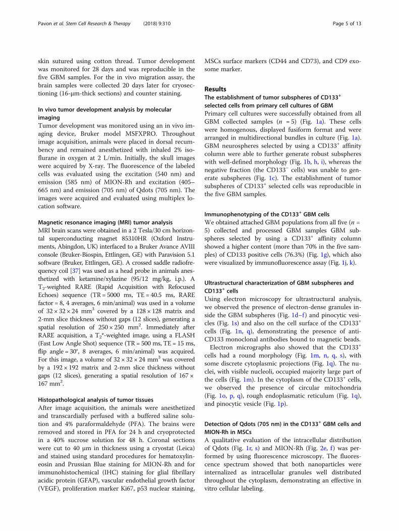
ResultsThe establishment of tumor subspheres of CD133+
selected cells from primary cell cultures of GBMPrimary cell cultures were successfully obtained from allGBM collected samples (n = 5) (Fig. 1a). These cellswere homogenous, displayed fusiform format and werearranged in multidirectional bundles in culture (Fig. 1a).GBM neurospheres selected by using a CD133+ affinitycolumn were able to further generate robust subsphereswith well-defined morphology (Fig. 1b, h, i), whereas thenegative fraction (the CD133− cells) was unable to gen-erate subspheres (Fig. 1c). The establishment of tumorsubspheres of CD133+ selected cells was reproducible inthe five GBM samples.
Immunophenotyping of the CD133+ GBM cellsWe obtained attached GBM populations from all five (n =5) collected and processed GBM samples GBM sub-spheres selected by using a CD133+ affinity columnshowed a higher content (more than 70% in the five sam-ples) of CD133 positive cells (76.3%) (Fig. 1g), which alsowere visualized by immunofluorescence assay (Fig. 1j, k).
Ultrastructural characterization of GBM subspheres andCD133+ cellsUsing electron microscopy for ultrastructural analysis,we observed the presence of electron-dense granules in-side the GBM subspheres (Fig. 1d–f ) and pinocytic vesi-cles (Fig. 1s) and also on the cell surface of the CD133+
cells (Fig. 1n, q), demonstrating the presence of anti-CD133 monoclonal antibodies bound to magnetic beads.Electron micrographs also showed that the CD133+
cells had a round morphology (Fig. 1m, n, q, s), withsome discrete cytoplasmic projections (Fig. 1q). The nu-clei, with visible nucleoli, occupied majority large part ofthe cells (Fig. 1m). In the cytoplasm of the CD133+ cells,we observed the presence of circular mitochondria(Fig. 1o, p, q), rough endoplasmatic reticulum (Fig. 1q),and pinocytic vesicle (Fig. 1p).
Detection of Qdots (705 nm) in the CD133+ GBM cells andMION-Rh in MSCsA qualitative evaluation of the intracellular distributionof Qdots (Fig. 1r, s) and MION-Rh (Fig. 2e, f ) was per-formed by using fluorescence microscopy. The fluores-cence spectrum showed that both nanoparticles wereinternalized as intracellular granules well distributedthroughout the cytoplasm, demonstrating an effective invitro cellular labeling.
Pavon et al. Stem Cell Research & Therapy (2018) 9:310 Page 5 of 13
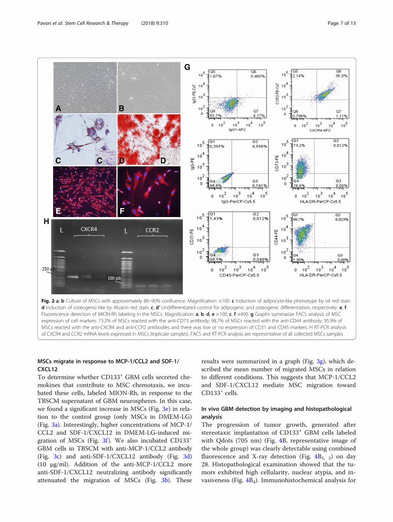
Isolation, culture of MSCsAfter three passages of culture, the MSC population fromUC samples became more morphologically homogeneous.These cell populations mostly exhibited a fibroblast-likecell profile (Fig. 2a, b). The process of differentiation ofMSCS into adipocyte-like was demonstrated by the oil redcytochemical test, which exhibited, in red, lipid droplets(Fig. 2c); the differentiation of MSCs into osteoblast-likecells was also confirmed, showing a strong cytochemicalpattern of Alizarin Red, which indicated the presence ofcalcium deposits (Fig. 2d). Thus, we confirmed that thecultured cells demonstrated multipotentiality, by givingrise to osteoblasts and adipocytes when exposed toadequate differentiating conditions. FACS analysis showedthe cells were strongly positive for the typical mesenchy-mal markers, such as CD29, CD44 (hyaluronic receptor),CD73, CD90, CD105 (endoglin), CD166, low or noexpression of MHC class I antigens, HLA-DR andhematopoietic cell markers (CD14, CD31, CD34, CD45and CD106), and absence of MHC class II antigens (29)(Fig. 2g).
MSCs express the chemokine receptors CCR2 and CXCR4Before performing specific studies, we verified that thecultured cells were negative for CD31 and CD45 surfacemarkers and positive for CD44 and CD73 surface markers(Fig. 2g).To study the role of chemokine receptors in MSCmigration toward CD133+ GBM cells, we examined theexpression of homing markers (the receptors for MCP-1/CCL2 and SDF-1/CXCL12, respectively) in MSCs, whichco-expressed CXCR4 and CCR2 (95.9%) (Fig. 2g) by FACSanalysis. To confirm this data, we identified the transcrip-tion of CXCR4 and CCR2 mRNAs (Fig. 2h) of MSCs byRT-PCR analysis.
CD133+ GBM cells express MCP-1/CCL2 and SDF-1/CXCL12We postulated that these chemokines, released byCD133+ cells, could be potential mediators of MSC migra-tion. To test this hypothesis, we examined their expressionusing RT-PCR. We observed that CD133+ GBM cells ex-press the transcripts for MCP-1/CCL2 mRNAs (Fig. 3h)and SDF-1/CXCL12 mRNAs (Fig. 3i).
Fig. 1 a The establishment of human GBM primary cell culture. b Isolation of tumor neurospheres derived from GBM primary cell culture. dPurification of GBM cells from tumor subspheres using CD133 microbeads. Immunophenotypic characterization by using flow cytometry toevaluate the efficiency of magnetic cell separation for the antigenic marker, CD133 (76.3%). e–h CD133+GBM cells were able to further generatesubspheres, compared with the absence of subspheres obtained from CD133− fractions (c). e, f GBM subspheres visualized by inverted microscopy. g,h GBM subspheres visualized by fluorescence microscopy. i–k TEM of the GBM subspheres. l–q TEM of the CD133+ stem cells. n = nucleus, c =cytoplasm, mi = mitochondria, rer = rough endoplasmic reticulum, pv = pinocytic vesicles, v = vacuoles, arrow = electron-dense granules ormagnetic beads. Scale: i–k 5.0 μm, l 2.0 μm, m–q 1.0 μm. r Fluorescence detection of Qdots (705 nm) labeling in the CD133+ GBM cells. sFluorescence detection of Qdots (705 nm) labeling in the CD133+ GBM cells and DAPI. Magnification: ×400. All figures are representative onesfrom assays performed at least five times
Pavon et al. Stem Cell Research & Therapy (2018) 9:310 Page 6 of 13
MSCs migrate in response to MCP-1/CCL2 and SDF-1/CXCL12To determine whether CD133+ GBM cells secreted che-mokines that contribute to MSC chemotaxis, we incu-bated these cells, labeled MION-Rh, in response to theTBSCM supernatant of GBM neurospheres. In this case,we found a significant increase in MSCs (Fig. 3e) in rela-tion to the control group (only MSCs in DMEM-LG)(Fig. 3a). Interestingly, higher concentrations of MCP-1/CCL2 and SDF-1/CXCL12 in DMEM-LG-induced mi-gration of MSCs (Fig. 3f ). We also incubated CD133+
GBM cells in TBSCM with anti-MCP-1/CCL2 antibody(Fig. 3c) and anti-SDF-1/CXCL12 antibody (Fig. 3d)(10 μg/ml). Addition of the anti-MCP-1/CCL2 moreanti-SDF-1/CXCL12 neutralizing antibody significantlyattenuated the migration of MSCs (Fig. 3b). These
results were summarized in a graph (Fig. 3g), which de-scribed the mean number of migrated MSCs in relationto different conditions. This suggests that MCP-1/CCL2and SDF-1/CXCL12 mediate MSC migration towardCD133+ cells.
In vivo GBM detection by imaging and histopathologicalanalysisThe progression of tumor growth, generated afterstereotaxic implantation of CD133+ GBM cells labeledwith Qdots (705 nm) (Fig. 4B, representative image ofthe whole group) was clearly detectable using combinedfluorescence and X-ray detection (Fig. 4B1, 2) on day28. Histopathological examination showed that the tu-mors exhibited high cellularity, nuclear atypia, and in-vasiveness (Fig. 4B4). Immunohistochemical analysis for
Fig. 2 a, b Culture of MSCs with approximately 80–90% confluence. Magnification: ×100. c Induction of adipocyte-like phenotype by oil red stain.d Induction of osteogenic-like by Alizarin red stain. c, d′ Undifferentiated control for adipogenic and osteogenic differentiation, respectively. e, fFluorescence detection of MION-Rh labeling in the MSCs. Magnification: a, b, d, e ×100; c, f ×400. g Graphs summarize FACS analysis of MSCexpression of cell markers: 73.2% of MSCs reacted with the anti-CD73 antibody; 98.7% of MSCs reacted with the anti-CD44 antibody; 95.9% ofMSCs reacted with the anti-CXCR4 and anti-CCR2 antibodies and there was low or no expression of CD31 and CD45 markers. H RT-PCR analysisof CXCR4 and CCR2 mRNA levels expressed in MSCs (triplicate samples). FACS and RT-PCR analysis are representative of all collected MSCs samples
Pavon et al. Stem Cell Research & Therapy (2018) 9:310 Page 7 of 13

GFAP confirmed tumor formation originated in the glia(Fig. 4B5) and vascular proliferation (Fig. 4B6).
Tumorigenesis and MSCsThe in vivo study began with the exclusive stereotaxicimplantation of 1 × 10⁴ MSCs labeled MION-Rh (condi-tion A), which, as expected, was not capable of generatetumor. These cells were visualized outside the brainparenchyma surface by using combined fluorescence andX-ray detection (Fig. 4A1, 2) and by the Prussian Bluehistochemical assay, which demonstrated the presenceof iron (MION-Rh) (Fig. 4A3). This analysis was used asthe control of the study (these cells alone did not gener-ate brain tumor).The progression of tumor growth, generated after
stereotaxic implantation of 1 × 10⁴ CD133+ GBM cellslabeled with Qdots (705 nm) (condition B) was showedin Fig. 4b. The tumor was identified by using combinedfluorescence and X-ray detection (Fig. 4B1, 2) and by thePrussian Blue histochemical analysis (Fig. 4B3). This ana-lysis served as control group in the process of compari-son of tumor development of the C and D conditions.
Interestingly, when 1 × 10⁴ MSCs labeled MION-Rhwere implanted together with 1 × 10⁴ CD133+ GBM cellslabeled Qdot 750 nm (condition C), the tumor displayedsignificant progression on the contralateral side, inwhich also was evidenced migration of MSCs labeledMION-Rh (Fig. 4C4; representative image of the wholegroup). The tumor was identified by using combinedfluorescence and X-ray detection (Fig. 4C3; representa-tive image of the whole group) and by IHC analysis,which demonstrated significant aggressiveness, glialinvasiveness (Fig. 4C6; representative image of the wholegroup), vascular proliferation (Fig. 4C7) and the detec-tion of a high number of cycling cells (Fig. 4C5; repre-sentative image of the whole group).To determine the contribution to MSC chemotaxis of
chemokine-secreting CD133+ GBM cells, we injected1 × 104MSCs into the caudal vein of the animals, afterthe tumor had been established for 28 days (conditionD) (Figs. 4D and 5; representative image of the wholegroup). Corroborating the results of in vitro migrationassays, MSCs labeling MION-Rh were able to cross theblood-brain barrier (Figs. 4D1 and 5c, o), co-locatingCD133+ cells (Figs. 4D3, 5a) and promoting their
Fig. 3 Representative figure of migration assays of MSCs, in transwell dishes, in different conditions placed in the lower chambers: a MSCs notlabeled [control], b conditioned medium supplemented with specific neutralized antibodies (anti-MCP-1 and anti-SDF-1), c conditioned mediumsupplemented with specific neutralized antibodies (anti-MCP-1), d conditioned medium supplemented with specific neutralized antibodies (anti-SDF-1), e CD133+ cell culture supernatants (TBSCM), f chemokines MCP-1 and SDF-1, g graph summarized of mean number of migrated MSCs inrelation to different conditions, h RT-PCR analysis of MCP-1/CCL2 mRNA levels expressed in CD133+ GBM cells (n = 5; GBM1 GBM2 GBM3 GBM4GBM5). i RT-PCR analysis of SDF-1/CXCL12 mRNA levels expressed in CD133+ GBM cells (n = 5; GBM1 GBM2 GBM3 GBM4 GBM5). L50: Ladder50 bp; LM low mass ladder; R positive control (human reference total RNA Clontech)
Pavon et al. Stem Cell Research & Therapy (2018) 9:310 Page 8 of 13

proliferation (Figure 4D2 and Fig. 5b), when we followthe development of the tumor for 20 days.IHC analysis confirmed tumor dissemination (Figs. 4D4,
5 and 5n, q–t), glial invasiveness (Figs. 4D6, 5u), vascularproliferation (Fig. 4D7), and IHC staining patters of p53(Fig. 5v), when compared to the study situation that didnot receive MSCs.This study condition was also monitored using MRI
analysis (Fig. 5; representative image of the whole group)to evaluate migration of MSCs labeled with MION-Rh, ananoparticle suitable for the study, due to its magneticcharacter, in addition to fluorescence. We observed “dark”hypointense zones in the T2*-weighted images (Fig. 5d–k),
which revealed the process of migration of MSCs towardCD133+ GBM cells, this outcome also was visualized bypresence of iron (MION-Rh) in the histochemical analysis(Fig. 5m). MSCs also were visualizes by cell proliferationassay (Fig. 5o and expression of CD44 (Fig. 5p) and CD73(Fig. 5q) positive cell surface markers by IHC. These re-gions were also, respectively, positives for CD63 (Fig. 5w,x) and CD9 (Fig. 5y, z, z′) exosomes markers, which,probably, were secreted by MSCs (Fig. 5q, r, y, z).
DiscussionThe tropism of MSCs toward GBM makes these cells as ahighly attractive vehicle for the delivery of therapeutic
Fig. 4 Tumorigenesis study for stereotaxic implantation of the cells in different conditions: A 1 × 10⁴ MSCs labeled MION-Rh; B 1 × 10⁴ CD133+
GBM cells labeled Qdots(705 nm); C 1 × 10⁴ MSCs labeled MION-Rh added 1 × 10⁴ CD133+ GBM cells labeled Qdots(705 nm); D implantation of1 × 104 CD133+ GBM cells labeled Qdots (705 nm), after the establishment of the GBM (28 days), was made the infusion in caudal vein 1 × 10⁴
MSCs (MION-Rh); the development of tumor was followed for 20 days. A1, C1, D1 MSCs labeled MION-Rh and visualized by fluorescencedetection. B1, C2, D2 CD133
+ GBM cells labeled Qdot 705 nm and visualized by fluorescence detection. A2, B2, C3, D3 MSCs labeled MION-Rh andCD133+ GBM cells labeled Qdot 705 nm using combined fluorescence and X-ray detection. A3, C4 IHC analysis for Prussian blue staining of theMSCs labeled with MION-Rh. B4, D4, D5 Hematoxylin and eosin staining. B5, C6, D6 IHC analysis for GFAP. C5 IHC analysis for Ki67; B6, C7, D7 IHCanalysis for VEGF. Red arrow: site of stereotaxic implantation of MSCs labeled MION-Rh. Green arrow: site of stereotaxic implantation of CD133+
GBM cells labeled Qdot (705 nm).Green circle evidenced proliferation: MSCs and CD133+ GBM cells. These images are representative of allcollected MSCs and GBM samples
Pavon et al. Stem Cell Research & Therapy (2018) 9:310 Page 9 of 13

products directly to tumor. Our results demonstrated thatpossibly this tropism should be governed by CD133+
GBM cells. However, the molecular events that governMSCs homing to CD133+ GBM cells and their effects ontumor development are unclear.Herein, we isolated CD133+ GBM cells, which were
appropriately obtained from the establishment of tumorsubspheres from primary cell cultures of GBM and char-acterized by immunophenotype and ultrastructural as-pects described elsewhere [33, 34].In our study, we demonstrated that specific chemokines,
such as MCP-1/CCL2 and SDF-1/CXCL12 (receptorsCCR2 and CXCR4 expression in MSCs from hUCB),
mediate the migration of MSCs toward CD133+ GBMcells in vitro.MCP-1/CCL2 is a member of the cytokine/chemokine
superfamily that regulates migration and infiltration ofmonocytes/macrophages to tumor sites, [38, 39], therebyinhibiting anti-tumor immune responses [38] and promot-ing tumorigenesis and metastasis of the gliomas in vivo[40]. Moreover, addition of the anti-MCP-1/CCL2 neutral-izing antibody significantly attenuated the migration ofMSCs toward CD133+ cell culture supernatants (TBSCM).The chemokine MCP-1/CCL2 plays an important role inthe regulation of stem cell trafficking [41]. Recent studiesdemonstrate that the progress of GBM is driven by stem
Fig. 5 Implantation of 1 × 10⁴ CD133+ GBM cells labeled Qdots (705 nm), after the establishment of the GBM (28 days), was made the infusion incaudal vein 1 × 10⁴ MSCs (MION-Rh); the development of tumor was followed for 20 days. a MSCs labeled MION-Rh and CD133+ GBM cellslabeled Qdot 705 nm using combined fluorescence and X-ray detection. b CD133+ GBM cells labeled Qdot 705 nm and visualized by fluorescencedetection. c MSCs labeled MION-Rh and visualized by fluorescence detection. d–h, i–l MRI (T2*-weighted images) of animal brain monitoring of theprocess of migration of MSCs, which were able to cross the blood-brain barrier of the animal and migrated to the tumor region, promoting GBM cellproliferation. l MRI (T2*-weighted images) of animal brain without stereotaxic implantation of cells (control group). Red circle showed migration assaysof MSCs and green circle evidenced tumor propagation. m IHC analysis for Prussian blue staining of the MSCs labeled with MION-Rh. n,r, s, t Hematoxylin and eosin staining. u IHC analysis for GFAP. o IHC analysis for Ki67; v IHC analysis for p53; p IHC analysis for CD44staining of the MSCs; q IHC analysis for CD73 staining of the MSCs; w, x IHC analysis for CD63 staining of the MSCs-derived exosomes;y–z′ IHC analysis for CD9 staining of the MSCs-derived exosomes. These images are representative of all collected MSCs and GBM samples
Pavon et al. Stem Cell Research & Therapy (2018) 9:310 Page 10 of 13

cells, critical promoters of tumor growth, invasion, andneovascularization [42]. CXCR4 has been found to be up-regulated in CD133+ GBM stem cells upon activation withSDF-1/CXCL12, a CXCR4 ligand [43]. There is evidencethat disruption of CXCR4 results in a reduction of GBMstem cell markers and reduction in tumor cell prolifera-tion [44]. Therefore, in our results in vitro, MCP-1/CCL2and SDF-1/CXCL12 might play an important role inMSCs homing toward CD133+ cells.Park et al. [45] already stated that SDF-1(CXCL12)/
CXCR4 could be involved in the recruitment of MSCs toU-251MG glioma cells lines and that overexpression ofCXCR4 might be a useful tool for stem cell-based gliomatherapy. These authors performed the animal model usingmigration assay by injection of MSCs directly in the brain,the contralateral site of gliomas, and did not observetumor dissemination after 10 days post-injection.Considering the relevance of the CD133+ model [5–7],
our group used this methodology to generate tumor invivo. Different from Park et al. [45], we infused MSCs inthe caudal vein of the animals, which were able to crossthe blood-brain barrier and co-located with CD133+ GBMinitiating cells, obtained from tumor subspheres from pri-mary cell cultures of GBM. Following the migration pro-tocols for 20 days, we validated the chemotactic effect ofMCP-1/CCL2 and SDF-1/CXCL12 in mediating the mi-gration of MSCs toward CD133+ GBM cells, and we ob-served tumor development, glial invasiveness, vascularproliferation and detection of a high number of cyclingcells, when compared to the study situation that did not
receive MSCs. MRI analysis confirmed the process of mi-gration of MSCs toward CD133+ GBM cells and intensebrain tumor dissemination. These findings assume thatchemokines mediate MSC migration toward CD133+
GBM cells and that this could promote tumor develop-ment and metastatic proliferation.Interestingly, in the study conditions, where MSCs
were implanted together with CD133+ GBM cells, sig-nificant tumor progression was also displayed whencompared to condition B, which was generated byimplantation of CD133+ GBM cells only.Pavon et al. [33] showed that CD133+ GBM cells express
molecular signatures of MSCs. Therefore, we hypothesizethat CD133+ cells, due to their MSC-like properties, re-cruit MSCs, and sustain tumor growth, which is affectedby the tumor microenvironment created by the non-neo-plastic stroma composed of inflammatory [34, 46]. MSCsrelease many promigratory chemokines, which facilitatetumor progression including proliferation, senescence,angiogenesis, epithelial mesenchymal transition, immuneevasion, and metastasis [47, 48].These events could be modulated by recruited
MSCs-derived exosome, here in our study demon-strated by expression tetraspanin CD9/CD63 protein[49], which apparently could be involved in tumorcell invasion and consequently tumor dissemination(schematic representation described of Fig. 6). How-ever, other studies on biological effects mediated bythese vesicles need to be developed to prove thisfinding.
Fig. 6 Schematic representation demonstrating that chemokines mediate MSC migration toward CD133+ stem cell of GBM and scanning electronmicroscopy of exosome, secreted by MSCs, promoting tumor dissemination
Pavon et al. Stem Cell Research & Therapy (2018) 9:310 Page 11 of 13

Therefore, tumor growth effect of MSCs tropismtoward GBM remains controversial: (i) CD133+ GBMcells maintain only a subset of primary GBM; probably,CD133− cells also participate in the process of modulat-ing the tropism and (ii) the intrinsic factor such as doseof MSCs and timing of implantation should be tested infuture trials.Different studies reported either MSC anti-tumor
activity or their support to tumor growth. Behaan et al.[50] and Motaln and Turnsek [51] demonstrated thatthe using of MSCs as cellular vectors for modulating cy-tokines and cytokine receptors’ signaling in GBM couldbeen more efficient at inhibiting GBM progression.Nevertheless, it is still controversial whether this tropismof MSCs toward the tumor area is associated with GBMpromotion or suppression [52]. Okamoto et al. [53] indi-cated that MSCs were capable of stimulating GBM cellproliferation through a paracrine effect mediated byTGFB1. These findings provide novel insights to betterunderstand the relationship between CD133+ GBM cellsand MSCs, raising awareness in the use of MSCs as ther-apies for gliomas [54]. These studies, however, exploredinsufficient in vivo results. In our work, we demon-strated of tumorigenicity of CD133+ cells in conjunctionto with the migration of MSCs toward GBM andsuggested, strongly, MSCs contribution to tumor devel-opment, invasion, and metastatic dissemination.
ConclusionWe suggest that the MSC-like properties of CD133+ GBMcells confer proangiogenic and anti-apoptotic characteris-tics that may sustain tumor growth. Thus, tumor progres-sion may be directed by reciprocal interaction betweenstromal cells and tumor cells, by chemotactic action ofMCP-1/CCL2 and SDF-1/CXCL12 and probable, effect ofMSC-derived exosome, to create an appropriate environ-ment for tumor aggressiveness. These findings may behardly evaluated in any future in treatment strategies thatuse MSCs as vehicles for drug delivery into glioma tumors.
AcknowledgementsWe acknowledge support from the Department of Neurology and Neurosurgery,Hospital of São Paulo (HSP), Federal University of São Paulo (UNIFESP), São Paulo,SP, Brazil, and FAPESP.
FundingThe study was funded by Fundação de Amparo à Pesquisa do Estado de SãoPaulo (FAPESP) (process no. 11/50542-9).
Availability of data and materialsData sharing not applicable to this article as no datasets were generated oranalyzed during the current study.
Authors’ contributionsLFP, TTS, AVS, and LCM carried out the cellular and molecular biology studies.EFC and FRC carried out the experimental model. JMM, FFP, AT, and SMparticipated in the MRI tumor analysis. JGS and PB carried out molecularimaging assay. SMFM, DMO, SRCT, and AMCT participated in the designof the study. LFP, MAPN, and SC conceived of the study, participated in
its design and coordination, and drafted the manuscript. All authors readand approved the final manuscript.
Ethics approvalUNIFESP ethics committee approved the study. Informed consent was obtainedfrom all patients (CEP: 09/687/CAAE: 0140.0.028.000-07) and ethical standards ofexperimental animal (CEUA: 1686-13).
Consent for publicationNot applicable.
Competing interestsThe authors declare that they have no competing interests.
Publisher’s NoteSpringer Nature remains neutral with regard to jurisdictional claims in publishedmaps and institutional affiliations.
Author details1Department of Neurosurgery, Federal University of São Paulo, São Paulo,Brazil. 2 Experimental Research Center, Hospital Israelita Albert Einstein, SãoPaulo, Brazil. 3Discipline of Nephrology, Federal University of São Paulo, SãoPaulo, Brazil. 4Laboratory of Molecular Biology, Butantan Institute, São Paulo,Brazil. 5Centre of Excellence in New Target Discovery (CENTD), ButantanInstitute, São Paulo, Brazil. 6Department of Genetics and Morphology,University of Brasília, Brasília, Brazil. 7Pediatric Oncology Institute, Grupo deApoio ao Adolescente e à Criança com Câncer (GRAACC), Federal Universityof São Paulo, São Paulo, Brazil. 8São Carlos Institute of Physics, São PauloUniversity, São Carlos, Brazil. 9Laboratory of Cellular and MolecularNeurosurgery, Federal University of São Paulo, Rua Napoleão de Barros, n.626 –Vila Clementino, São Paulo, SP 04024-002, Brazil.
Received: 31 January 2018 Revised: 11 September 2018Accepted: 16 October 2018
References1. Ostrom QT, Gittleman H, Liao P, et al. CBTRUS statistical report: primary
brain and central nervous system tumors diagnosed in the United States in2007–2011. Neuro Oncol. 2014;16(Suppl 4):iv1–63.
2. Schittenhelm J. Recent advances in subtyping tumors of the centralnervous system using molecular data. Expert Rev Mol Diagn. 2017;17(1):83–94.
3. Thomas AA, Brennan CW, De Angelis LM, et al. Emerging therapies forglioblastoma. JAMA Neurol. 2014;71(11):1437–44.
4. Siegel RL, Miller KD, Jemal A. Cancer statistics, 2015. CA Cancer J Clin. 2015;65(1):5–29.
5. Singh SK, Clarke ID, Terasaki M, et al. Identification of a cancer stem cell inhuman brain tumors. Cancer Res. 2003;63:5821–8.
6. Liu G, Yuan X, Zeng Z, et al. Analysis of gene expression andchemoresistance of CD133+ cancer stem cells in glioblastoma. Mol Cancer.2016;5:67.
7. Beier D, Hau P, Proescholdt M, et al. CD133(+) and CD133(−) glioblastoma-derived cancer stem cells show differential growth characteristics andmolecular profiles. Cancer Res. 2007;67(9):4010–5.
8. Liu Q, Nguyen DH, Dong Q, et al. Molecular properties of CD133+glioblastoma stem cells derived from treatment-refractory recurrent braintumors. J Neuro-Oncol. 2009;94(1):1–19.
9. Parker Kerrigan BC, Shimizu Y, Andreeff M, et al. Mesenchymal stromal cellsfor the delivery of oncolytic viruses in gliomas. Cytotherapy. 2017;4:445–57.
10. Hamada H, Kobune M, Nakamura K, et al. Mesenchymal stem cells (MSC) astherapeutic cytoreagents for gene therapy. Cancer Sci. 2005;96:149–56.
11. Phinney DG, Isakova I. Plasticity and therapeutic potential of mesenchymalstem cells in the nervous system. Curr Pharm Des. 2005;11:1255–65.
12. Studeny M, Marini FC, Champlin RE, et al. Bone marrow-derivedmesenchymal stem cells as vehicles for interferon-{beta} delivery intotumors. Cancer Res. 2005;62:3603–8.
13. Nakamura K, Ito Y, Kawano Y, et al. Antitumor effect of geneticallyengineered mesenchymal stem cells in a rat glioma model. Gene Ther.2004;11:1155–64.
14. Nakamizo A, Marini F, Amano T, et al. Human bone marrow-derived mesenchymalstem cells in the treatment of gliomas. Cancer Res. 2005;65:3307–18.
Pavon et al. Stem Cell Research & Therapy (2018) 9:310 Page 12 of 13

15. Doucette T, Rao G, Yang Y, et al. Mesenchymal stem cells display tumor-specific tropism in an RCAS/Ntv-a glioma model. Neoplasia. 2011;13(8):716–25.
16. Kidd S, Spaeth E, Dembinski JL, et al. Direct evidence of mesenchymal stemcell tropism for tumor and wounding microenvironments using in vivobioluminescent imaging. Stem Cells. 2009;10:2614–23.
17. Kosztowski T, Zaidi HA, Quiñones-Hinojosa A. Applications of neural andmesenchymal stem cells in the treatment of gliomas. Expert Rev AnticancerTher. 2009;9(5):597–612.
18. Mikheeva SA, Mikheev AM, Petit A, et al. TWIST1 promotes invasion throughmesenchymal change in human glioblastoma. Mol Cancer. 2010;20(9):194.
19. Wu X, Hu J, Zhou L, et al. In vivo tracking of superparamagnetic iron oxidenanoparticle-labeled mesenchymal stem cell tropism to malignant gliomasusing magnetic resonance imaging. Laboratory investigation. J Neurosurg.2008;108(2):320–9.
20. Kang SG, Jeun SS, Lim JY, et al. Cytotoxicity of human umbilical cord blood-derived mesenchymal stem cells against human malignant glioma cells.Childs Nerv Syst. 2008;24(3):293–302.
21. Greco SJ, Rameshwar P. Mesenchymal stem cells in drug/gene delivery:implications for cell therapy. Ther Deliv. 2012;3:997–1004.
22. Matuskova M, Hlubinova K, Pastorakova A, et al. HSV-tk expressingmesenchymal stem cells exert by stander effect on human glioblastomacells. Cancer Lett. 2010;290:58–67.
23. Thery C, Zitvogel L, Amigorena S. Exosomes: composition, biogenesis andfunction. Nat Rev Immunol. 2002;2(8):569–79.
24. Schey KL, Luther JM, Rose KL. Proteomics characterization of exosomecargo. Methods. 2015;87:75–82.
25. Zhang X, Yuan X, Shi H, et al. Exosomes in cancer: small particle, big player.J Hematol Oncol. 2015;8:83.
26. Schichor C, Birnbaum T, Etminan N, et al. Vascular endothelial growth factorA contributes to glioma-induced migration of human marrow stromal cells(hMSC). Exp Neurol. 2006;199(2):301–10.
27. Birnbaum T, Roider J, Schankin CJ, et al. Malignant gliomas actively recruitbone marrow stromal cells by secreting angiogenic cytokines. J Neuro-Oncol. 2007;83(3):241–7.
28. Kim SS, Yoo SW, Park TS, et al. Neural induction with neurogenin1 increasesthe therapeutic effects of mesenchymal stem cells in the ischemic brain.Stem Cells. 2008;26(9):2217–28.
29. Rempel SA, Dudas S, Ge S, et al. Identification and localization of thecytokine SDF1 and its receptor, CXC chemokine receptor 4, to regionsof necrosis and angiogenesis in human glioblastoma. Clin Cancer Res.2000;6:102–11.
30. Zhou Y, Larsen PH, Hao C, et al. CXCR4 is a major chemokine receptor onglioma cells and mediates their survival. J BiolChem. 2002;277:49481–7.
31. Louis DN, Perry A, Reifenberger G, et al. The 2016 World HealthOrganization Classification of tumors of the central nervous system: asummary. Acta Neuropathol. 2016;131(6):803–20.
32. Lenkiewicz M, Li N, Singh SK. Culture and isolation of brain tumor initiatingcells. Curr Protoc Stem Cell Biol. 2009;Chapter 3:Unit3.3.
33. Pavon LF, Marti LC, Sibov TT, et al. In vitro analysis of neurospheres derivedfrom glioblastoma primary culture: a novel methodology paradigm. FrontNeurol. 2014;4:214.
34. Pavon LF, Sibov TT, de Oliveira DM, et al. Mesenchymal stem cell-likeproperties of CD133+ glioblastoma initiating cells. Oncotarget. 2016;7(26):40546–57.
35. Sibov TT, Pavon LF, Oliveira DM, et al. Characterization of adherent umbilicalcord blood stromal cells regarding passage, cell number, and nano-biomarkingutilization. Cell Reprogram. 2010;12(4):391–403.
36. Sibov TT, Severino P, Marti LC, et al. Mesenchymal stem cells from umbilicalcord blood: parameters for isolation, characterization and adipogenicdifferentiation. Cytotechnology. 2012;64(5):511–21.
37. Papoti D, Vidoto ELG, Martins MJ, et al. Effects of crossing saddle coilconductors: electric length X mutual inductance. Concepts Magn Reson.2010;37B:193–201.
38. Mizutani K, Sud S, McGregor NA, et al. The chemokine CCL2 increasesprostate tumor growth and bone metastasis through macrophage andosteoclast recruitment. Neoplasia. 2009;11:1235–42.
39. Gabrilovich DI, Nagaraj S. Myeloid-derived suppressor cells as regulators ofthe immune system. Nat Rev Immunol. 2009;9:162–74.
40. Ostrand-Rosenberg S. Myeloid-derived suppressor cells: more mechanismsfor inhibiting antitumor immunity. Cancer Immunol Immunother. 2010;59(10):1593–600.
41. Leung SY, Wong MP, Chung LP, et al. Monocyte chemoattractant protein-1expression and macrophage infiltration in gliomas. Acta Neuropathol. 1997;93:518–27.
42. Ehtesham M, Winston JA, Kabos P, et al. CXCR4 expression mediates gliomacell invasiveness. Oncogene. 2006;25:2801–6.
43. Ehtesham M, Mapara KY, Stevenson CB, et al. CXCR4 mediates the proliferationof glioblastoma progenitor cells. Cancer Lett. 2009;274:305–12.
44. Gatti M, Pattarozzi A, Bajetto A, et al. Inhibition of CXCL12/CXCR4 autocrine/paracrine loop reduces viability of human glioblastoma stem-like cells affectingself-renewal activity. Toxicology. 2013;314:209–20.
45. Park SA, Ryu CH, Kim SM, et al. CXCR4 transfected human umbilical cordbloodderived mesenchymal stem cells exhibitenhanced migratory capacitytoward gliomas. Oncol. 2011;38(1):97–103.
46. Bergfeld SA, De Clerck YA. Bone marrow-derived mesenchymal stem cellsand the tumor microenvironment. Cancer Metastasis Rev. 2010;29:249–61.
47. Purvaba J, Sarvaiya DG, Ulasov I, et al. Chemokines in tumor progressionand metastasis. Oncotarget. 2013;4(12):2171–85.
48. Avnet S, Di Pompo G, Chano T, et al. Cancer-associated mesenchymalstroma fosters the stemness of osteosarcoma cells in response to intratumoralacidosis via NF-κB activation. Int J Cancer. 2017;140(6):1331–45.
49. Zöller M. Tetraspanins: push and pull in suppressing and promoting metastasis.Nat Rev Cancer. 2009;9:40–55.
50. Behnan J, Isakson P, Joel M, et al. Recruited brain tumor-derived mesenchymalstem cells contribute to brain tumor progression. Stem Cells. 2014;32:1110–23.
51. Motaln H, Turnsek TL. Cytokines play a key role in communication betweenmesenchymal stem cells and brain cancer cells. Protein Pept Lett. 2015;22(4):322–31.
52. Bajetto A, Pattarozzi A, Corsaro A, et al. Different effects of human umbilicalcord mesenchymal stem cells on glioblastoma stem cells by direct cellinteraction or via released soluble factors. Front Cell Neurosci. 2017;11:312.
53. Rodini CO, Golçalves da Silva PB, Assoni AF, et al. Mesenchymal stem cellsenhance tumorigenic properties of human glioblastoma through independentcell-cell communication mechanisms. Oncotarget. 2018;9(37):24766–77.
54. Vieira de Castro J, Gomes ED, Granja S, et al. Impact of mesenchymal stemcells' secretome on glioblastoma pathophysiology. J Transl Med. 2017;15(1):200.
Pavon et al. Stem Cell Research & Therapy (2018) 9:310 Page 13 of 13


![Enhanced Expression of ABCB1 and Nrf2 in CD133-Positive ...downloads.hindawi.com/journals/sci/2020/8868849.pdf · pies/stresses, and the metastasis of cancer cells [5–11]. CD133](https://img.pdfslide.net/doc/110x75/5fbcf848095d1e282b57d0c3/enhanced-expression-of-abcb1-and-nrf2-in-cd133-positive-piesstresses-and-the.jpg)
![Prominin-1/CD133, a neural and hematopoietic stem cell ... · 350 μCi/ml [35S] Easytag express protein labeling mix (PerkinElmer Life Sciences, Boston, Mass.; 1175.0 Ci/ mmol). After](https://img.pdfslide.net/doc/110x75/5fbac4981ba23c3b6517e31f/prominin-1cd133-a-neural-and-hematopoietic-stem-cell-350-ciml-35s-easytag.jpg)