Embed Size (px)
Citation preview

INSIGHT INTO ADENOVIRUS PROGRAMMED DISASSEMBLY FROM
CRYOEM: THE STRUCTURES OF AD2ts1 AND THE AD35f+DEFENSIN HD5
COMPLEX
By
Mariena Silvestry Ramos
Dissertation
Submitted to the Faculty of the
Graduate School of Vanderbilt University
in partial fulfillment of the requirements for
the degree of
DOCTOR OF PHILOSOPHY
in
Molecular Physiology and Biophysics
August, 2009
Nashville, Tennessee
Approved:
Professor Albert H. Beth
Professor Charles E. Cobb
Professor Terence S. Dermody
Professor Hassane S. Mchaourab
Professor Jens Meiler

To my mom, dad and sister, I love you dearly. It is because of you that I am here
today. To Raúl, my best friend and companion. To my Ague, who I’ve missed
every day since May of 1995. To CazMan ... keep on beating Ad.

iii
ACKNOWLEDGEMENTS
I would like to thank my mentor, Phoebe Stewart, who gave me the
chance to do fun, exciting and cutting edge research in her lab. Her faith and
support were always there for me. I admire her positive attitude, especially at
times where it was hard to find a pattern or explanation to a result. Her ideas and
energy were critical at every step of the road. And the quality of research which I
have done in her lab is superb.
I am grateful for starting my thesis work under the direction of Dr. Susan
Sabán. She was the one that got me “hooked” on cryoEM and her technical and
teaching skills set an example to follow. Susan is an outstanding microscopist
and her attention to detail and insight served as an inspiration all thru the years.
Susan has been a great friend and tremendous source of support and inspiration.
I have been truly blessed for having her as a friend, lab-mate and mentor.
I am indebted to Dewight Williams, Jian Shi and Mike Lewis for helping
me get out of trouble while learning cryoEM. Dewight and Jian were instrumental
in speeding the data acquisition with their SAM software, for that I cannot thank
them enough. Steffen Lindert has been a great lab-mate along the way. Without
him I would not have been able to collect and process the great amount of data
for these studies in a short amount of time. Steffen has been a good friend and a
great source of scientific and technical questions, all of which made me a better,
more inquisitive researcher. Thanks for all the great German chocolate (ist sehr
gut!).

iv
I thank all the wonderful collaborators that I was fortunate to work with
while doing my thesis and non-thesis related projects. Drs. Glen Nemerow,
Jason Smith and Christopher Wiethoff who are remarkable scientists and all of
our conversations were a tremendous learning experience. Drs. David Curiel,
Dmitry Shayakhmetov and Leonard Rome were instrumental in providing non-
thesis related projects that have made a big impact on my scientific development.
I acknowledge the support from the Center for Structural Biology and the
Molecular Biophysics Training Program, which funded my research during my
second year Vanderbilt. Dr. Walter Chazin has been of great help in this process
and I thank him for his interest in my research and career growth. Also, Jarrod
Smith, Carrie Lee Kennedy and Matt Binkley who were very nice in assisting with
the computer processing needs at the CSB and VAMPIRE. And also the friends
at the Jerome lab, Jay, Denny and Mary, thanks for accommodating my grid-
baking needs.
I thank my family: my mami and papi are the source of everyday
inspiration. Their hard work and wisdom have been with me always. I cannot
thank them enough for their support and encouragement. My sister provided a
much needed relief and source of laughter along this path. I thank her for being
there at all the stages of my graduate career (gracias por las Medallas y
Coronas). My grandma Regina who is extremely supportive, loving, and fun. My
Aunt María, Aunt Margie, Uncle Georgie and Uncle Willie and my cousins
Gloriena, Billie Joe and Brian, who have made me so happy and provided much
needed prayers, strength and love. My friends: Sully, Delly, Mayda, Mayra C and

v
Mayra A, Leri, Noelia, Ama, Ale, Leo, Renier, Willie, Karinna, Chuck, Sepan,
Ezelle and Julia, Ida, El “Coro” and “los corillos Boricuas”. All of you made me
laugh along this journey and I cannot thank you enough for being the funniest,
most awesome friends ever. I thank Raul’s parents, Lianny and José Raúl.
Words cannot express how blessed I am to know you. I cannot thank you enough
for your calls, prayers, visits, and encouragement. You are an example of what I
aspire to be as a Christian and as a researcher. And Raúl: you came into my life
unexpectedly and you’ve changed it in so many good ways. You have taught me
so much, through words and actions. You are my best friend, companion and
boyfriend. You are my blessing. I thank God for each and every one of you. I love
you all.
Last but not least I thank the Choir of St. Henry’s Catholic Church in
Bellevue. When my spirit was broken your music was always there to lift me up. I
thank Mr. John and Ruth Adrian, and Mrs. Mary Corby, Julie Schwartz and Lee
Suttle, three of the most beautiful voices I have ever heard. Shellie for being a
really cool gal, I wish you a long and prosperous career in publishing, you
deserve it! I will miss the choir so very much. Finally I thank the Allens for all their
help, you are a wonderful family.

vi
TABLE OF CONTENTS
Page
DEDICATION ........................................................................................................ii
ACKNOWLEDGEMENTS..................................................................................... iii
LIST OF TABLES .................................................................................................ix
LIST OF FIGURES ............................................................................................... x
LIST OF ABBREVIATIONS .................................................................................xii
Chapter
I. INTRODUCTION ..................................................................................... 1
Adenovirus capsid composition ............................................................... 1
External capsid proteins ................................................................. 2
Additional internal capsid proteins................................................... 8 Viral Core: Structure and components ............................................ 9
Adenovirus Cell Entry and Capsid Dismantling..................................... 11 Aim 1: Structure of the Ad2ts1 Mutant .................................................. 13 Ad Immunity Mediated by Human Defensins ........................................ 15 Aim 2: Structure of the Ad35f Vector Complexed with HD5 Defensin... 16 Concluding Remarks............................................................................. 16
II. MATERIALS AND METHODS.............................................................. 19
Preparation and isolation of Ad2ts1 ...................................................... 19 Cryoelectron microscopy of Ad2ts1....................................................... 20 Image processing of Ad2ts1.................................................................. 20 Difference map analysis of Ad2ts1........................................................ 22 Quantification of core-plus-capsid and capsid-only average intensities in cryoEM particle images......................................................................... 26 Membrane disruption assay .................................................................. 27 Preparation and isolation of Ad35f ........................................................ 27 Cryoelectron Microscopy of Ad35f+HD5 ............................................... 28 Image Processing of Ad35f+HD5.......................................................... 28
III. AIM 1: CRYOELECTRON MICROSCOPY STRUCTURE OF THE ADENOVIRUS TYPE 2 TEMPERATURE-SENSITIVE MUTANT 1 REVEALS INSIGHT INTO THE CELL ENTRY DEFECT ................................................................. 30
Introduction ........................................................................................... 30

vii
Adenovirus structure and organization: Viral components and their locations............................................. 30
Adenovirus type 2 temperature-sensitive mutant and its role in elucidating Ad cell entry .......................................... 32
Results.................................................................................................. 33 CryoEM structure reveals differences in the Ad2ts1 core compared to the mature Ad core............................... 33 The Ad2ts1 penton base is anchored to the viral core .................. 44 Density inside the Ad2ts1 hexon cavities is assigned as preprotein VI.................................................................................. 44
Assignments for preproteins IIIa and VIII on the inner capsid....... 45 Hexon shields protein VI and preprotein VI membrane lytic activity.................................................................................... 53
Discussion............................................................................................. 55 The Ad core condenses and becomes less symmetrically ordered during maturation............................... 55 Vertex release may require a conformational change that is blocked in Ad2ts1................................................... 56
IV. AIM2: CRYOEM STRUCTURE OF AN ADENOVIRUS-HUMAN α- DEFENSIN COMPLEX: INSIGHT INTO BINDIG SITES CRITICAL FOR NEUTRALIZATION ............................................................................... 61 Introduction ........................................................................................... 61
Defensins ...................................................................................... 61 Modes of defensin neutralization................................................... 62 Elucidating defensin mode of action against adenovirus............... 63
Results .................................................................................................. 65 Stoichiometry of the Ad35f/HD5 interaction .................................. 65 CryoEM structural study of the Ad35f+HD5 complex .................... 78 Difference mapping allows visualization of HD5 on the Ad capsid ................................................................................ 71 HD5 has multiple binding sites on the top of the hexons in the Ad capsid............................................................................................ 74 HD5 coats the fiber and tops of the penton base in the Ad capsid ...................................................................................... 80
Discussion............................................................................................. 86
V. SUMMARY AND CONCLUSIONS........................................................ 97 Implications for Adenovirus cell entry from our cryoEM study of Ad2ts1 ..................................................................................... 97 Future directions for Ad2ts1 .......................................................... 98 Ad-mediated gene delivery.......................................................... 100 Antiviral peptides: human Ads and defensins.............................. 103 Results and implications from the Ad35f+HD5 studies................ 104 Future directions for Ad-defensin ................................................ 105

viii
Appendices ......................................................................................... 108 Appendix 1 .................................................................................... 108 Appendix 2 .................................................................................... 145 Appendix 3 .................................................................................... 146 Appendix 4 .................................................................................... 147 References.......................................................................................... 148

ix
LIST OF TABLES
Table Page
2.1 Difference mapping steps............................................................................. 23
2.2 Composition and mass of the protein/DNA core of human Ad5.................... 25

x
LIST OF FIGURES
Figure Page
1.1 Diagram depicting the composition of the Ad capsid ...................................... 3
1.2 Penton base conformational change induced by fiber peptide binding........... 5
1.3 Location of protein IX within the Ad capsid..................................................... 7
3.1 CryoEM structures of Ad2ts1 and Ad35f reveal a major structural difference in the core of the virion ....................................................................................... 35
3.2 The main differences between Ad2ts1 and Ad35f are
on the interior of the icosahedral capsid ....................................................... 38
3.3 Average radial density distributions of the Ad2ts1 and Ad35f structures ...... 40
3.4 Masking of the capsid-only and
core-plus-capsid regions of a particle image ................................................ 43
3.5 Preprotein VI is assigned to density within
the cavity of every hexon trimer in Ad2ts1.................................................... 47
3.6 Density assigned to protein IX and
preproteins IIIa and VIII are found within Ad2ts1 .......................................... 50
3.7 The secondary structure prediction algorithms Jufo, Psipred, and
Sam, were used to obtain a three-state prediction for Ad5 protein VIII......... 52
3.8 Hexon binding shields protein VI and
preprotein VI membrane lytic activity............................................................ 54
3.9 Diagram of proposed cell entry events for Ad2 and Ad5 vs. Ad2ts1............. 58
4.1 Stoichiometry of HD5 binding to Ad5............................................................ 67
4.2 CryoEM structures of Ad35f+HD5 compared to Ad35f ................................. 70
4.3 Difference map analysis of three cryoEM structures with a calculated pseudoatomic facet of hexons and pentons ....................................................... 73

xi
4.4 Difference density corresponding to both HD5 and the hexon surface loops missing from the crystal structure above each of the four unique hexon positions within the asymmetric unit of the 67% Ad35f+HD5 structure.............................. 77
4.5 Difference density corresponding to both HD5 and the hexon surface loops above each of the four unique hexon positions within the asymmetric unit of the 12% Ad35f+HD5 structure.................................................................................. 78
4.6 Difference density from the Ad35f structure corresponding to the missing loops in the hexon crystal structure .................................................................... 79
4.7 Difference density in the vertex region in both the Ad35f+HD5 67% and 12% structures and the Ad35f structure...................................................................... 81
4.8 Side views of the vertex region in the Ad35f+HD5 67% and 12% structures and the Ad35f structure ...................................................................................... 84
4.9 Postulated critical neutralization site for HD5 based on a combined cryoEM and sequence analysis ....................................................................................... 90
4.10 Infectivity assay for various Ad types in the presence of HD5 .................... 92
4.11 Close up view the postulated critical binding site of HD5 involving the Ad fiber N-terminal region and near the penton base RGD loop.............................. 95

xii
ABBREVIATIONS
Ad/Ads = adenovirus/adenoviruses
Ad2ts1 = Adenovirus type 2 temperature-sensitive mutant 1
SCID = severe combined immune deficiency
CryoEM = cryoelectron microscopy
HVR/HVRs = hyper-variable region/regions
Aa/aa’s = amino acid/amino acids
TP = terminal protein
HD5 = human α-defensin 5
HNP1 = human neutrophil peptide 1
CAR = Coxsackievirus and Adenovirus receptor
RGD loop = Arginine, Glycine and Aspartic Acid loop
FSC = Fourier Shell Correlation
SulfoB = Sulforhodamine B
1
CHAPTER I
Introduction
Adenoviruses are large non-enveloped viruses that cause a variety of
infections, particularly in the ocular, respiratory, excretory and gastrointestinal
systems. These infections are usually non fatal, except in immunocompromised
individuals. Ads are used in approximately a third of all ongoing gene and
vaccine delivery trials (http://www.wiley.co.uk/genmed/clinical). In addition, Ads
have been an important discovery tool in molecular biology, for processes such
as RNA splicing and virus-induced tumor formation. Though the lifecycle of Ad is
well characterized, structural detail of events such as maturation, cell entry and
disassembly intermediates is lacking.
Adenovirus capsid composition
Adenoviruses are composed of 11 proteins, a virally encoded protease
and a double stranded linear 36,000 bp DNA genome. Ads were first identified in
the 1950s as the causative agent of acute respiratory infections in military
recruits (8). These viruses were first visualized by negative-stain electron
microscopy in 1950’s and their icosahedral shape was noted.
The capsid diameter is over 900Å without taking into account the fiber
protein protruding from each vertex of the icosahedron. To date there have been
51 different types of human Ads identified which are classified into 6 species,
named A through F, based on their ability to agglutinate blood cells, DNA
2
sequence similarities, and host immune response. Viruses in species C, which
includes Ad 2 and 5, are among the best characterized types of Ad.
External capsid proteins
The Ad capsid is composed of 3 major proteins: the trimeric hexon found
in 240 trimers, the pentameric penton base found in 12 pentamers, and the
trimeric fiber also found in 12 trimers. The penton base and trimeric fiber together
form the penton located at each icosahedral vertex. The outer capsid also
contains protein IX while the inner capsid surface of Ad is coated with proteins
IIIa, VI and VIII. The core of the virus does not follow icosahedral symmetry and
contains the core proteins V, VII, mu, terminal protein, Ad protease and the DNA
(Figure 1.1).
The adenoviral proteins are named according to their migration pattern on
SDS-PAGE gels. Hexon, a homotrimer of protein II, is produced in the host cell
cytoplasm and its atomic resolution structure has been determined (87). This
structure shows that the hexon monomer is rich in β strands. The homotrimer,
which forms the majority of the icosahedral capsid, consists of a
pseudohexagonal base with a triangular top containing the flexible hypervariable
regions (HVRs). The 6Å resolution cryoEM structure of the Ad35f vector shows
density for the missing HVR loops (124).

3
Figure 1.1 Diagram depicting the composition of the Ad capsid. The icosahedrally shaped virus is composed of 11 proteins, the virally encoded protease and the 36kb double stranded DNA genome. The major capsid components, hexon, penton base, and fiber form the icosahedral shell. Protein IX stabilizes the capsid on the outside. Proteins IIIa, VI and VIII associate with the inner capsid surface. The core of the virus is formed by proteins V, VII, Mu, TP and the protease (not depicted), together with the DNA. The core does not follow the icosahedral symmetry of the capsid. The hexon, penton base and fiber knobs are depicted as crystal structures. Adapted with permission from (80).

4
An atomic resolution structure of the penton base pentamer (protein III),
alone and in complex with a fiber peptide (protein IV) has been determined (124).
The overall topology of the penton base is similar to that of hexon, a structure
rich in β-strands. The penton base crystal structure lacks the RGD-loop, which
was not resolved due to flexibility (Figure 1.2). There is a slight conformational
change at the top of the penton base that occurs when the fiber binds (124). In
the 6Å resolution cryoEM structure of Ad35f the density for the missing RGD-
loops is observed as well as the conformation of the fiber-bound penton base
(89) (Figure 1.2).

5
Figure 1.2 Penton base conformational change induced by fiber peptide binding. Crystal structures of the penton base complexed with the fiber peptide (A; PDB ID: 1X9T) or alone (B; PDB ID: 1X9P) show a slight conformational change at the top of the penton base when the fiber peptide binds (A vs. B, yellow helices). The two crystal structures of the penton base were docked into the 6Å resolution cryoEM structure of the Ad35f vector (gray mesh). The cryoEM structure agrees with the conformation of the penton base with the bound fiber peptide (A). The red asterisks denote the region of the flexible RGD loops. Adapted with permission from (89).
** **

6
The third major capsid component is the fiber and it is a trimer of protein
IV. It is non-covalently bound to the penton base and is responsible for
interacting with the CAR or CD46 receptors. The length of the fiber varies
according to the serotype, adding 120 to 315Å to the maximal capsid radius (80).
Atomic resolution structures of a portion of the fiber shaft, the head (or knob),
and the head complexed with Ad receptors have been determined (27, 55, 105,
106). Because the fiber shaft is highly flexible, it is difficult to reconstruct the full
length protein by cryoEM or to determine an atomic resolution structure of the full
length protein by X-ray crystallography.
The outer capsid protein, IX, is described as a cement protein and viral
targeting studies indicate that modification of its C-terminus alters Ad tropism
(26). No high-resolution structure of this protein is currently available, but
secondary structure prediction algorithms indicate that of all the Ad proteins,
protein IX is the only one with a predicted coiled-coil (89).Twelve copies of
protein IX are found per facet and form four trimeric clusters with their N-terminal
domains and three tetrameric helical bundles with their C-terminal domains
(Figure 1.3). In the 6Å resolution cryoEM structure of Ad35f, a coiled-coil is
observed in the facet between hexons. A cryoEM structure of enhanced green
fluorescent protein tagged-protein IX confirms this location (67).
The 6Å resolution cryoEM study and difference mapping analysis has
shown that the remaining capsid associated proteins are internal. Their roles in
the Ad lifecycle including assembly and programmed disassembly during cell
entry are beginning to be understood.

7
Figure 1.3. Location of protein IX (red) within the Ad capsid. A portion of the facet containing three hexons in position 3 (blue) and three hexons in position 4 (purple) is shown. Four copies of the N-terminal domain of protein IX form trimeric clusters, while the C-terminal regions form three tetrameric helical bundles. Scale bar is 50Å. Adapted with permission from (89).

8
Additional internal capsid proteins
The inner capsid associated components, proteins IIIa, VI and VIII, are
processed by the virally-encoded protease. Temperature-sensitive mutant
viruses for protein IIIa, such as the Adts112 mutant, are defective in viral
assembly and maturation, thus protein IIIa’s role has been linked to these
important processes. The protease removes the C-terminal 15 aa of the Ad5
protein IIIa precursor. Analysis of the Ad35f cryoEM structure with secondary
structure prediction algorithms resulted in the assignment of protein IIIa to highly
α-helical regions on the inner surface (89). Structure prediction algorithms
indicate that protein IIIa has the highest propensity to form α-helices of all the
adenoviral proteins, with 14 predicted alpha helices with a length of 10 or more
residues (89). A structural study involving tagging of the N-terminus of protein IIIa
with short peptides, such as six-histidine or FLAG peptides, supports the cryoEM
assignment and indicates that protein IIIa’s N-terminus faces the inner capsid
surface beneath the icosahedral vertices (91).
The second inner capsid protein is VI, which acts as the membrane lytic
factor of Ad (118). This protein is produced as a 250 aa precursor. The protease
removes 44 residues from precursor protein VI; 33 from the N-terminus and the
last 11 C-terminal residues to yield mature VI in Ad2 and Ad5. The C-terminal 11
residues serve as a cofactor for the protease and increase its proteolytic activity
by 300-fold (36, 49). Mass spectrometry studies of Ad5 indicate that there are
approximately 360 copies of VI within the Ad capsid (58). In the Ad35f structure
that contains the mature form of VI, rods of density presumably α-helices, are

9
assigned to VI and are located within the hexon cavity (89). Besides mediating
endosomalysis, protein VI has been linked to other events in Ad lifecycle,
including protein import the trimeric hexons to the host cell nucleus during Ad
assembly (119).
The third internal capsid protein is protein VIII which is present in 120
copies per capsid or 2 copies per asymmetric unit. The Ad35f structural analysis
resulted in assignment of protein VIII to two elongated triangularly shaped
regions located below the hexon facet, with one position in the middle of the facet
and a second position closer the vertex and near protein IIIa. Protein VIII has
been associated with capsid stabilization as an Ad5 mutant for this protein is
more thermolabile than wildtype Ad5 (61). The protease cleaves the Ad5 protein
VIII precursor at two sites. It is not known whether all of the cleaved fragments
remain within the capsid.
Currently, there is no atomic resolution structure for the three internal
capsid components, proteins IIIa, VI and VIII, or their precursor forms. Their
locations and copy numbers in the virion have been deduced from biochemical,
mass spectrometry and cryoEM studies.
Viral Core: Structure and components
Electron microscopy experiments in the 1970’s described the core as
being composed of spherical particles with a condensed center from which
twisted filaments or loops of DNA emanate (9, 21, 44, 81). The core contains five
proteins, namely V, VII, mu, TP and the protease together with the ~36,000 bp

10
genomic dsDNA. CryoEM studies have indicated that the core does not follow
icosahedral symmetry, which results in weakly reconstructed density (98, 122).
There is a debate on whether the Ad core is incorporated once the capsid is
assembled or if the capsid is assembled around a pre-formed core (122).
Early cross-linking studies of the Ad proteins indicated that protein V
associates with a dimer of protein VI (16). Therefore it is possible that in
immature Ad virions the core and internal capsid surface are connected. Protein
VII is the major core protein. It is processed by the viral protease and binds the
Ad genome non-specifically to aid in its condensation (122). Two copies of TP
are covalently linked to the viral genome and this protein has been proposed to
participate in viral replication, possibly acting as a primer for DNA replication (20,
45, 77). Mu is synthesized as a 79 amino acid precursor (protein X) that is
processed by the protease at both the N- and C-terminal ends. Mature mu is
19 aa long and is known to associate with the viral DNA in vitro, suggesting that
mu helps condense DNA in the core, but its function is not clearly understood
(15, 16, 50).
Taking into account the copy number and molecular weight for the core
components (proteins V, VII, TP, mu, 20-70 copies of the Ad encoded protease,
and the genome) the expected mass of the core is approximately 44MDa
(Chapter 2, Table 2.2). Because cryoEM single particle reconstruction relies on
image averaging and the core does not follow the same symmetry as the
capsid it is only partially reconstructed. Cryoelectron tomography relies on
tilting of the specimen holder and can generate three-dimensional structures

11
generally to a resolution of 50Å, thus a high-resolution characterization of the
Ad core probably cannot be achieved with either three-dimensional EM
technique. The core cannot be characterized by NMR as it exceeds the size
limitations of this technique (< 100,000 kDa). Because the core does not follow
the icosahedral symmetry of the capsid it would also be averaged away in X-
ray crystallographic studies. Therefore determining a high-resolution structure
of the Ad core is beyond the capabilities of current structural approaches.
Adenovirus Cell Entry and Capsid Dismantling
Ad enters host cells by interacting with two receptors at the cell surface.
The first receptor is the coxsackievirus and adenovirus receptor (CAR), which is
utilized by most Ad types. Some Ad types interact with CD46 instead of CAR
(80). It is the head or knob of the fiber that interacts with CAR or CD46. Atomic
resolution structures of the fiber head in complex with either CAR or CD46 have
been determined (27, 55, 63, 105). Fiber binds to either CAR or CD46 with high
affinity; dissociation constants are in the nM range (1 nM for the fiber-CAR
association and 5-15 nM for the fiber-CD46 association) (57, 80). This high-
affinity interaction keeps Ad attached to the host cell. CAR is a member of the
immunoglobulin superfamily and is involved in the formation of tight junctions in
epithelial cells (80). Most Ads use CAR as the primary receptor including Ads of
species A, C, E and F, while Ads in species B and D interact with CD46 or sialic
acid (80).

12
The second Ad receptor is αv integrins (αvβ3 or αvβ5, and in some cases
αMβ2 or α5β1), which mediate Ad internalization. Binding of Ad to integrins was
described in 1993 (117). The RGD-binding αvβ3 or αvβ5 interact with the RGD
loop of the penton base. This interaction triggers signaling pathways in the cell
leading to receptor-mediated endocytosis of the virus, via clathrin-coated vesicles
(23, 80, 96, 116, 117). The affinity of the penton base-integrin interaction is lower
(55 nM) than that of fiber with CAR or CD46 (80, 106). Interaction of integrins
activates rearrangement of the cytoskeleton prior to virus internalization (through
PI3 kinase and Rho GTPase activation) (116, 117). Once the clathrin-coated
vesicle containing Ad is released from the host cell membrane it is delivered to
the early endosome, where the environment of this compartment acidified (pH 6-
6.5). Acidification of the endosome is thought to trigger the release of the viral
proteins at the vertices of the capsid (118). Although some studies indicate that
the fibers may fall off earlier than penton base, heat denaturation and low pH-
induced disassembly studies indicate that the penton base and 25% of the
hexons (presumably the peripentonal hexons) are released together with protein
IIIa and 80% of protein VI (37, 118).
Release of protein VI from the inside of the capsid is required for
endosomal escape, as protein VI is the membrane lytic factor of Ad (118).
Although the precise mechanism of protein VI-mediated membrane
destabilization is not understood, residues spanning aa 36-53 are necessary for
membrane lytic activity and are predicted to form and amphipathic α-helix (118).
The amphipathic helix may inset into the endosomal membrane, and the

13
concerted action of a high number of copies of protein VI (~300) may disrupt the
membrane by a carpeting, or membrane coating, mechanism (37, 118).
After the virus escapes form endosomes it interacts with motor proteins in
the cytosol, such as dynein, which translocate the virus from the endosome to
the nuclear pore complex (NPC) (60). Hexons dock at the NPC protein
CAN/Nup214. At least two other Ad proteins (VII and terminal protein (TP))
interact with either the NPC or nuclear components to release the Ad genome
into the nucleus (12, 21, 60, 86, 122). Three non-Ad proteins assist in the
process of genome delivery, histone H1, HscC70, and CRM1, a nuclear export
protein, after which Ad genome transcription begins. The viral proteins are
synthesized in the cytoplasm, and are shuttled into the nucleus for assembly of
progeny virions. Precursor VI, which as nuclear export and import signals, binds
hexons in the cytoplasm and shuttles them into the nucleus for virion assembly
(119).
Aim 1: Structure of the Ad2ts1 Mutant
Ad2ts1 is one of a series of chemically-generated Ad mutants first
described in 1976. The mutational study was aimed at identifying and describing
the Ad genome and the encoded proteins by genetic complementation (6, 111,
112) . In the case of Ad2ts1, the defect was traced to a point mutation in the Ad
protease, a proline to leucine substitution at position 137 (P137L) (83, 111). This
point mutation does not interfere with the proteolytic capacity of the protease, as
the mutant protease is able to process the precursor proteins when isolated. The

14
problem is that the protease is not packaged in the virus when produced at the
non-permissive temperature (39°C) (83). Ad2ts1 particles produced at the non-
permissive temperature (Ad2ts1-39°C) have a cell entry defect. They are able to
enter host cells but cannot escape the endosome and are either recycled to the
cell membrane or targeted for degradation in the lysosome. Isolated Ad2ts1-39°C
particles contain the precursor forms of the Ad proteins IIIa, VI, VII, VIII, X (mu)
and TP (111).
In Ad2ts1-39°C, the membrane lytic protein VI is not released, and
endosomalysis does not take place. Isolated protein VI, in both its mature and
immature forms, can mediate membrane penetration in liposomes. This suggests
that the Ad2ts1-39°C defect is related to disassembly of the virion during cell
entry. This prompted us to investigate the structure of Ad2ts1-39°C by cryoEM
single particle methods. We envisioned that studying the structure of this mutant
virus would increase our understanding of the normal events in assembly,
maturation and programmed disassembly of this important human pathogen.
In Chapter 3 I present a 10.5Å resolution cryoEM structure of the Ad2ts1-
39°C mutant. This structure shows that precursor proteins IIIa, VI and VIII are
associated with both the inner capsid surface and the core of Ad2ts1-39°C. In
contrast, in the mature Ad35f structure these three proteins are associated with
the capsid and are clearly separated from the core. Of special interest is the
location and structure of precursor protein VI and how it relates to Ad2ts1-39°C’s
inability to undergo programmed disassembly. We present a new model of
capsid disassembly for mature Ad2 and Ad5. The critical vertex disassembly step

15
is blocked in Ad2ts1-39°C. It involves a role for the protease in priming the virion
for programmed disassembly during cell entry, including the release of the fiber
and protein VI.
Ad Immunity Mediated by Human Defensins
An important step in successful delivery of a transgene or therapeutic
agent is to bypass the host’s immune system. Ad encounters a plethora of
immune system components once it enters its host, one of which is defensin.
Defensins are small cationic peptides with potent immune activity that in humans
are classified into two groups, alpha and beta defensins, according to the pattern
of their disulphide bonds. Defensins are effective against various microbes,
including bacteria and both enveloped and non-enveloped viruses, such as HIV-
1, influenza A virus, and human papillomavirus (32, 56).
Defensins are expressed by a variety of cells, including neutrophils,
Paneth cells, and in the urinary and airway tracts. The mechanism by which
defensins disrupt infections is currently not well understood, but in the case of
bacteria, fungi and enveloped viruses, is thought to involve membrane disruption.
The mechanism of defensin action against Ad has been partially characterized
and involves stabilization of the capsid, via direct interaction of HD5 defensins
with the Ad capsid. Defensin inhibition of this non-enveloped virus is achieved at
low micromolar concentrations (IC50 of 3-4 μM), with complete inhibition at
concentrations greater than 10 μM by preventing the release of the vertex
components of the Ad capsid. We present a new model of capsid disassembly

16
for mature Ad2 and Ad5. The critical vertex disassembly step is blocked in
Ad2ts1-39°C.
Aim 2: Structure of the Ad35f Vector Complexed with HD5 Defensin
Since the mechanism of defensin action against enveloped and non-
enveloped viruses appears to differ, we wanted to structurally characterize the
interaction of human α-defensin HD5 complexed with the Ad35f vector. The
Ad35f vector is composed of the Ad5 capsid pseudotyped with the short Ad35
fiber. The short fiber is advantageous for high-resolution cryoEM studies.
In Chapter 4 I present two structures of Ad35f complexed with human α-
defensin HD5. Difference density analysis revealed multiple binding sites on the
outer capsid surface. These include sites on the hexon towers and central
depression, as well as on the penton base and fiber. The structural analysis
together with the information on which Ad serotypes are neutralized by HD5 and
which are not, led to a hypothesis regarding the critical HD5 binding site for
neutralization. A model for Ad neutralization by HD5 is presented that involves
capsid stabilization and prevention of protein VI release.
Concluding Remarks
In Chapter 2 of the thesis the materials and methods used for these
studies are described. Chapter 3 presents the results of Aim I, focusing on the
role of the immature capsid proteins of Ad2ts1 in preventing release of the
membrane lytic factor at the appropriate time during Ad entry into host cells.
Based on our findings we propose that the membrane lytic factor, protein VI,

17
must be processed by the Ad protease in order to separate from the core and be
ready to dissociate from the capsid once the vertex proteins are released in the
endosome. Packaging of the protease and timely cleavage of the precursor
proteins are critical for producing a fully infectious virion.
Chapter 4 presents two cryoEM structures at 9.6Å and 11.8Å resolution of
the Ad35f vector complexed with human α-defensin HD5. The higher resolution
structure has less defensin density coating the outer capsid surface. The lower
resolution structure has significantly more defensin density. We have analyzed
the possible defensin binding sites in both structures. HD5 density is observed
interacting with hexons, penton bases and fibers in multiple sites on the virion
surface. We propose a model of neutralization for Ads in which the defensin
interferes with the loss of the fiber resulting in a “locking shut” of the capsid that
prevents the release of the fiber and of the membrane lytic protein VI.
Chapter 5 presents the conclusions and future directions of these
research projects and an overview of Ad programmed disassembly based on our
findings. Supplemental chapters S1, S2, and S3 show the results of three
additional research projects that I contributed to during my graduate research
experience. Chapter S1 presents the 6Å resolution structure of the Ad35f vector.
At this resolution α-helices of 10 or more residues are observed as rods of
density. This was confirmed by docking the crystal structure of hexon and penton
base into the cryoEM density map. The subnanometer resolution (< 10Å) cryoEM
structure allowed a detailed analysis of the capsid associated proteins for which
no atomic resolution structures are available (89). Chapter S2 describes a
18
cryoEM structure of an Ad5 vector complexed with the blood coagulation factor
X. Factor X binds with high affinity (229 pM) to the hexon protein. Our cryoEM
study enabled visualization of the factor X binding site on the virion (52). Chapter
S3 describes a 38Å resolution structure of the vault ribonucleoprotein tagged with
a cysteine rich tag to localize the major vault protein’s C-terminus. In addition to
this Cys-rich tag, IgG and EGF tags were tested for their ability to target vaults to
cancer cells. Because of their hollow interior and relatively large size (700Å x
410Å) vaults are proposed as nanodelivery particles that could be engineered to
carry molecular cargo and to target and treat specific cells, such as epithelial
cancer cells (54). Chapter S4 presents the series of scripts used to perform
difference density mapping for Aims 1 and 2.

19
CHAPTER II
Materials and Methods
Contribution note: The Nemerow group prepared the Ad2ts1 and Ad35f samples
used for data collection. Steffen Lindert helped with data acquisition and initial
image processing for both aims.
Preparation and isolation of Ad2ts1
A549 cells were maintained in Dulbecco’s complete modified Eagle’s
medium (DMEM) supplemented with 10 mM HEPES, 2 mM L-glutamine, 1 mM
sodium pyruvate, 0.1 mM nonessential amino acids, 100 U of penicillin G/ml, 0.3
mg of gentamicin/ml, and 10% fetal bovine serum. The temperature-sensitive
mutant Ad2ts1 was propagated in A459 cells at the non-permissive temperature
of 39.5°C. Cells were infected at a multiplicity of infection of 300 particles/cell
with Ad2ts1 that had been previously grown at the permissive temperature of
33°C. Infected cells were harvested after nearly complete cytopathic effect,
approximately 60 h post-infection. Virus was purified from freeze/thaw lysates by
two rounds of CsCl density gradient ultracentrifugation, dialyzed against Tris
buffer at pH 8.0 (50mM Tris, 130mM NaCl, and 3mM KCl) and immediately
prepared for cryoelectron microscopy. Ad2ts1 particles were compared to wild-
type Ad5 particles for the presence of unprocessed (preprotein) capsid proteins
as determined by SDS-PAGE.

20
Cryoelectron microscopy of Ad2ts1
CryoEM grids were prepared as described by Saban et al. (88). Briefly, 6
μl samples of Ad2ts1 at a concentration of 614 μg/ml were applied to Quantifoil
R2/4 holey carbon grids (Quantifoil Micro Tools GmbH) and vitrified using the
Vitrobot cryo-fixation device (FEI Company). Data collection was performed on
an FEI Polara (300 kV; FEG) transmission cryoelectron microscope operated at
liquid nitrogen temperature and 300 kV with a Gatan UltraScan 4kx4k charge-
coupled device (CCD) camera. Eight datasets were collected for a total of 7,218
cryoelectron micrographs. Datasets 1 through 4 were collected manually and
datasets 5 through 8 were collected using SAM, a semiautomatic data collection
routine (95).The absolute magnification for all datasets was ~398kX.
Image processing of Ad2ts1
A total of 5,544 particle images were selected from the micrographs using
the automatic selection program VIRUS (1). The particles were binned to 2502
pixels (4.6Å pixels) for the initial rounds of refinement, and later to 5002 or 7502
for processing with finer pixel sizes (3.1Å and 1.55Å, respectively). The initial
microscope defocus and astigmatism parameters were determined with
CTFFIND3 (74), and later refined with FREALIGN (39). During the initial stages
of refinement only the orientational parameters (translation center and three
Euler angles) were refined. In later rounds of refinement, the absolute
magnification was also refined on a per particle image basis. The cryoEM
structure of Ad35f (89) filtered to 12Å resolution was used as the starting model
for refinement. A modified version of FREALIGN was used to allow the input of

21
externally determined particle centers (89). After each round of refinement
several reconstructions were calculated with various thresholds for the “phase
residual” parameter calculated by FREALIGN, which is a weighted correlation
coefficient between particle and reference (39). The reconstruction with the
highest resolution for the icosahedral capsid (radii 300-463Å) as assessed by the
Fourier Shell Correlation (FSC) 0.5 threshold was selected as the input map for
the subsequent round of refinement. After the final round of refinement, two types
of Ad2ts1 density maps were calculated: one including only the capsid radial
shell (300-463Å) for resolution assessment, and the other including all radial
shells so that the reconstruction would include the core and fibers.
The final reconstruction presented in the figures includes 890 particle
images, corresponding to 16% of the data. Additional reconstructions were also
calculated with up to 4,229 particles, or 76% of the data. The reconstructions
based on larger data subsets are nearly identical to the highest resolution
reconstruction based on 16% of the data, except that they have slightly worse
resolutions. The map based on 76% of the data has an estimated resolution of
11.7Å at the FSC 0.5 threshold, while the map based on 16% of the data has a
resolution of 10.5Å at the FSC 0.5 threshold. In an attempt to examine the core
structure, “core-only” reconstructions of Ad2ts1 and Ad35f were calculated with
Frealign by setting the outer radius of the map to 300Å or 325Å. We applied
various low pass filters with resolution cut offs in the range of 20-100Å to the
core-only maps and examined them with UCSF Chimera. No prominent,
reproducible features were observed in either the Ad2ts1 or Ad35f core

22
reconstructions. A temperature factor (B= -300Å2) was applied to the highest
resolution Ad2ts1 and Ad35f reconstructions to restore high-resolution contrast
using the BFACTOR program (http://emlab.rose2.brandeis.edu/grigorieff). The
FSC and radial density plots were generated with MatLab.
Difference map analysis of Ad2ts1
A pseudoatomic facet composed of 18 copies of the Ad5 hexon trimer
(PDB ID: 1P30) (87) and three copies of the Ad2 penton base pentamer with
fiber peptide (PDB ID: 1X9T) (124) was generated. Optimal docked positions for
hexon and penton base were found with the “Fit Model in Map” function of UCSF
Chimera (82). The pseudoatomic facet was filtered to the same resolution as the
Ad2ts1 or control Ad35f (89) cryoEM structure and subtracted from the cryoEM
density map with IMAGIC (104) to reveal density for the minor capsid proteins.
Table 2.1 summarizes the steps involved during the difference mapping
calculations. All graphics figures were produced with UCSF Chimera.

23
Table 2.1. Difference mapping steps
Step #
Description
1 Names and variables are defined for subsequent steps
2 Cuts region of experimental map for later steps; filters control map to match
the experimental map resolution
3 Adjusts control map dimensions to match experimental map dimensions
4 Cuts region of control map matching those of the experimental map
5 Uses UCSF Chimera to coarsely position a facet onto the experimental map
6 Saves individual coordinates of coarsely fitted facet from step 5 for use
during step 7
7 Uses UCSF Chimera to accurate fit the facet into the experimental map
8 The fitted facet coordinates are stored relative to the experimental map
9 Converts facet to EM Density (MRC/CCP4); filters and fits the MRC facet
properly into the experimental map
10 Normalizes all the maps; the mean and standard deviation values for each
map are set equal
11 Applies appropriate threshold values to all the maps (experimental, control
and facet) to ensure that the subtraction process is performed correctly
12 Continuation of step 11
13 Continuation of step 11
14 Difference maps are calculated (Map – Map and Map – Facet)

24
The following assumptions were made for the mass calculations of the
Ad2ts1 and Ad5 cores. Monomer copy number per virion: protein II (hexon), 720;
protein III (penton base), 60; protein IIIa, 60; protein IV (fiber), 36; protein V, 170;
protein VI, 369; protein VII, 633; protein VIII, 120; protein IX, 240; protein X (mu),
125; terminal protein, 2; protease, 43 for mature Ad and 9 for Ad2ts1. The copy
number estimates for proteins V, VI, VII are derived from mass spectrometry
(58). Biochemical estimates are used for protein X (mu) (12, 50) and terminal
protein (12, 84). The protease copy number of 43 in mature Ad is an average of
three estimates, 10, 50, or 70 copies per virion (3, 9, 65), and the protease copy
number in Ad2ts1 is reduced 5-fold (9). Since the copy number of L1-52K in the
mature Ad virion is controversial (18, 48), and since the copy number in the
Ad2ts1 virion has been estimated as just one to two molecules (48), we left this
protein out of the mass calculations. We assumed a mass of 23.7MDa for the
36kb dsDNA Ad2 and Ad5 genomes. Our calculations indicate that the mass of
the Ad2ts1 core is ~46MDa vs. ~44MDa for mature Ad5 core (a 5%difference).
When the internal capsid proteins are included in the calculation, the mass of the
Ad2ts1 core is ~63MDa vs. ~59MDa for mature Ad5 (a 7% difference). See table
2.2 for details.

25
Table 2.2 Composition and mass of the protein/DNA core of human Ad5
aThe protein masses were calculated using
(http://www.sciencegateway.org/tools/proteinmw.htm) and the DNA mass is estimated assuming a molecular weight of 330Da per base.
bThe copy numbers for V and VII are from mass spectrometry (58).
cThe copy number for protein X(mu) is from (12, 50) and for TP is from (12, 84) and (84).
dThe protease copy number in the mature virion is an average of three estimates of 10, 50 or 70 copies (3, 65, 66). The protease copy number in the immature core of Ad2ts1 is estimated as 5-fold lower based on a biochemical estimate (3).
Core Component
Processed by the Ad protease
Copy Number
Precursor Mass (kDa)a
Mature Mass (kDa)
Total Mass
In Immature Core of Ad2ts1 (kDa)
Total Mass
In Mature Core (kDa)
Vb
N
170
41
41
6,970
6,970
VIIb
Y
633
22
19
13,926
12,027
X(mu)c
Y
125 9
2
1,125
250
TPc
Y
2
76
37
152
74
Proteased
N
43
(mature)
9 (Ad2ts1)
23
23
207
989
dsDNA
35,938bp
N/A
1
23,719
23,719
23,719
23,719
Total
46,099
44,029

26
Quantification of core-plus-capsid and capsid-only average intensities in cryoEM particle images
Subsets of 100 cryoEM particle images included in the highest resolution
Ad2ts1 and Ad35f reconstructions were selected for analysis of their 2D
projection density. A third subset of 100 Ad2ts1 particle images excluded from
the highest resolution reconstruction, but included in the reconstruction based on
76% of the data was also evaluated. IMAGIC (104) was used to translationally
center the selected particle images according to the refined FREALIGN centers
(x, y). The centered particles were normalized (with the IMAGIC Norm-Variance
command) and inverted (with the IMAGIC Arithmetic-with-image command,
Invert subcommand) so that protein density would have positive intensity values.
Then pixels with negative intensity values were set to zero (with the IMAGIC
Arithmetic-with-image command, Threshold subcommand). A circular mask
(radius=300Å) was applied to generate core-plus capsid images, and radial
masks (inner radius=300Å, outer radius=463Å) were applied to generate capsid-
only images (with the IMAGIC Arithmetic-with-image command, Circle and Ring
subcommands). The core-plus-capsid images contain projection information from
the core as well as from the top and bottom capsid surfaces. The capsid-only
images contain projection information from only the capsid around the outer edge
of the particle image (Chapter 3, Figure 3.4). The IMAGIC Survey command was
used to calculate the average intensities for the core-plus-capsid and capsid-only
images.
27
Membrane disruption assay
Ad-mediated membrane disruption was assessed by the dequenching of
sulforhodamine B (Sulfo B) fluorescence upon its release from the liposomes.
Fluorescence intensity was monitored using a Tecan Genios microplate reader
equipped with 535/20 nm excitation and 585/20 nm emission filters, respectively.
Liposomes were diluted to a final phospholipid concentration of 2 µM in 100 µl of
buffer in 96-well plates (catalogue no. 3904; Costar). Various amounts of virus or
recombinant Ad proteins in 5 mM HEPES (pH 7.0) were added to the liposomal
solutions preequilibrated at 37°C, and the kinetics of membrane disruption were
monitored by the increase in SulfoB fluorescence. One hundred percent dye
release was determined by adding Triton X-100 to liposomes at a final
concentration of 0.2% (wt/vol). The percentage of SulfoB release was calculated
by the equation %SulfoB released = 100 x [(Fmeas – F0)/(Ftx100 – F0)], where Fmeas
is the maximum fluorescence intensity measured for each sample, F0 is the
fluorescence intensity in the absence of virus or protein, and Ftx100 is the
fluorescence intensity in the presence of 0.2% Triton X-100.
Preparation and isolation of Ad35f
The Ad35F vector consisting of the Ad5 capsid with the pseudotyped
Ad35 fiber was prepared as described in (89). The virus was propagated in A 549
cells and purified by standard CsCl centrifugation (89). Viral protein concentration
was determined by the Bio-Rad Protein Assay (Bio-Rad, Richmond, CA) with a

28
bovine serum albumin standard and used to calculate the viral particle
concentration as described by Smith and Nemerow, 2008 (10).
Cryoelectron Microscopy of Ad35f+HD5
CryoEM grids were prepared as described by Saban, et al. (89). The
complex of Ad35f and HD5 defensin was prepared by incubating HD5 (Peptides
International, Inc; Louisville, KY) at a concentration of 20μM with 200 μg/mL
concentration of Ad35f for 30 minutes at room temperature. 6 µl samples of
Ad35f-HD5 were applied to C-flat 2/4-4C carbon coated grids (Electron
Microscopy Sciences) and vitrified using the Vitrobot cryo-fixation device (FEI
Company) or in-house plunging device. Data collection was performed on an
FEI Polara (300 kV; FEG) transmission cryoelectron microscope operated at
liquid nitrogen temperature and 300 kV with a Gatan UltraScan 4kx4k charge-
coupled device camera. A total of 3,000 micrographs were collected in 6 datasets
using SAM, an in-house semiautomatic data collection routine (95). The absolute
magnification for all datasets was ~398kX.
Image Processing of Ad35f+HD5
A total of 3000 particles were selected using the particle program VIRUS
(1). The particles were binned to 3202 pixels for the initial rounds of particle
refinement, then, binned to 6402 and later to 9602 for image processing with a
1.63Å pixel size. The defocus and astigmatism parameters were determined
using the program CTFFIND3 (74). All datasets were initially refined using the
previously published structure of the Ad35f vector (89) filtered to 12Å resolution

29
as the starting model for the program FREALIGN (39). All datasets were refined
using a modified code of FREALIGN to allow the input of externally determined
particle center. A subset of 348 particles was included in the final structure with a
resolution of 9.6Å at 0.5 FSC based on the Fourier Shell Correlation method. A
pseudoatomic capsid was built using the Ad5 hexon (PDB ID: 1P30) (84) and the
Ad2 penton base (PDB ID: 1X9T) (124) coordinates. Docking of the hexon and
penton base coordinates was also used to refine the pixel size of the cryoEM
structure. Optimal docked positions for hexon and penton base were found with
the “Fit Model in Map” routine of UCSF Chimera (82). Two difference density
maps were calculated for data analysis. The first difference density map
calculation was done by subtracting the Ad35f cryoEM structure filtered to 9.6Å
from the Ad35f+HD5 cryoEM structure. The second difference map was
calculated by subtracting facet of 18 hexons and 3 pentons from the 11.8 Å
Ad35f+HD5 structure. A B factor of -300Å2 was applied to both the Ad35f-HD5
and Ad35f structures using the B-Factor software (38). All graphics figures were
generated with UCSF Chimera (82). The FSC plot was generated with the
MatLab software.

30
CHAPTER III
AIM 1: CRYOELECTRON MICROSCOPY STRUCTURE OF THE ADENOVIRUS TYPE 2 TEMPERATURE-SENSITIVE MUTANT 1 REVEALS
INSIGHT INTO THE CELL ENTRY DEFECT
Introduction
Adenovirus structure and organization: viral components and their locations
CryoEM studies of Ad combined with atomic resolution structures of
component proteins (hexon, penton base, fiber and protease) have led to a
detailed structural model for the mature Ad virion (80) . While the Ad protein
capsid is icosahedral, the core does not follow the overall symmetry of the
particle, and thus the core is not well represented in cryoEM structures (98). The
core is composed of the 36kb dsDNA genome complexed with four viral proteins
(V, VII, mu, and terminal protein (TP)) and the virally encoded cysteine protease.
The core of the mature virion may also contain a few copies of the L1-52K
protein (18), a possible scaffolding protein that is present in higher copy numbers
in assembling virions (48). The capsid contains the major capsid proteins, hexon,
penton base and fiber, together with four minor capsid proteins (IIIa, VI, VIII, and
IX). CryoEM difference mapping analyses have led to revised assignments for
the locations of the minor capsid proteins with protein IX on the exterior and the
other three proteins on the inner capsid surface (29, 89). A scanning
transmission electron microscopy (STEM) study indicated that four trimers of
protein IX stabilize the group-of-nine hexons in the center of each facet (33).
However more recent cryoEM studies indicated that only the N-terminal domain
31
of protein IX forms these trimeric assemblies (88, 89), while the C-terminal
domain, which has a long predicted α-helix with strong propensity for coiled coil
formation, associates in helical bundles at the facet edges (89). Two cryoEM
studies support the assignment of the tetrameric helical bundle on the capsid
exterior to the C-terminal domain of protein IX (30, 67). Curiously twelve
monomers of protein IX per facet assemble into four trimers with their N-terminal
domains and three tetramers with their C-terminal domains.
The internal location for protein IIIa below the penton base and
surrounding peripentonal hexons was confirmed by a study of virions with N-
terminally tagged protein IIIa (91). Although the locations for proteins VI and VIII
have not been experimentally confirmed, these proteins are more than likely on
the internal side of the capsid as there is no remaining unassigned cryoEM
density on the exterior of the capsid. In addition, proteins VI and VIII are two of
the viral proteins that are produced in precursor form and cleaved by the viral
protease during maturation of the assembled virion (65). The protease is
presumed to be packaged within the interior of the virion, and therefore the
assignment of proteins VI and VIII to the interior of the capsid where they would
be accessible to the protease is logical. Density within the internal cavity of all
240 hexon trimers in the Ad capsid has been assigned to protein VI on the basis
of biochemical and temperature-sensitive studies (89, 119).
Ad cell entry begins with attachment of the Ad fiber to either CAR (7) or
CD46 (34), which serve as the primary attachment receptors for Ad on most cell
types (80). Internalization via clathrin-mediated endocytosis is triggered by

32
association of the Ad penton base with αv integrins (117). Escape from the
endosome is facilitated by the membrane lytic activity of protein VI, which is
released from the virion in the low pH environment of the early endosome (118).
The stepwise dismantling of the Ad virion during cell entry has been described
biochemically but has not been fully characterized structurally (37). After
endosomal escape, the partially uncoated Ad virion is transported along
microtubules (100) to the nucleus where the viral genome is inserted into the
nucleus via a nuclear pore complex.
Adenovirus type 2 temperature-sensitive mutant and its role in elucidating Ad cell entry
Propagation of an Ad2 temperature-sensitive mutant (Ad2ts1) at non-
permissive temperatures (>39°C) results in the synthesis of virions that have an
uncoating defect (68, 75, 77, 111). Although these Ad2ts1 particles are capable
of interacting with CAR and undergoing internalization via association with αv
integrins, they are unable to escape the early endosome and thus are targeted
for degradation in lysosomes (35, 37). The Ad2ts1 genetic defect is a point
mutation (P137L) in the protease that is linked to a defect in packaging into the
virion (83). In wild-type Ad virions, the protease is activated inside nascent virions
by the viral DNA as well as an 11 amino acid peptide from the C-terminal end of
protein VI (65). The Ad protease mediates the maturational cleavage of six
structural proteins: IIIa, VI, VII, VIII, mu, and TP, as well as the presumed
scaffolding protein L1-52K (22, 66, 114). In Ad2ts1 particles these cleavages do
not occur. The presence of the precursor forms of these proteins in Ad2ts1 is
associated with greater capsid stability (10, 118). Here we present a cryoEM

33
structural study of the Ad2ts1 particles that provides insight into the cell entry
defect of this temperature-sensitive mutant. Comparison of the Ad2ts1 structure
with that of a mature Ad virion indicates that the major differences are in the
interior of the virion. Difference density analysis shows that precursor proteins
IIIa and VI connect the inner capsid to the core, preventing the release of the
membrane lytic protein VI. Retention of preVI in the capsid results in Ad
degradation in lysosomes.
Results
CryoEM structure reveals differences in the Ad2ts1 core compared to the mature
Ad core
A 10.5Å resolution cryoEM structure of the immature Ad2ts1 virion was
calculated from 890 particle images selected from a total set of 5,544. Only a
relatively low percentage (16%) of the Ad2ts1 particle images was included in the
final, highest resolution reconstruction. Additional reconstructions were
calculated including 50-76% of the data, however these had lower resolutions.
This suggests either structural heterogeneity between the Ad2ts1 particles or
variable signal-to-noise ratios in the cryoelectron micrographs. A comparison of
selected particle images included in the 16% map vs. those rejected from the
16% map but included in the 76% map indicates that there is variation in the
signal-to-noise ratio. On average the 76% map includes noisier particle images
than are included in the highest resolution, 16% map. We suspect that the long
(~360Å) and flexible Ad2 fiber present on the Ad2ts1 virions may lead to varying

34
thicknesses of vitreous ice on the cryoEM grids. This would in turn lead to
variation in the signal-to-noise ratios in the micrographs. The highest resolution
Ad2ts1 cryoEM structure has an estimated resolution of 10.5Å at the Fourier
shell correlation (FSC) 0.5 threshold (Figure 3.1) (85). A cryoEM structure of the
mature Ad35f virion at 6.9Å resolution (FSC 0.5) is shown for comparison (89).
The Ad35f vector has an Ad5 capsid pseudotyped with an Ad35 fiber, which is
relatively short (~130Å). Except for the different fibers, the Ad35f cryoEM
structure serves as a reasonable comparison structure for Ad2ts1. Excluding
fiber, the Ad2 and Ad5 structural proteins have identities of 86 to 100%.
Therefore the major differences between Ad2ts1 and Ad35f are the fibers (Ad2
vs. Ad35), the variable hexon surface loops, and the presence of preproteins in
Ad2ts1.

35

36
Figure 3.1. CryoEM structures of Ad2ts1 and Ad35f reveal a major structural difference in the core of the virion. (A) Cropped view of the Ad2ts1 reconstruction. The crop plane is colored by the density value, with the strongest density in red and the weakest in green. The protein/DNA containing core displays predominantly strong density (red). (B) Cropped view of the Ad35f reconstruction (89) with the crop plane colored as in (A). Both structures are shown filtered to 10.5Å resolution. Scale bar, 100Å. (C) An FSC plot indicating a resolution range for Ad2ts1 of 10.5Å to 8.6Å (10.5Å at FSC 0.5; 9.5Å at FSC 0.3; and 8.6Å at FSC 0.143). The resolution range for Ad35f is 6.9Å to 5.2Å (6.9Å at FSC 0.5; 6.1Å at FSC 0.3; and 5.2Å at FSC 0.143).

37
When both the Ad2ts1 and Ad35f cryoEM structures are filtered to the
same resolution (10.5Å), their outer icosahedral capsid structures appear
essentially indistinguishable, but the core regions differ considerably (Figures
3.1 and 3.2). When the structures are colored by their reconstructed density
values, as in Figure 3.1, the Ad35f core (green to yellow) appears weaker than
the surrounding capsid (green to red, with red representing the strongest
reconstructed density values). In contrast, the Ad2ts1 core (red) appears
stronger than its surrounding capsid (green to red). When the two structures are
normalized to have the same mean and standard deviations, the average
reconstructed density value in the Ad2ts1 core is 44% greater (more dense) than
that in the Ad35f core. This effect is also evident in the average radial density
profiles of the Ad2ts1 and Ad35f structures (Figure 3.3). When the two profiles
are normalized on the icosahedral capsids, the core of the Ad2ts1 reconstruction
is significantly denser. These findings indicate that either the immature core of
Ad2ts1 is more ordered than the mature core of Ad35f or there is significantly
more molecular mass within the Ad2ts1 core.

38

39
Figure 3.2. The main differences between Ad2ts1 and Ad35f are on the interior of the icosahedral capsid. (A) Views of the penton base with a segment of protruding fiber. The fiber shaft of both virions is flexible and thus only short portions are reconstructed. Both structures are shown filtered to 10.5Å resolution and radially color coded (300Å = red; 480Å = blue). (B) Outer views of the vertex regions showing a penton base with five surrounding hexons. (C) Side views of the two vertex regions at similar isosurface contour levels. The Ad2ts1 capsid (yellow to blue) is closely associated with and connects to the core of the virion (red). In contrast, the Ad35f capsid is separated from the core by a gap in the density. (D) Inner views of the vertex regions showing the core density for Ad2ts1 and the resolved internal capsid density below the penton base and surrounding hexons for Ad35f. Scale bars, 50Å.

40
Figure 3.3. Average radial density distributions of the Ad2ts1 and Ad35f structures. Profiles for Ad2ts1 (solid line) and Ad35f (dashed line) were calculated with the IMAGIC-5 Threed-radial density-options routine. The two profiles were normalized in the radial shell (370-463Å) indicated by the bracket and corresponding to the outer portion of the icosahedral capsid.

41
The proteome of the mature Ad5 virion has been analyzed in detail by
mass spectrometry (18, 58, 62). Numerous biochemical and PAGE analyses (43,
47, 76, 77) have indicated that the protein composition of Ad2ts1 is similar to that
of the mature virion but with precursor forms of multiple viral proteins and a ~5-
fold reduction in the encapsidation of protease. We estimated the total molecular
mass of the Ad2ts1 and Ad35f cores by considering the copy numbers and
molecular masses of the core components in both their immature forms (for
Ad2ts1) and mature forms (for Ad35f). We also assumed that all of the small
cleavage products generated by the Ad protease would be released from the
virion. These calculations indicate a difference of 5 to 7% in total molecular mass
between the Ad2ts1 and Ad5 cores depending on whether or not the inner capsid
proteins (IIIa, VI, and VIII) are included in the calculation (Table 2.2). Although
the mature core may have a slightly smaller total molecular mass than the
immature core of Ad2ts1, our calculations indicate that the mass difference is not
great enough to explain the significantly stronger reconstructed density of the
Ad2ts1 core. The results of the molecular mass calculations for the Ad2ts1 and
Ad35f cores are supported by an analysis of the cryoEM particle images of
Ad2ts1 and Ad35f. Using a subset of particle images included in either the
highest resolution Ad2ts1 reconstruction or the Ad35f reconstruction, we
quantitated the average signal intensity from the core-plus-capsid within a radius
of 300Å and the average intensity from the capsid-only within the radial shell of
300 to 463Å (Figure 3.4). The ratio of the core-plus-capsid average intensity to
the capsid-only average intensity in the two-dimensional projection images is

42
essentially the same for Ad2ts1 and Ad35f, indicating that the Ad2ts1 core has
approximately the same total molecular mass as the mature Ad core. Our
working hypothesis for the observed core difference in the two structures is that
the Ad2ts1 has an increased level of icosahedral order.

43
Figure 3.4. Masking of the capsid-only and core-plus-capsid regions of a particle image. (A) A projection of the Ad2ts1 reconstruction at 10.5Å resolution viewed along a 3-fold symmetry axis is shown to simulate a particle image. The signal-to-noise ratio of this projection image is significantly better than that of the cryoEM particle images. (B) The simulated Ad2ts1 particle image with inner and outer radial masks applied to isolate the capsid-only region. (C) The simulated Ad2ts1 particle image with a circular mask applied to isolate the core-plus-capsid region. Since the particle image represents a projection of the density within the whole particle, the central region of the particle image contains information from both the core and the capsid.

44
The cryoEM reconstruction also shows that the Ad2ts1 core extends to
and appears to connect with the capsid (Figure 3.2 C). CryoEM reconstructions
of mature Ad virions, in contrast, show the core to be separated from the capsid
with a prominent gap below the capsid inner surface. These observations
indicate that the Ad core condenses, or undergoes a structural rearrangement,
during the maturation process. This conclusion is supported by a study showing
that Ad2ts1 chromatin is more resistant to micrococcal nuclease digestion than
mature Ad chromatin (76). Since the Ad2ts1 core appears to be connected to the
capsid, condensation of the genome may be inhibited. The immature core might
possess a greater degree of icosahedral order by virtue of its association with the
capsid, and this could lead to a denser core in the reconstruction even without a
significant difference in total mass.
The Ad2ts1 penton base is anchored to the viral core
One of the Ad2ts1 reported phenotypes is its failure to release the fibers
during cell entry (34), while wild-type Ad virions are thought to lose their fibers or
vertices early in the cell entry pathway (37). This phenotype is somewhat difficult
to explain because neither the penton base nor the fiber is cleaved by the viral
protease, and thus Ad2ts1 contains the same forms of these proteins as in Ad2.
Comparison of the vertex regions of the Ad2ts1 and Ad35f cryoEM structures
reveals no obvious difference between the outer proteins of the virion that could
explain this property (Figure 3.2 A and B). The crystal structure of the Ad2
penton base with the N-terminal region of fiber (124) can be fit equally well into
the vertex regions of the Ad2ts1 and Ad35f cryoEM structures. When the vertex
45
region is viewed from inside the virion, well-resolved density below the penton
base of Ad35f is observed, while only the dense core of Ad2ts1 can be seen
(Figure 3.2 D). The density below the penton base includes protein IIIa (89, 91),
which presumably interacts with the N-terminal tails of penton base (aa 1-51)
missing from the crystal structure. Preprotein IIIa is cleaved by the protease, with
the C-terminal 15 residues removed for both Ad2 and Ad5. The sequence of
preprotein IIIa from these two serotypes indicates that there are 3 Arg residues in
the C-terminal peptide removed by the protease, disconnecting capsid and core.
We speculate that these positively charged Arg residues may interact with the
viral dsDNA genome. After protease cleavage of protein IIIa, the C-terminal Arg-
rich peptide may remain associated with the core. The cryoEM structure of
Ad2ts1 indicates that penton base is anchored to the viral core, presumably via
the precursor form of protein IIIa.
Density inside the Ad2ts1 hexon cavities is assigned as preprotein VI
To more fully compare the structures of the minor capsid components in
Ad2ts1 and Ad35f, we docked the atomic resolution structures of hexon (87) and
penton base with the N-terminal fiber peptide (124) into the cryoEM density and
generated difference maps for both virions. The Ad2ts1 difference map shows
density on the external capsid surface corresponding to the fiber shaft, the RGD-
containing loop of penton base, surface loops of hexon missing from the crystal
structure, and protein IX (Figure 3.5 A). In addition, the difference map also
reveals density inside the cavity of every hexon trimer in the shape of a “plug”,
which connects to the viral core (Figure 3.5 B and C). Less prominent density

46
within the Ad35f hexons has also been assigned to protein VI on the basis of
both biochemical and molecular genetic information (89, 119). Preprotein VI is
cleaved by the protease, removing 33 aa from the N-terminus and 11 aa from the
C-terminus for both Ad2 and Ad5. We tentatively assign the plug density found
inside the cavity of every hexon in Ad2ts1 as the precursor form of protein VI.

47

48
Figure 3.5. Preprotein VI is assigned to density within the cavity of every hexon trimer in Ad2ts1. (A) An enlarged facet (gray) consisting of docked crystal structures of 18 hexon trimers and 3 penton base pentamers is shown filtered to 10.5Å resolution. The facet is superimposed on the Ad2ts1 difference density, which is radially color coded as in Figure 3.2. The external difference density includes the protruding fiber shaft, surface loops of hexon and penton base missing from their respective crystal structures, and density assigned to protein IX. (B) Cropping away the top ~80Å of the facet reveals difference density inside of every hexon trimer (green). Similar density within the Ad35f hexons has been assigned to protein VI (89). The density within every hexon of Ad2ts1 is tentatively assigned as preprotein VI. (C) Side cropped view of a peripentonal hexon within the facet (gray) with the internal difference density in the shape of a “plug”, which connects to the Ad2ts1 core (yellow to red). Scale bars, 100Å.

49
Assignments for preproteins IIIa and VIII on the inner capsid
On the exterior capsid surface the Ad2ts1 difference map reveals density
assigned to the N- and C-terminal domains of protein IX (Figure 3.5 A). The
dense core of Ad2ts1 complicates the analysis of the internal capsid proteins;
however, strong density is observed at the locations assigned to mature proteins
IIIa and VIII in Ad35f (Figure 3.6 B). While the mature forms of proteins IIIa and
VIII are separated from the core in Ad35f, the precursor forms in Ad2ts1 appear
to be anchored to the core. Both preproteins IIIa and VIII are cleaved by the
protease, which could lead to their separation from the core. Mass spectrometry
experiments indicate that the Ad5 preprotein VIII is cleaved at two sites with
resulting fragment 1 (12.1 kDa) and fragment 3 (7.6 kDa) retained in the mature
virion (58). It is not clear whether fragment 2 of protein VIII is retained or
released. The resolution of the Ad35f cryoEM structure, 6.9Å, is high enough to
reveal α-helices within protein IIIa and the density assigned to protein VIII (89).
The observation of one relatively long (~35Å) α-helix in the protein VIII density
(Figure 3.6 C) is consistent with the secondary structure prediction of a ~22 aa
α-helix in fragment 1 (Figure 3.7). The more moderate resolution of the Ad2ts1
cryoEM structure, 10.5Å, does not show clearly resolved α-helices. Nevertheless,
Ad2ts1 does contain a density rod at the same site, suggesting that the N-
terminal region of preprotein VIII (aa1-111) is associated with the capsid.

50

51
Figure 3.6. Density assigned to protein IX and preproteins IIIa and VIII are found within Ad2ts1. (A) The Ad2ts1 difference map (filtered to 10.5Å) and the Ad35f difference map (filtered to 10.5Å resolution) are shown superimposed on a portion of the facet (gray). The density assigned to the N-terminal domain of protein IX is outlined with triangles, and that assigned to the C-terminal domain of protein IX is within ovals (89). (B) Cropping away the top ~100Å reveals internal difference density below the penton base and hexons assigned to preproteins IIIa (oval) and VIII (rectangle) in Ad2ts1, and the mature forms of proteins IIIa and VIII in Ad35f. (C) Rotating by 180° and enlarging one copy of preprotein VIII (left, transparent) or mature protein VIII (right, transparent), shows the density rod assigned to the predicted 22aa α-helix (red) in fragment 1 of protein VIII. In panel C the Ad35f density is shown filtered to 6.9Å so that α-helical rod is well resolved. The internal Ad2ts1 difference density in panels B and C is shown with a relatively high isosurface contour level (0.65σ versus 0.37σ in panel A) so that the density assigned to precursor proteins can be resolved from the core. The Ad35f difference density is shown with an isosurface contour level of 0.85σ to reveal the α-helical nature of the C-terminal domain of protein IX, as well as proteins IIIa and VIII. In panels A and B the Ad2ts1 and Ad35f difference density maps are shown radially color coded as in Figure 3.2. Scale bar, 50 Å.

52
Figure 3.7. The secondary structure prediction algorithms Jufo (72, 73), Psipred (51), and Sam (13, 53) were used to obtain predictions for Ad5 protein VIII. To increase confidence in the secondary structure prediction, a three-state consensus prediction of the three methods was created by averaging the individual predictions (96). Alpha-helical regions are indicated by red “H” symbols, β-strand regions by blue “S” symbols, and random coil regions by green “C” symbols. The two Ad protease cleavage sites are indicated by vertical bars. The consensus sequence indicates an α-helix of ~22 residues in fragment 1, or two closely spaced α-helices of 6 and 15 residues with a gap of 1 residue.

53
Hexon shields protein VI and preprotein VI membrane lytic activity
Protein VI has long been referred to as a hexon-associated protein (28,
70). More recently, protein VI has been identified as a membrane lytic factor of
Ad (118). Both recombinant protein VI and its precursor form possess membrane
lytic activity in liposome disruption assays. Here we present data showing that
purified hexon inhibits the membrane lytic activity of both protein VI and
preprotein VI (Figure 3.8). Inhibition was saturable with a maximal reduction in
membrane lytic activity of ~60%. These results indicate that the membrane lytic
domains of protein VI and preprotein VI are equivalently shielded by hexon.

54
Figure 3.8. Hexon binding blocks protein VI and preprotein VI membrane lytic activity. Recombinant protein VI or preprotein VI was incubated with increasing amounts of purified hexon for 30 minutes before addition to SulfoB-containing liposomes at 37ºC. The percentage of SulfoB released was measured 15 min after protein addition to liposomes. Protein VI (black circles) or preprotein VI (white circles) was present at a final concentration of 20 nM. The plateau at ~40% of SulfoB released suggests that hexon does not completely block the membrane lytic activity of protein VI or preprotein VI.

55
Discussion
The Ad core condenses and becomes less symmetrically ordered during maturation
The Ad2ts1 cryoEM structure indicates that the core of this mutant virus is
connected to the icosahedral capsid and that the core may have partial
icosahedral order. As the wild-type Ad core undergoes maturation, which
involves the proteolytic cleavage of precursors of seven core and inner-capsid
proteins, the core condenses and separates from the icosahedral outer capsid
leading to a less icosahedrally arranged structure for the DNA/protein viral core.
The moderate resolution (~10Å) of the Ad2ts1 cryoEM structure does not allow
us to visualize precisely which precursor proteins are mediating the capsid/core
association in Ad2ts1. However, two studies have found that capsid protein VI
interacts with the core protein V (16, 68). Therefore, we think that preprotein VI,
which is present in ~369 copies per virion (58), binds to both hexon in the capsid
and protein V in the core and contributes to the observed capsid/core connection
in Ad2ts1. This conclusion is in agreement with observations by Mirza and Weber
that preprotein VI is associated with Ad2ts1 cores isolated by pyridine treatment,
but protein VI is not associated with mature cores (76, 77). Preprotein IIIa, which
is present in 60 copies per virion and which is underneath the vertices, is also
likely to contribute to the observed Ad2ts1 capsid/core connection. The protein
IIIa precursor may be anchored to the Ad2ts1 core by its arginine-rich C-terminal
peptide region. The protease removes the arginine-rich region, and thus the
mature protein IIIa may be more closely associated with the capsid than the core.

56
Vertex release may require a conformational change that is blocked in Ad2ts1
A conformational change in the vertex region has been proposed after the
interaction of Ad with integrins on the cell surface (36). Alternatively,
conformational changes in the vertex proteins may be triggered by the low pH
environment of the endosome. These conformational changes are thought to
lead to release of the vertex proteins, including penton base, fiber, protein IIIa,
and the membrane lytic protein VI (118). Comparison of the Ad2ts1 cryoEM
structure with that of Ad35f, which contains the mature Ad5 structural proteins
with an Ad35 fiber, indicates no obvious differences on the outer surface of the
icosahedral capsid. However, differences are found on the inner surface of the
capsid below the penton base. Docking of the crystal structure into the cryoEM
structures shows that the N-terminal 51 aa tails of penton base, which are
missing from the crystal structure, are oriented toward the viral core (29, 88, 89).
We have previously postulated that the N-terminal tails of penton base interact
with protein IIIa in the mature Ad virion (89). This interaction also likely occurs in
Ad2ts1 with preprotein IIIa. Anchoring of the penton base to the Ad2ts1 core via
the preprotein IIIa may block the proposed conformational change in the vertex
region (Figure 3.9). Additionally, anchoring of the hexons to the immature core
by preprotein VI may also contribute to the higher stability of Ad2ts1. The stability
of Ad2ts1 can be partially overcome by high temperature. In vitro uncoating
assays with Ad2ts1 indicate that ~50% of penton base, fiber, and protein IIIa is
released at neutral pH and 50°C, while ~100% of preprotein VI is retained in the
virion (118). In contrast, wild-type Ad5 virions exposed to the same pH release

57
the vertex proteins (penton base, fiber, and protein IIIa) together with protein VI
and at a lower temperature ~45°C. Another study comparing the uncoating of
Ad2ts1 and Ad5 found that for Ad5 at neutral pH, protein VI and fiber both
dissociated at 49°C. For Ad2ts1 fiber dissociation occurred at 49°C, but
preprotein VI dissociation was only observed above 67°C (10). This uncoating
information together with the Ad2ts1 structure indicates that vertex release is
inhibited in Ad2ts1 and that even if this inhibition is overcome with high
temperature (50 to 67°C), preprotein VI remains firmly anchored to the immature
core.

58
Figure 3.9. Diagram of proposed cell entry events for Ad2 and Ad5 vs. Ad2ts1. (A) One vertex of an Ad2 or Ad5 virion is shown schematically (left). The location of protein IIIa (magenta ovals) underneath the penton base is as assigned by a cryoEM study (89) and confirmed by a peptide tagging study (91). The position of protein VI (red) within the cavity of every hexon trimer is as assigned by cryoEM (89). We propose that a critical step of disassembly is either release of the fiber or release of the fiber/penton base complex (middle). Vertex release is associated with release of 25% of the hexons and 80% of protein VI (right), as shown by Wiethoff et al. (118) for Ad5 at temperatures over 45°C. (B) One vertex of an Ad2ts1 virion is shown schematically, with preprotein IIIa (black ovals) and preprotein VI (orange) anchored to the immature DNA/protein core (cyan). The association of preprotein IIIa with both the N-terminal tails of penton base and the viral core may impede the release of the vertex proteins (fiber, penton base and preprotein IIIa). In addition, anchoring of preprotein VI to the core may block its release.

59
It has been shown previously that both protein VI and preprotein VI have
membrane lytic activity (118). We present data indicating that hexon can shield
the membrane lytic activity of both the mature and preprotein forms of protein VI
equally well. Thermal disassembly studies indicate that mature wild-type Ad
virions release ~80% of penton base with ~25% of the hexons in the Ad capsid,
and a large percentage (~80%) of protein VI (118) following heating at 43°C and
higher temperatures. If the cryoEM density assignment for protein VI is correct
and protein VI is associated with all of the hexons in the capsid, then homotypic
interactions between protein VI monomers would explain the simultaneous
release of a high percentage of this protein (Figure 3.9 A) (89). Uncoating
assays indicate the preprotein VI is not released from Ad2ts1 even at 60°C (pH
7.4). The cryoEM structure indicates that preprotein VI is associated with both
the capsid hexons and the immature core (Figure 3.5 B) and presumably the
interaction with the core is stronger. Since preprotein VI is not released, Ad2ts1
cannot escape from the endosome in a timely manner and is targeted to
lysosomes for degradation (36).
In conclusion, the cryoEM structure of Ad2ts1 reveals a virion in which the
core protein/DNA has not condensed and separated from the capsid as in mature
Ad. The interaction between the capsid and core is presumably mediated by one
or more of the precursor proteins, with IIIa and VI being likely candidates.
Preprotein IIIa has an arginine rich C-terminal peptide tail, which is cleaved by
the protease and which may associate with the dsDNA viral genome. Protein VI
associates with core protein V (16, 68), and preprotein VI is retained in pyridine

60
cores (77), making it a second likely candidate. The cryoEM observation that the
Ad2ts1 capsid and core are associated is consistent with functional studies
showing that Ad2ts1 is more stable than wild-type mature virions (43, 118).
The finding that Ad2ts1 fails to release its vertex proteins (36), combined
with the cryoEM observation of no obvious differences between the external
surface of Ad2ts1 and a mature Ad virion, indicates that the capsid/core
connection plays a role in the disassembly defect of Ad2ts1. Our structural
analyses suggest that vertex release and disassembly are impeded in Ad2ts1 by
linkage of the penton base to preprotein IIIa and the immature core.

61
CHAPTER IV
AIM 2: CRYOEM STRUCTURE OF AN ADENOVIRUS-HUMAN α-DEFENSIN COMPLEX: INSIGHT INTO BINDING SITES CRITICAL FOR
NEUTRALIZATION
Introduction
Defensins
Defensins are small cationic peptides that present one of the first lines of
protection against pathogens. They are expressed in a variety of cells and are
active against a wide range of microbes, including Gram-positive and -negative
bacteria, enveloped viruses such as HIV and human influenza A virus and non-
enveloped viruses such as adenovirus and human papillomavirus.
Human defensins are classified into two groups, alpha and beta
defensins, according to their pattern of disulfide bonds. Both alpha and beta
defensins are short polypeptides (18 to 45 residues), cationic and have three
intramolecular disulfide bonds with different disulfide pairings. Human alpha
defensins are expressed in a variety of cells including neutrophils, Paneth cells,
and epithelial cells of the small intestine and airways. Beta defensins are mostly
expressed in epithelial cells. Both types of defensins are constitutively active, but
can be upregulated upon exposure to a pathogen.

62
Modes of defensin neutralization
Defensins act in a variety of ways to neutralize infections caused by
bacteria, fungi, and enveloped and non-enveloped viruses. Human β-defensin
hBD-2 neutralizes E. coli by preventing bacterial adhesion. Bacterial flagellin
appears to be involved in the induction of hBD-2 synthesis and neutralizes not
only E. coli, but also Salmonella, among other pathogens (78, 92, 93, 102).
Isolated E. coli flagellin induces hBD-2 mRNA expression in Caco-2 cells (92).
Flagellin, the structural protein of the bacterial flagellum, participates in bacterial
adhesion and stimulates production of interleukin-8 and nitric oxide in addition to
hBD-2.
For HIV, human β-defensins interfere with infection in various stages of
the lifecycle. Defensins hBD-2 and hBD-3, which are expressed in the mouth,
appear to interfere with HIV reverse transcription, as no cDNA provirus can be
detected when these defensins are expressed. hBD-2 and hBD-3 might also
interact with receptors necessary for HIV cell uptake (such as CXCR4), and thus
prevent internalization of the virus. Human α-defensins also interfere with HIV
infection (14, 19, 32, 56, 115, 121).
In addition to preventing adhesion and/or internalization of pathogens,
defensins and related peptides are known to disrupt intracellular signaling of the
pathogen or to form pores to puncture the membrane of a pathogen (14, 19, 24,
25, 41, 42, 46, 59, 71, 79, 90). For adenovirus, which lacks a lipid envelope, the

63
mechanism of defensin neutralization is likely to differ from that used to
neutralize other pathogens.
Elucidating defensin mode of action against adenovirus
The Nemerow group has shown that human α-defensins HNP1 and HD5
neutralize Ad5 infection by direct association with the capsid (10). Species A, B
and C Ads appear to be neutralized by HD5 but not species D Ads (JG Smith
and GR Nemerow, Figure 4.10, unpublished results). Using Ad5 vectors
expressing enhanced green fluorescent protein (eGFP), Smith and Nemerow
established that HNP1 inhibits Ad5 infection up to 20-fold with an IC50 between 5
and 6 μM. HD5 is more potent and has an IC50 of 3-4 μM. It also completely
blocks Ad5 infection at high concentration (>5 μM), unlike HNP1. They also
found that HD5 inhibits an early step in Ad cell entry, but does not interfere with
virus attachment and internalization. Ad5, in the presence of neutralizing levels of
HD5, is efficiently internalized but does not escape the endosome and is targeted
for degradation.
Smith and Nemerow showed that disassembly of the vertex region and
release of the membrane lytic protein VI is inhibited by two human α-defensins
(10). Release of protein VI is necessary in order for Ad to escape from the
endosome. The Ad/defensin complex is similar to the temperature-sensitive Ad2
mutant, Ad2ts1, in that it cannot release protein VI in the endosome and thus is
targeted for degradation in the lysosome. When neutralizing concentrations of
HD5 are added to Ad at 4°C and in the absence of cells, nearly complete

64
inhibition of viral infection is observed when the Ad/HD5 mixture is warmed to
37°C and introduced to A549 cells. When HD5 is added after Ad is warmed to
37°C and introduced to A549 cells, partial inhibition is observed. These results
point to an inhibition of Ad infection at an early step of the Ad lifecycle as HD5
exhibits a time-dependent inhibitory effect. Normally Ad2 reaches the nuclear
pore complex within 45min of binding to the cell surface.
In another experiment designed to test viral disruption of the endosomal
membrane, ribotoxin α-sarcin was mixed with Ad in the presence or absence of
HD5 and HNP1. The toxin alone cannot enter cells. After metabolic labeling with
35S-Met, cell samples were tested for incorporation of 35S-Met into newly
synthesized proteins. When the experiment is performed with wild-type Ad
without defensin, the toxin gets into cells and 35S-Met incorporation is reduced by
90% with 50 ng virus/ml corresponding to 4000 particles/cell. With the Ad2ts1
mutant, 100 ng virus/ml causes only a 25% reduction in 35S-Met incorporation.
Ad in the presence of HD5 or HNP1 behaves similarly to Ad2ts1. These results
indicate that Ad complexed with either HD5 or HNP1 is impeded in its ability to
disrupt the endosomal membrane.
Incubation of Ad5 at temperatures between 43°C to 73°C causes the loss
of both fiber and protein VI. When Ad2ts1 produced at non-permissive
temperature is incubated at the same temperatures, the fiber is lost in a similar
manner as Ad5, but protein VI is retained and only released at higher
temperatures. When Ad5 is incubated with 5μM HD5, the fiber is partially
stabilized with the capsid and protein VI is retained as in Ad2ts1 (10).

65
The precise mechanism of how human α-defensins might stabilize Ad
virions and block infection remained elusive after the biochemical studies of the
Nemerow group, prompting us to determine a cryoEM structure of Ad in complex
with HD5 in an attempt to shed light on the mechanism of neutralization.
Results
Stoichiometry of the Ad35f/HD5 interaction
Before we began the cryoEM study, the Nemerow laboratory estimated
the stoichiometry of HD5 binding to Ad5. We wanted to confirm that HD5 bound
with a high enough copy number to be observed by cryoEM at ~10Å resolution.
The initial stoichiometry experiments involved mixing Ad with HD5 labeled with
Alexa Fluor 488, but unfortunately labeled HD5 was found to aggregate.
Therefore, the final stoichiometry experiment was performed with unlabeled HD5.
High-resolution Tricine gels, combined with a very sensitive protein stain (Deep
Purple) and a sensitive imaging system (GE Healthcare Typhoon) were used to
determine the stoichiometry of the complex (JG Smith and GR Nemerow,
personal communication). HD5 was bound to Ad on ice, followed by removal of
unbound HD5 with a histodenz density gradient. The sample was then reduced
and boiled prior to separation of the proteins by SDS-PAGE and quantification of
bound HD5 by comparison with the major structural proteins of Ad (hexon and
penton base).
Saturation was achieved at approximately 10 μM, or ~1,000 HD5
molecules per Ad virion as indicated by the highest plateau in Figure 4.1. A

66
lower plateau was also observed in the 6-8 μM range, which corresponds to
~500 HD5 molecules per Ad virion. These plateaus may indicate the presence of
both high affinity and low affinity binding sites. Because of the difficulties
associated with determining stoichiometry by gel quantification, these results are
probably accurate to within a factor of two (GR Nemerow, personal
communication). Therefore, a concentration of HD5 of 20 μM was used to form
the complex with Ad for the cryoEM study. This HD5 concentration is within the
highest plateau in the stoichiometry plot (Figure 4.1), and is above the
concentration found in an independent experiment for complete neutralization of
Ad5 (5 μM) (10).

67
Figure 4.1. Stoichiometry of HD5 binding to Ad5. Unlabeled HD5 was complexed with Ad5 and quantitated by high-resolution gel analysis. Saturation was achieved at ~10 μM, or 1000 HD5 molecules per Ad virion (Unpublished results, JG Smith and GR Nemerow). There is also a lower plateau at 6-8 μM, or ~500 HD5 molecules per virion. The observation of two plateaus might indicate both high and low affinity binding sites for HD5 on the Ad virion. However, there is not much difference in the “high” and “low” affinity values and the error bars are large. Therefore, this result will be confirmed before publication.

68
CryoEM structural study of the Ad35f+HD5 complex
For our cryoEM study we used the Ad35f vector, which consists of the Ad5
capsid pseudotyped with the Ad35 fiber. This vector is neutralized by HD5 (97),
and the short Ad35 fiber is ideal for cryoEM studies as the sample can be
embedded in relatively thin ice (86). Sample embedded in the thinnest possible
ice results in cryoEM particle images with the best signal-to-noise ratio.
Cryoelectron micrographs with better contrast are obtained for Ad35f than for
Ads with long fibers. A high-resolution structure of the Ad35f vector (6.9Å at FSC
0.5) has been determined, and this structure provides a control for the
Ad35f+HD5 cryoEM structures presented here (89).
We found that Ad samples with concentrations in the range of 150 to 200
μg/mL (1-1.3 nM) are suitable for cryoEM studies. These concentrations lead to
nicely spaced virions in the holes of a cryoEM grid and are ideal for data
collection. To ensure a high occupancy of HD5 on all possible binding sites of the
virion, we prepared an Ad35f/HD5 mixture with an HD5 concentration of 20 μM
and an Ad concentration of 1.1 nM (160 μg/mL).
Figure 4.2 presents the cryoEM structures of Ad35f both with and without
HD5. Two cryoEM structures of Ad35f+HD5 were determined, each including a
different percentage of particle images. We found that the highest resolution was
achieved with only a small percentage of the Ad35f+HD5 dataset. The
Ad35f+HD5 structure based on 12% of the data (348 out of 3000 particle images)
has a resolution of 9.6Å at the FSC 0.5 threshold. In contrast, the Ad35f structure

69
based on 67% of the data (2032 out of 3000 particle images) has a lower
resolution of 11.8Å (FSC 0.5). Visually, it appears that the Ad35f+HD5 67% map
has much more bound HD5 than the Ad35f+HD5 12% map. We hypothesize that
HD5 binds heterogeneously to the virion and that selection for the highest
resolution data resulted in a cryoEM structure composed of a subset of particle
images. It is possible that the Ad35f+HD5 12% structure contains high affinity
HD5 binding sites. Alternatively, the 12% structure may contain the most
icosahedrally positioned HD5. The data selection is based on a FREALIGN
phase residual, which measures how well particle images agree with a matching
projection of the cryoEM structure. It seems that a thick coating of HD5 on the Ad
virion lowers the resolution of the structure.
The Ad35f+HD5 67% structure shows a significant amount of extra density
around the fiber and above the penton base compared to Ad35f (Figure 4.2).
These additional sites mostly form a second layer of defensin molecules on the
Ad capsid surface, and therefore may be lower affinity sites. This observation is
consistent with results of Smith and Nemerow who noted that defensins interact
with the vertex region of the virion. In an attempt to localize the critical binding
sites of HD5 on the Ad capsid, a difference mapping analysis was performed for
both the Ad35f+HD5 67% map and the Ad35f+HD5 12% map.

70
Figure 4.2 CryoEM structures of Ad35f+HD5 compared to Ad35f. (A) CryoEM structure of Ad35f+HD5 including 67% of the data (left), 12% of the data (center), and Ad35f without HD5 (right). The 3 structures are radially color coded with the fibers and penton base in red (radii >463Å) and the hexons in orange to green (radii 463 Å to 369Å). (B) Enlarged view of the vertex regions of the Ad35f+HD5 and Ad35f structures. Scale bars, 100Å. (C) An FSC plot indicating a resolution for the Ad35f+HD5 structure with 67% of the data of 11.8Å (FSC 0.5); for the Ad35f+HD5 structure with 12% of the data, of 9.6Å; and a resolution for the Ad35f structure of 6.9Å (FSC 0.5).

71
Difference mapping allows visualization of HD5 on the Ad capsid
The Ad35f+HD5 structure based on 67% of the data shows extra density
attributable to HD5 on the fiber and on the top of the penton bases and hexons.
With the Ad35f+HD5 structure based on 12% of the data it is less clear whether
there is density that can be attributed to HD5. However, the small mass of an
HD5 monomer (3839 Da) compared to the large mass of the Ad virion (150 MDa)
might make it difficult to discern HD5 in the Ad/HD5 complex. To identify the
defensin binding sites on the Ad capsid, difference maps were calculated by
subtracting a pseudoatomic facet from each cryoEM structure.
The pseudoatomic facet consists of 18 hexons and 3 penton bases and is
built by fitting the crystal structures of the Ad5 hexon (PDB ID: 1P30) and Ad2
penton base and fiber peptide (PDB ID: 1X9T) into the cryoEM density. The
fitting was performed with the UCSF Chimera routine “Fit Model into Map” (82).
Since the Ad35f vector contains the Ad5 capsid proteins (except for the fibers), it
is reasonable to use the Ad5 hexon and Ad2 penton base crystal structures to
build the pseudoatomic facet. The Ad2 and Ad5 penton bases sequences are
98% identical. Once the pseudoatomic facet is built, it is then converted into a
density map with the pdb2mrc command in EMAN2 (92). The map subtraction
step is performed using the IMAGIC-5 “Threed-Two-Volumes-Operations” routine
(104). The subtraction of a calculated density map from the cryoEM structure
allows for a more uniform difference map than the subtraction of an experimental
cryoEM map from another cryoEM map. There is no “ringing” in a calculated
density map of the facet as there is in an experimental cryoEM map induced by

72
imperfect correction of the defocus and astigmatism in the original cryoelectron
micrographs. Subtraction of a pseudoatomic facet from Ad35f (without HD5)
allows for visualization of the hexon and penton base loops and fiber segments
not observed in the X-ray crystal structures (Figure 4.3 C). The difference
mapping analysis also reveals the density for protein IX on the outer-capsid
surface. Protein IX cannot be included in the pseudoatomic facet, as no atomic
resolution structure of it has been determined. In the Ad35f+HD5 difference maps
(Figure 4.3, A and B) we find that HD5 coats the outer-capsid surface, in
particular on top of the hexons, and at the icosahedral vertices on top of the
penton base and around the fiber.

73
Figure 4.3. Difference map analysis of three cryoEM structures with a calculated pseudoatomic facet of hexons and pentons. A facet (gray) was generated by fitting the atomic resolution structures of 18 copies of the Ad5 hexon (PDB: 1P30) and three copies Ad2 penton base and N-terminal fiber peptide complex (PDB: 1X9T) into an icosahedral facet of the Ad35f+HD5 structures (A and B) and the Ad35f structure (C). In A and B the density on top of the facet corresponds to HD5 as well as the missing residues of hexon, penton base, fiber and protein IX. In C, the density on top of the facet corresponds only to the missing residues of hexon, penton base, fiber and protein IX. Scale bar is 100Å.
74
HD5 has multiple binding sites on the top of the hexons in the Ad capsid
To take a closer look at the defensin binding sites on hexon, we
manually docked the HD5 crystal structure (PDB ID: 1ZMP) into the Ad35f+HD5
difference maps. Hexons are distributed in four unique positions within the
asymmetric unit of the icosahedral Ad capsid. Hexons in position 1 are
immediately adjacent to the penton base. Hexons in position 2 are located next
to hexon 1 and contact another hexon 2 at the icosahedral 2-fold axes. Hexons in
position 3 are found at the center of the icosahedral facet, at the icosahedral 3-
fold axes. Hexons in position 4 surround hexons in position 3. These hexon
positions have been defined previously by Saban et al., 2006 (89). Figures 4.4
and 4.5 show the difference density observed above each unique hexon position
for both the 67% and 12% Ad35f+HD5 structures.
Difference density above the hexons in the Ad35f structure without
defensin is shown for comparison in Figure 4.6. We note that the Ad35f
difference density above hexons appears more regular than the Ad35f+HD5
difference density. The Ad35f difference density corresponds to the loops
missing from the Ad5 hexon crystal structure (45 residues) (87). Some of these
residues form hyper-variable regions (HVRs), which are the most divergent
regions among hexons of different types (87, 89). The largest missing region
spans residues 136 to 164, which forms a loop at the top of hexon and which
contain several glutamic and aspartic acid residues (PDB ID: 1P30). As the same
loops are missing for each hexon (in positions 1-4), it is not surprising that the
Ad35f difference density over each hexon is almost the same. In contrast, the

75
Ad35f+HD5 difference density is more variable above each of the four hexon
positions. We conclude that HD5 binds to the flexible loops at the top of hexon
and likely induces conformational changes. HD5 may alter the conformation of
these loops in multiple ways, leading to less regular difference density as
observed in the Ad35f+HD5 difference maps.
In the 67% Ad35f+HD5 difference map, hexons in positions 1 and 2 have
a thick coating of the difference density, while hexons in positions 3 and 4 are
more moderately coated (Figure 4.4). The difference density was displayed as a
mesh for manual docking of the HD5 crystal structure (PDB ID: 1ZMP) (101).
Multiple HD5 monomers were docked into the observed difference density,
resulting in an average of 12.5 HD5 molecules above each hexon. Fifteen HD5
molecules were fit above hexon 1, 13 HD5 molecules were fit above hexon 2,
and 11 HD5 molecules were fit above hexons 3 and 4. Given the intermediate
resolution of the cryoEM structures (9-12Å), and the heterogeneous binding
pattern of HD5 to the capsid, it is not possible to fit HD5 with the precision of
atomic resolution. These docked HD5 positions are only meant to give a general
sense of how HD5 might bind to the hexons.
Binding at the top of the hexons is expected, since HD5 has an overall
positive charge, and the depression at the center of the hexon trimer is
negatively charged. CryoEM structures of Ad in complex with blood coagulation
factor X (fX) have shown density attributable to the positively charged factor fX
Gla domain at this same central depression of hexon (2, 52, 108, 110). Some of
76
the loops missing from the Ad5 crystal structure are also negatively charged such
as aa 136-164, presenting additional reasonable binding sites for HD5 on hexon.
In the 12% Ad35f+HD5 structure, hexons in positions 1 and 2 also
display more difference density than hexons 3 and 4 (Figure 4.5), but overall the
amount of difference density is significantly less than in the 67% Ad35f+HD5
structure. As for the 67% structure, manual docking with the HD5 crystal
structure was performed. Twelve HD5 molecules were docked above the hexons
in positions 1 and 2. Nine and 7 HD5 molecules were docked above the hexons
in positions 3 and 4, respectively. Defensins docked in the 12% Ad35f+HD5
structure might represent the highest-affinity binding sites above the hexons. Our
manual docking experiment indicated that an average of 10 HD5 molecules
might be bound to hexons in the 12% Ad35f+HD5 structure. An average of 12.5
HD5 molecules might be bound to hexons in the 67% Ad35f+HD5 structure.
There are 240 hexon trimers in the Ad capsid; therefore, we estimate that there
are 2,400 to 3,000 possible HD5 binding sites just on hexons of the Ad capsid.
However, we suspect that only a fraction of these possible sites are occupied on
any one Ad virion because of the overlapping nature of the modeled sites.
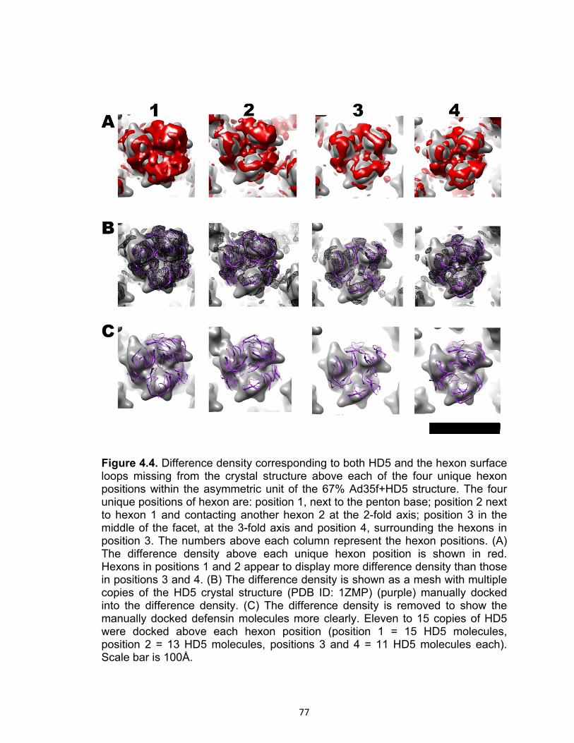
77
Figure 4.4. Difference density corresponding to both HD5 and the hexon surface loops missing from the crystal structure above each of the four unique hexon positions within the asymmetric unit of the 67% Ad35f+HD5 structure. The four unique positions of hexon are: position 1, next to the penton base; position 2 next to hexon 1 and contacting another hexon 2 at the 2-fold axis; position 3 in the middle of the facet, at the 3-fold axis and position 4, surrounding the hexons in position 3. The numbers above each column represent the hexon positions. (A) The difference density above each unique hexon position is shown in red. Hexons in positions 1 and 2 appear to display more difference density than those in positions 3 and 4. (B) The difference density is shown as a mesh with multiple copies of the HD5 crystal structure (PDB ID: 1ZMP) (purple) manually docked into the difference density. (C) The difference density is removed to show the manually docked defensin molecules more clearly. Eleven to 15 copies of HD5 were docked above each hexon position (position 1 = 15 HD5 molecules, position 2 = 13 HD5 molecules, positions 3 and 4 = 11 HD5 molecules each). Scale bar is 100Å.

78
Figure 4.5. Difference density corresponding to both HD5 and the hexon surface loops above each of the four unique hexon positions within the asymmetric unit of the 12% Ad35f+HD5 structure. The four unique positions of hexon are indicated above each column. (A) The difference density above each unique hexon position is shown in magenta. (B) The difference density is shown as a mesh with multiple copies of the HD5 crystal structure (PDB ID: 1ZMP) (purple) manually docked. (C) The difference density is removed to show the manually docked defensin molecules more clearly. An average of 10 copies of HD5 were docked above each hexon position (position 1 = 12 HD5 molecules, position 2 = 12 HD5 molecules, position 3 = 9 HD5 molecules and position 4 = 7 HD5 molecules). Scale bar is 100Å.

79
Figure 4.6. Difference density from the Ad35f structure corresponding to the missing loops in the hexon crystal structure. The four unique positions of hexon are indicated above each column. Arrows indicate density assigned to protein IX, next to hexons in position 2, 3 and 4. (A) The difference density above each unique hexon position is shown in blue. (B) The difference density is shown as a mesh. Note that the difference density above each unique hexon position is similar. Scale bar is 100Å.

80
HD5 coats the fiber and tops of the penton base in the Ad capsid
Difference density attributable to HD5 is not only observed above the
hexons, but also at the vertex region, above the penton base and coating the Ad
fiber. Figure 4.7 shows the difference density at the vertex region for both the
Ad35+HD5 67% and 12% structures. The Ad35f+HD5 67% structure shows
extensive difference above the penton base, while the 12% Ad35f+HD5 structure
has significantly less difference density. Figure 4.7 also shows the difference
density corresponding to the Ad5 penton base RGD-containing loop (residues
297-374) and the Ad35 fiber. The pseudoatomic facet used for difference
mapping contains the Ad2 penton base in complex with an Ad2 fiber peptide
(PDB ID: 1X9T). This structure is missing the RGD loops of the penton base
(residues 297 to 374) as well as the fiber shaft and knob (29, 106, 124).
Therefore, the control Ad35f difference density corresponds to the penton base
RGD-containing loop and the Ad35f fiber shaft. The fiber knob is flexible and not
completely reconstructed.
In their biochemical studies, Smith and Nemerow found that Ad, in the
presence of neutralizing levels of HD5, was efficiently taken up by host cells
suggesting that HD5 does not interfere the association of Ad with αv integrins
(10). There are numerous negatively charged residues in the RGD loop of the
Ad2 penton base and in the Ad35 fiber shaft. However, we suspect that only a
fraction of these possible sites are occupied on any one Ad virion because of the
overlapping nature of the modeled sites.

81
Figure 4.7. Difference density in the vertex region in both the Ad35f+HD5 67% and 12% structures and the Ad35f structure. (A) Top and side views of the vertex from the Ad35f+HD5 67% structure. (B) Top and side views of the vertex from the Ad35f+HD5 12% structure. (C) Top and side views of the vertex from Ad35f showing difference density corresponding to the RGD-containing loop of the penton base and the fiber shaft. Scale bar is 100Å.

82
As performed for the difference density over the hexons, copies of the
HD5 crystal structure were manually docked into the difference density in the
vertex region (Figure 4.8). This difference density displays 5-fold averaging due
to the icosahedral symmetry imposed during the reconstruction process. There is
a structural mismatch between the 5-fold penton base and 3-fold fiber, therefore
the fiber density is inappropriately averaged in the cryoEM structures (89). To
help interpret the difference density corresponding to the fiber, 4 triple β-spiral
repeats from the Ad2 fiber (PDB ID: 1QIU) were manually docked into the
difference density. The Ad35f fiber shaft is predicted to have 6 triple β-spiral
repeats but the full length of the fiber is not reconstructed due to flexibility. During
positioning of the HD5 molecules, the sites corresponding to the RGD loops were
avoided.
In our models, HD5 density appears to surround the fiber shaft in the 67%
Ad35f+HD5 structure, while only very few copies of HD5 appear to bind in the
12% Ad35f+HD5 structure. Fifteen HD5 monomers were docked into the
difference density observed above the penton base and surrounding the fiber in
the Ad35f+HD5 67% structure, while avoiding the RGD-loop region. In the 12%
Ad35f+HD5 structure only 5 HD5 monomers were docked into the observed
difference density. Oligomerization of HD5 molecules might occur (JG Smith,
personal communication), which could explain the thick layer of the difference
density in the Ad35f+HD5 67% structure.
There are 12 vertices per virion; therefore, our modeling studies indicate
60 to 180 possible HD5 binding sites on the vertices of one Ad virion. There

83
could also be additional HD5 binding sites on the rest of the fiber shaft and the
fiber knob, which are not completely reconstructed due to flexibility. Binding of
HD5 to Ad does not interfere with the Ad-CAR/Ad-CD46 interaction or with the
Ad-integrin interaction, which triggers viral internalization. Based on these
cryoEM results, we conclude that defensins neutralize Ad by locking the fiber to
the penton base. Fiber loss may be a first key step during Ad programmed
disassembly; therefore, locking the fiber to the capsid may prevent release of the
membrane lytic protein VI.
.

84

85
Figure 4.8. Side views of the vertex region in the Ad35f+HD5 67% and 12% structures and the Ad35f structure. (A) The difference density (mesh) from the Ad35f+HD5 67% structure is shown with 4 fiber shaft repeats (green) from crystal structure of the Ad2 fiber (PDB ID: 1QIU). In addition, 15 copies of the HD5 crystal structure (purple) were manually docked into the observed difference density. The difference density is removed in the right panel. (B) Same as (A) but for the Ad35f+HD5 12% structure. Five copies of HD5 were docked within the observed difference density. (C) The difference density from the Ad35f structure corresponding to the RGD-loops of the penton base and the fiber shaft. The crystal structure of the Ad2 penton base (yellow) and N-terminal fiber peptide (green) with red at the position of residues E18 and D19 is shown. Cyan bars represent the missing RGD loops of the penton base. The cryoEM density for penton base (gray) is shown in panels A and B. The light blue arrows point to the position of the RGD loop in all 3 cryoEM structures. Scale bar is 100Å.

86
Discussion
A possible mechanism of Ad neutralization by HD5 is to stabilize the
vertices of the capsid, as suggested by Smith and Nemerow, though the critical
binding sites for neutralization remained elusive (10). Given that the Ad5/HD5
complex enters the endosome but then is targeted for degradation, we
considered which possible HD5 binding sites identified by cryoEM might stabilize
the Ad virion and prevent release of the membrane lytic protein VI.
We examined all of the possible HD5 binding sites on the Ad capsid
identified by the cryoEM difference mapping analysis and considered which of
these sites may be related to the increased stability of the Ad/HD5 particle.
Multiple possible HD5 binding sites are found around the hexon towers and at
the central depression of the hexon trimer. There are many negatively charged
residues and flexible loops in these locations, making them ideal surfaces for
binding of numerous copies of the positively charged HD5 molecule. We
observed more HD5 bound to the hexons in position 1 than the other 3 hexon
positions of the asymmetric unit. The hexon 1 position is also called the
peripentonal hexon position, as it is adjacent to the penton base in the vertex.
We also observe significant HD5 binding over the penton base. It is possible that
multiple copies of HD5 form a bridge between the peripentonal hexons (those at
position 1) and the penton base. The cryoEM structures represent an icosahedral
average of the particle images included in the reconstruction. Therefore, the HD5
connection between the peripentonal hexons and the penton base would be
difficult to visualize unless it was present in the same manner in the majority of

87
the asymmetric units and particle images. Since not all of the peripentonal
hexons and penton bases are connected by HD5 bridges, it is not obvious how
HD5 binding to the tops of the hexons could significantly increase the stability of
the Ad virion.
Multiple possible HD5 binding sites are also found above the penton base
and around the fiber shaft. There are many negatively charged residues, as well
as one flexible surface loop (the RGD loop) at the top of penton base and
numerous flexible loops along the fiber shaft, which could be ideal binding sites
for the positively charged HD5 molecule. Following the manual docking of HD5 in
the vertex region difference density, we carefully examined the sites where HD5
binds. In both the Ad35f+HD5 67% and 12% structures HD5 is docked in the
vicinity of the fiber N-terminal peptide at the top of the penton base.
In the crystal structure of the Ad2 penton base and fiber peptide, the N-
terminal fiber peptide (aa 10-19) binds to the top of the penton base near the
flexible RGD loop. A conserved hydrophobic stretch of fiber residues (FNPVYPY,
aa 11-17 in Ad2) forms the interaction region with the top of the penton base
(Figure 4.9). Following this conserved fiber region is a stretch of four residues
that are negatively charged or polar in many Ad types including Ad2, Ad5 and
Ad35, but positively charged in other Ad types including Ad17 and Ad37, which
are not neutralized by HD5. We considered whether HD5 binding at this site,
when it is negatively charged as in the Ad35 fiber, may lead to enhanced Ad
stability.

88
An early step in Ad programmed disassembly is the loss of the fiber
concurrent with, or followed shortly by, the loss of the penton base and the
peripentonal hexons (37, 118). If the loss of the fiber is a necessary first step (as
a primer) for the subsequent loss of other viral components, binding of the HD5
to the N-terminal fiber region may stabilize the Ad virion by stabilizing the
fiber/penton base complex. In support of this idea, it has been noted that
fiberless Ad5 vectors are less stable than Ads with fiber (57, 109). These Ad5
fiberless vectors tend to release their viral DNA genome prematurely.
Our working hypothesis is that HD5 binding to the negatively charged and
polar fiber residues, aa 18-21 in Ad35, stabilizes the fiber/penton base complex
and thereby stabilizes the Ad virion and leads to its inability to escape the
endosome. These four residues are near the top of the penton base and in the
vicinity of the fiber shaft (Figure 4.9). When HD5 is bound at this proposed
critical site involving the N-terminal region of the fiber, it is also likely to interact
with residues in the RGD loop of the penton base, therefore stabilizing the
fiber/penton base complex. This hypothesis also explains why some types of Ad
are neutralized by HD5 and other types, like those of species D, are not
neutralized by HD5. We speculate that there is a correlation between non-
neutralization by HD5 and a positively charged sequence after the conserved
penton base interaction region of the Ad fiber.
The Nemerow group has tested various types of human Ads to determine
whether or not they are neutralized by HD5 (Figure 4.10). Ad2 and Ad5, which
belong to species C Ads, are strongly neutralized by HD5. Similarly, species A

89
and B Ads are also neutralized by HD5. The Ad35f vector, which is Ad5
pseudotyped with the Ad35 fiber, is also neutralized by HD5 (J. Smith and G.
Nemerow, personal communication). None of the species D Ads that have been
tested thus far are neutralized by HD5. The fiber sequence is known for all but
two of the Ads types tested (Ad23p and Ad51p). So far there is a perfect
correlation between non-neutralization by HD5 and a positively charged fiber
sequence after the conserved penton base interaction region (Figure 4.10). All of
the species D types tested that are not neutralized by HD5 and for which we
know the fiber sequence, contain a GYAR fiber sequence after the conserved
hydrophobic stretch. This sequence has a positively charged arginine (R)
residue, which would likely repel HD5 away from this site.

90
Figure 4.9. Postulated critical neutralization site for HD5 based on a combined cryoEM and sequence analysis. (A) Three fiber N-terminal peptides (green) are shown above the penton base (multicolor) (PDB ID: 1X9T). (B) Sequence alignment of the N-terminal regions of the fiber for Ad types 2, 5, 35 which are neutralized by HD5, and types 17, and 37, which are not neutralized by HD5. Ad types 2, 5 and 35 have a negatively charged and polar sequence at positions 18-21 in the N-terminal fiber sequence (outlined with red box). Ad types 17 and 37, both species D viruses, contain a positively charged sequence of residues at this position (outlined with a blue box). Residues highlighted in green are conserved among different Ad species and form the penton base association region shown as a green ribbon in panel A.

91
A negatively charged and polar sequence after the conserved penton
base interaction region, such as DTET in the Ad2 and Ad5 fibers or EDES in the
Ad35 fiber, would form a favorable binding pocket for HD5 at the top of the
penton base. Difference density is observed in this vicinity in both the
Ad35f+HD5 12% and 67% structures (Figure 4.11). Without an atomic resolution
structure of an Ad/HD5 complex we cannot precisely define the HD5 binding
sites on the Ad capsid. The size of HD5 (with molecular dimensions of 17Å x 22Å
x 31Å) is similar to the resolutions of the Ad35f+HD5 cryoEM structures (9-12Å).
Thus, visualizing the precise orientations of the HD5 molecules relative to the Ad
capsid is not feasible in these cryoEM structures. Also, cryoEM structure
determination relies on image averaging. If HD5 binds in multiple overlapping
sites as we suspect, then we would observe only an average of the bound HD5
rather than discrete binding sites. HD5 binding probably does not follow
icosahedral symmetry and, therefore, during the symmetry averaging process
some of the HD5 density will be inappropriately averaged. In addition, if HD5
binds to flexible surface loops of Ad as the cryoEM results indicate, this is likely
to cause conformational changes further complicating our ability to resolve
discrete HD5 binding sites. Despite all of these factors, the intermediate
resolution cryoEM structures of Ad35f+HD5, combined with difference mapping
using a pseudoatomic facet, have enabled us to visualize the general areas of
HD5 binding on the Ad capsid.

92
Figure 4.10. Infectivity assay for various Ad types in the presence of HD5. The Ad types and their species classifications are indicated at the bottom of the figure. We hypothesize, based on our cryoEM study together with the biochemical and cell entry analyses performed in the Nemerow lab, that the residues 18-EDES-21 for Ad35f, and similar negatively charged sequences in other Ad types, form the critical neutralization binding site for HD5. The sequence of this N-terminal fiber region is shown at the bottom of the figure. The complete fiber sequences for types 23p and 51p have not been determined. The concentration of HD5 was 15μM in the neutralization assay. Note than in some cases HD5 (for example Ad23p and Ad28p) enhances Ad infectivity. Unpublished data from the Nemerow laboratory (J.G. Smith and G.R. Nemerow).

93
Manual docking with the HD5 crystal structure into the cryoEM difference
density suggests that it is plausible for one or more of the arginine residues on
the surface of HD5 to interact with the negatively charged region of the fiber and
with the flexible RGD loop of penton base. This interaction may “trap” the fiber
and anchor it to the penton base. After the Ad/HD5 complex enters the host cell
endosome, we speculate that the trapped fiber cannot be released from the virion
preventing release of the vertex proteins. Since release of the vertex proteins
including protein VI is critical for escape from the endosome, HD5 effectively
blocks the Ad capsid from undergoing programmed disassembly and therefore
blocks the viral course of infection.
Our cryoEM study of an Ad/HD5 complex has led to the hypothesis that
the critical binding site for HD5 neutralization involves the negatively charged and
polar fiber residues, aa 18-21 in Ad35, at the top of the penton base. This
hypothesis is currently being tested in the Nemerow laboratory with chimeric Ad
vectors containing either negatively or positively charged residues at this site in
the fiber protein. The combined structural, biochemical, and neutralization studies
indicate that HD5 stabilizes the Ad vertex region and prevents viral disassembly
in the endosome. This neutralization mechanism may also be relevant for other
non-enveloped viruses. Defensins neutralize several non-enveloped viruses in
addition to Ad, such as human papillomavirus (HPV) and rotaviruses. If the
defensin neutralization mechanism can be determined for Ad, this knowledge
may be applicable for other non-enveloped viruses. Understanding the
94
mechanism of action of defensins may also help in the development of new
effective treatments against Ad infections.
In the case of Ad, rather than forming membrane pores or interfering with
the interaction between the pathogen and host cell receptors, HD5 appears to
coat the capsid and interfere with programmed disassembly. This allows the host
cell enough time to degrade the virion in the lysosome and thus block the normal
course of viral infection.

95
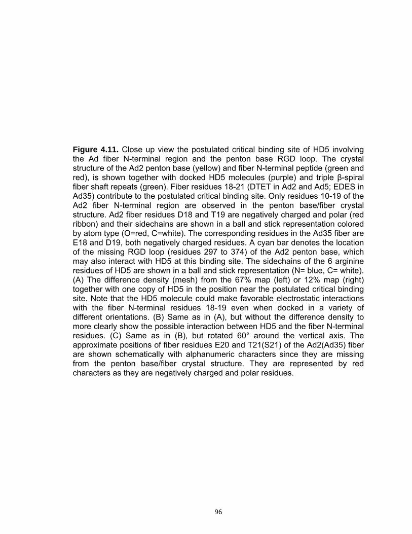
96
Figure 4.11. Close up view the postulated critical binding site of HD5 involving the Ad fiber N-terminal region and the penton base RGD loop. The crystal structure of the Ad2 penton base (yellow) and fiber N-terminal peptide (green and red), is shown together with docked HD5 molecules (purple) and triple β-spiral fiber shaft repeats (green). Fiber residues 18-21 (DTET in Ad2 and Ad5; EDES in Ad35) contribute to the postulated critical binding site. Only residues 10-19 of the Ad2 fiber N-terminal region are observed in the penton base/fiber crystal structure. Ad2 fiber residues D18 and T19 are negatively charged and polar (red ribbon) and their sidechains are shown in a ball and stick representation colored by atom type (O=red, C=white). The corresponding residues in the Ad35 fiber are E18 and D19, both negatively charged residues. A cyan bar denotes the location of the missing RGD loop (residues 297 to 374) of the Ad2 penton base, which may also interact with HD5 at this binding site. The sidechains of the 6 arginine residues of HD5 are shown in a ball and stick representation (N= blue, C= white). (A) The difference density (mesh) from the 67% map (left) or 12% map (right) together with one copy of HD5 in the position near the postulated critical binding site. Note that the HD5 molecule could make favorable electrostatic interactions with the fiber N-terminal residues 18-19 even when docked in a variety of different orientations. (B) Same as in (A), but without the difference density to more clearly show the possible interaction between HD5 and the fiber N-terminal residues. (C) Same as in (B), but rotated 60° around the vertical axis. The approximate positions of fiber residues E20 and T21(S21) of the Ad2(Ad35) fiber are shown schematically with alphanumeric characters since they are missing from the penton base/fiber crystal structure. They are represented by red characters as they are negatively charged and polar residues.

97
CHAPTER V
Summary and Conclusions
Implications for Adenovirus cell entry from our cryoEM study of Ad2ts1
The cryoEM studies presented here provide insight into the structural
events leading to escape of adenovirus from the endosome, a critical step during
its lifecycle. First, we studied Ad2ts1, a mutant generated in the 1970’s, which
represents a fully assembled but immature Ad. Ad2ts1 contains the immature
forms of both capsid-associated proteins (IIIa, VI and VIII) and core-associated
proteins (VII, mu and terminal protein) that depend on the Ad protease for
maturation (17, 43, 69, 70, 99, 111, 113, 119). When produced at non-permissive
temperature (39°C), the Ad2ts1 mutant incorporates a 5-fold reduced copy
number of the viral protease due to a proline to leucine substitution at position
137 of its sequence. This defect causes the virus to be targeted for degradation
in the lysosome or recycled to the cell membrane (6, 37).
The structure of the Ad2ts1 mutant reveals a connection between the
capsid and core formed by the uncleaved precursor proteins. Based on this
finding, we propose that Ad2ts1 cannot undergo the necessary conformational
changes that result in successful disassembly of the vertex and release of the
membrane lytic factor protein VI for endosomalysis. Failure of the Ad2ts1 mutant
to disassemble in the endosome highlights a critical role for the Ad protease in
viral maturation (6, 37, 43, 83, 111, 112). The Ad protease primes the virus for
disassembly in the host cell by separating the core and inner capsid surface,

98
allowing the penton bases and peripentonal hexons along with protein VI to
dissociate from the virus in the endosome.
Ad2ts1 contains precursor VI, which is processed by the virally-encoded
protease, yet it is fully functional and exhibits similar membrane-lytic activity as
mature VI (118). Our structural study shows that the precursor proteins present in
Ad2ts1 lock the capsid from the inside. Locking of the capsid and association of
precursor protein VI with the core appear to interfere with the release of the Ad
proteins at the appropriate time.
Future directions for Ad2ts1
CryoEM relies on image averaging, and reaching high-resolution is
dependent on the degree of uniformity or homogeneity of the imaged sample.
The Ad2ts1 cryoEM structure contains only ~16% of the collected data, possibly
due to heterogeneity in the sample (96). In comparison, the Ad35f 6Å structure
includes more than one third of the collected particle images. Estimates of the
copy number of protease in the mature Ad virion and in Ad2ts1 differ. It has been
estimated that Ad2ts1 contains fivefold fewer copies of the protease than wild-
type mature virions (3). A study of the Ad protease in 1990 by Anderson noted
that low levels of protease could be detected. This finding suggests that there is
a degree of leakiness, or incomplete penetrance, of the Ad2ts1 phenotype, which
results in an incorporation defect. There could be different populations of the
Ad2ts1 virions with different protease copy numbers, which cannot be
distinguished visually. Some virions might have no protease and remain in a

99
completely immature state, while other virions might contain 1 to 2 copies of the
protease and undergo limited proteolytic processing. Such a scenario would give
rise to a heterogeneous Ad2ts1 sample for cryoEM studies. Having different
populations of immature Ad means that some virions might undergo programmed
disassembly, resembling mature Ad, while others will be arrested at the step of
endosomal escape.
To reach subnanometer resolution and facilitate a more in depth
interpretation of the Ad2ts1 structure, it would be ideal to collect Ad2ts1 data
from a more homogeneous sample. Producing a more homogeneous Ad2ts1
sample could be achieved by deleting the Ad protease gene from the Ad2ts1
genome or inserting a stop codon in the protease ORF. Uniformity of the
immature Ad2ts1 sample would then allow a higher number of particles to be
included in the reconstruction to possibly achieve a high resolution structure,
facilitating assignment of the density for the precursor proteins.
Another way to understand Ad maturation and disassembly would be to
investigate the interaction between hexon and protein VI, in both its mature and
precursor forms by cryoEM. Protein VI is a ~23 kDa protein and would not be
feasible for cryoEM analysis as an isolated protein. However, the hexon trimer is
over 300 kDa, and a hexon/protein VI complex or hexon/precursor VI should be
amenable to single particle cryoEM analysis. A high-resolution structure of the
hexon-VI complex could facilitate interpretation of the Ad2ts1 density and deduce
which regions of protein VI might be interacting with the core components. Even
if only moderate resolution structures of the hexon-VI and hexon-precursor VI

100
complexes were obtained, difference mapping analyses could provide
information on the position of the cleaved N- and C- termini. Changes in the
conformation between mature and immature VI have been postulated as a way
of preventing the release of protein VI from the virus while maturation occurs.
Concordantly, our studies show that hexon shields the lytic function of protein VI
by ~60% (69, 70, 118). Structures of the hexon-VI and hexon-precursor VI
complexes would help us understand the viral maturation steps related to the
protein VI proteolytic cleavage. These studies might also enable us to determine
whether the predicted amphipathic helix of protein VI, which is critical for
membrane lytic activity (aa 36-53), is shielded inside the hexon cavity. If so,
protein VI must dissociate from hexon to perform its lytic function.
Ad-mediated gene delivery
Gene therapy, which is in essence the delivery of a corrective gene to
affected tissues, emerged as a discipline in the late 1970’s and 80’s due to
advances in genetic manipulation. The original goal of replacing a defective gene
has expanded into the delivery of genetic material for the treatment of various
diseases; possible oncolytic therapies include delivery of tumor suppressors and
immunomodulating factors (5, 26, 31, 64, 94, 103, 107, 120). Various vectors are
being developed to deliver therapeutic genes and they belong to one of two
classes: non-viral and viral vectors. Non-viral vectors (such as DNA-containing
liposomes and plasmids) have the advantage of by-passing the immune
response elicited by viral vectors, but the effectiveness of delivery is low when
compared to viral-based therapies.

101
Viral vectors, including retroviruses, pox viruses, adeno-associated virus,
and adenovirus, have evolved efficient ways to deliver genetic material to target
cells. Adenovirus has emerged as a powerful vector for gene delivery and
currently a third of all ongoing gene therapy clinical trials in the US use Ad as the
vector (5, 26, 31).
Among the serious considerations when using Ad vectors is the rapid
immune response that the capsid proteins produce in the host. Since no
integration of the Ad genome occurs, boosting doses of Ad vectors are required
for continued therapeutic treatment. In the late 1990’s a costly lesson was
learned when a young man, Jesse Gelsinger was provided a high dose of Ad as
treatment for an X-linked deficiency, ornithine transcarbamylase. His death
halted gene therapy trials temporarily in order to determine the safe-dosage
conditions.
To date, Ads are being explored to treat several diseases, including heart
failure and cancer. Recombinant human Ads are being used in clinical gene
therapy trails aiming to treat heart failure due to damage to cardiomyocytes
caused by coronary artery disease, inflammation or hypertension. Treatment of
these cells is facilitated by the expression of CAR in the cardiovascular system
(64), in particular on cardiomyocytes and vascular endothelial cells. The goals of
this approach are to ensure that healthy cardiomyocytes are not adversely
affected and to provide a “cure” to those cardiomyocytes that have been affected
but not lost by arterial disease or hypertension (40, 64, 103). Ad-mediated gene
delivery in the cardiovascular system has been recently studied, for the

102
prevention of cardiac arrhythmias (by providing Ad carrying the beta-adrenergic
receptor kinase inhibitor gene), and for delivery of angiogenic growth factors (like
hepatocyte growth factor) (40, 64, 103). These therapies could potentially help in
the regeneration of damaged cardiomyocytes and improve recovery of patients
suffering from different forms of heart disease.
In cancer, Ad-based vectors can be used to provide a variety of therapies
and boost the results of chemotherapy. Several studies have been conducted
using Ad as the vector for the treatment of cancer, including the delivery of tumor
suppressors (p53, p21, Rb), immunomodulating cytokines (such as IL-2, TNFα,
etc), or to stimulate antigen production for tumor vaccines and inhibitors of
angiogenesis (4, 5, 94, 120).
Despite the significant progress in this area, much work remains to be
done to optimize Ad vectors and create new vectors that can evade the strong
immune response which hampers Ad-based therapies. Our cryoEM studies of Ad
have suggested several possible approaches for improving Ad vectors. Hexon
has been shown to bind to blood coagulation factor X, an important factor in the
cascade of clot production, which targets Ads towards the liver (4, 52). If the
organ targeted for therapy is not the liver, mutations to the hexon protein could
be introduced to avoid delivery to the liver. The Ad-factor X binding sites appears
to be at the hexon towers, which are highly variable and could possibly tolerate
sequence alterations. A second avenue is to modify the outer-capsid protein IX,
which helps to stabilize hexon-hexon interactions. Modifications to this protein
(such as labeling with GFP) appear to be well tolerated and could be used to re-

103
target the virus to the desired organ or tissue. A third important alteration would
be to evade the innate immune system by preventing association of Ads with
human defensins. Human α-defensins were recently shown to inhibit Ad infection
by binding to the Ad capsid and preventing disassembly (10). We have shown
that a possible important site for defensin binding is at the interface of the penton
base and fiber proteins and if mutations were to be produced in this region, Ad
vectors may be able to evade defensin-mediated neutralization.
Antiviral peptides: human Ads and defensins
The cryoEM structural study presented in Chapter 4 enabled us to
propose a mechanism for how human α-defensin HD5 neutralizes Ad infection.
Human α-defensin HD5 exhibits potent anti-adenoviral activity through binding
and stabilization of the capsid resulting in failure of the virion to escape the
endosome during cell entry (10). Stabilization of the vertex in the capsid appears
to block disassembly of the capsid as these components are among the first to
be released upon entry of the virus into host cells (10). Human papillomavirus
appears to be arrested at a similar stage as adenovirus when exposed to
defensins (11). Therefore, understanding how defensins neutralize Ad infection
may impact an understanding of defensin-mediated neutralization of other non-
enveloped viruses. Also knowledge of the way in which defensins neutralize Ad
may pave the way for the next generation of Ad vectors that by-pass detection by
the innate immune system.

104
Results and implications from the Ad35f+HD5 studies
The cryoEM study on the interaction of a human defensin with adenovirus
enabled us to hypothesize possible sites for virus neutralization. A defensin-
mediated immune response has been described for both bacteria and enveloped
viruses. For human adenovirus, a non-enveloped virus, Smith and Nemerow,
have shown that defensins HD5 and HNP-1 inhibit Ad infection causing the
neutralized virus to behave in a similar manner as Ad2ts1 produced at 39°C (10).
The Ad-defensin interaction was shown (a) to be direct, (b) to target the incoming
virus for degradation in the lysosome and (c) to prevent disassembly of the
capsid by stabilization at the vertex region. Unpublished studies have found that
HD5 is effective at preventing infection of species A, B and C Ads but not of
species D (Chapter 4, Figure 4.9). HD5 binds to the Ad capsid, but the site(s)
important for neutralization remained elusive. This prompted our investigation
into the structure of human Ad complexed with HD5 defensin.
The cryoEM structure of the Ad35f vector complexed with HD5 shows that
HD5 binds to hexons, penton bases, and fibers of the capsid. The main sites of
defensin binding on the Ad capsid are on the towers and central depressions of
the hexons (which are negatively charged), along the fiber shaft and the tops of
the penton base at the interface where the penton base and fiber interact. A
detailed analysis of the Ad35f-HD5 structural results, together with biochemical
and sequence analysis of the Ad serotypes that are neutralized by defensin and
those that are not neutralized, led us to hypothesize that binding of defensin to
the interface formed by the penton base and fiber serves as a key site for

105
neutralization. Density assigned to HD5 is observed near the N-terminal region of
the fiber. Residues 18 to 21 of the Ad35 fiber form a negatively charged pocket.
Modeling of the HD5 atomic resolution structure into the difference density
indicates that this pocket can fit HD5. As HD5 is predominantly positively
charged, it would have charge complementarity with the pocket created by the N-
terminal region of the Ad35 fiber. In contrast, the corresponding residues of non-
neutralized Ads, such as Ad17 and Ad37 (species D Ads), have positively
charged residues in this pocket which would repel HD5. Currently, the Nemerow
group is testing this site by creating chimeric Ads, in which the N-terminal portion
of the fiber from a defensin-neutralized species is swapped with that of a non-
neutralized species. We anticipate that these studies will definitively test the
hypothesis of the critical neutralization site. If the hypothesis is refuted, then the
Ad-defensin structure will be re-examined for other possible critical sites.
This mechanism of defensin neutralization of Ad differs from that used by
defensins to neutralize bacteria, fungi and enveloped viruses. Defensins appear
to stabilize the Ad capsid and prevent the release of the membrane-lytic factor in
the endosome, thus targeting the virus for lysosomal degradation.
Future directions for Ad-defensin
The cryoEM structure of the Ad35f+HD5 complex reveals binding of HD5
to the outer capsid of Ad and no other noticeable changes to the Ad structure.
The resolution of 9.6Å facilitates data interpretation, but given the small size of
HD5 it is difficult to uniquely position HD5 relative to the Ad capsid in all of its

106
multiple binding sites. Specifically, we cannot determine which surface of HD5 is
interacting with the Ad capsid proteins. The cryoEM structure of Ad35f+HD5
allows for visualization of the general areas of binding for HD5 on the Ad surface,
but not their precise positioning. A high-resolution structure of the Ad/defensin
complex might allow precise analyses of the interaction between this peptide and
Ad. If all of the HD5 binding sites are on flexible loops, then even a high-
resolution structure might not reveal the precise interaction between peptide and
Ad. Using the moderate resolution cryoEM structure of this complex and our
knowledge of the electrostatic surface of HD5, we propose that HD5 binds to a
negatively charged pocket of residues located at the interface of the fiber and
penton base of Ad. This region of residues on the Ad surface may serve as one
important location for neutralization of Ad infection.
To allow a more detailed understanding of the Ad-defensin interaction, a
significantly higher number of cryoEM particle images could be collected and
averaged. An atomic-resolution structure of adenovirus might be within reach in
the near future, as four near-atomic (3.8-4.5Å) resolution cryoEM structures
(rotavirus, Epsilon 15 bacteriophage, GroEL and cytoplasmic polyhedrosis virus)
were reported last year (123). These near-atomic resolution cryoEM structures
involved averaging one million to several million asymmetric units. Higher
resolution data would facilitate a more precise modeling of the defensin
interaction with the fiber and penton base surfaces. This information might also
lead to an understanding of how similar defensins neutralize other non-
enveloped viruses. By carefully examining the binding regions of HD5 on the Ad

107
surface combined with sequence analysis, locations with similar
structure/organization in other non-enveloped viruses could be mutated to test for
loss of HD5 neutralization. Additionally other defensins (such as HNP-1 and
human beta defensins) could be studied in complex with Ad by cryoEM, as well
as complexes of HD5 with the isolated penton (the complex formed by the
penton base and fiber proteins). This would likely reveal a more detailed view of
how HD5 stabilizes the penton base and fiber complex.
CryoEM structures of adenovirus have increased our understanding of
the cell entry and programmed disassembly as presented in the two aims of this
thesis. The cryoEM structure of the Ad2ts1 mutant highlights the importance of
the incorporation of the protease for priming Ad virions for disassembly. Studies
of the Ad+HD5 complex enabled us to hypothesize how this innate immune
peptide neutralizes infection. These studies provide us with a more detailed
understanding on how Ad disassembly is exquisitely controlled, and moreover,
how interference with disassembly leads to degradation of the virus in
lysosomes. It is of particular interest for the gene delivery field how Ads are
neutralized by defensins, which are a first line of defense for the host. These and
future studies may pave the way for the next generation of Ad vectors that can
avert neutralization by defensins and more successfully delivery the therapeutic
gene.

108
Appendix 1
Difference Mapping Scripts
Developed by Mariena Silvestry Ramos and Phoebe L. Stewart

109
Scripts for Difference Map Analysis
Notes:
1. The following scripts work with the IMAGIC-5 release from 080206.
IMAGIC commands can change depending on the release date, thus the
command lines that call for each IMAGIC routine may vary from what is
presented here. The order and/or specifications for the answers may vary
too depending on the IMAGIC version/release.
2. To use the following difference mapping scripts several components are
needed:
a. Experimental Map (the best cryoEM structure that has been
calculated) in MRC (CCP4) format (possibly in 7503 if the
adenovirus sample is of the capsid alone or 6403 or 9603 if it is a
complex).
b. Control Map in MRC format with dimensions of 7503 (for
adenovirus, this varies according to the sample).
c. Pixel sizes for both the experimental and control maps at their
respective dimensions.
d. A “top” directory to which these scripts and the input maps should
be copied.
e. When using these scripts for adenovirus difference mapping, you
will need an initial PDB facet.

110
DM-script # 1 – Prepares the maps
#!/bin/csh -f
echo " "
echo "DIFFERENCE MAP STEP 1"
echo "Please have full experimental map and control map in starting directory"
echo "Both maps should be in CCP4 format"
echo "Experimental map dimensions should be 6403"
echo "Control map should be 7503 "
echo "If Experimental map is at 9603, then comment out BLOWUP section"
echo " And fix input name to the next subfile section"
echo " "
# EDIT the following lines about the input
set TOP = (for example: /janus1/msilvest/diff_map_test1)
set MAINMAP = experimental_map.mrc
set CTRLMAP = control_map.mrc
set MAINPIX = pixel size for experimental map at 9603
set MAINPIX_640 = pixel size for experimental map at 6403
set CTRLPIX = pixel size for experimental map at 7503
set MAINRESN = final resolution of experimental map
set FRACTION = 2*CTRLPIX/MAINRESN
# Calculate fraction 2*CTRLPIX/MAINRESN
#Generate settings
set MAINMAPIMG = `echo $MAINMAP | awk -F. ' '`
set MAINMAPIMG_960 = $MAINMAPIMG"_960"
set MAINMAPIMG_750 = $MAINMAPIMG"_750"
set MAINMAPIMG_CH = $MAINMAPIMG"_ch"
set MAINMAPMRC_CH = $MAINMAPIMG"_ch.mrc"

111
set CTRLMAPIMG = `echo $CTRLMAP | awk -F. '{print $1}'`
set CTRLMAPIMG_FILT = $CTRLMAPIMG"_filt"
set CTRLMAPIMG_FILT_ZOOM = $CTRLMAPIMG"_filt_zoom"
# Automatic main map conversion
echo "Performing main map conversion"
cd $TOP
echo "Performing main map conversion"
cd $TOP
/programs/imagic/imagic/stand/em2em.e << END1
CCP4
IMAGIC
3D
$MAINMAP
$MAINMAPIMG
$MAINPIX,$MAINPIX,$MAINPIX
YES
END1
/programs/imagic/imagic/threed/blowup3d.e << END2
BLOWUP
$MAINMAPIMG
$MAINMAPIMG_960
960,960,960
END2
/programs/imagic/imagic/threed/subfile3d.e << END3
$MAINMAPIMG_960
$MAINMAPIMG_750

112
CENTER
750,750,750
0,0,0
END3
rm -f *960*
echo "*************************************************"
echo "Please proceed with dm-step2"
DM-script # 2 – Cuts chunks of the experimental and control maps for display in later steps and filters control map
#Generate settings
set MAINMAPIMG = `echo $MAINMAP | awk -F. '{print $1}'`
set MAINMAPIMG_750 = $MAINMAPIMG"_750"
set MAINMAPIMG_CH = $MAINMAPIMG"_ch"
set MAINMAPMRC_CH = $MAINMAPIMG"_ch.mrc"
set CTRLMAPIMG = `echo $CTRLMAP | awk -F. '{print $1}'`
set CTRLMAPIMG_FILT = $CTRLMAPIMG"_filt"
set CTRLMAPIMG_FILT_ZOOM = $CTRLMAPIMG"_filt_zoom"
/programs/imagic/imagic/threed/subfile3d.e << END1
$MAINMAPIMG_750
$MAINMAPIMG_CH
GENERAL
115,175,190
520,520,520
0,0,0
END1
/programs/imagic/imagic/stand/em2em.e << END2

113
IMAGIC
CCP4
3D
$MAINMAPIMG_CH
$MAINMAPMRC_CH
$MAINPIX,$MAINPIX,$MAINPIX
YES
END2
# Automatic control map conversion
echo "Performing control map conversion"
/programs/imagic/imagic/stand/em2em.e << END3
CCP4
IMAGIC
3D
$CTRLMAP
$CTRLMAPIMG
$CTRLPIX,$CTRLPIX,$CTRLPIX
YES
END3
/programs/imagic/imagic/threed/fft3d.e << END4
FILTER
$CTRLMAPIMG
$CTRLMAPIMG_FILT
GAUSS
$FRACTION
END4
echo "*****************************************************"

114
echo " Please proceed with dm-step3"
DM-script # 3 - Adjusts control map dimensions to match experimental map dimensions
#!/bin/csh -f
echo " "
echo "DF-MAP STEP 3"
echo " "
# EDIT the following lines about the input (refer to previous steps to name files accordingly)
# IMPORTANT!!!! ENTER ZOOMF MANUALLY (CTRLPIX/MAINPIX)
# MAIN MAP is now the 750 version of the 640 version of your starting map
set TOP =
set MAINMAP =
set CTRLMAP =
set MAINPIX =
set CTRLPIX = CTRLPIX/MAINPIX
set MAINRESN =
set ZOOMF =
#Generate settings
set MAINMAPIMG = `echo $MAINMAP | awk -F. '{print $1}'`
set MAINMAPIMG_CH = $MAINMAPIMG"_ch"
set MAINMAPMRC_CH = $MAINMAPIMG"_ch.mrc"
set CTRLMAPIMG = `echo $CTRLMAP | awk -F. '{print $1}'`
set CTRLMAPIMG_FILT = $CTRLMAPIMG"_filt"
set CTRLMAPIMG_FILT_ZOOM = $CTRLMAPIMG"_filt_zoom"
set CTRLMAPIMG_FILT_ZOOM_TRANS = $CTRLMAPIMG"_filt_zoom_trans"

115
set CTRLMAPIMG_FILT_ZOOM_TRANS_CH = $CTRLMAPIMG"_filt_zoom_trans_ch"
set CTRLMAPMRC_FILT_ZOOM_TRANS_CH =
$CTRLMAPIMG"_filt_zoom_trans_ch.mrc"
/programs/imagic/imagic/threed/blowup3d.e << END1
ZOOM
$CTRLMAPIMG_FILT
$CTRLMAPIMG_FILT_ZOOM
$ZOOMF
END1
echo "******************************************************"
echo "Please continue to dm-step4"
DM-script # 4 – Cuts chunk of control map for later steps
#!/bin/csh -f
echo " "
echo "DF-MAP STEP 4"
echo " "
# EDIT the following lines about the input (refer to previous steps to name files accordingly)
set TOP =
set MAINMAP =
set CTRLMAP =
set MAINPIX =
set CTRLPIX =
set MAINRESN =
#Generate settings

116
set MAINMAPIMG = `echo $MAINMAP | awk -F. '{print $1}'`
set MAINMAPIMG_CH = $MAINMAPIMG"_ch"
set MAINMAPMRC_CH = $MAINMAPIMG"_ch.mrc"
set CTRLMAPIMG = `echo $CTRLMAP | awk -F. '{print $1}'`
set CTRLMAPIMG_FILT = $CTRLMAPIMG"_filt"
set CTRLMAPIMG_FILT_ZOOM = $CTRLMAPIMG"_filt_zoom"
set CTRLMAPIMG_FILT_ZOOM_TRANS = $CTRLMAPIMG"_filt_zoom_trans"
set CTRLMAPIMG_FILT_ZOOM_TRANS_CH = $CTRLMAPIMG"_filt_zoom_trans_ch"
set CTRLMAPMRC_FILT_ZOOM_TRANS_CH
= $CTRLMAPIMG"_filt_zoom_trans_ch.mrc"
echo "Aligning zoomed control map to cryo map"
/programs/imagic/imagic/threed/align3d.e << END1
$CTRLMAPIMG_FILT_ZOOM
$MAINMAPIMG
YES
$CTRLMAPIMG_FILT_ZOOM_TRANS
NO
YES
0,0,0
NO
NO
10
scratch3d
END1
rm -f scratch3d.*
/programs/imagic/imagic/threed/subfile3d.e << END2

117
$CTRLMAPIMG_FILT_ZOOM_TRANS
$CTRLMAPIMG_FILT_ZOOM_TRANS_CH
GENERAL
115,175,190
520,520,520
0,0,0
END2
/programs/imagic/imagic/stand/em2em.e << END3
IMAGIC
CCP4
3D
$CTRLMAPIMG_FILT_ZOOM_TRANS_CH
$CTRLMAPMRC_FILT_ZOOM_TRANS_CH
$MAINPIX,$MAINPIX,$MAINPIX
YES
END3
echo "The following two files are ready to look at in Chimera"
echo "Remember to set origin to 0,0,0"
echo $MAINMAPMRC_CH
echo $CTRLMAPMRC_FILT_ZOOM_TRANS_CH
echo "Please continue to df-step5"
DM-script # 5 – Uses UCSF Chimera to coarsely position facet in PDB format within experimental map
# INSTRUCTIONS
# Sit at alpha, beta or gamma and login
# cd to dir with threed...ch.mrc and coarserefn_facet_v1.pdb
# NOTE: You will need to cp coarserefn_facet_v1.pdb from another dir
118
# Edit the lines below to have the correct map chunk filename
# and correct voxel size
# Start chimera
# Get a command line window with favorites, command line
# In the Chimera command line type the following command:
# source df-step5
# When the Side view window opens, then it is time to do the following
# move x 40 (adjust number until facet is aligned in x
# move y 100 (adjust number until facet is aligned in y
# move z 90 (adjust number until facet is aligned in z
# write relative 0 1 coarserefn_facet_v2.pdb
# then close session and see df-step6
#
open experimental_map_chunk.mrc
volume #0 origin 0,0,0
volume #0 voxelsize ADD the correct voxel site here
volume #0 level 0.03
window
open coarserefn_facet_v1.pdb
~show #1
ribbon #1
window
~select 0
select 1
start Side View
#move x 10
#move y 10

119
#move z 10
#write relative 0 1 coarserefn_facet_v2.pdb
DM-script # 6 - Saves individual coordinate files for each capsomer from the coarsely positioned facet from step 5
#!/bin/csh -f
# EDIT SECTION BELOW and RUN THIS IN A TERMINAL WINDOW
echo " "
echo "DF-STEP6 SELECT CAPSID PROTEINS FROM FACET PDB"
echo " "
# EDIT the following lines about the input (refer to previous steps to name files accordingly)
set TOP =
set MAINMAPMRC_CH = experimental_map_chunk.mrc
set FACET_COARSE = coarserefn_facet_v2.pdb
set MAINPIX = experimental map pixel size
set MAINRESN = experimental map resolution
#Generate settings
set MAINMAP_CH = `echo $MAINMAPMRC_CH | awk -F. '{print $1}'`
echo "Selecting out hexon and penton coordinates from facet pdb file"
cat $FACET_COARSE | grep -v '^CONECT' > temp1.pdb
cat temp1.pdb | grep -v '^HELIX' > temp2.pdb
cat temp2.pdb | grep -v '^SHEET' > temp3.pdb
cat temp3.pdb | grep -v '^TER' > temp4.pdb
head -n 21002 temp4.pdb | tail -n 21002 > hex1a_pre_refn.pdb
head -n 42004 temp4.pdb | tail -n 21002 > hex2a_pre_refn.pdb
head -n 63006 temp4.pdb | tail -n 21002 > hex3a_pre_refn.pdb
head -n 84008 temp4.pdb | tail -n 21002 > hex4a_pre_refn.pdb

120
head -n 105010 temp4.pdb | tail -n 21002 > hex1b_pre_refn.pdb
head -n 126012 temp4.pdb | tail -n 21002 > hex2b_pre_refn.pdb
head -n 147014 temp4.pdb | tail -n 21002 > hex3b_pre_refn.pdb
head -n 168016 temp4.pdb | tail -n 21002 > hex4b_pre_refn.pdb
head -n 189018 temp4.pdb | tail -n 21002 > hex1c_pre_refn.pdb
head -n 210020 temp4.pdb | tail -n 21002 > hex2c_pre_refn.pdb
head -n 231022 temp4.pdb | tail -n 21002 > hex3c_pre_refn.pdb
head -n 252024 temp4.pdb | tail -n 21002 > hex4c_pre_refn.pdb
head -n 273026 temp4.pdb | tail -n 21002 > hex1d_pre_refn.pdb
head -n 294028 temp4.pdb | tail -n 21002 > hex2d_pre_refn.pdb
head -n 315030 temp4.pdb | tail -n 21002 > hex1e_pre_refn.pdb
head -n 336032 temp4.pdb | tail -n 21002 > hex2e_pre_refn.pdb
head -n 357034 temp4.pdb | tail -n 21002 > hex1f_pre_refn.pdb
head -n 378036 temp4.pdb | tail -n 21002 > hex2f_pre_refn.pdb
head -n 396063 temp4.pdb | tail -n 18027 > pena_pre_refn.pdb
head -n 414090 temp4.pdb | tail -n 18027 > penb_pre_refn.pdb
head -n 432117 temp4.pdb | tail -n 18027 > penc_pre_refn.pdb
rm temp1.pdb
rm temp2.pdb
rm temp3.pdb
rm temp4.pdb
echo "*************************************************************"
echo "Please continue with df-step7"
DM-script # 7 – Uses UCSF Chimera to select and refine the positions of individual hexons and penton bases in the facet
# INSTRUCTIONS
# Sit at alpha, beta or gamma and login

121
# cd to dir with threed...ch.mrc and hex1a_pre_refn.pdb etc.
# Edit the lines below to have the correct map chunk filename
# and correct voxel size
# Start chimera
# Get a command line window with favorites, command line
# In the Chimera command line type the following command:
# source df-step7
# When the Side view window opens, then it is time to do the following
# Click on Fit button in Fit Model in Map
# Click on Fit button in Fit Model in Map a second time
#
############ REPEAT FOR SECOND PROTEIN ########
# select #2
# Click on Fit button in Fit Model in Map
# Click on Fit button in Fit Model in Map a second time
#
############ REPEAT FOR THIRD PROTEIN ########
# select #3
# Click on Fit button in Fit Model in Map
# Click on Fit button in Fit Model in Map a second time
#
############ REPEAT FOR REST OF THE PROTEINS UNTIL ########
# select #21
# Click on Fit button in Fit Model in Map
# Click on Fit button in Fit Model in Map a second time
#
########### FINAL STEPS #######################

122
# at command line type
# source df-step8
# In Chimera below the command line you will see a message about
# which file it wrote. When it has finished writing the penc file
# you may close chimera and move on to df-step9.
########### NOTE ######################################
# NOTE - the Fit Model in Map result is the same
# even when the map is coarsely displayed, at 8 or 4 or 1
#
open experimental_map_chunk.mrc
volume #0 origin 0,0,0
volume #0 voxelsize ADD the correct voxel site here
volume #0 level 0.03
window
open hex1a_pre_refn.pdb
open hex2a_pre_refn.pdb
open hex3a_pre_refn.pdb
open hex4a_pre_refn.pdb
open hex1b_pre_refn.pdb
open hex2b_pre_refn.pdb
open hex3b_pre_refn.pdb
open hex4b_pre_refn.pdb
open hex1c_pre_refn.pdb
open hex2c_pre_refn.pdb
open hex3c_pre_refn.pdb
open hex4c_pre_refn.pdb
open hex1d_pre_refn.pdb

123
open hex2d_pre_refn.pdb
open hex1e_pre_refn.pdb
open hex2e_pre_refn.pdb
open hex1f_pre_refn.pdb
open hex2f_pre_refn.pdb
open pena_pre_refn.pdb
open penb_pre_refn.pdb
open penc_pre_refn.pdb
~show #1-21
ribbon #1-21
window
~select 0
select 1
select #1
start Fit Model in Map
start Side View
DM-script # 8 – The docked coordinates for each hexon and penton base in step 7 are saved relative to the experimental map
# INSTRUCTIONS
# When you have completed all of the manual fits in chimera (df-step7)
# then at Chimera command line type the following
# source df-step8
# and the fit coordinates will be saved to 21 files
write relative 0 1 hex1a_post_refn.pdb
write relative 0 2 hex2a_post_refn.pdb
write relative 0 3 hex3a_post_refn.pdb
write relative 0 4 hex4a_post_refn.pdb

124
write relative 0 5 hex1b_post_refn.pdb
write relative 0 6 hex2b_post_refn.pdb
write relative 0 7 hex3b_post_refn.pdb
write relative 0 8 hex4b_post_refn.pdb
write relative 0 9 hex1c_post_refn.pdb
write relative 0 10 hex2c_post_refn.pdb
write relative 0 11 hex3c_post_refn.pdb
write relative 0 12 hex4c_post_refn.pdb
write relative 0 13 hex1d_post_refn.pdb
write relative 0 14 hex2d_post_refn.pdb
write relative 0 15 hex1e_post_refn.pdb
write relative 0 16 hex2e_post_refn.pdb
write relative 0 17 hex1f_post_refn.pdb
write relative 0 18 hex2f_post_refn.pdb
write relative 0 19 pena_post_refn.pdb
write relative 0 20 penb_post_refn.pdb
write relative 0 21 penc_post_refn.pdb
DM-script # 9 – Converts all the hexon and penton base PDB files to a density map (MRC/CCP4 format), filters the calculated map to the appropriate resolution. Note that the PDB2MRC command with the “center” option in this script applies an unwanted translation to the calculated map. Therefore this script also aligns the calculated map with the experimental map using the IMAGIC threed-align (align3d.e) command.
#!/bin/csh -f
echo " "
echo "DF-MAP STEP 9"
echo "Please have map chunk and 18 hexon and 3 penton pdb files in the starting directory"
echo "The hexon pdbs should be named with the following convention"

125
echo "hex1a_post_refn.pdb"
echo "The penton pdbs should be named with the following convention"
echo "pena_post_refn.pdb"
echo " "
# EDIT the following lines about the input (refer to previous steps to name files accordingly)
set TOP =
set SAMP = name of the experimental map
set DIFN = difference map test number (if you have run other diffmap rounds)
set MAP_CHUNK = experimental map chunk
set MAINPIX = experimental map pixel size
set RESN = experimental map resolution
set BOX = 520
cd $TOP
echo "Putting all hexon coords into one file"
set HEXPDB = "facet_18hexon_"$SAMP"_"$DIFN".pdb"
set HEXMRC = "facet_18hexon_"$SAMP"_"$DIFN".mrc"
set HEXIMG = "facet_18hexon_"$SAMP"_"$DIFN
set HEXIMG_TRANS = "facet_18hexon_"$SAMP"_"$DIFN"_trans"
set HEXMRC_TRANS = "facet_18hexon_"$SAMP"_"$DIFN"_trans.mrc"
cat hex1a_post_refn.pdb | grep '^ATOM' > $HEXPDB
cat hex2a_post_refn.pdb | grep '^ATOM' >> $HEXPDB
cat hex3a_post_refn.pdb | grep '^ATOM' >> $HEXPDB
cat hex4a_post_refn.pdb | grep '^ATOM' >> $HEXPDB
cat hex1b_post_refn.pdb | grep '^ATOM' >> $HEXPDB
cat hex2b_post_refn.pdb | grep '^ATOM' >> $HEXPDB
cat hex3b_post_refn.pdb | grep '^ATOM' >> $HEXPDB
cat hex4b_post_refn.pdb | grep '^ATOM' >> $HEXPDB

126
cat hex1c_post_refn.pdb | grep '^ATOM' >> $HEXPDB
cat hex2c_post_refn.pdb | grep '^ATOM' >> $HEXPDB
cat hex3c_post_refn.pdb | grep '^ATOM' >> $HEXPDB
cat hex4c_post_refn.pdb | grep '^ATOM' >> $HEXPDB
cat hex1d_post_refn.pdb | grep '^ATOM' >> $HEXPDB
cat hex2d_post_refn.pdb | grep '^ATOM' >> $HEXPDB
cat hex1e_post_refn.pdb | grep '^ATOM' >> $HEXPDB
cat hex2e_post_refn.pdb | grep '^ATOM' >> $HEXPDB
cat hex1f_post_refn.pdb | grep '^ATOM' >> $HEXPDB
cat hex2f_post_refn.pdb | grep '^ATOM' >> $HEXPDB
echo "Putting all penton coords into one file"
set PENTPDB = "facet_3penton_"$SAMP"_"$DIFN".pdb"
set PENTMRC = "facet_3penton_"$SAMP"_"$DIFN".mrc"
set PENTIMG = "facet_3penton_"$SAMP"_"$DIFN
set PENTIMG_TRANS = "facet_3penton_"$SAMP"_"$DIFN"_trans"
set PENTMRC_TRANS = "facet_3penton_"$SAMP"_"$DIFN"_trans.mrc"
cat pena_post_refn.pdb | grep '^ATOM' > $PENTPDB
cat penb_post_refn.pdb | grep '^ATOM' >> $PENTPDB
cat penc_post_refn.pdb | grep '^ATOM' >> $PENTPDB
echo "Making complete facet with hexon and penton pdbs"
set FACETPDB = "facet_18hex3pent_"$SAMP"_"$DIFN".pdb"
set FACETMRC = "facet_18hex3pent_"$SAMP"_"$DIFN".mrc"
set FACETIMG = "facet_18hex3pent_"$SAMP"_"$DIFN
set FACETIMG_TRANS = "facet_18hex3pent_"$SAMP"_"$DIFN"_trans"
set FACETMRC_TRANS = "facet_18hex3pent_"$SAMP"_"$DIFN"_trans.mrc"
cat $HEXPDB > $FACETPDB
cat $PENTPDB >> $FACETPDB
127
echo "Converting complete facet with penton and hexon pdb files to mrc"
/programs/EMAN/bin/pdb2mrc $FACETPDB $FACETMRC apix=$MAINPIX res=$RESN box=$BOX center
/programs/imagic/imagic/stand/em2em.e << END7
CCP4
IMAGIC
3D
$FACETMRC
$FACETIMG
$MAINPIX,$MAINPIX,$MAINPIX
YES
END7
echo "Aligning facet MRC to map chunk"
/programs/imagic/imagic/threed/align3d.e << END10
$FACETIMG
$MAP_CHUNK
YES
$FACETIMG_TRANS
NO
YES
0,0,200
NO
NO
10
scratch3d3
END10
rm -f scratch*
/programs/imagic/imagic/stand/em2em.e << END13

128
IMAGIC
CCP4
3D
$FACETIMG_TRANS
$FACETMRC_TRANS
$MAINPIX,$MAINPIX,$MAINPIX
YES
END13
echo " "
echo "*********************************************"
echo "The following files are ready for Chimera"
echo "Remember to set origin to 0 0 0 for all MRC files"
echo $MAP_CHUNK
echo $FACETMRC_TRANS
echo "Select Thresh level for each map in Chimera"
echo "Then continue with df-step10"
DM-script # 10 – Normalizes the cryoEM maps and calculated facet
#!/bin/csh -f
echo " "
echo "DF-MAP STEP 10"
echo " "
echo " Remember to change thresh value to appropriate value"
echo " as obtained from Chimera in step "
# EDIT the following lines about the input (refer to previous steps to name files accordingly)
set TOP =
129
set SAMP =
set DIFN =
set MAP_CHUNK =
set MAP_CTRL =
set MAINPIX =
set RESN =
set BOX = 520
set FACET = facet_18hex3pent_
# Settings from df-step9
set MAP1 = `echo $MAP_CHUNK | awk -F. '{print $1}'`
set MAP_IMG = $MAP1
set MAP_CTRLMRC = $MAP_CTRL".mrc"
set FACETPDB = "facet_18hex3pent_"$SAMP"_"$DIFN".pdb"
set FACETMRC = "facet_18hex3pent_"$SAMP"_"$DIFN".mrc"
set FACETIMG = "facet_18hex3pent_"$SAMP"_"$DIFN
set FACETIMG_TRANS = "facet_18hex3pent_"$SAMP"_"$DIFN"_trans"
set FACETMRC_TRANS = "facet_18hex3pent_"$SAMP"_"$DIFN"_trans.mrc"
set FACETIMG_TRANS_NORM = "facet_18hex3pent_"$SAMP"_"$DIFN"_trans_norm"
set FACETMRC_TRANS_NORM = "facet_18hex3pent_"$SAMP"_"$DIFN"_trans_norm.mrc"
# Settings for df-step10
set MAP_IMG_NORM = $MAP1"_norm"
set MAP_MRC_NORM = $MAP1"_norm.mrc"
set MAP_IMG_NORM_ADD = $MAP1"_norm_add"
set MAP_IMG_NORM_ADD_THR = $MAP1"_norm_add_thr"
set MAP_CTRL_NORM = $MAP_CTRL"_norm"
set MAP_CTRL_NORM_MRC = $MAP_CTRL"_norm.mrc"

130
set MAP_CTRL_NORM_ADD = $MAP_CTRL"_norm_add"
set MAP_CTRL_NORM_ADD_THR = $MAP_CTRL"_norm_add_thr"
set FACETIMG_TRANS_THR = "facet_18hex3pent_"$SAMP"_"$DIFN"_trans_thr"
set FACETIMG_TRANS_THR_NORM = "facet_18hex3pent_"$SAMP"_"$DIFN"_trans_thr_norm"
set DIFFIMG_CRYO_MIN_FACET = "diff_cryo_min_facet_"$SAMP"_"$DIFN
set DIFFMRC_CRYO_MIN_FACET = "diff_cryo_min_facet_"$SAMP"_"$DIFN".mrc"
set DIFFIMG_CRYO_MIN_MAP_CTRL = "diff_cryo_min_map_ctrl_"$SAMP"_"$DIFN
set DIFFMRC_CRYO_MIN_MAP_CTRL = "diff_cryo_min_map_ctrl_"$SAMP"_"$DIFN".mrc"
set DIFFIMG_MAP_CTRL_MIN_CRYO = "diff_map_ctrl_min_cryo_"$SAMP"_"$DIFN
set DIFFMRC_MAP_CTRL_MIN_CRYO = "diff_map_ctrl_min_cryo_"$SAMP"_"$DIFN".mrc"
cd $TOP
/programs/imagic/imagic/threed/norm3d.e << END1
$MAP_IMG
$MAP_IMG_NORM
NORM
10.0
END1
/programs/imagic/imagic/threed/norm3d.e << END2
$MAP_CTRL
$MAP_CTRL_NORM
NORM
10.0

131
END2
/programs/imagic/imagic/threed/norm3d.e << END3
$FACETIMG_TRANS
$FACETIMG_TRANS_NORM
NORM
10.0
END3
echo "*******************************************************"
echo " Preparing facet for ccp4 conversion and normalization"
echo "*******************************************************"
/programs/imagic/imagic/stand/headers.e << END4
$FACETIMG_TRANS
WRITE
INDEX
61
INTERACTIVE
0
END4
/programs/imagic/imagic/stand/em2em.e << END 5
IMAGIC
CCP4
3D
$FACETIMG_TRANS_NORM
$FACETMRC_TRANS_NORM
$MAINPIX,$MAINPIX,$MAINPIX
YES
END5

132
echo "***************************************************"
echo "Please continue with df-step11"
DM-script # 11 – Steps 11 to 13 are necessary to apply the appropriate threshold values to all the maps (experimental, control and facet) to ensure that the subtraction process is performed correctly.
#!/bin/csh -f
echo " "
echo "DF-MAP STEP 10"
echo " "
echo " Remember to change thresh value to appropriate value"
echo " as obtained from Chimera in step "
# EDIT the following lines about the input (refer to previous steps to name files accordingly)
set TOP =
set SAMP =
set DIFN =
set MAP_CHUNK =
set MAP_CTRL =
set MAINPIX =
set RESN =
set BOX = 520
set FACET = facet_18hex3pent_
# Settings from df-step9
set MAP1 = `echo $MAP_CHUNK | awk -F. '{print $1}'`
set MAP_IMG = $MAP1
set MAP_CTRLMRC = $MAP_CTRL".mrc"
set FACETPDB = "facet_18hex3pent_"$SAMP"_"$DIFN".pdb"

133
set FACETMRC = "facet_18hex3pent_"$SAMP"_"$DIFN".mrc"
set FACETIMG = "facet_18hex3pent_"$SAMP"_"$DIFN
set FACETIMG_TRANS = "facet_18hex3pent_"$SAMP"_"$DIFN"_trans"
set FACETMRC_TRANS = "facet_18hex3pent_"$SAMP"_"$DIFN"_trans.mrc"
set FACETIMG_TRANS_NORM = "facet_18hex3pent_"$SAMP"_"$DIFN"_trans_norm"
set FACETMRC_TRANS_NORM = "facet_18hex3pent_"$SAMP"_"$DIFN"_trans_norm.mrc"
# Settings for df-step10
set MAP_IMG_NORM = $MAP1"_norm"
set MAP_MRC_NORM = $MAP1"_norm.mrc"
set MAP_IMG_NORM_ADD = $MAP1"_norm_add"
set MAP_IMG_NORM_ADD_THR = $MAP1"_norm_add_thr"
set MAP_CTRL_NORM = $MAP_CTRL"_norm"
set MAP_CTRL_NORM_MRC = $MAP_CTRL"_norm.mrc"
set MAP_CTRL_NORM_ADD = $MAP_CTRL"_norm_add"
set MAP_CTRL_NORM_ADD_THR = $MAP_CTRL"_norm_add_thr"
set FACETIMG_TRANS_THR = "facet_18hex3pent_"$SAMP"_"$DIFN"_trans_thr"
set FACETIMG_TRANS_THR_NORM = "facet_18hex3pent_"$SAMP"_"$DIFN"_trans_thr_norm"
set DIFFIMG_CRYO_MIN_FACET = "diff_cryo_min_facet_"$SAMP"_"$DIFN
set DIFFMRC_CRYO_MIN_FACET = "diff_cryo_min_facet_"$SAMP"_"$DIFN".mrc"
set DIFFIMG_CRYO_MIN_MAP_CTRL = "diff_cryo_min_map_ctrl_"$SAMP"_"$DIFN
set DIFFMRC_CRYO_MIN_MAP_CTRL = "diff_cryo_min_map_ctrl_"$SAMP"_"$DIFN".mrc"
set DIFFIMG_MAP_CTRL_MIN_CRYO = "diff_map_ctrl_min_cryo_"$SAMP"_"$DIFN

134
set DIFFMRC_MAP_CTRL_MIN_CRYO = "diff_map_ctrl_min_cryo_"$SAMP"_"$DIFN".mrc"
cd $TOP
/programs/imagic/imagic/stand/em2em.e << END 1
IMAGIC
CCP4
3D
$MAP_IMG_NORM
$MAP_MRC_NORM
$MAINPIX,$MAINPIX,$MAINPIX
YES
END1
/programs/imagic/imagic/stand/em2em.e << END 2
IMAGIC
CCP4
3D
$MAP_CTRL_NORM
$MAP_CTRL_NORM_MRC
$MAINPIX,$MAINPIX,$MAINPIX
YES
END2
echo "***************************************************"
echo "Please continue with df-step12"

135
DM-script # 12 - Steps 11 to 13 are necessary to apply the appropriate threshold values to all the maps (experimental, control and facet) to ensure that the subtraction process is performed correctly.
#!/bin/csh -f
echo " "
echo "DF-MAP STEP 10"
echo " "
echo " Remember to change thresh value to appropriate value"
echo " as obtained from Chimera in step "
# EDIT the following lines about the input (refer to previous steps to name files accordingly)
set TOP =
set SAMP =
set DIFN =
set MAP_CHUNK =
set MAP_CTRL =
set MAINPIX =
set RESN =
set BOX = 520
set FACET = facet_18hex3pent_
# Settings from df-step9
set MAP1 = `echo $MAP_CHUNK | awk -F. '{print $1}'`
set MAP_IMG = $MAP1
set MAP_CTRLMRC = $MAP_CTRL".mrc"
set FACETPDB = "facet_18hex3pent_"$SAMP"_"$DIFN".pdb"
set FACETMRC = "facet_18hex3pent_"$SAMP"_"$DIFN".mrc"
set FACETIMG = "facet_18hex3pent_"$SAMP"_"$DIFN
set FACETIMG_TRANS = "facet_18hex3pent_"$SAMP"_"$DIFN"_trans"

136
set FACETMRC_TRANS = "facet_18hex3pent_"$SAMP"_"$DIFN"_trans.mrc"
set FACETIMG_TRANS_NORM = "facet_18hex3pent_"$SAMP"_"$DIFN"_trans_norm"
set FACETMRC_TRANS_NORM = "facet_18hex3pent_"$SAMP"_"$DIFN"_trans_norm.mrc"
# Settings for df-step10
set MAP_IMG_NORM = $MAP1"_norm"
set MAP_MRC_NORM = $MAP1"_norm.mrc"
set MAP_IMG_NORM_ADD = $MAP1"_norm_add"
set MAP_IMG_NORM_ADD_THR = $MAP1"_norm_add_thr"
set MAP_CTRL_NORM = $MAP_CTRL"_norm"
set MAP_CTRL_NORM_MRC = $MAP_CTRL"_norm.mrc"
set MAP_CTRL_NORM_ADD = $MAP_CTRL"_norm_add"
set MAP_CTRL_NORM_ADD_THR = $MAP_CTRL"_norm_add_thr"
set FACETIMG_TRANS_THR = "facet_18hex3pent_"$SAMP"_"$DIFN"_trans_thr"
set FACETIMG_TRANS_THR_NORM = "facet_18hex3pent_"$SAMP"_"$DIFN"_trans_thr_norm"
set DIFFIMG_CRYO_MIN_FACET = "diff_cryo_min_facet_"$SAMP"_"$DIFN
set DIFFMRC_CRYO_MIN_FACET = "diff_cryo_min_facet_"$SAMP"_"$DIFN".mrc"
set DIFFIMG_CRYO_MIN_MAP_CTRL = "diff_cryo_min_map_ctrl_"$SAMP"_"$DIFN
set DIFFMRC_CRYO_MIN_MAP_CTRL = "diff_cryo_min_map_ctrl_"$SAMP"_"$DIFN".mrc"
set DIFFIMG_MAP_CTRL_MIN_CRYO = "diff_map_ctrl_min_cryo_"$SAMP"_"$DIFN
set DIFFMRC_MAP_CTRL_MIN_CRYO = "diff_map_ctrl_min_cryo_"$SAMP"_"$DIFN".mrc"
cd $TOP
/programs/imagic/imagic/stand/em2em.e << END 4

137
IMAGIC
CCP4
3D
$MAP_CTRL_NORM
$MAP_CTRL_NORM_MRC
$MAINPIX,$MAINPIX,$MAINPIX
YES
END4
echo "***************************************************"
echo "Now look at MRC files for cryomap, map ctrl and facet"
echo "Decide on add values for difference map calculation"
DM-script # 13 - Steps 11 to 13 are necessary to apply the appropriate threshold values to all the maps (experimental, control and facet) to ensure that the subtraction process is performed correctly.
#!/bin/csh -f
echo " "
echo "DF-MAP STEP 10"
echo " "
echo " Remember to change thresh value to appropriate value"
echo " as obtained from Chimera in step "
# EDIT the following lines about the input (refer to previous steps to name files accordingly)
set TOP =
set SAMP =
set DIFN =
set MAP_CHUNK =
set MAP_CTRL =

138
set MAINPIX =
set RESN =
set BOX = 520
set FACET = facet_18hex3pent_
# Settings from df-step9
set MAP1 = `echo $MAP_CHUNK | awk -F. '{print $1}'`
set MAP_IMG = $MAP1
set MAP_CTRLMRC = $MAP_CTRL".mrc"
set FACETPDB = "facet_18hex3pent_"$SAMP"_"$DIFN".pdb"
set FACETMRC = "facet_18hex3pent_"$SAMP"_"$DIFN".mrc"
set FACETIMG = "facet_18hex3pent_"$SAMP"_"$DIFN
set FACETIMG_TRANS = "facet_18hex3pent_"$SAMP"_"$DIFN"_trans"
set FACETMRC_TRANS = "facet_18hex3pent_"$SAMP"_"$DIFN"_trans.mrc"
# Settings for df-step10
set MAP_IMG_NORM = $MAP1"_norm"
set MAP_IMG_NORM_ADD = $MAP1"_norm_add"
set MAP_IMG_NORM_ADD_THR = $MAP1"_norm_add_thr"
set MAP_CTRL_NORM = $MAP_CTRL"_norm"
set MAP_CTRL_NORM_ADD = $MAP_CTRL"_norm_add"
set MAP_CTRL_NORM_ADD_THR = $MAP_CTRL"_norm_add_thr"
set FACETIMG_TRANS_THR = "facet_18hex3pent_"$SAMP"_"$DIFN"_trans_thr"
set FACETIMG_TRANS_THR_NORM = "facet_18hex3pent_"$SAMP"_"$DIFN"_trans_thr_norm"
set DIFFIMG_CRYO_MIN_FACET = "diff_cryo_min_facet_"$SAMP"_"$DIFN
set DIFFMRC_CRYO_MIN_FACET = "diff_cryo_min_facet_"$SAMP"_"$DIFN".mrc"
set DIFFIMG_CRYO_MIN_MAP_CTRL = "diff_cryo_min_map_ctrl_"$SAMP"_"$DIFN

139
set DIFFMRC_CRYO_MIN_MAP_CTRL = "diff_cryo_min_map_ctrl_"$SAMP"_"$DIFN".mrc"
set DIFFIMG_MAP_CTRL_MIN_CRYO = "diff_map_ctrl_min_cryo_"$SAMP"_"$DIFN
set DIFFMRC_MAP_CTRL_MIN_CRYO = "diff_map_ctrl_min_cryo_"$SAMP"_"$DIFN".mrc"
cd $TOP
/programs/imagic/imagic/threed/inc3dmenu.e << END2
ARITHMETIC
ADD
$MAP_IMG_NORM
$MAP_IMG_NORM_ADD
Write the appropriate value here as determined by using UCSF Chimera
END2
/programs/imagic/imagic/threed/inc3dmenu.e << END3
THRESH
$MAP_IMG_NORM_ADD
$MAP_IMG_NORM_ADD_THR
BELOW
0
END3
/programs/imagic/imagic/threed/inc3dmenu.e << END5
ARITHMETIC
ADD
$MAP_CTRL_NORM
$MAP_CTRL_NORM_ADD
Write the appropriate value here as determined by using UCSF Chimera

140
END5
/programs/imagic/imagic/threed/inc3dmenu.e << END6
THRESH
$MAP_CTRL_NORM_ADD
$MAP_CTRL_NORM_ADD_THR
BELOW
0
END6
rm -f *750*.img
rm -f *750*.hed
rm -f *pre*pdb
echo "***************************************************"
echo "Now you are ready to perform the difference maps"
DM-script # 14 – Calculates difference maps
#!/bin/csh -f
echo " "
echo "DF-MAP STEP 10"
echo " "
echo " Remember to change thresh value to appropriate value"
echo " as obtained from Chimera in step "
# EDIT the following lines about the input
set TOP =
set SAMP =
set DIFN = dm4

141
set MAP_CHUNK =
set MAP_CTRL =
set MAINPIX =
set RESN =
set BOX = 520
set FACET = facet_18hex3pent_
# Settings from df-step10
set MAP1 = `echo $MAP_CHUNK | awk -F. '{print $1}'`
set MAP_IMG = $MAP1
set MAP_MRC = $MAP1".mrc"
set MAP_CTRLMRC = $MAP_CTRL".mrc"
set FACETPDB = "facet_18hex3pent_"$SAMP"_"$DIFN".pdb"
set FACETMRC = "facet_18hex3pent_"$SAMP"_"$DIFN".mrc"
set FACETIMG = "facet_18hex3pent_"$SAMP"_"$DIFN
set FACETIMG_TRANS = "facet_18hex3pent_"$SAMP"_"$DIFN"_trans"
set FACETMRC_TRANS = "facet_18hex3pent_"$SAMP"_"$DIFN"_trans.mrc"
# Settings for df-step11
set MAP_IMG_NORM = $MAP1"_norm"
set MAP_IMG_NORM_ADD = $MAP1"_norm_add"
set MAP_IMG_NORM_ADD_THR = $MAP1"_norm_add_thr"
set MAP_CTRL_NORM = $MAP_CTRL"_norm"
set MAP_CTRL_NORM_ADD = $MAP_CTRL"_norm_add"
set MAP_CTRL_NORM_ADD_THR = $MAP_CTRL"_norm_add_thr"
set FACETIMG_TRANS = "facet_18hex3pent_"$SAMP"_"$DIFN"_trans"

142
set FACETIMG_TRANS_NORM = "facet_18hex3pent_"$SAMP"_"$DIFN"_trans_norm"
set DIFFIMG_CRYO_MIN_FACET = "diff_cryo_min_facet_"$SAMP"_"$DIFN
set DIFFMRC_CRYO_MIN_FACET = "diff_cryo_min_facet_"$SAMP"_"$DIFN".mrc"
set DIFFIMG_CRYO_MIN_MAP_CTRL = "diff_cryo_min_map_ctrl_"$SAMP"_"$DIFN
set DIFFMRC_CRYO_MIN_MAP_CTRL = "diff_cryo_min_map_ctrl_"$SAMP"_"$DIFN".mrc"
set DIFFIMG_MAP_CTRL_MIN_CRYO = "diff_map_ctrl_min_cryo_"$SAMP"_"$DIFN
set DIFFMRC_MAP_CTRL_MIN_CRYO = "diff_map_ctrl_min_cryo_"$SAMP"_"$DIFN".mrc"
cd $TOP
echo "**************************************"
echo "Creating difference maps"
echo "**************************************"
/programs/imagic/imagic/threed/inc3dmenu.e << END10
TWO3DIM
SUBTRACT
$MAP_IMG
$FACETIMG_TRANS_NORM
$DIFFIMG_CRYO_MIN_FACET
END10
/programs/imagic/imagic/threed/inc3dmenu.e << END12
TWO3DIM
SUBTRACT

143
$MAP_IMG
$MAP_CTRL_NORM_ADD_THR
$DIFFIMG_CRYO_MIN_MAP_CTRL
END12
/programs/imagic/imagic/threed/inc3dmenu.e << END14
TWO3DIM
SUBTRACT
$MAP_CTRL_NORM_ADD_THR
$MAP_IMG
$DIFFIMG_MAP_CTRL_MIN_CRYO
END14
/programs/imagic/imagic/stand/em2em.e << END15
IMAGIC
CCP4
3D
$DIFFIMG_MAP_CTRL_MIN_CRYO
$DIFFMRC_MAP_CTRL_MIN_CRYO
$MAINPIX,$MAINPIX,$MAINPIX
YES
END15
/programs/imagic/imagic/stand/em2em.e << END11
IMAGIC
CCP4
3D
$DIFFIMG_CRYO_MIN_FACET

144
$DIFFMRC_CRYO_MIN_FACET
$MAINPIX,$MAINPIX,$MAINPIX
YES
END11
/programs/imagic/imagic/stand/em2em.e << END13
IMAGIC
CCP4
3D
$DIFFIMG_CRYO_MIN_FACET
$DIFFMRC_CRYO_MIN_MAP_CTRL
$MAINPIX,$MAINPIX,$MAINPIX
YES
END13
echo " "
echo "******************************************************************"
echo "The following file is ready for Chimera"
echo "Remember to set origin to 0 0 0 for all MRC files"
echo $DIFFMRC_CRYO_MIN_FACET
echo $DIFFMRC_CRYO_MIN_MAP_CTRL
echo $DIFFMRC_MAP_CTRL_MIN_CRYO
echo "Cut chunks of the diff-density to easy viewing in Chimera at 1,1,1"

145
Appendix 2
Adenovirus serotype 5 hexon is critical for virus infection of hepatocytes in vivo
O. Kalyuzhniy , N.C. Di Paolo, M. Silvestry, S.E. Hofherr, M.A. Barry,
P.L. Stewart, and D.M. Shayakhmetov
Proceedings of the National Academy of Sciences U S A. Apr 8;105(14):5483-8.
2008

146
Appendix 3
Visualization of alpha-helices in a 6-angstrom resolution cryoelectron microscopy
structure of adenovirus allows refinement of capsid protein assignments
Susan D. Saban, Mariena Silvestry, Glen R. Nemerow, and Phoebe L. Stewart
Journal of Virology Dec;80(24):12049-59. 2006

147
Appendix 4
Targeting vault nanoparticles to specific cell surface receptors
V.A. Kickhoefer, M. Han, S. Raval-Fernandes, M.J. Poderycki, R.J. Moniz, D.
Vaccari, M. Silvestry, P.L. Stewart, K.A. Kelly, and L.H. Rome
ACS Nano. Jan 27;3(1):27-36. 2009

148
REFERENCES
1. Adiga, U., W. T. Baxter, R. J. Hall, B. Rockel, B. K. Rath, J. Frank, and R. Glaeser. 2005. Particle picking by segmentation: a comparative study with SPIDER-based manual particle picking. J Struct Biol 152:211-220.
2. Alba, R., A. C. Bradshaw, A. L. Parker, D. Bhella, S. N. Waddington, S.
A. Nicklin, N. van Rooijen, J. Custers, J. Goudsmit, D. H. Barouch, J. H. McVey, and A. H. Baker. 2009. Identification of coagulation factor (F)X binding sites on the adenovirus serotype 5 hexon: effect of mutagenesis on FX interactions and gene transfer. Blood. In Press
3. Anderson, C. W. 1990. The proteinase polypeptide of adenovirus
serotype 2 virions. Virology 177:259-272. 4. Baker, A. H., J. H. McVey, S. N. Waddington, N. C. Di Paolo, and D. M.
Shayakhmetov. 2007. The influence of blood on in vivo adenovirus bio-distribution and transduction. Mol Ther 15:1410-1416.
5. Bangari, D. S., and S. K. Mittal. 2006. Current strategies and future
directions for eluding adenoviral vector immunity. Curr Gene Ther 6:215-226.
6. Begin, M., and J. Weber. 1975. Genetic analysis of adenovirus type 2. I.
Isolation and genetic characterization of temperature-sensitive mutants. J Virol 15:1-7.
7. Bergelson, J. M., J. A. Cunningham, G. Droguett, E. A. Kurt-Jones, A.
Krithivas, J. S. Hong, M. S. Horwitz, R. L. Crowell, and R. W. Finberg. 1997. Isolation of a common receptor for Coxsackie B viruses and adenoviruses 2 and 5. Science 275:1320-1323.
8. Bernard M. Fields, D. M. K., Peter M. Howley, Robert M. Chanock,
Joseph L. Melnick, Thomas P. Monath, Bernard Roizman and Stephen E. Straus. 1996. Fields Virology, p. 2111-2172. In B. N. Fields (ed.), Fields Virology, 3rd ed, vol. 2. Lippincott-Raven, Philadelphia.
9. Brown, D. T., M. Westphal, B. T. Burlingham, U. Winterhoff, and W.
Doerfler. 1975. Structure and composition of the adenovirus type 2 core. J Virol 16:366-387.
10. Buck, C. B. 2008. Defensins' offensive play: exploiting a viral achilles'
heel. Cell Host Microbe 3:3-4. 11. Buck, C. B., P. M. Day, C. D. Thompson, J. Lubkowski, W. Lu, D. R.
Lowy, and J. T. Schiller. 2006. Human alpha-defensins block

149
papillomavirus infection. Proc Natl Acad Sci U S A 103:1516-1521. 12. Challberg, M. D., S. V. Desiderio, and T. J. Kelly, Jr. 1980. Adenovirus
DNA replication in vitro: characterization of a protein covalently linked to nascent DNA strands. Proc Natl Acad Sci U S A 77:5105-5109.
13. Chandonia, J. M., and M. Karplus. 1999. New methods for accurate
prediction of protein secondary structure. Proteins 35:293-306. 14. Chang, T. L., J. Vargas, Jr., A. DelPortillo, and M. E. Klotman. 2005.
Dual role of alpha-defensin-1 in anti-HIV-1 innate immunity. J Clin Invest 115:765-773.
15. Chatterjee, P. K., M. E. Vayda, and S. J. Flint. 1986. Identification of
proteins and protein domains that contact DNA within adenovirus nucleoprotein cores by ultraviolet light crosslinking of oligonucleotides 32P-labelled in vivo. J Mol Biol 188:23-37.
16. Chatterjee, P. K., M. E. Vayda, and S. J. Flint. 1985. Interactions among
the three adenovirus core proteins. J Virol 55:379-386. 17. Chatterjee, P. K., U. C. Yang, and S. J. Flint. 1986. Comparison of the
interactions of the adenovirus type 2 major core protein and its precursor with DNA. Nucleic Acids Res 14:2721-2735.
18. Chelius, D., A. F. Huhmer, C. H. Shieh, E. Lehmberg, J. A. Traina, T.
K. Slattery, and E. Pungor, Jr. 2002. Analysis of the adenovirus type 5 proteome by liquid chromatography and tandem mass spectrometry methods. J Proteome Res 1:501-513.
19. Cole, A. M., and A. L. Cole. 2008. Antimicrobial polypeptides are key
anti-HIV-1 effector molecules of cervicovaginal host defense. Am J Reprod Immunol 59:27-34.
20. Coombs, D. H., A. J. Robinson, J. W. Bodnar, C. J. Jones, and G. D.
Pearson. 1979. Detection of covalent DNA-protein complexes: the adenovirus DNA-terminal protein complex and HeLa DNA-protein complexes. Cold Spring Harb Symp Quant Biol 43 Pt 2:741-753.
21. Corden, J., H. M. Engelking, and G. D. Pearson. 1976. Chromatin-like
organization of the adenovirus chromosome. Proc Natl Acad Sci U S A 73:401-404.
22. Cotten, M., and J. M. Weber. 1995. The adenovirus protease is required
for virus entry into host cells. Virology 213:494-502.

150
23. Cuzange, A., J. Chroboczek, and B. Jacrot. 1994. The penton base of human adenovirus type 3 has the RGD motif. Gene 146:257-259.
24. Daher, K. A., M. E. Selsted, and R. I. Lehrer. 1986. Direct inactivation of
viruses by human granulocyte defensins. J Virol 60:1068-1074. 25. Dietrich, D. E., X. Xiao, D. V. Dawson, M. Belanger, H. Xie, A.
Progulske-Fox, and K. A. Brogden. 2008. Human alpha- and beta-defensins bind to immobilized adhesins from Porphyromonas gingivalis. Infect Immun 76:5714-5720.
26. Dmitriev, I. P., E. A. Kashentseva, and D. T. Curiel. 2002. Engineering
of adenovirus vectors containing heterologous peptide sequences in the C terminus of capsid protein IX. J Virol 76:6893-6899.
27. Durmort, C., C. Stehlin, G. Schoehn, A. Mitraki, E. Drouet, S. Cusack,
and W. P. Burmeister. 2001. Structure of the fiber head of Ad3, a non-CAR-binding serotype of adenovirus. Virology 285:302-312.
28. Everitt, E., L. Lutter, and L. Philipson. 1975. Structural proteins of
adenoviruses. XII. Location and neighbor relationship among proteins of adenovirion type 2 as revealed by enzymatic iodination, immunoprecipitation and chemical cross-linking. Virology 67:197-208.
29. Fabry, C. M., M. Rosa-Calatrava, J. F. Conway, C. Zubieta, S. Cusack,
R. W. Ruigrok, and G. Schoehn. 2005. A quasi-atomic model of human adenovirus type 5 capsid. EMBO J 24:1645-1654.
30. Fabry, C. M., M. Rosa-Calatrava, C. Moriscot, R. W. Ruigrok, P.
Boulanger, and G. Schoehn. 2009. The C-terminal domains of adenovirus serotype 5 protein IX assemble into an antiparallel structure on the facets of the capsid. J Virol 83:1135-1139.
31. Flotte, T. R. 2007. Gene therapy: the first two decades and the current
state-of-the-art. J Cell Physiol 213:301-305. 32. Furci, L., F. Sironi, M. Tolazzi, L. Vassena, and P. Lusso. 2007. Alpha-
defensins block the early steps of HIV-1 infection: interference with the binding of gp120 to CD4. Blood 109:2928-2935.
33. Furcinitti, P. S., J. van Oostrum, and R. M. Burnett. 1989. Adenovirus
polypeptide IX revealed as capsid cement by difference images from electron microscopy and crystallography. EMBO J 8:3563-3570.
34. Gaggar, A., D. M. Shayakhmetov, and A. Lieber. 2003. CD46 is a cellular receptor for group B adenoviruses. Nat Med 9:1408-1412.

151
35. Gastaldelli, M., N. Imelli, K. Boucke, B. Amstutz, O. Meier, and U. F. Greber. 2008. Infectious adenovirus type 2 transport through early but not late endosomes. Traffic 9:2265-2278.
36. Greber, U. F., P. Webster, J. Weber, and A. Helenius. 1996. The role of
the adenovirus protease on virus entry into cells. EMBO J 15:1766-1777. 37. Greber, U. F., M. Willetts, P. Webster, and A. Helenius. 1993. Stepwise
dismantling of adenovirus 2 during entry into cells. Cell 75:477-486. 38. Grigorieff, N., Bfactor:
http://emlab.rose2.brandeis.edu/grigorieff/download_b.html. 39. Grigorieff, N. 2007. FREALIGN: high-resolution refinement of single
particle structures. J Struct Biol 157:117-125. 40. Gupta, D., E. J. Molina, J. Palma, J. P. Gaughan, W. Long, and M.
Macha. 2008. Adenoviral beta-adrenergic receptor kinase inhibitor gene transfer improves exercise capacity, cardiac contractility, and systemic inflammation in a model of pressure overload hypertrophy. Cardiovasc Drugs Ther 22:373-381.
41. Gura, T. 2001. Innate immunity. Ancient system gets new respect.
Science 291:2068-2071. 42. Hancock RE, S. H. 2006. Antimicrobial and host-defense peptides as new
anti-infective therapeutic strategies. Nat Biotechnol 24:1551-1557. 43. Hannan, C., L. H. Raptis, C. V. Dery, and J. Weber. 1983. Biological and
structural studies with an adenovirus type 2 temperature-sensitive mutant defective for uncoating. Intervirology 19:213-223.
44. Harpst, J. A., J. F. Ennever, and W. C. Russell. 1977. Physical
properties of nucleoprotein cores from adenovirus type 5. Nucleic Acids Res 4:477-490.
45. Harter, M. L., J. B. Lewis, and C. W. Anderson. 1979. Adenovirus type 2
terminal protein: purification and comparison of tryptic peptides with known adenovirus-coded proteins. J Virol 31:823-835.
46. Harvey, S. A., E. G. Romanowski, K. A. Yates, and Y. J. Gordon. 2005.
Adenovirus-directed ocular innate immunity: the role of conjunctival defensin-like chemokines (IP-10, I-TAC) and phagocytic human defensin-alpha. Invest Ophthalmol Vis Sci 46:3657-3665.
47. Hassell, J. A., and J. Weber. 1978. Genetic analysis of adenovirus type 2. VIII. Physical locations of temperature-sensitive mutations. J Virol

152
28:671-678. 48. Hasson, T. B., D. A. Ornelles, and T. Shenk. 1992. Adenovirus L1 52-
and 55-kilodalton proteins are present within assembling virions and colocalize with nuclear structures distinct from replication centers. J Virol 66:6133-6142.
49. Honkavuori, K. S., B. D. Pollard, M. S. Rodriguez, R. T. Hay, and G. D.
Kemp. 2004. Dual role of the adenovirus pVI C terminus as a nuclear localization signal and activator of the viral protease. J Gen Virol 85:3367-3376.
50. Hosokawa, K., and M. T. Sung. 1976. Isolation and characterization of
an extremely basic protein from adenovirus type 5. J Virol 17:924-934. 51. Jones, D. T. 1999. Protein secondary structure prediction based on
position-specific scoring matrices. J Mol Biol 292:195-202. 52. Kalyuzhniy, O., N. C. Di Paolo, M. Silvestry, S. E. Hofherr, M. A. Barry,
P. L. Stewart, and D. M. Shayakhmetov. 2008. Adenovirus serotype 5 hexon is critical for virus infection of hepatocytes in vivo. Proc Natl Acad Sci U S A 105:5483-5488.
53. Karplus, K., K. Sjolander, C. Barrett, M. Cline, D. Haussler, R.
Hughey, L. Holm, and C. Sander. 1997. Predicting protein structure using hidden Markov models. Proteins Suppl 1:134-139.
54. Kickhoefer, V. A., M. Han, S. Raval-Fernandes, M. J. Poderycki, R. J.
Moniz, D. Vaccari, M. Silvestry, P. L. Stewart, K. A. Kelly, and L. H. Rome. 2009. Targeting vault nanoparticles to specific cell surface receptors. ACS Nano 3:27-36.
55. Kirby, I., E. Davison, A. J. Beavil, C. P. Soh, T. J. Wickham, P. W.
Roelvink, I. Kovesdi, B. J. Sutton, and G. Santis. 2000. Identification of contact residues and definition of the CAR-binding site of adenovirus type 5 fiber protein. J Virol 74:2804-2813.
56. Kuhn, L., D. Trabattoni, C. Kankasa, K. Semrau, P. Kasonde, F.
Lissoni, M. Sinkala, M. Ghosh, C. Vwalika, G. M. Aldrovandi, D. M. Thea, and M. Clerici. 2005. Alpha-defensins in the prevention of HIV transmission among breastfed infants. J Acquir Immune Defic Syndr 39:138-142.
57. Legrand, V., D. Spehner, Y. Schlesinger, N. Settelen, A. Pavirani, and M. Mehtali. 1999. Fiberless recombinant adenoviruses: virus maturation and infectivity in the absence of fiber. J Virol 73:907-919.

153
58. Lehmberg, E., J. A. Traina, J. A. Chakel, R. J. Chang, M. Parkman, M. T. McCaman, P. K. Murakami, V. Lahidji, J. W. Nelson, W. S. Hancock, E. Nestaas, and E. Pungor, Jr. 1999. Reversed-phase high-performance liquid chromatographic assay for the adenovirus type 5 proteome. J Chromatogr B Biomed Sci Appl 732:411-423.
59. Lehrer RI, L. A., Ganz T. 1993. Defensins: antimicrobial and cytotoxic
peptides of mammalian cells. Annu Rev Immunol 11:105-128. 60. Leopold, P. L., and R. G. Crystal. 2007. Intracellular trafficking of
adenovirus: many means to many ends. Adv Drug Deliv Rev 59:810-821. 61. Liu, G. Q., L. E. Babiss, F. C. Volkert, C. S. Young, and H. S.
Ginsberg. 1985. A thermolabile mutant of adenovirus 5 resulting from a substitution mutation in the protein VIII gene. J Virol 53:920-925.
62. Liu, Y.-H., G. Vellekamp, G. Chen, U. A. Mirza, D. Wylie, B.
Twarowska, J. T. Tang, F. W. Porter, S. Wang, T. L. Nagabhushan, and B. N. Pramanik. 2003. Proteomic study of recombinant adenovirus 5 encoding human p53 by matrix-assisted laser desorption/ionization mass spectrometry in combination with database search. International Journal of Mass Spectrometry 226:55-69.
63. Lortat-Jacob, H., E. Chouin, S. Cusack, and M. J. van Raaij. 2001.
Kinetic analysis of adenovirus fiber binding to its receptor reveals an avidity mechanism for trimeric receptor-ligand interactions. J Biol Chem 276:9009-9015.
64. Ly, H., Y. Kawase, R. Yoneyama, and R. J. Hajjar. 2007. Gene therapy
in the treatment of heart failure. Physiology (Bethesda) 22:81-96. 65. Mangel, W. F., M. L. Baniecki, and W. J. McGrath. 2003. Specific
interactions of the adenovirus proteinase with the viral DNA, an 11-amino-acid viral peptide, and the cellular protein actin. Cell Mol Life Sci 60:2347-2355.
66. Mangel, W. F., D. L. Toledo, M. T. Brown, J. H. Martin, and W. J.
McGrath. 1996. Characterization of three components of human adenovirus proteinase activity in vitro. J Biol Chem 271:536-543.
67. Marsh, M. P., S. K. Campos, M. L. Baker, C. Y. Chen, W. Chiu, and M.
A. Barry. 2006. Cryoelectron microscopy of protein IX-modified adenoviruses suggests a new position for the C terminus of protein IX. J Virol 80:11881-11886.
68. Matthews, D. A., and W. C. Russell. 1998. Adenovirus core protein V is
154
delivered by the invading virus to the nucleus of the infected cell and later in infection is associated with nucleoli. J Gen Virol 79 ( Pt 7):1671-1675.
69. Matthews, D. A., and W. C. Russell. 1994. Adenovirus protein-protein
interactions: hexon and protein VI. J Gen Virol 75 ( Pt 12):3365-3374. 70. Matthews, D. A., and W. C. Russell. 1995. Adenovirus protein-protein
interactions: molecular parameters governing the binding of protein VI to hexon and the activation of the adenovirus 23K protease. J Gen Virol 76 ( Pt 8):1959-1969.
71. McPhee JB, H. R. 2005. Function and therapeutic potential of host
defence peptides. J Pept Sci 11:677-687. 72. Meiler, J., and D. Baker. 2003. Coupled prediction of protein secondary
and tertiary structure. Proc Natl Acad Sci U S A 100:12105-12110. 73. Meiler, J., J. J. Prompers, W. Peti, C. Griesinger, and R. Bruschweiler.
2001. Model-free approach to the dynamic interpretation of residual dipolar couplings in globular proteins. J Am Chem Soc 123:6098-6107.
74. Mindell, J. A., and N. Grigorieff. 2003. Accurate determination of local
defocus and specimen tilt in electron microscopy. J Struct Biol 142:334-347.
75. Mirza, A., and J. Weber. 1980. Infectivity and uncoating of adenovirus
cores. Intervirology 13:307-311. 76. Mirza, M. A., and J. Weber. 1982. Structure of adenovirus chromatin.
Biochim Biophys Acta 696:76-86. 77. Mirza, M. A., and J. Weber. 1979. Uncoating of adenovirus type 2. J Virol
30:462-471. 78. Mondel, M., B. O. Schroeder, K. Zimmermann, H. Huber, S. Nuding, J.
Beisner, K. Fellermann, E. F. Stange, and J. Wehkamp. 2009. Probiotic E. coli treatment mediates antimicrobial human beta-defensin synthesis and fecal excretion in humans. Mucosal Immunol 2:166-172.
79. Mookherjee N, H. R. 2007. Cationic host defence peptides: innate
immune regulatory peptides as a novel approach for treating infections. Cell Mol Life Sci 64:922-933.
80. Nemerow, G. R., L. Pache, V. Reddy, and P. L. Stewart. 2009. Insights into adenovirus host cell interactions from structural studies. Virology 384:380-388.

155
81. Nermut, M. V., J. A. Harpst, and W. C. Russell. 1975. Electron microscopy of adenovirus cores. J Gen Virol 28:49-58.
82. Pettersen, E. F., T. D. Goddard, C. C. Huang, G. S. Couch, D. M.
Greenblatt, E. C. Meng, and T. E. Ferrin. 2004. UCSF Chimera--a visualization system for exploratory research and analysis. J Comput Chem 25:1605-1612.
83. Rancourt, C., H. Keyvani-Amineh, S. Sircar, P. Labrecque, and J. M.
Weber. 1995. Proline 137 is critical for adenovirus protease encapsidation and activation but not enzyme activity. Virology 209:167-173.
84. Rekosh, D. M., W. C. Russell, A. J. Bellet, and A. J. Robinson. 1977.
Identification of a protein linked to the ends of adenovirus DNA. Cell 11:283-295.
85. Rosenthal, P. B., and R. Henderson. 2003. Optimal determination of
particle orientation, absolute hand, and contrast loss in single-particle electron cryomicroscopy. J Mol Biol 333:721-745.
86. Russell, W. C., and B. Precious. 1982. Nucleic acid-binding properties of
adenovirus structural polypeptides. J Gen Virol 63 (Pt 1):69-79. 87. Rux, J. J., P. R. Kuser, and R. M. Burnett. 2003. Structural and
phylogenetic analysis of adenovirus hexons by use of high-resolution x-ray crystallographic, molecular modeling, and sequence-based methods. J Virol 77:9553-9566.
88. Saban, S. D., R. R. Nepomuceno, L. D. Gritton, G. R. Nemerow, and P.
L. Stewart. 2005. CryoEM structure at 9A resolution of an adenovirus vector targeted to hematopoietic cells. J Mol Biol 349:526-537.
89. Saban, S. D., M. Silvestry, G. R. Nemerow, and P. L. Stewart. 2006.
Visualization of alpha-helices in a 6-angstrom resolution cryoelectron microscopy structure of adenovirus allows refinement of capsid protein assignments. J Virol 80:12049-12059.
90. Salvatore M, G.-S. A., Ruchala P, Lehrer RI, Chang T, Klotman ME.
2007. alpha-Defensin inhibits influenza virus replication by cell-mediated mechanism(s). J Infect Dis 196:835-843.
91. San Martin, C., J. N. Glasgow, A. Borovjagin, M. S. Beatty, E. A.
Kashentseva, D. T. Curiel, R. Marabini, and I. P. Dmitriev. 2008. Localization of the N-terminus of minor coat protein IIIa in the adenovirus capsid. J Mol Biol 383:923-934.

156
92. Schlee, M., J. Wehkamp, A. Altenhoefer, T. A. Oelschlaeger, E. F. Stange, and K. Fellermann. 2007. Induction of human beta-defensin 2 by the probiotic Escherichia coli Nissle 1917 is mediated through flagellin. Infect Immun 75:2399-2407.
93. Schneider, J. J., A. Unholzer, M. Schaller, M. Schafer-Korting, and H.
C. Korting. 2005. Human defensins. J Mol Med 83:587-595. 94. Seth, P. 2005. Vector-mediated cancer gene therapy: an overview.
Cancer Biol Ther 4:512-517. 95. Shi, J., D. R. Williams, and P. L. Stewart. 2008. A Script-Assisted
Microscopy (SAM) package to improve data acquisition rates on FEI Tecnai electron microscopes equipped with Gatan CCD cameras. J Struct Biol 164:166-169.
96. Silvestry, M., S. Lindert, J. G. Smith, O. Maier, C. M. Wiethoff, G. R.
Nemerow, and P. L. Stewart. 2009. Cryo-electron microscopy structure of adenovirus type 2 temperature-sensitive mutant 1 reveals insight into the cell entry defect. J Virol 83:7375-7383.
97. Smith, J. G., and G. R. Nemerow. 2008. Mechanism of adenovirus
neutralization by Human alpha-defensins. Cell Host Microbe 3:11-19. 98. Stewart, P. L., R. M. Burnett, M. Cyrklaff, and S. D. Fuller. 1991. Image
reconstruction reveals the complex molecular organization of adenovirus. Cell 67:145-154.
99. Sung, M. T., T. M. Cao, M. A. Lischwe, and R. T. Coleman. 1983.
Molecular processing of adenovirus proteins. J Biol Chem 258:8266-8272. 100. Suomalainen, M., M. Y. Nakano, S. Keller, K. Boucke, R. P. Stidwill,
and U. F. Greber. 1999. Microtubule-dependent plus- and minus end-directed motilities are competing processes for nuclear targeting of adenovirus. J Cell Biol 144:657-672.
101. Szyk, A., Z. Wu, K. Tucker, D. Yang, W. Lu, and J. Lubkowski. 2006.
Crystal structures of human alpha-defensins HNP4, HD5, and HD6. Protein Sci 15:2749-2760.
102. Takahashi, A., A. Wada, K. Ogushi, K. Maeda, T. Kawahara, K.
Mawatari, H. Kurazono, J. Moss, T. Hirayama, and Y. Nakaya. 2001. Production of beta-defensin-2 by human colonic epithelial cells induced by Salmonella enteritidis flagella filament structural protein. FEBS Lett 508:484-488.

157
103. Tamamori-Adachi, M., H. Takagi, K. Hashimoto, K. Goto, T. Hidaka, U. Koshimizu, K. Yamada, I. Goto, Y. Maejima, M. Isobe, K. I. Nakayama, N. Inomata, and S. Kitajima. 2008. Cardiomyocyte proliferation and protection against post-myocardial infarction heart failure by cyclin D1 and Skp2 ubiquitin ligase. Cardiovasc Res 80:181-190.
104. van Heel, M., G. Harauz, E. V. Orlova, R. Schmidt, and M. Schatz.
1996. A new generation of the IMAGIC image processing system. J Struct Biol 116:17-24.
105. van Raaij, M. J., E. Chouin, H. van der Zandt, J. M. Bergelson, and S.
Cusack. 2000. Dimeric structure of the coxsackievirus and adenovirus receptor D1 domain at 1.7 A resolution. Structure 8:1147-1155.
106. van Raaij, M. J., A. Mitraki, G. Lavigne, and S. Cusack. 1999. A triple
beta-spiral in the adenovirus fibre shaft reveals a new structural motif for a fibrous protein. Nature 401:935-938.
107. Vasudevan S, Y. J., Osapay G, Tran P, Tai K, Liang W, Kumar V,
Selsted ME, Cocco MJ. 2008. Synthesis, structure, and activities of an oral mucosal alpha-defensin from rhesus macaque. J Biol Chem 283:35869-35877.
108. Vigant, F., D. Descamps, B. Jullienne, S. Esselin, E. Connault, P.
Opolon, T. Tordjmann, E. Vigne, M. Perricaudet, and K. Benihoud. 2008. Substitution of hexon hypervariable region 5 of adenovirus serotype 5 abrogates blood factor binding and limits gene transfer to liver. Mol Ther 16:1474-1480.
109. Von Seggern DJ, C. C., Fleck SK, Stewart PL, Nemerow GR. 1999. A
helper-independent adenovirus vector with E1, E3, and fiber deleted: structure and infectivity of fiberless particles. J Virol. 73:1601-1608.
110. Waddington, S. N., J. H. McVey, D. Bhella, A. L. Parker, K. Barker, H.
Atoda, R. Pink, S. M. Buckley, J. A. Greig, L. Denby, J. Custers, T. Morita, I. M. Francischetti, R. Q. Monteiro, D. H. Barouch, N. van Rooijen, C. Napoli, M. J. Havenga, S. A. Nicklin, and A. H. Baker. 2008. Adenovirus serotype 5 hexon mediates liver gene transfer. Cell 132:397-409.
111. Weber, J. 1976. Genetic analysis of adenovirus type 2 III. Temperature
sensitivity of processing viral proteins. J Virol 17:462-471. 112. Weber, J., M. Begin, and G. Khittoo. 1975. Genetic Analysis of
Adenovirus Type 2 II. Preliminary Phenotypic Characterization of Temperature-Sensitive Mutants. J Virol 15:1049-1056.

158
113. Webster, A., R. T. Hay, and G. Kemp. 1993. The adenovirus protease is
activated by a virus-coded disulphide-linked peptide. Cell 72:97-104. 114. Webster, A., S. Russell, P. Talbot, W. C. Russell, and G. D. Kemp.
1989. Characterization of the adenovirus proteinase: substrate specificity. J Gen Virol 70 ( Pt 12):3225-3234.
115. Weinberg, A., M. E. Quinones-Mateu, and M. M. Lederman. 2006. Role
of human beta-defensins in HIV infection. Adv Dent Res 19:42-48. 116. Wickham, T. J., E. J. Filardo, D. A. Cheresh, and G. R. Nemerow.
1994. Integrin alpha v beta 5 selectively promotes adenovirus mediated cell membrane permeabilization. J Cell Biol 127:257-264.
117. Wickham, T. J., P. Mathias, D. A. Cheresh, and G. R. Nemerow. 1993.
Integrins alpha v beta 3 and alpha v beta 5 promote adenovirus internalization but not virus attachment. Cell 73:309-319.
118. Wiethoff, C. M., H. Wodrich, L. Gerace, and G. R. Nemerow. 2005.
Adenovirus protein VI mediates membrane disruption following capsid disassembly. J Virol 79:1992-2000.
119. Wodrich, H., T. Guan, G. Cingolani, D. Von Seggern, G. Nemerow,
and L. Gerace. 2003. Switch from capsid protein import to adenovirus assembly by cleavage of nuclear transport signals. EMBO J 22:6245-6255.
120. Yang D, L. Z., Tewary P, Chen Q, de la Rosa G, Oppenheim JJ. 2007.
Defensin participation in innate and adaptive immunity. Curr Pharm Des 13:3131-3139.
121. Yedery, R. D., and K. V. Reddy. 2005. Antimicrobial peptides as
microbicidal contraceptives: prophecies for prophylactics--a mini review. Eur J Contracept Reprod Health Care 10:32-42.
122. Zhang, W., and R. Arcos. 2005. Interaction of the adenovirus major core
protein precursor, pVII, with the viral DNA packaging machinery. Virology 334:194-202.
123. Zhou, Z. H. 2008. Towards atomic resolution structural determination by
single-particle cryo-electron microscopy. Curr Opin Struct Biol 18:218-228. 124. Zubieta, C., G. Schoehn, J. Chroboczek, and S. Cusack. 2005. The
structure of the human adenovirus 2 penton. Mol Cell 17:121-135.