Embed Size (px)
Citation preview
Bronchoscopy
TSDA Boot Camp
September 11-14, 2014
Chapel Hill, NC
General Thoracic Faculty
• Rick Feins, MD,
Program Director
• Carolyn Jones, MD
Course Director
• John Nesbitt, MD
Course Director
• Mark Adams, RDCS
• Manjit Bains, MD
• D. Pat Burney
• Mark Ferguson, MD
• Sean Kwon, MD
• Mark Onaitis, MD
• Lana Schumacher, MD
• Mithran Sukumar, MD
• Norman Snow, MD
• Darryl Weiman, MD
• Walter Wolfe, MD
• Stephen Yang, MD

Goals
– Laryngeal and Airway Anatomy
– Flexible Bronchoscopy
• Basics
• Indications/ Contraindications
– Rigid Bronchoscopy
• Basics
• Indications/ Contraindications

The Larynx
Epiglottis
To the
Esophagus
Cuneiform and
corniculate cartilage
Aryepiglottic
fold
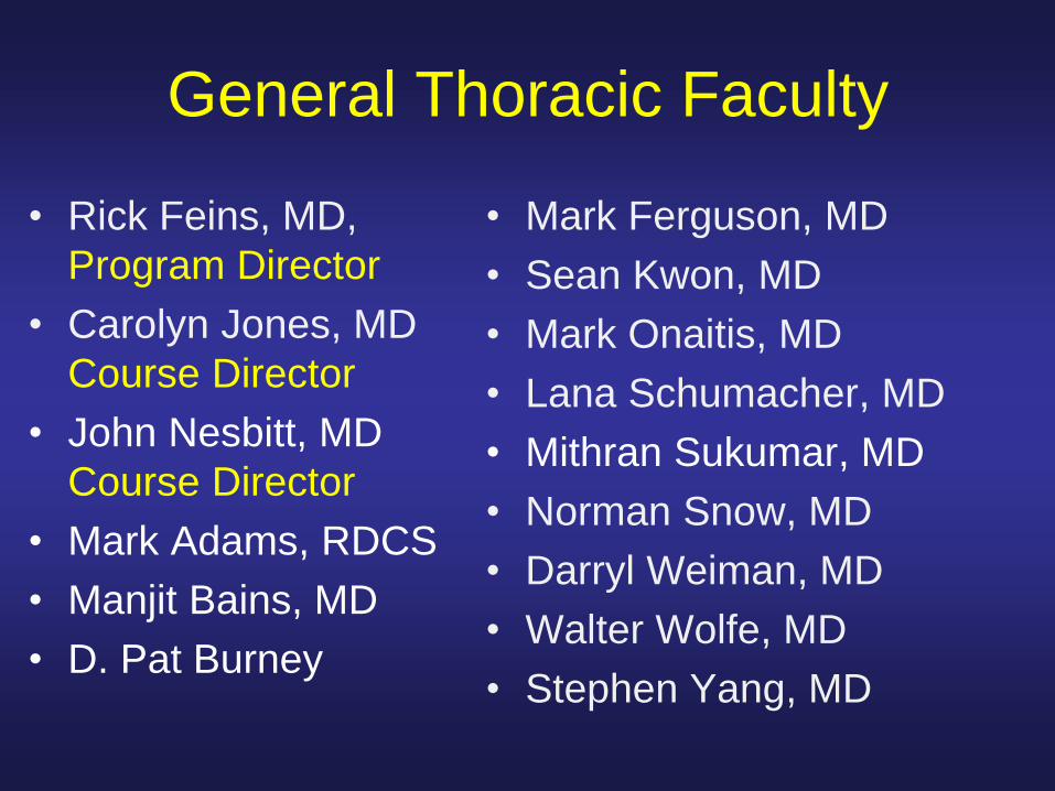
The Larynx: Anatomy
Structural rigidity provided by: The epiglottis, Thyroid cartilage and Cricoid cartilage
www.throat-cancer-symptoms.com/ www.yoursurgery.com

The Larynx II
Tracheal Cartilaginous
Rings
True
Vocal
Cords
False
Vocal
Cords
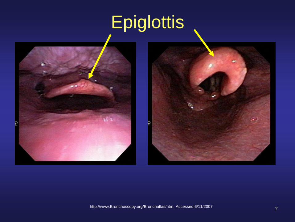
7
Epiglottis
http://www.Bronchoscopy.org/Bronchatlas/htm. Accessed 6/11/2007

http://www.Bronchoscopy.org/Bronchatlas/htm. Accessed 6/11/2007
The Larynx
open for inspiration and closed for swallowing
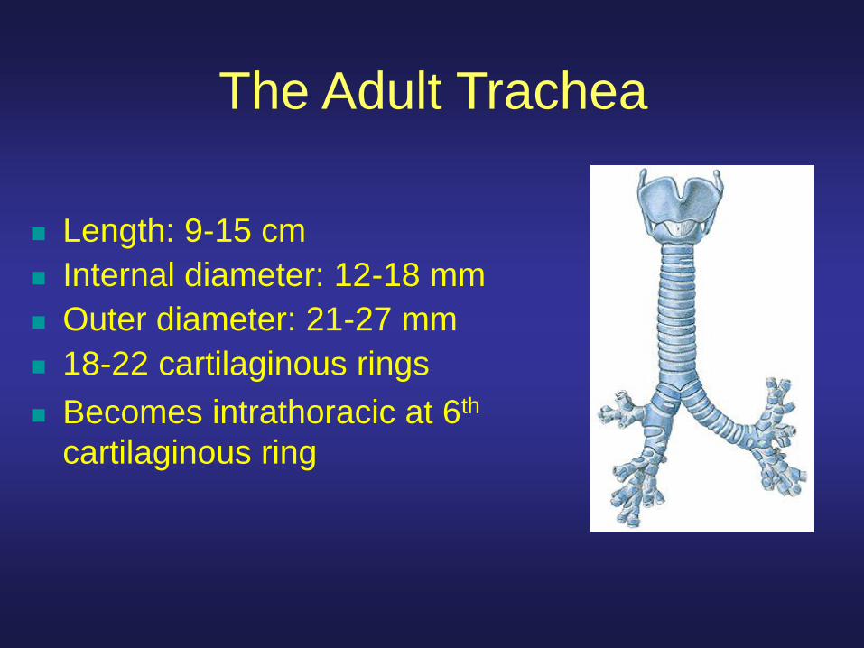
The Adult Trachea
Length: 9-15 cm
Internal diameter: 12-18 mm
Outer diameter: 21-27 mm
18-22 cartilaginous rings
Becomes intrathoracic at 6th
cartilaginous ring

Tracheal dimensions – adult male
•Average cross-sectional
area is 2.8 cm2
•Upper limits of normal
Transverse diameter of 25
mm and AP diameter of 27
mm
•The lower limit of normal
for both diameters is about
13 mm
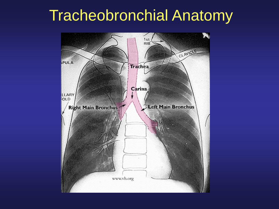
Tracheobronchial Anatomy
www.vh.org

12
The Carina
Posterior
posterior From front of
patient
LMB
RMB
BI, All Rights Reserved, 2005 13
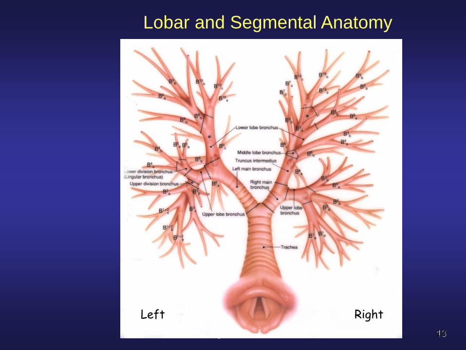
Lobar and Segmental Anatomy
Left Right

Right Bronchial Anatomy
• Right main bronchus
– 2cm long, I.D. 10-16mm
• Right upper lobe
• Bronchus intermedius
–Middle lobe
–Lower lobe
10/27/2005 BI, All Rights Reserved, 2005 15
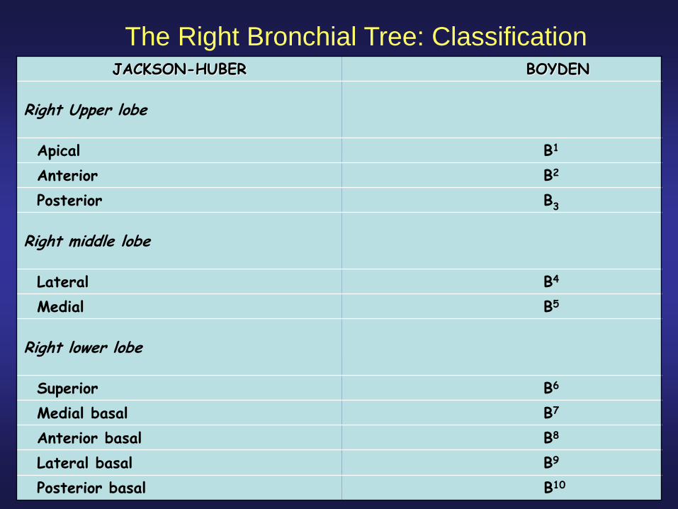
The Right Bronchial Tree: Classification JACKSON-HUBER BOYDEN
Right Upper lobe
Apical B1
Anterior B2
Posterior B3
Right middle lobe
Lateral B4
Medial B5
Right lower lobe
Superior B6
Medial basal B7
Anterior basal B8
Lateral basal B9
Posterior basal B10
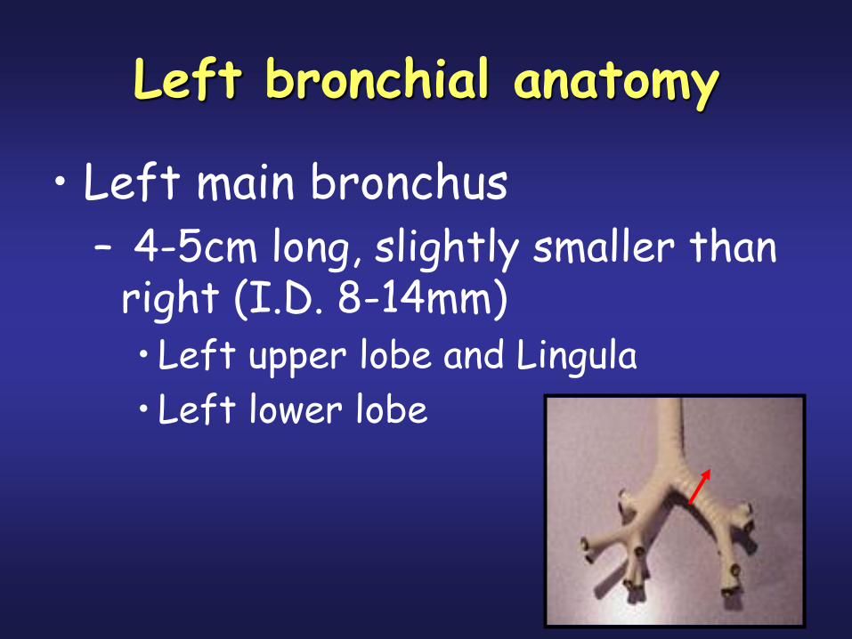
Left bronchial anatomy
• Left main bronchus – 4-5cm long, slightly smaller than
right (I.D. 8-14mm) • Left upper lobe and Lingula
• Left lower lobe

17
Left Bronchial Tree- nomenclatures
Left upper lobe
Upper division
Apical-posterior B1 & 3
Anterior B2
Lingular/division
Superior B4
Inferior B5
Left lower lobe
Superior B6
Anteromedial B7&8
Lateral basal B9
Posterior basal B10
JACKSON-HUBER BOYDEN

Flexible Bronchoscopy
Courtesy of Robert Garland RRT
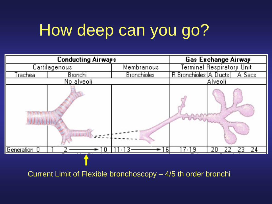
How deep can you go?
Current Limit of Flexible bronchoscopy – 4/5 th order bronchi

Flexible Bronchoscopy- Indications
• Diagnostic – Non massive hemoptysis
– Stridor /Localized wheeze
– Suspected TEF
– Chest trauma
– Pulmonary nodule/ mass
– Mediastinal / hilar Lymphadenopathy
– Pneumonia • Immunocompromised
• Nosocomial
• Non-resolving
– Evaluation rejection
• Therapeutic – Central Airway Obstruction
• Laser photoresection
• Electrocautery
• Argon plasma coagulation
• Brachytherapy
• Photodynamic therapy
– FB removal
– Balloon Dilatation
– Pulmonary toilet
– Endotracheal Intubation
– Percutaneous dilatational tracheostomy
– Metallic Stent placement

Flexible Bronchoscopy-
Contraindications • Inspection
– Life-threatening arrhythmia
– Refractory Hypoxemia
– Inability to cooperate with procedure
– Recent MI or unstable angina
• Biopsy – Serum creatinine >3
– Platelets <50,000
– Uncorrected coagulopathy
– Pulmonary HTN
– SVC syndrome

Rigid Bronchoscopy
Courtesy of Robert Garland RRT
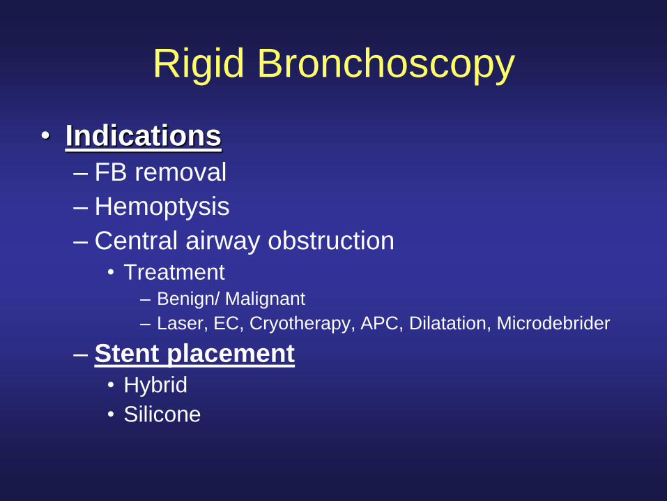
Rigid Bronchoscopy
• Indications
– FB removal
– Hemoptysis
– Central airway obstruction • Treatment
– Benign/ Malignant
– Laser, EC, Cryotherapy, APC, Dilatation, Microdebrider
– Stent placement • Hybrid
• Silicone
Rigid Bronchoscopy
• Contraindication
– Unstable CV status
– Refractory hypoxemia
– Maxilo-facial trauma
– Limited ROM of head and neck
– C-spine instability
– Inexperience