Embed Size (px)
Citation preview
Tumor Endothelial Cells
Andrew C. Dudley
The Department of Cellular and Molecular Physiology, The University of North Carolina at Chapel Hill, ChapelHill, North Carolina 27599; Lineberger Comprehensive Cancer Center, Chapel Hill, North Carolina 27599;and McAllister Heart Institute, Chapel Hill, North Carolina 27599
Correspondence: [email protected]
The vascular endothelium is a dynamic cellular “organ” that controls passage of nutrientsinto tissues, maintains the flow of blood, and regulates the trafficking of leukocytes. Intumors, factors such as hypoxia and chronic growth factor stimulation result in endothelialdysfunction. For example, tumor blood vessels have irregular diameters; they are fragile,leaky, and blood flow is abnormal. There is now good evidence that these abnormalitiesin the tumor endothelium contribute to tumor growth and metastasis. Thus, determiningthe biological basis underlying these abnormalities is critical for understanding the patho-physiology of tumor progression and facilitating the design and delivery of effective antian-giogenic therapies.
INTRODUCTION: BLOOD VESSELSTRUCTURE AND FUNCTION
Endothelial Cells, Smooth Muscle Cells,and Basement Membrane
A continuous layer of endothelial cells (ECs)lines the heart, arterioles, capillaries, veins,
and lymphatics. Aird characterizes the endothe-lium as a dynamic, functioning organ (Aird2006). The endothelium is highly specializedand varies considerably from tissue to tissueand organ to organ. For example, the kidney’sglomerulus is a fenestrated capillary tuft that fil-ters blood to form urine whereas the blood–brain barrier endothelium is characterized byjunctional proteins that restrict passage of sol-utes into the central nervous system. Irrespec-tive of its tissue of origin, the endotheliumperforms several critical functions includingregulating the passage of nutrients, oxygen,
and other solutes from the bloodstream to thetissues, regulating the flow of blood by main-taining a nonthrombogenic surface, and con-trolling the trafficking of leukocytes into andout of the tissues.
The structure of the normal vascular endo-thelium is hierarchical. Arteries branch to arte-rioles, which then form thin-walled capillaries.Smooth muscle cells (SMCs) wrap around largevessel endothelium and provide vessel stabilityand paracrine/juxtacrine cues to the underlyingECs. SMCs also express contractile proteinsthat regulate vessel diameter. The finer capilla-ries are surrounded by perivascular cells calledpericytes that also provide vessel stability (Hir-schi and D’Amore 1996). Genetic depletionof PDGFB (a major pericyte growth factor) orits receptor results in loss of pericytes, vesselleakage, and hemorrhage (Hellstrom et al.2001). All blood vessels have a proteinaceous
Editors: Michael Klagsbrun and Patricia D’Amore
Additional Perspectives on Angiogenesis available at www.perspectivesinmedicine.org
Copyright # 2012 Cold Spring Harbor Laboratory Press; all rights reserved; doi: 10.1101/cshperspect.a006536
Cite this article as Cold Spring Harb Perspect Med 2012;2:a006536
1
ww
w.p
ersp
ecti
vesi
nm
edic
ine.
org
on August 1, 2019 - Published by Cold Spring Harbor Laboratory Press http://perspectivesinmedicine.cshlp.org/Downloaded from
basement membrane or extracellular matrix(ECM) usually rich in collagens, laminin, andfibronectin. The ECM provides support andstability but can also signal through interactionswith integrins expressed on the EC surface(Hynes 2009).
HOW NEW BLOOD VESSELS ARE FORMED
Blood vessels are dynamic structures. Newvessels are formed when needed (e.g., duringwound healing), whereas old ones are prunedaway. Neovascularization occurs by three mainprocesses: angiogenesis, vasculogenesis, andintussusception. These same processes are reca-pitulated during pathophysiological angiogene-sis found in tumors; however, the key regulatorypathways controlling blood vessel growth,branching, and morphology in tumor vesselsare faulty. In addition, some of the same proc-esses regulating blood vessel patterning andgrowth during development of the embryoreappear in tumor angiogenesis (Baudinoet al. 2002). A fourth process termed arte-riogenesis involves an increase in the diam-eter of preexisting arterioles that remodel andform collaterals, but this process is not well-described in the tumor vasculature and is notdiscussed here.
Angiogenesis
In the adult, new blood vessels arise from preex-isting ones by angiogenesis. Angiogenesis ischaracterized by dissolution of the ECM, ECmitoses, and sprouting. Vascular patterning iscontrolled by gradients of angiogenic factorsthat guide and unite immature endothelial tipcells (Gerhardt et al. 2003). For example, aVEGF/Dll4/notch axis is a key regulator of ves-sel sprouting (Hellstrom et al. 2007). Surpris-ingly, the neuronal and vascular systems sharecommon guidance cues (Klagsbrun and Eich-mann 2005). An intrinsic pathway using localexpression gradients of sFLT-1 that directsemerging sprouts away from the parent vesselwas recently proposed (Chappell et al. 2009).After a new vessel is formed, the basementmembrane and pericytes add stability to the
nascent vascular tree. Angiogenesis is a tightlycontrolled, self-regulating, reversible process.For example, the formation of new blood vesselshas evolved so that products generated duringECM remodeling can inhibit EC proliferationand migration, thus fine-tuning the formationof new vascular structures (Kalluri 2003).
Vasculogenesis
In contrast to angiogenesis, vasculogenesisoccurs mainly during development when pro-genitor cells (angioblasts) committed to thevascular lineage differentiate to form an imma-ture vascular plexus in the embryo (Jin and Pat-terson 2009). In the yolk sac, a bi-potent stemcell called the hemangioblast is thought to dif-ferentiate to form both hematopoietic cellsand ECs (Lacaud et al. 2004). Aggregates of me-sodermal cells within the yolk sac form a bloodisland that gives rise to ECs at the periphery andhematopoietic cells in the center. Shared expres-sion of a number of different markers supportsthe concept that ECs and hematopoietic cellshave a common ancestor; however, unequivocalevidence for the existence of the hemangioblastis still debated. Strong evidence for the genera-tion of hematopoietic cells through hemogenicendothelium was recently presented (Lancrinet al. 2009). Bona fide ECs for postnatal vasculo-genesis can be isolated from the peripheralblood of adults, but the origin of these circulat-ing ECs remains elusive (Yoder et al. 2007;Melero-Martin et al. 2008). Furthermore, therole of circulating ECs in tumor angiogenesisremains controversial.
Intussusceptive Angiogenesis
An alternative and rapid mechanism for a newvessel to form is through intussusceptive angio-genesis (IA). During IA, the capillary wall formstransvascular tissue pillars and extends into thelumen splitting the vessel on the long axis whilemaintaining intact circulation (Burri et al.2004). Because the process occurs by reorgan-ization of existing cells, IA allows for the rapidincrease in capillary density in the developingembryo and perhaps in tumors (Patan et al.
A.C. Dudley
2 Cite this article as Cold Spring Harb Perspect Med 2012;2:a006536
ww
w.p
ersp
ecti
vesi
nm
edic
ine.
org
on August 1, 2019 - Published by Cold Spring Harbor Laboratory Press http://perspectivesinmedicine.cshlp.org/Downloaded from
2001). For example, intermittent changes inblood flow and sheer stress in the tumor vascu-lature may induce remodeling and IA on thetime scale of minutes (Patan et al. 1996).Although IA does occur in the vasculature ofgrowing tumors, it has been largely unexploredas a mechanism for creating new tumor bloodvessels or as a process that could be inhibitedto thwart tumor growth.
TUMOR BLOOD VESSELS APPEARABNORMAL IN VIVO
It was noticed long ago that tumor bloodvessels were abnormal morphologically. Three-dimensional scanning electron microscopy ofvascular plaster casts showed chaotic networksof tortuous endothelium lacking the normalhierarchical arrangement of artery-arteriole-capillary (Warren et al. 1978; Konerding et al.1999). Poor tumor vessel stability may becaused by defects in pericytes, which are inlower abundance and are loosely attached com-pared to normal vessels (Baluk et al. 2005).These changes in vessel stability can affect bloodflow. For example, some tumor vessels are notperfused with blood altogether, whereas othershave chaotic blood flow that may reverse direc-tions. New techniques have allowed for accu-rately measuring blood flow in tumors at the
single-capillary resolution (Kamoun et al.2010). The density of blood vessels may increasein bursts during early tumor formation whiledecreasing in larger tumors when tumor growthoutpaces the rate of blood vessel formation.Poor tumor vessel quality rather than vesselabundance may therefore be a better prognosticindicator of tumor progression or perhapsmetastasis. Dvorak has nicely classified tumorvessels into six types based on their morphology(Nagy et al. 2009). For example, “mother ves-sels” are greatly enlarged, tortuous, and thin-walled, whereas “glomeruloid microvascularproliferations” are tangles of tiny vessels withirregular ordered pericytes and multilayeredbasement membranes.
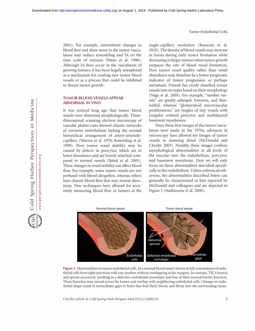
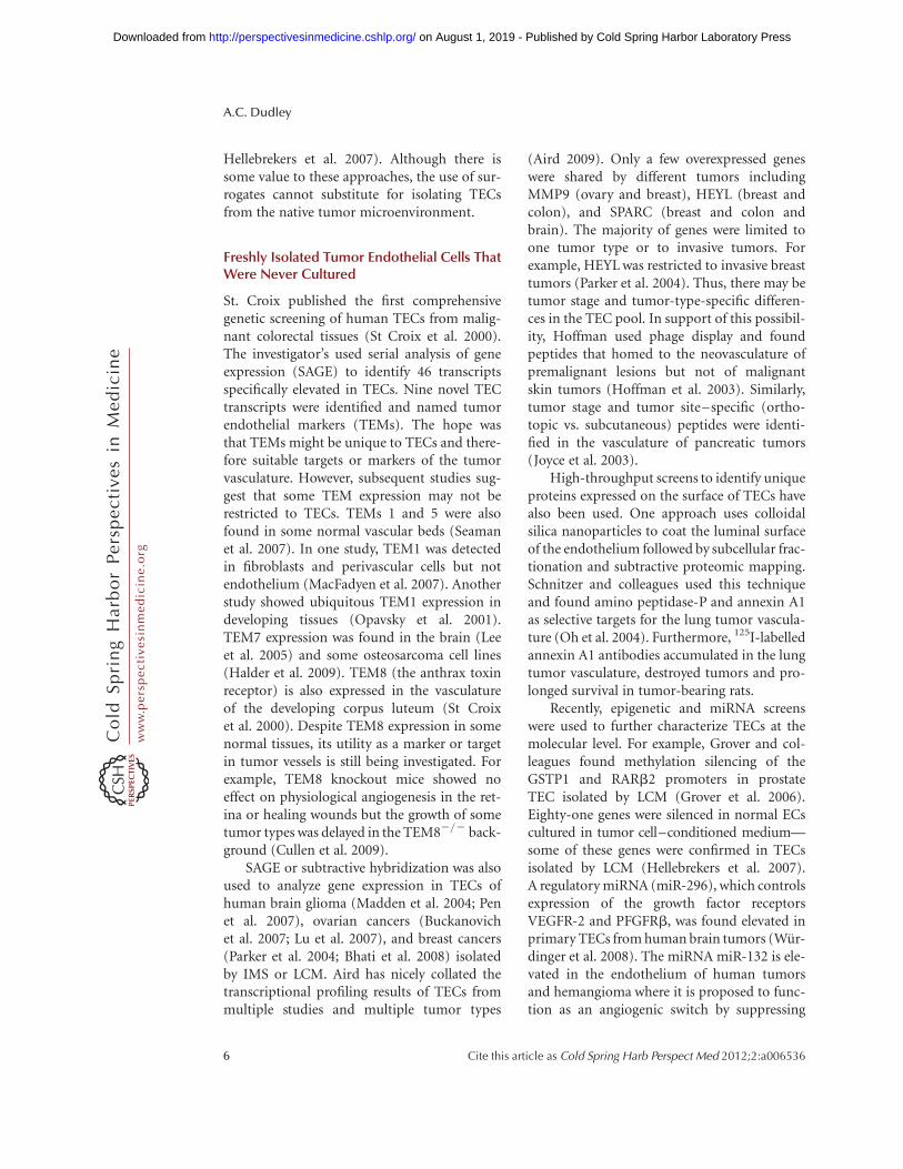
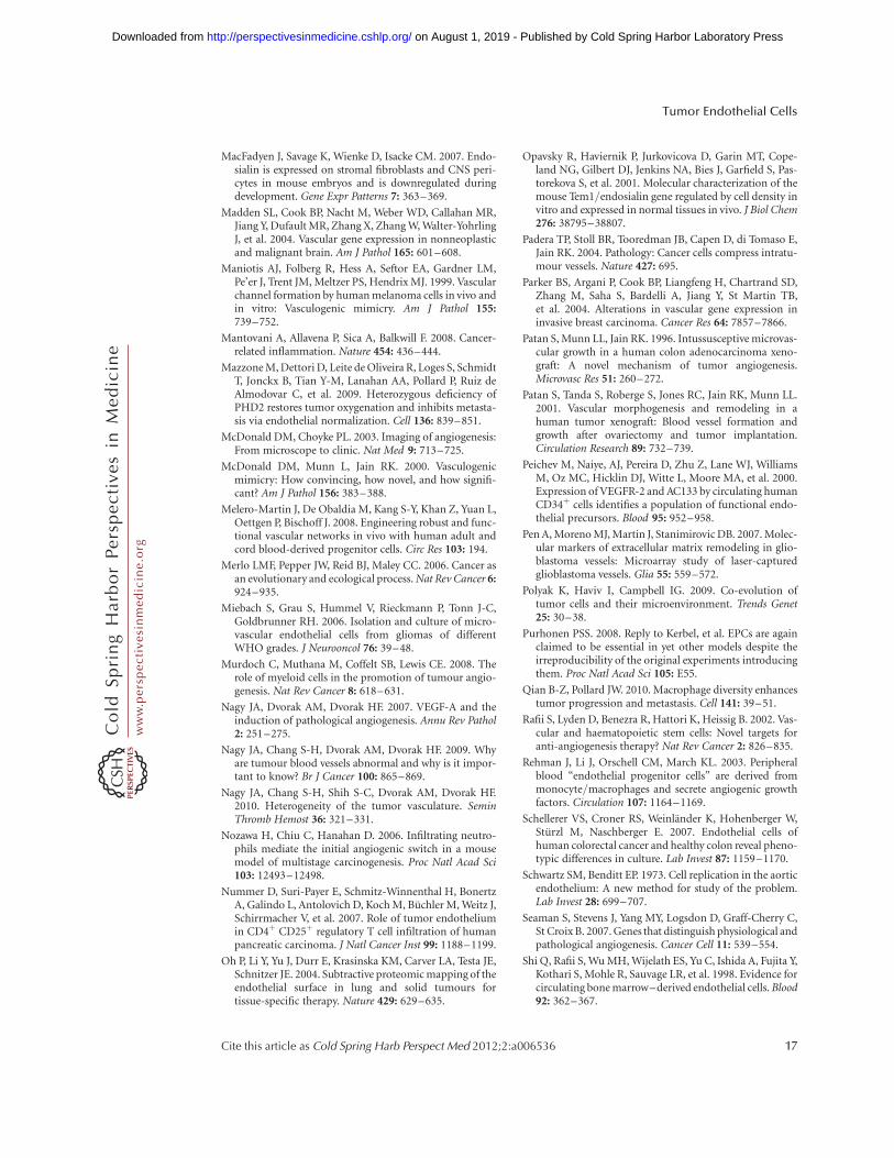
Since those first images of the tumor vascu-lature were made in the 1970s, advances inmicroscopy have allowed for images of tumorvessels in stunning detail (McDonald andChoyke 2003). Notably, these images confirmmorphological abnormalities at all levels ofthe vascular tree: the endothelium, pericytes,and basement membrane. Here we will onlyfocus on those abnormalities described specifi-cally in the endothelium. Unless referenced oth-erwise, the abnormalities described below cangenerally be characterized as first reported byMcDonald and colleagues and are depicted inFigure 1 (Hashizume et al. 2000).
Endothelialcells
Intercellulargaps
Defective endothelialmonolayer
Abnormalsprouts
Normal blood vessel Tumor blood vessel
Figure 1. Abnormalities in tumor endothelial cells. In a normal blood vessel (shown at left) a monolayer of endo-thelial cells form tight junctions with one another without overlapping at the margins. In contrast, TECs branchand sprout excessively resulting in a defective endothelial monolayer and loss of their normal barrier function.These branches may extend across the lumen and overlap with neighboring endothelial cells. Changes in endo-thelial shape result in intercellular gaps or holes that leak fluid, blood, and fibrin into the surrounding tissue.
Tumor Endothelial Cells
Cite this article as Cold Spring Harb Perspect Med 2012;2:a006536 3
ww
w.p
ersp
ecti
vesi
nm
edic
ine.
org
on August 1, 2019 - Published by Cold Spring Harbor Laboratory Press http://perspectivesinmedicine.cshlp.org/Downloaded from
Defective Endothelial Monolayer
In normal tissues the ECs form a continuousand uniform monolayer with few cytoplasmicprojections. Tumor endothelial cells (TECs)are of irregular shape and size; they have ruffledmargins and long, fragile cytoplasmic projec-tions extending outward and across the vessellumen. The tips of some branched TECs maypenetrate the lumen creating openings or smallintercellular gaps in the vessel wall. These open-ings allow extravasated erythrocytes to pool atthe periphery of tumor blood vessels forming“blood lakes” which are not anastomosed withthe vasculature. The appearance of tumor endo-thelium is described as “mosaic” because ofspotty CD31 immunoreactivity in vivo (diTomaso et al. 2005). The reasons for this aretwofold: ultrastructural analysis of the tumorvessel lining shows that individual TEC lackCD31 expression and in other areas of the vesselwall the TECs are absent altogether. Where thereare gaps between adjacent TECs, blood canmake direct contact with the exposed basementmembrane. Nearby tumor cells may also fillthese exposed gaps (Chang et al. 2000). Tumorcells that express VE-cadherin, fill in these gaps,and masquerade as endothelium have beenreported (Maniotis et al. 1999), but this topicremains controversial (McDonald et al. 2000).
Large Intercellular Openings and Holes
Similar to small intercellular gaps betweenneighboring TEC, larger openings (�1.5 mM)in the walls of tumor blood vessels are also visi-ble by scanning electron microscopy. Theselarger openings lack the structural features,including the finger-like projections often ob-served in inflamed endothelium that couldmediate their closure. Furthermore, high TECturnover and motility may not allow for properformation of intercellular junctions and base-ment membranes resulting in these larger holes.Transcellular holes (�0.5 mM), fenestrae, andchannels are also common in the tumor vascu-lature. Together, these small endothelial gapsand larger openings are probably responsiblefor hemorrhage and plasma leakage observedin most tumors. Holes in the tumor vasculature
could also act as a conduit for the passage oftumor cells into the circulation.
Abnormal Sprouts
Tumor vessels have thin cytoplasmic projec-tions extending across the vessel lumen. Theseprojections resemble tip-like filopodia observedduring intussusceptive growth but they mayoverlap with one another and form loose con-nections. The origin of these sprouts may bethe oxygen seeking tip cells at the leading edgeof sprouting vessels in hypoxic regions of thetumor microenvironment. Carmeliet and col-leagues recently proposed a molecular basisfor these abnormal sprouts (Mazzone et al.2009). For example, tip cells of tumor vesselsin mice haploinsufficient for the oxygen sensorPHD2 appear quiescent and adjust their shapeand phenotype to restore oxygen supply. Molec-ular therapies targeting PHD2 may “normalize”tumor vessels improving perfusion while amel-iorating the hypoxia that can promote invasionand metastasis (Sullivan and Graham 2007).
What Are the Causes of These Abnormalities?
Tumors are caricatures of a dysfunctional“organ.” For example, metabolic pathways arecorrupted, tumor cells deprive nearby tissuesof nutrients, there is a buildup of toxic wasteproducts such as lactate resulting in acidosis,and there are areas of nonperfusion resultingin focal regions of hypoxia. If one considersthat the vasculature is a supply line for oxygenand nutrients but also a conduit for the removalof waste products, then abnormalities in theblood vessels themselves are a major contribu-tor to the abnormal microenvironment intumors. Not only do cancer cells thrive in thisenvironment, but selection pressure created bythese microenvironmental bottlenecks mayactually contribute to their propagation (Merloet al. 2006).
As an example, most tumors are character-ized by the expression of high concentrationsof VEGF-A. VEGF alone is a potent vasodilatorthat can promote fluid leakage and high inter-stitial pressures, abnormal branching morpho-genesis, and small gaps and fissures in the
A.C. Dudley
4 Cite this article as Cold Spring Harb Perspect Med 2012;2:a006536
ww
w.p
ersp
ecti
vesi
nm
edic
ine.
org
on August 1, 2019 - Published by Cold Spring Harbor Laboratory Press http://perspectivesinmedicine.cshlp.org/Downloaded from
vasculature (Nagy et al. 2007). Thus, chronicVEGF stimulation in tumors promotes sprout-ing and excessive branching of tip cells leadingto irregularities in the TEC monolayer andloss of barrier function. Irregularities in theTEC lining surrounding these vessel protru-sions impairs blood flow resulting in hypoxiaand hypoperfusion. Tumor vessels are alsosqueezed and compressed by overlying tumorcells, which creates biomechanical tension,strain, and changes in blood flow (Paderaet al. 2004). This chaotic pattern of blood flowcan alter endothelial shape, size, and differentia-tion perhaps through aberrant expression offlow-mediated transcription factors (De Valand Black 2009). Notably, endothelial dysfunc-tion in tumors is not a dead end. All cell typesfound within the perivascular niche either indirect contact with the endothelium or depend-ent on soluble vascular-derived factors (e.g.,pericytes and trafficking leukocytes) for theirgrowth and differentiation might be adverselyaffected.
STUDIES OF ISOLATED TUMORENDOTHELIAL CELLS
For many years, it was assumed that TECs weresimilar to their normal counterparts irrespec-tive of the obvious morphological abnormal-ities of tumor blood vessels in situ. In fact, thetheoretical success of antiangiogenic (specifi-cally antiendothelial) strategies in cancer de-pends to some extent on TECs remainingstabile and not altering their phenotype overtime. However, recent studies have shown thatTEC are more complex and labile than expectedchallenging the assumption that TEC are nor-mal. Collectively, these studies show morpho-logical, pathophysiological, cytogenetic, epi-genetic, and gene expression changes in theTEC pool. Despite this new knowledge, overallour understanding of TEC biology has beenhampered by several technical limitations,which are described below.
First, a common method for isolating“pure” populations of TECs is immunomag-netic separation (IMS) with antibody-coatedbeads. Common cell-surface markers used to
isolate TEC are CD31 (Hida et al. 2004),ICAM-2 (Dudley et al. 2008a), and CD146 (StCroix et al. 2000). However, these markers areshared by endothelial cells from virtually all vas-cular beds (capillary, venous, arterial, and lym-phatic); thus, heterogeneity in the isolatedpopulation of TECs is unavoidable. The fidelityof IMS is also not 100% so contamination byfibroblasts and tumor cells is problematic.Diphtheria toxin has been successfully used toeliminate human tumor cells from cultures ofmouse TECs (Arbiser et al. 1999; Hida et al.2004). The most proficient way to ensure purityof isolated TECs is to prepare clonal popula-tions, but this can be challenging because ECsplated at clonal density will often undergo sen-escence. Laser capture microdissection (LCM)is also used to isolate TECs but the cells cannotbe cultured. Similar to IMS, there are problemswith contaminating pericytes and tumor cellsthat copurify with TECs.
Second, the conditions for culturing TECsin vitro are not well defined. TECs may beadapted to the tumor microenvironment (e.g.,chaotic blood flow, acidosis, hypoxia, nutrientdeprivation), which cannot be faithfully repli-cated in vitro. There may be unique mediarequirements and combinations of growth andattachment factors. For example, in one study,the use of oncofetal fibronectin was necessaryto maintain the phenotype of TECs isolatedfrom Lewis lung carcinoma (LLC) (Allportand Weissleder 2003). Thus, following pheno-typic drift, TECs placed in culture may no lon-ger resemble TECs in vivo.
Despite the challenges, several groups haveisolated and characterized (i.e., high-through-put gene expression analyses) TECs fromhuman tumors and from tumors implanted inmice. These studies typically fall into two cate-gories: those that isolated and characterizedTECs that were never cultured, and those thatused isolated and culture expanded TECs fortheir analyses. Other studies have attempted tocreate or identify “TEC” lines in vitro by cultur-ing normal ECs with tumor cell conditionedmedia or by screening mouse cell lines for theexpression of markers already known to be ex-pressed by TECs (Walter-Yohrling et al. 2004;
Tumor Endothelial Cells
Cite this article as Cold Spring Harb Perspect Med 2012;2:a006536 5
ww
w.p
ersp
ecti
vesi
nm
edic
ine.
org
on August 1, 2019 - Published by Cold Spring Harbor Laboratory Press http://perspectivesinmedicine.cshlp.org/Downloaded from
Hellebrekers et al. 2007). Although there issome value to these approaches, the use of sur-rogates cannot substitute for isolating TECsfrom the native tumor microenvironment.
Freshly Isolated Tumor Endothelial Cells ThatWere Never Cultured
St. Croix published the first comprehensivegenetic screening of human TECs from malig-nant colorectal tissues (St Croix et al. 2000).The investigator’s used serial analysis of geneexpression (SAGE) to identify 46 transcriptsspecifically elevated in TECs. Nine novel TECtranscripts were identified and named tumorendothelial markers (TEMs). The hope wasthat TEMs might be unique to TECs and there-fore suitable targets or markers of the tumorvasculature. However, subsequent studies sug-gest that some TEM expression may not berestricted to TECs. TEMs 1 and 5 were alsofound in some normal vascular beds (Seamanet al. 2007). In one study, TEM1 was detectedin fibroblasts and perivascular cells but notendothelium (MacFadyen et al. 2007). Anotherstudy showed ubiquitous TEM1 expression indeveloping tissues (Opavsky et al. 2001).TEM7 expression was found in the brain (Leeet al. 2005) and some osteosarcoma cell lines(Halder et al. 2009). TEM8 (the anthrax toxinreceptor) is also expressed in the vasculatureof the developing corpus luteum (St Croixet al. 2000). Despite TEM8 expression in somenormal tissues, its utility as a marker or targetin tumor vessels is still being investigated. Forexample, TEM8 knockout mice showed noeffect on physiological angiogenesis in the ret-ina or healing wounds but the growth of sometumor types was delayed in the TEM82/2 back-ground (Cullen et al. 2009).
SAGE or subtractive hybridization was alsoused to analyze gene expression in TECs ofhuman brain glioma (Madden et al. 2004; Penet al. 2007), ovarian cancers (Buckanovichet al. 2007; Lu et al. 2007), and breast cancers(Parker et al. 2004; Bhati et al. 2008) isolatedby IMS or LCM. Aird has nicely collated thetranscriptional profiling results of TECs frommultiple studies and multiple tumor types
(Aird 2009). Only a few overexpressed geneswere shared by different tumors includingMMP9 (ovary and breast), HEYL (breast andcolon), and SPARC (breast and colon andbrain). The majority of genes were limited toone tumor type or to invasive tumors. Forexample, HEYL was restricted to invasive breasttumors (Parker et al. 2004). Thus, there may betumor stage and tumor-type-specific differen-ces in the TEC pool. In support of this possibil-ity, Hoffman used phage display and foundpeptides that homed to the neovasculature ofpremalignant lesions but not of malignantskin tumors (Hoffman et al. 2003). Similarly,tumor stage and tumor site–specific (ortho-topic vs. subcutaneous) peptides were identi-fied in the vasculature of pancreatic tumors(Joyce et al. 2003).
High-throughput screens to identify uniqueproteins expressed on the surface of TECs havealso been used. One approach uses colloidalsilica nanoparticles to coat the luminal surfaceof the endothelium followed by subcellular frac-tionation and subtractive proteomic mapping.Schnitzer and colleagues used this techniqueand found amino peptidase-P and annexin A1as selective targets for the lung tumor vascula-ture (Oh et al. 2004). Furthermore, 125I-labelledannexin A1 antibodies accumulated in the lungtumor vasculature, destroyed tumors and pro-longed survival in tumor-bearing rats.
Recently, epigenetic and miRNA screenswere used to further characterize TECs at themolecular level. For example, Grover and col-leagues found methylation silencing of theGSTP1 and RARb2 promoters in prostateTEC isolated by LCM (Grover et al. 2006).Eighty-one genes were silenced in normal ECscultured in tumor cell–conditioned medium—some of these genes were confirmed in TECsisolated by LCM (Hellebrekers et al. 2007).A regulatory miRNA (miR-296), which controlsexpression of the growth factor receptorsVEGFR-2 and PFGFRb, was found elevated inprimary TECs from human brain tumors (Wur-dinger et al. 2008). The miRNA miR-132 is ele-vated in the endothelium of human tumorsand hemangioma where it is proposed to func-tion as an angiogenic switch by suppressing
A.C. Dudley
6 Cite this article as Cold Spring Harb Perspect Med 2012;2:a006536
ww
w.p
ersp
ecti
vesi
nm
edic
ine.
org
on August 1, 2019 - Published by Cold Spring Harbor Laboratory Press http://perspectivesinmedicine.cshlp.org/Downloaded from
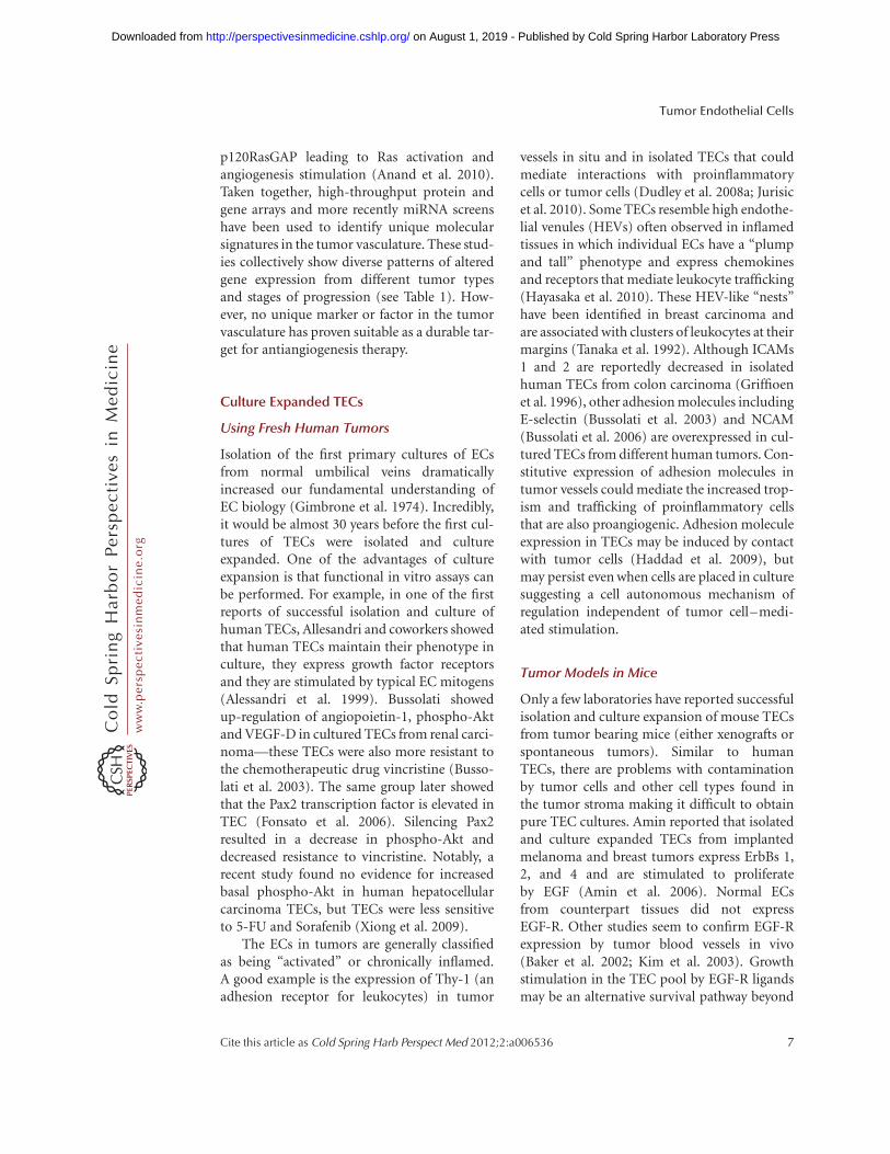
p120RasGAP leading to Ras activation andangiogenesis stimulation (Anand et al. 2010).Taken together, high-throughput protein andgene arrays and more recently miRNA screenshave been used to identify unique molecularsignatures in the tumor vasculature. These stud-ies collectively show diverse patterns of alteredgene expression from different tumor typesand stages of progression (see Table 1). How-ever, no unique marker or factor in the tumorvasculature has proven suitable as a durable tar-get for antiangiogenesis therapy.
Culture Expanded TECs
Using Fresh Human Tumors
Isolation of the first primary cultures of ECsfrom normal umbilical veins dramaticallyincreased our fundamental understanding ofEC biology (Gimbrone et al. 1974). Incredibly,it would be almost 30 years before the first cul-tures of TECs were isolated and cultureexpanded. One of the advantages of cultureexpansion is that functional in vitro assays canbe performed. For example, in one of the firstreports of successful isolation and culture ofhuman TECs, Allesandri and coworkers showedthat human TECs maintain their phenotype inculture, they express growth factor receptorsand they are stimulated by typical EC mitogens(Alessandri et al. 1999). Bussolati showedup-regulation of angiopoietin-1, phospho-Aktand VEGF-D in cultured TECs from renal carci-noma—these TECs were also more resistant tothe chemotherapeutic drug vincristine (Busso-lati et al. 2003). The same group later showedthat the Pax2 transcription factor is elevated inTEC (Fonsato et al. 2006). Silencing Pax2resulted in a decrease in phospho-Akt anddecreased resistance to vincristine. Notably, arecent study found no evidence for increasedbasal phospho-Akt in human hepatocellularcarcinoma TECs, but TECs were less sensitiveto 5-FU and Sorafenib (Xiong et al. 2009).
The ECs in tumors are generally classifiedas being “activated” or chronically inflamed.A good example is the expression of Thy-1 (anadhesion receptor for leukocytes) in tumor
vessels in situ and in isolated TECs that couldmediate interactions with proinflammatorycells or tumor cells (Dudley et al. 2008a; Jurisicet al. 2010). Some TECs resemble high endothe-lial venules (HEVs) often observed in inflamedtissues in which individual ECs have a “plumpand tall” phenotype and express chemokinesand receptors that mediate leukocyte trafficking(Hayasaka et al. 2010). These HEV-like “nests”have been identified in breast carcinoma andare associated with clusters of leukocytes at theirmargins (Tanaka et al. 1992). Although ICAMs1 and 2 are reportedly decreased in isolatedhuman TECs from colon carcinoma (Griffioenet al. 1996), other adhesion molecules includingE-selectin (Bussolati et al. 2003) and NCAM(Bussolati et al. 2006) are overexpressed in cul-tured TECs from different human tumors. Con-stitutive expression of adhesion molecules intumor vessels could mediate the increased trop-ism and trafficking of proinflammatory cellsthat are also proangiogenic. Adhesion moleculeexpression in TECs may be induced by contactwith tumor cells (Haddad et al. 2009), butmay persist even when cells are placed in culturesuggesting a cell autonomous mechanism ofregulation independent of tumor cell–medi-ated stimulation.
Tumor Models in Mice
Only a few laboratories have reported successfulisolation and culture expansion of mouse TECsfrom tumor bearing mice (either xenografts orspontaneous tumors). Similar to humanTECs, there are problems with contaminationby tumor cells and other cell types found inthe tumor stroma making it difficult to obtainpure TEC cultures. Amin reported that isolatedand culture expanded TECs from implantedmelanoma and breast tumors express ErbBs 1,2, and 4 and are stimulated to proliferateby EGF (Amin et al. 2006). Normal ECsfrom counterpart tissues did not expressEGF-R. Other studies seem to confirm EGF-Rexpression by tumor blood vessels in vivo(Baker et al. 2002; Kim et al. 2003). Growthstimulation in the TEC pool by EGF-R ligandsmay be an alternative survival pathway beyond
Tumor Endothelial Cells
Cite this article as Cold Spring Harb Perspect Med 2012;2:a006536 7
ww
w.p
ersp
ecti
vesi
nm
edic
ine.
org
on August 1, 2019 - Published by Cold Spring Harbor Laboratory Press http://perspectivesinmedicine.cshlp.org/Downloaded from
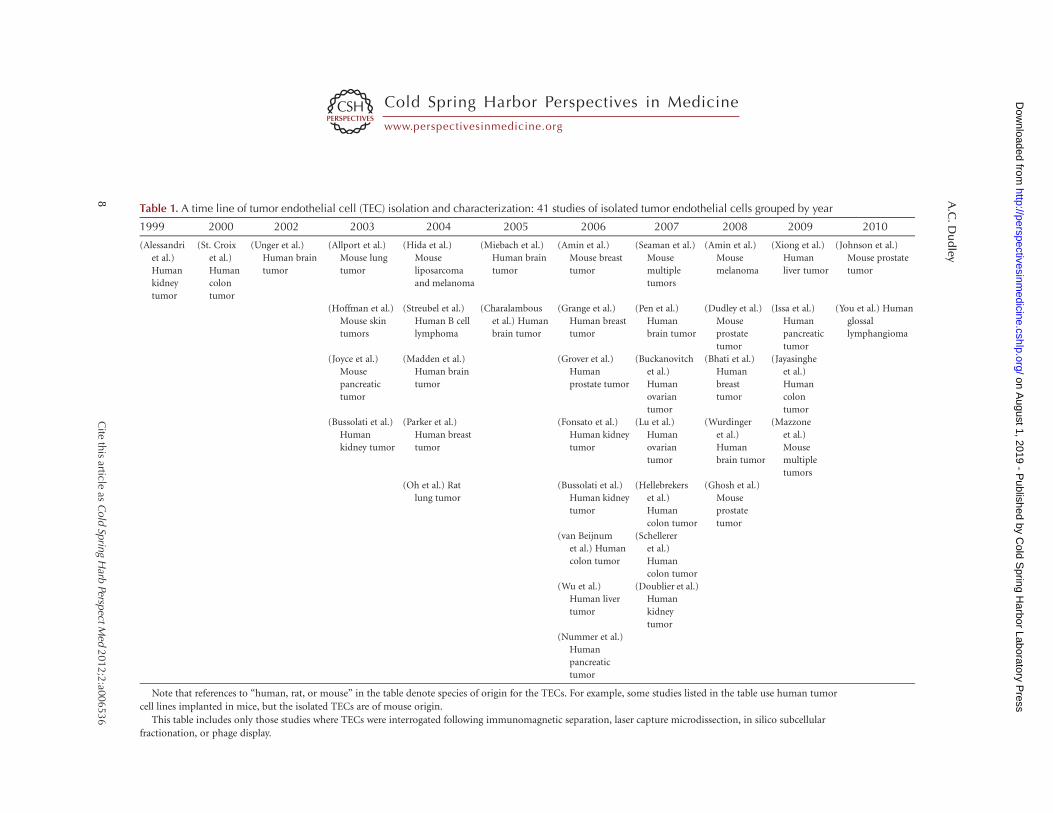
Table 1. A time line of tumor endothelial cell (TEC) isolation and characterization: 41 studies of isolated tumor endothelial cells grouped by year
1999 2000 2002 2003 2004 2005 2006 2007 2008 2009 2010
(Alessandri
et al.)
Human
kidney
tumor
(St. Croix
et al.)
Human
colon
tumor
(Unger et al.)
Human brain
tumor
(Allport et al.)
Mouse lung
tumor
(Hida et al.)
Mouse
liposarcoma
and melanoma
(Miebach et al.)
Human brain
tumor
(Amin et al.)
Mouse breast
tumor
(Seaman et al.)
Mouse
multiple
tumors
(Amin et al.)
Mouse
melanoma
(Xiong et al.)
Human
liver tumor
(Johnson et al.)
Mouse prostate
tumor
(Hoffman et al.)
Mouse skin
tumors
(Streubel et al.)
Human B cell
lymphoma
(Charalambous
et al.) Human
brain tumor
(Grange et al.)
Human breast
tumor
(Pen et al.)
Human
brain tumor
(Dudley et al.)
Mouse
prostate
tumor
(Issa et al.)
Human
pancreatic
tumor
(You et al.) Human
glossal
lymphangioma
(Joyce et al.)
Mouse
pancreatic
tumor
(Madden et al.)
Human brain
tumor
(Grover et al.)
Human
prostate tumor
(Buckanovitch
et al.)
Human
ovarian
tumor
(Bhati et al.)
Human
breast
tumor
(Jayasinghe
et al.)
Human
colon
tumor
(Bussolati et al.)
Human
kidney tumor
(Parker et al.)
Human breast
tumor
(Fonsato et al.)
Human kidney
tumor
(Lu et al.)
Human
ovarian
tumor
(Wurdinger
et al.)
Human
brain tumor
(Mazzone
et al.)
Mouse
multiple
tumors
(Oh et al.) Rat
lung tumor
(Bussolati et al.)
Human kidney
tumor
(Hellebrekers
et al.)
Human
colon tumor
(Ghosh et al.)
Mouse
prostate
tumor
(van Beijnum
et al.) Human
colon tumor
(Schellerer
et al.)
Human
colon tumor
(Wu et al.)
Human liver
tumor
(Doublier et al.)
Human
kidney
tumor
(Nummer et al.)
Human
pancreatic
tumor
Note that references to “human, rat, or mouse” in the table denote species of origin for the TECs. For example, some studies listed in the table use human tumor
cell lines implanted in mice, but the isolated TECs are of mouse origin.
This table includes only those studies where TECs were interrogated following immunomagnetic separation, laser capture microdissection, in silico subcellular
fractionation, or phage display.
A.C
.Du
dley
8C
iteth
isarticle
asC
old
Sprin
gH
arbPersp
ectM
ed2012;2
:a006536
www.perspectivesinmedicine.org
on August 1, 2019 - P
ublished by Cold S
pring Harbor Laboratory P
ress http://perspectivesinm
edicine.cshlp.org/D
ownloaded from
the canonical VEGF/bFGF axis. Specific inhib-ition of EGF-R in TECs in vivo can impairangiogenesis and growth of tumor cells thatdo not express EGF-R (Amin et al. 2008).
A principle of antiangiogenesis therapies incancer is that the tumor endothelium is normaland will not change over time or develop drugresistance. However, Hida reported abnormalcentrosomes and aneuploid chromosomes inTECs isolated from human melanoma and lip-osarcoma implanted in mice (Hida et al. 2004).These chromosomal abnormalities were notclonal nor were they derived from humangenetic material incorporated into mouse chro-mosomes. Of note, overstimulation of endothe-lial cultures by growth factors including bFGFand VEGF was recently reported to induce cen-trosome duplication in ECs (Taylor et al. 2010).On the other hand, Streubel detected primaryand secondary translocations in ECs that wereidentical to those found in follicular lymphomasuggesting either a tumor cell of origin for TECsor sharing of DNA between tumor cells and ECs(Streubel et al. 2004). Horizontal transfer ofgenetic material via apoptotic bodies betweentumor cells and ECs has been reported (Ehnforset al. 2009). Loss of function of gatekeepergenes in the stroma (e.g., p53) may also underliethese cytogenetic abnormalities (Hill et al. 2005;Dudley et al. 2008b).
Our laboratory has reported unusual pat-terns of differentiation in TECs isolated fromspontaneous prostate tumors in TRAMP(transgenic adenocarcinoma of the mouse pros-tate) mice. For example, prostate tumor TECsunexpectedly differentiate to form bone andcartilage (Dudley et al. 2008a). Ectopic micro-vascular calcification was also detected alongthe capillary lumens of human prostate cancers.Breast tumors are also characterized by microcal-cifications in the vasculature (Tse et al. 2008).Unexpected patterns of differentiation in tumorvessels may arise as vascular cells (and other stro-mal cells) coevolve with tumor cells and switchtheir phenotype (Polyak et al. 2009). For exam-ple, lineage switches and coexpression oflymphatic, endothelial, and fibroblast markershave been observed in the blood vessels of othermalignancies (Breiteneder-Geleff et al. 1999;
Wang et al. 2004). Pathophysiological conditionsin the tumor microenvironment includingaberrant expression of growth and differentiationfactors may control the fate, differentiation,and mesenchymal transition of TECs (Verfaillie2008). One surprising finding is that TECsappear to “remember” the tumor microenviron-ment from which they were isolated. For exam-ple, Ghosh showed that isolated TECs fail toreorient their actin cytoskeleton when exposedto uniaxial cyclic strain and they display greatertraction forces in response to variations in ECMelasticity in vitro (Ghosh et al. 2008). These re-sults were related to high constitutive Rho andits downstream effector ROCK in TECs. TheECM in tumors is characterized by highly cross-linked collagen resulting in a “stiffer” matrix thatcan alter the pattern of signaling and focal adhe-sions to the underlying endothelium (Leventalet al. 2009). TECs may be reprogrammed oradapted to these conditions, which couldaccount for some of the structural and functionalabnormalities in the tumor vasculature.
TEC ABNORMALITIES MAY CONTRIBUTETO TUMOR PROGRESSION
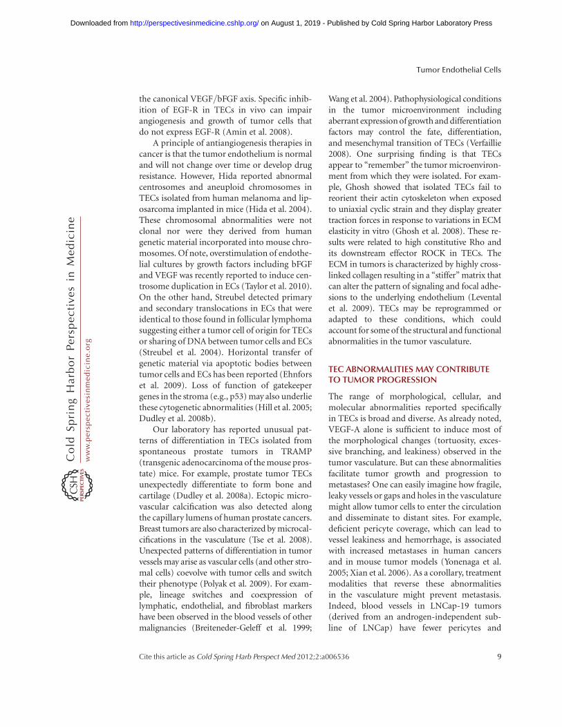
The range of morphological, cellular, andmolecular abnormalities reported specificallyin TECs is broad and diverse. As already noted,VEGF-A alone is sufficient to induce most ofthe morphological changes (tortuosity, exces-sive branching, and leakiness) observed in thetumor vasculature. But can these abnormalitiesfacilitate tumor growth and progression tometastases? One can easily imagine how fragile,leaky vessels or gaps and holes in the vasculaturemight allow tumor cells to enter the circulationand disseminate to distant sites. For example,deficient pericyte coverage, which can lead tovessel leakiness and hemorrhage, is associatedwith increased metastases in human cancersand in mouse tumor models (Yonenaga et al.2005; Xian et al. 2006). As a corollary, treatmentmodalities that reverse these abnormalitiesin the vasculature might prevent metastasis.Indeed, blood vessels in LNCap-19 tumors(derived from an androgen-independent sub-line of LNCap) have fewer pericytes and
Tumor Endothelial Cells
Cite this article as Cold Spring Harb Perspect Med 2012;2:a006536 9
ww
w.p
ersp
ecti
vesi
nm
edic
ine.
org
on August 1, 2019 - Published by Cold Spring Harbor Laboratory Press http://perspectivesinmedicine.cshlp.org/Downloaded from
increased tumor cell invasion compared to theparental cell line (Welen et al. 2009). Similarly,a change in TEC shape in PHD2þ/2 mice is suf-ficient to improve oxygenation while suppress-ing tumor invasion and metastases (Mazzoneet al. 2009). Jain has hypothesized that vesselleakiness impairs the delivery of chemothera-peutic drugs to the tumor site and thereforeblocking VEGF might “normalize” blood flowand improve drug delivery (Jain 2005). In amouse model, a single dose of Avastin (anti-VEGF antibodies) decreased microvessel den-sity, vessel permeability and interstitial pressurewhile intratumoral perfusion was improved(Dickson et al. 2007). Recent clinical studiessupport the concept that combining Avastinwith chemotherapy can lead to improved out-comes in patients with advanced rectal cancer(Willett et al. 2007).
Heterotypic interactions and cross talkbetween TECs and other cell types, particularlyleukocytes mobilized from the circulation,might also be affected by abnormalities in thetumor vasculature. As an example, some adhe-sion molecules may be decreased in the tumorvasculature allowing tumors to escape immunesurveillance because of impaired interactionsbetween T-lymphocytes and the blood vesselwall (Griffioen et al. 1996; Dirkx et al. 2006).Indeed, the penetration and efficacy of primedT cells for tumor immunotherapy was en-hanced when proinflammatory agents thatup-regulated ICAM and VCAM in the vascula-ture were coadministered (Garbi et al. 2004).
On the other hand, tumors resemble“wounds that never heal” and TECs may consti-tutively overexpress adhesion molecules forproinflammatory cells (Dvorak 1986). Thus, aperivascular positioning of leukocytes at theperiphery of tumor vessels is common (Dudleyet al. 2010). These proinflammatory cells ex-press most of the endothelial survival andmatrix remodeling factors required for angio-genesis. In one sense then, inflammation oractivation of TEC enables the conscription ofa diverse population of auxiliary cells that playa catalytic role during angiogenesis. Further-more, proinflammatory cells, particularly mac-rophages, not only stimulate angiogenesis, but
they may also enable metastasis (Qian andPollard 2010). Because the endothelium acts agatekeeper controlling the egress of proinflam-matory cells into the tissue (or tumor), blockingspecific interactions between TECs and theimmune cell infiltrate may indirectly impairangiogenesis and metastasis. Although manyof the chemokines that control leukocyte trop-ism are tumor cell–derived, TECs may be adirect source for many of these chemotactic fac-tors (Butler et al. 2010).
WHAT IS THE ORIGIN OF TUMORENDOTHELIUM?
Where does the tumor endothelium comefrom? The answer to this question has beenmore difficult to answer than expected. For along time, the ECs lining tumor vessels werethought to arise only by mitoses, sprouting, orsimple cooption of preexisting capillaries,whereas vasculogenesis occurred only duringembryonic development. However, a distortedvariation of each of these processes probablygenerates new endothelium in tumors. Theremay also be additional, unexpected sources forTECs. For example, Hendrix suggests that stem-like tumor cells may transdifferentiate to formendothelium (Hendrix et al. 2003). A mesen-chymal stem cell, with properties of endothelialcells, was shown to form pericytes and endothe-lium in hemangioma (Khan et al. 2008).
The turnover of the endothelium in normaltissues is low. It is estimated that only 0.1%–3%of all ECs turn over daily but this may declinewith age (Schwartz and Benditt 1973). Intumors, EC turnover greatly accelerates andmay be 20–2000 times the rate in normal tissues(Hobson and Denekamp 1984). In a seminalpaper, Folkman showed that normal adulttissues implanted in the chick chorioallantoicmembrane (CAM) did not promote neovascu-larization whereas implanted tumors rapidlystimulated the growth of new blood vesselsfrom the host (Ausprunk et al. 1975). Forthe next 20 years, angiogenesis was consideredthe sole or predominate source of new endothe-lium in tumors. However, a report by Asahara in1997 described putative circulating CD34þ/
A.C. Dudley
10 Cite this article as Cold Spring Harb Perspect Med 2012;2:a006536
ww
w.p
ersp
ecti
vesi
nm
edic
ine.
org
on August 1, 2019 - Published by Cold Spring Harbor Laboratory Press http://perspectivesinmedicine.cshlp.org/Downloaded from
VEGFR-2þ endothelial progenitor cells thatcould home to areas of damaged tissue andincorporate into sites of active angiogenesis(Asahara et al. 1997). A number of subsequentstudies identified similar cells circulating inblood that were localized to sites of angiogenesisin ischemic tissues and tumors (Shi et al. 1998;Peichev et al. 2000; Lyden et al. 2001). Thus,postnatal vasculogenesis was proposed as analternative route for new tumor blood vessels.However, hematopoietic cells (e.g., monocytes)share markers with ECs, they can be mobilizedinto circulation and they also home to sitesof neovascularization following injury (Rafiiet al. 2002; Rehman et al. 2003; Case et al.2007). Their proximity to the blood vessel andexpression of markers shared with bona fideendothelium has created confusion and dis-crepancies over the identification of circulatingECs in solid tumors; especially in rodents (Ker-bel et al. 2008; Purhonen 2008; Yoder andIngram 2009).
The functional differences distinguishinghematopoietic cells from bona fide endothe-lium are becoming clear. Yoder suggests thatonly the endothelial colony forming cells(ECFCs) can form vessel lumens, whereas hem-atopoietic cells generally do not (Yoder 2009).A minimum set of criteria for identifying ECFCsin blood or tissues was also proposed by Yoder(Yoder 2009). These same criteria should beapplied for identifying ECFCs in tumors usingrodent models. For example, bone marrow abla-tion followed by engraftment with GFPþ mar-row is routinely used to track circulating“ECs” in tumors—immunohistochemistry orFACS is used to identify the putative ECs(Aghi and Chiocca 2005). However, these assaysprovide no functional information about thenature of the recruited circulating cell type(s).Culture expansion and characterization (e.g.,lumen-forming abilities when reimplanted inmice) of GFPþ ECFCs from collagenasedigested tumor extracts would provide defini-tive proof for the existence of a circulating ECfor tumor angiogenesis in rodents.
Recently, the vessel wall itself has been pro-posed as a source for vascular endotheliumbecause it contains subpopulations of ECs
with properties similar to blood-derived ECFCs(Ingram et al. 2005). Thus, in contrast to a bonemarrow origin, there may be a local reservoir ofhighly proliferative endothelium proximal tothe tumor site. Vessel wall EPCs (VW-EPCs)are proposed to rest in a “vasculogenic zone”between the smooth muscle and advential layersat the periphery of large blood vessels (Tilkiet al. 2009). No studies to date have determinedwhether VW-EPCs might form the majority ofthe angiogenesis response in tumors, if thereare any unique properties in VW-EPCs thatcould be exploited as an antiangiogenesis strat-egy, or if VW-EPCs might mediate vascularrebound often observed following antiangio-genic therapies in the clinic (Bergers and Hana-han 2008; Ellis and Hicklin 2008).
ANTIANGIOGENIC IS NOT ALWAYSANTIENDOTHELIAL
Many of the original antiangiogenic strategies incancer were designed to be “antiendothelial” forit makes biological sense to directly target the“pipes” transporting blood, oxygen, and nu-trients to a growing tumor. However, anunexpected complication is that cell-to-cellheterogeneity, acquired resistance, and a multi-source origin for TECs might impinge on thesuccess of antiendothelial strategies. Althoughthe success and selectivity of antiangiogenesistherapies in tumors depend to some extent onthere being differences in TECs compared totheir counterparts, too much variation in theTEC pool can have the opposite effect. Oneway around this problem is to target multiplecell types simultaneously, including those nowknown to play auxiliary roles during tumorblood vessel formation. Pericytes, fibroblasts,and other mesenchymal-lineage cells in thestroma may be valuable indirect targets for anti-angiogenesis in tumors (Loeffler et al. 2006);however, hematopoietic cells, particularly thoseof the myelomonocytic lineages, have recentlyreceived great attention for this purpose (Mur-doch et al. 2008). The role of hematopoietic lin-eage cells in tumor angiogenesis and theirpotential as targets for antiangiogenic therapiesis briefly discussed below.
Tumor Endothelial Cells
Cite this article as Cold Spring Harb Perspect Med 2012;2:a006536 11
ww
w.p
ersp
ecti
vesi
nm
edic
ine.
org
on August 1, 2019 - Published by Cold Spring Harbor Laboratory Press http://perspectivesinmedicine.cshlp.org/Downloaded from
An early clue that hematopoietic cells mightfacilitate angiogenesis comes from studies in theAML-1-deficient embryos. These mice lackdefinitive hematopoiesis and show impairedangiogenesis in the head and pericardium thatcan be rescued by addition of hematopoieticcells expressing ANG-1 (Takakura et al. 2000).Later and in tumors, marrow-derived inflam-matory cells including neutrophils, macro-phages, and mast cells were shown to providethe majority of the proangiogenic factorMMP9 during angiogenesis (Coussens et al.2000).
In the last 10 years, a number of studieshave confirmed the role of proinflammatorycells in tumor angiogenesis and have identifiedthe molecular pathways linking inflammationand cancer (Mantovani et al. 2008). Macro-phages in particular appear to promote tumorinvasion, cancer initiation, and angiogenesis(Qian and Pollard 2010). Furthermore, mono-cytes/macrophages may create a “permissive”environment that facilitates the seeding of
metastases at distant sites, even before tumorcell arrival (Kaplan et al. 2005). There appearto be tumor-specific and distinct populationsof myeloid cells recruited to tumors that releaseangiogenic factors involved in matrix remodel-ing or that stimulate endothelial cells directly(Coffelt et al. 2010). For example, selectivedepletion of neutrophils (Nozawa et al. 2006),monocytes/macrophages (Lin et al. 2006), orTIE-2 monocytes (De Palma et al. 2005)impairs tumor growth and angiogenesis. Somemyeloid lineage cells (CD11bþ) are mobilizedby chemotherapies or radiation (Ahn andBrown 2008). Furthermore, their activity intumors might mediate resistance to antian-giogenic therapies by stimulating vascularrebound. Indeed, Ahn and colleagues showedenhancement of radiotherapy in tumors whenCD11bþ antibodies were administered systemi-cally (Ahn et al. 2010). Finally, hematopoieticcells may stimulate tumor blood vessel forma-tion in other, unexpected ways. For example,macrophages may participate during nascent
Reductionist view
Tumorcell Blood
vessel
Vessel wallprogenitor
Bone marrow–derived EPC
Transdifferentiatedmesenchymal
progenitor
Transdifferentiatedmyeloid cell
Tumor cellvasculogenic
mimicry
Endothelialcell
Contemporary view
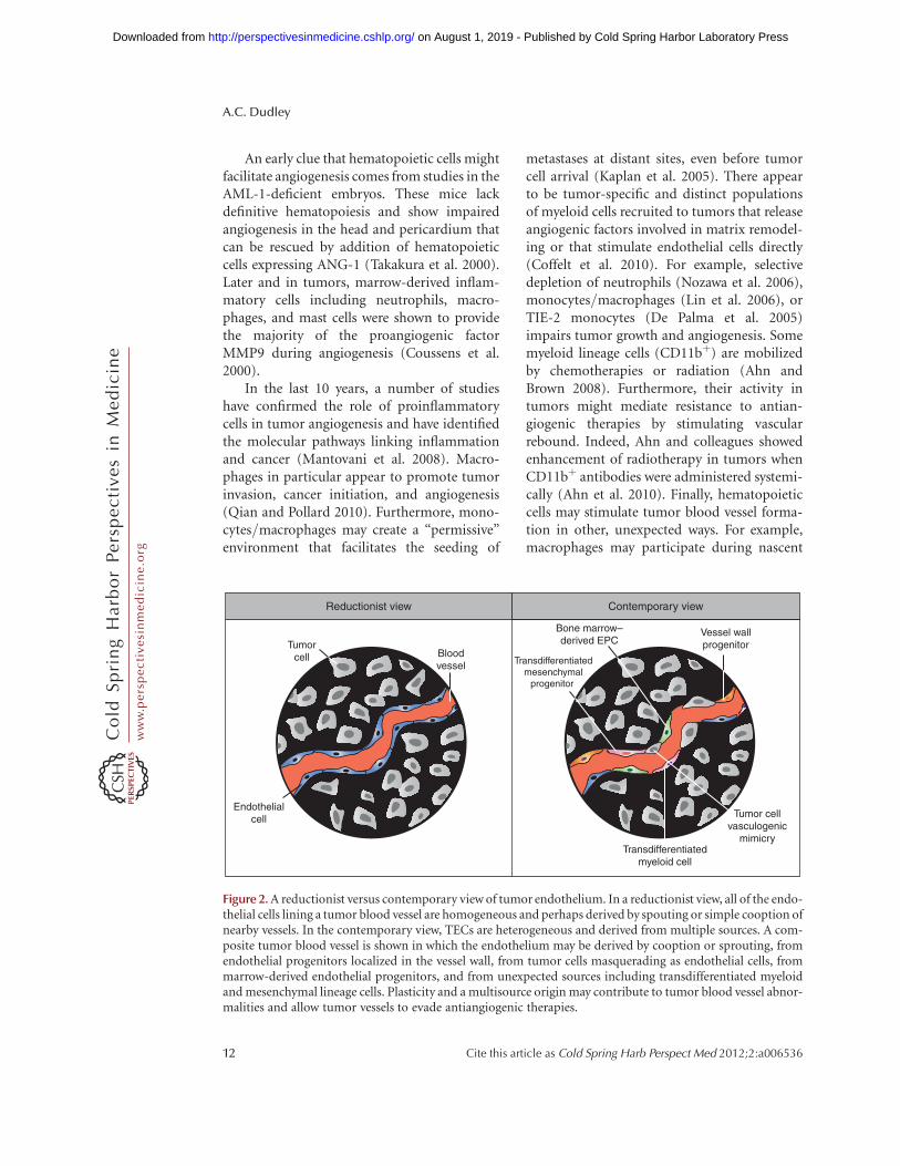
Figure 2. A reductionist versus contemporary view of tumor endothelium. In a reductionist view, all of the endo-thelial cells lining a tumor blood vessel are homogeneous and perhaps derived by spouting or simple cooption ofnearby vessels. In the contemporary view, TECs are heterogeneous and derived from multiple sources. A com-posite tumor blood vessel is shown in which the endothelium may be derived by cooption or sprouting, fromendothelial progenitors localized in the vessel wall, from tumor cells masquerading as endothelial cells, frommarrow-derived endothelial progenitors, and from unexpected sources including transdifferentiated myeloidand mesenchymal lineage cells. Plasticity and a multisource origin may contribute to tumor blood vessel abnor-malities and allow tumor vessels to evade antiangiogenic therapies.
A.C. Dudley
12 Cite this article as Cold Spring Harb Perspect Med 2012;2:a006536
ww
w.p
ersp
ecti
vesi
nm
edic
ine.
org
on August 1, 2019 - Published by Cold Spring Harbor Laboratory Press http://perspectivesinmedicine.cshlp.org/Downloaded from
vessel formation by providing guidance andmechanical cues that mediate anastomosisbetween branching tip cells (Fantin et al.2010). Transdifferentiation of CD45þ orCD11bþ myeloid lineage cells into lumen-forming endothelial-like cells in tumors wasdescribed by Bailey (Bailey et al. 2006) and byYang (Yang et al. 2004), respectively.
CONCLUSIONS AND PERSPECTIVES
Since Dr. Judah Folkman’s suggestion thattumors could be eradicated by targeting theblood vessels feeding them, the field of antian-giogenesis research in cancer has been metwith some surprises. For example, tumor vesselshave proven to be more complex and labile thanexpected and it was not predicted that TECsmight be cytogenetically abnormal or derivedfrom multiple sources (Fig. 2). Furthermore,there have been unexpected consequences ofVEGF inhibition including an up-regulationof compensatory angiogenic pathways (Craw-ford et al. 2009) and increased metastasis insome mouse tumor models (Ebos et al. 2009).Other obstacles include heterogeneity in thevascular bed and tumor-type or stage-specificdifferences in TEC that could ultimatelyimpinge on the effectiveness of the antiangio-genic therapies designed to target them (Bergerset al. 1999; Helfrich et al. 2010). There may beunique gene expression profiles in blood vesselsfrom different regions of the tumor microenvi-ronment or even in individual TECs of the samevessel (Chi et al. 2003). In a striking example, itwas recently suggested that some tumor vesselsmay lose their dependence on VEGF signalingaltogether theoretically rendering them refrac-tory to VEGF inhibition (Nagy et al. 2010).Our ability to isolate and better characterizeTEC from different tumors or during differentstages of tumor progression should be a valua-ble approach for finding new targets, beyondVEGF and its receptors, in vascular cells. Onthe other hand, an innovative approach thatincludes eliminating alternative cell types (e.g.,proinflammatory cells) that may not directlyform new blood vessels, but nevertheless playan important role during tumor angiogenesis
seems promising. Thus, TEC may be movingtargets and their phenotypic diversity or multi-source origin might impinge on the effective-ness of the drugs intended to target them, butthere are other approaches to consider towardthe goal of effective and durable antiangiogenicstrategies in cancer.
ACKNOWLEDGMENTS
A.C.D. is supported by a grant from theNational Cancer Institute of the National Insti-tutes of Health (CA140708). I thank Dr. Juan M.Melero-Martin for reviewing this manuscriptand Kristin Johnson for her excellent assistancewith the figures.
REFERENCES
Aghi M, Chiocca EA. 2005. Contribution of bone marrow–derived cells to blood vessels in ischemic tissues andtumors. Mol Ther 12: 994–1005.
Ahn G-O, Brown JM. 2008. Matrix metalloproteinase-9 isrequired for tumor vasculogenesis but not for angiogen-esis: Role of bone marrow–derived myelomonocyticcells. Cancer Cell 13: 193–205.
Ahn G-O, Tseng D, Liao C-H, Dorie MJ, Czechowicz A,Brown JM. 2010. Inhibition of Mac-1 (CD11b/CD18)enhances tumor response to radiation by reducing mye-loid cell recruitment. Proc Natl Acad Sci 107: 8363–8368.
Aird WC. 2006. Mechanisms of endothelial cell heterogene-ity in health and disease. Circ Res 98: 159–162.
Aird WC. 2009. Molecular heterogeneity of tumor endothe-lium. Cell Tissue Res 335: 271–281.
Alessandri G, Chirivi RG, Fiorentini S, Dossi R, BonardelliS, Giulini SM, Zanetta G, Landoni F, Graziotti PP, TuranoA, et al. 1999. Phenotypic and functional characteristicsof tumour-derived microvascular endothelial cells. ClinExp Metastasis 17: 655–662.
Allport JR, Weissleder R. 2003. Murine Lewis lungcarcinoma-derived endothelium expresses markers ofendothelial activation and requires tumor-specificextracellular matrix in vitro. Neoplasia 5: 205–217.
Amin DN, Hida K, Bielenberg DR, Klagsbrun M. 2006.Tumor endothelial cells express epidermal growth factorreceptor (EGFR) but not ErbB3 and are responsive toEGF and to EGFR kinase inhibitors. Cancer Res 66:2173–2180.
Amin DN, Bielenberg DR, Lifshits E, Heymach JV, Klags-brun M. 2008. Targeting EGFR activity in blood vesselsis sufficient to inhibit tumor growth and is accompaniedby an increase in VEGFR-2 dependence in tumor endo-thelial cells. Microvasc Res 76: 15–22.
Anand S, Majeti BK, Acevedo LM, Murphy EA, Muktha-varam R, Scheppke L, Huang M, Shields DJ, LindquistJN, Lapinski PE, et al. 2010. MicroRNA-132-mediated
Tumor Endothelial Cells
Cite this article as Cold Spring Harb Perspect Med 2012;2:a006536 13
ww
w.p
ersp
ecti
vesi
nm
edic
ine.
org
on August 1, 2019 - Published by Cold Spring Harbor Laboratory Press http://perspectivesinmedicine.cshlp.org/Downloaded from
loss of p120RasGAP activates the endothelium to facili-tate pathological angiogenesis. Nat Med 16: 909–914.
Arbiser JL, Raab G, Rohan RM, Paul S, Hirschi K, Flynn E,Price ER, Fisher DE, Cohen C, Klagsbrun M. 1999. Isola-tion of mouse stromal cells associated with a humantumor using differential diphtheria toxin sensitivity.Am J Pathol 155: 723–729.
Asahara T, Murohara T, Sullivan A, Silver M, van der Zee R,Li T, Witzenbichler B, Schatteman G, Isner JM. 1997. Iso-lation of putative progenitor endothelial cells for angio-genesis. Science 275: 964–967.
Ausprunk DH, Knighton DR, Folkman J. 1975. Vasculariza-tion of normal and neoplastic tissues grafted to the chickchorioallantois. Role of host and preexisting graft bloodvessels. Am J Pathol 79: 597–618.
Bailey AS, Willenbring H, Jiang S, Anderson DA, SchroederDA, Wong MH, Grompe M, Fleming WH. 2006. Myeloidlineage progenitors give rise to vascular endothelium.Proc Natl Acad Sci 103: 13156–13161.
Baker CH, Kedar D, McCarty MF, Tsan R, Weber KL, BucanaCD, Fidler IJ. 2002. Blockade of epidermal growth factorreceptor signaling on tumor cells and tumor-associatedendothelial cells for therapy of human carcinomas. AmJ Pathol 161: 929–938.
Baluk P, Hashizume H, McDonald DM. 2005. Cellularabnormalities of blood vessels as targets in cancer. CurrOpin Genet Dev 15: 102–111.
Baudino TA, McKay C, Pendeville-Samain H, Nilsson JA,Maclean KH, White EL, Davis AC, Ihle JN, ClevelandJL. 2002. c-Myc is essential for vasculogenesis and angio-genesis during development and tumor progression.Genes Dev 16: 2530–2543.
Bergers G, Hanahan D. 2008. Modes of resistance to anti-angiogenic therapy. Nat Rev Cancer 8: 592–603.
Bergers G, Javaherian K, Lo KM, Folkman J, Hanahan D.1999. Effects of angiogenesis inhibitors on multistagecarcinogenesis in mice. Science 284: 808–812.
Bhati R, Patterson C, Livasy CA, Fan C, Ketelsen D, Hu Z,Reynolds E, Tanner C, Moore DT, Gabrielli F, et al.2008. Molecular characterization of human breast tumorvascular cells. Am J Pathol 172: 1381–1390.
Breiteneder-Geleff S, Soleiman A, Kowalski H, Horvat R,Amann G, Kriehuber E, Diem K, Weninger W, TschachlerE, Alitalo K, et al. 1999. Angiosarcomas express mixedendothelial phenotypes of blood and lymphatic capilla-ries: Podoplanin as a specific marker for lymphatic endo-thelium. Am J Pathol 154: 385–394.
Buckanovich RJ, Sasaroli D, O’Brien-Jenkins A, Botbyl J,Hammond R, Katsaros D, Sandaltzopoulos R, LiottaLA, Gimotty PA, Coukos G. 2007. Tumor vascular pro-teins as biomarkers in ovarian cancer. J Clin Oncol 25:852–861.
Burri PH, Hlushchuk R, Djonov V. 2004. Intussusceptiveangiogenesis: Its emergence, its characteristics, and itssignificance. Dev Dyn 231: 474–488.
Bussolati B, Deambrosis I, Russo S, Deregibus MC, CamussiG. 2003. Altered angiogenesis and survival in humantumor-derived endothelial cells. FASEB J 17: 1159–1161.
Bussolati B, Grange C, Bruno S, Buttiglieri S, Deregibus MC,Tei L, Aime S, Camussi G. 2006. Neural-cell adhesionmolecule (NCAM) expression by immature and tumor-
derived endothelial cells favors cell organization intocapillary-like structures. Exp Cell Res 312: 913–924.
Butler JM, Kobayashi H, Rafii S. 2010. Instructive role of thevascular niche in promoting tumour growth and tissuerepair by angiocrine factors. Nat Rev Cancer 10: 138–146.
Case J, Mead LE, Bessler WK, Prater D, White HA, Saadat-zadeh MR, Bhavsar JR, Yoder MC, Haneline LS, IngramDA. 2007. Human CD34þAC133þVEGFR-2þ cells arenot endothelial progenitor cells but distinct, primitivehematopoietic progenitors. Exp Hematol 35: 1109–1118.
Cassoni P, Marrocco T, Bussolati B, Allia E, Munaron L,Sapino A, Bussolati G. 2006. Oxytocin induces prolifera-tion and migration in immortalized human dermalmicrovascular endothelial cells and human breast tumor-derived endothelial cells. Mol Cancer Res 4: 351–359.
Chang YS, di Tomaso E, McDonald DM, Jones R, Jain RK,Munn LL. 2000. Mosaic blood vessels in tumors: Fre-quency of cancer cells in contact with flowing blood.Proc Natl Acad Sci 97: 14608–14613.
Chappell JC, Taylor SM, Ferrara N, Bautch VL. 2009. Localguidance of emerging vessel sprouts requires solubleFlt-1. Dev Cell 17: 377–386.
Charalambous C, Pen LB, Su YS, Milan J, Chen TC, HofmanFM. 2005. Interleukin-8 differentially regulates migra-tion of tumor-associated and normal human brain endo-thelial cells. Cancer Res 65: 10347–10354.
Chi J-T, Chang HY, Haraldsen G, Jahnsen FL, TroyanskayaOG, Chang DS, Wang Z, Rockson SG, van de Rijn M,Botstein D, et al. 2003. Endothelial cell diversity revealedby global expression profiling. Proc Natl Acad Sci 100:10623–10628.
Coffelt SB, Lewis CE, Naldini L, Brown JM, Ferrara N, DePalma M. 2010. Elusive identities and overlapping phe-notypes of proangiogenic myeloid cells in tumors. Am JPathol 176: 1564–1576.
Coussens LM, Tinkle CL, Hanahan D, Werb Z. 2000.MMP-9 supplied by bone marrow–derived cells contrib-utes to skin carcinogenesis. Cell 103: 481–490.
Crawford Y, Kasman I, Yu L, Zhong C, Wu X, Modrusan Z,Kaminker J, Ferrara N. 2009. PDGF-C mediates theangiogenic and tumorigenic properties of fibroblastsassociated with tumors refractory to anti-VEGF treat-ment. Cancer Cell 15: 21–34.
Cullen M, Seaman S, Chaudhary A, Yang MY, Hilton MB,Logsdon D, Haines DC, Tessarollo L, St Croix B. 2009.Host-derived tumor endothelial marker 8 promotes thegrowth of melanoma. Cancer Res 69: 6021–6026.
De Palma M, Venneri MA, Galli R, Sergi Sergi L, Politi LS,Sampaolesi M, Naldini L. 2005. Tie2 identifies a hemato-poietic lineage of proangiogenic monocytes required fortumor vessel formation and a mesenchymal populationof pericyte progenitors. Cancer Cell 8: 211–226.
De Val S, Black BL. 2009. Transcriptional control of endo-thelial cell development. Dev Cell 16: 180–195.
Dickson PV, Hamner JB, Sims TL, Fraga CH, Ng CYC, Raja-sekeran S, Hagedorn NL, McCarville MB, Stewart CF,Davidoff AM. 2007. Bevacizumab-induced transientremodeling of the vasculature in neuroblastoma xeno-grafts results in improved delivery and efficacy of systemi-cally administered chemotherapy. Clin Cancer Res 13:3942–3950.
A.C. Dudley
14 Cite this article as Cold Spring Harb Perspect Med 2012;2:a006536
ww
w.p
ersp
ecti
vesi
nm
edic
ine.
org
on August 1, 2019 - Published by Cold Spring Harbor Laboratory Press http://perspectivesinmedicine.cshlp.org/Downloaded from
Dirkx AEM, oude Egbrink MGA, Castermans K, van derSchaft DWJ, Thijssen VLJL, Dings RPM, Kwee L, MayoKH, Wagstaff J, Bouma-ter Steege JCA, et al. 2006. Anti-angiogenesis therapy can overcome endothelial cellanergy and promote leukocyte-endothelium interactionsand infiltration in tumors. FASEB J 20: 621–630.
di Tomaso E, Capen D, Haskell A, Hart J, Logie JJ, Jain RK,McDonald DM, Jones R, Munn LL. 2005. Mosaic tumorvessels: Cellular basis and ultrastructure of focal regionslacking endothelial cell markers. Cancer Res 65: 5740–5749.
Doublier S, Ceretto M, Lupia E, Bravo S, Bussolati B,Camussi G. 2007. The proangiogenic phenotype oftumor-derived endothelial cells is reverted by the overex-pression of platelet-activating factor acetylhydrolase. ClinCancer Res 13: 5710–5718.
Dudley AC, Khan ZA, Shih S-C, Kang S-Y, Zwaans BMM,Bischoff J, Klagsbrun M. 2008a. Calcification of multipo-tent prostate tumor endothelium. Cancer Cell 14:201–211.
Dudley AC, Shih S-C, Cliffe AR, Hida K, Klagsbrun M.2008b. Attenuated p53 activation in tumour-associatedstromal cells accompanies decreased sensitivity to etopo-side and vincristine. Br J Cancer 99: 118–125.
Dudley AC, Udagawa T, Melero-Martin JM, Shih S-C, Cura-tolo A, Moses MA, Klagsbrun M. 2010. Bone marrow is areservoir for pro-angiogenic myelomonocytic cells butnot endothelial cells in spontaneous tumors. Blood 116:3367–3371.
Dvorak HF. 1986. Tumors: Wounds that do not heal. Simi-larities between tumor stroma generation and woundhealing. N Engl J Med 315: 1650–1659.
Ebos JML, Lee CR, Cruz-Munoz W, Bjarnason GA, Chris-tensen JG, Kerbel RS. 2009. Accelerated metastasis aftershort-term treatment with a potent inhibitor of tumorangiogenesis. Cancer Cell 15: 232–239.
Ehnfors J, Kost-Alimova M, Persson NL, Bergsmedh A, Cas-tro J, Levchenko-Tegnebratt T, Yang L, Panaretakis T,Holmgren L. 2009. Horizontal transfer of tumor DNAto endothelial cells in vivo. Cell Death Differ 16: 749–757.
Ellis LM, Hicklin DJ. 2008. Pathways mediating resistance tovascular endothelial growth factor–targeted therapy.Clin Cancer Res 14: 6371–6375.
Fantin A, Vieira JM, Gestri G, Denti L, Schwarz Q, Prykhoz-hij S, Peri F, Wilson SW, Ruhrberg C. 2010. Tissuemacrophages act as cellular chaperones for vascular anas-tomosis downstream of VEGF-mediated endothelial tipcell induction. Blood 116: 829–840.
Fonsato V, Buttiglieri S, Deregibus MC, Puntorieri V, Busso-lati B, Camussi G. 2006. Expression of Pax2 in humanrenal tumor-derived endothelial cells sustains apoptosisresistance and angiogenesis. Am J Pathol 168: 706–713.
Garbi N, Arnold B, Gordon S, Hammerling GJ, Ganss R.2004. CpG motifs as proinflammatory factors renderautochthonous tumors permissive for infiltration anddestruction. J Immunol 172: 5861–5869.
Gerhardt H, Golding M, Fruttiger M, Ruhrberg C, Lund-kvist A, Abramsson A, Jeltsch M, Mitchell C, Alitalo K,Shima D, et al. 2003. VEGF guides angiogenic sproutingutilizing endothelial tip cell filopodia. J Cell Biol 161:1163–1177.
Ghosh K, Thodeti CK, Dudley AC, Mammoto A, KlagsbrunM, Ingber DE. 2008. Tumor-derived endothelial cellsexhibit aberrant Rho-mediated mechanosensing andabnormal angiogenesis in vitro. Proc Natl Acad Sci 105:11305–11310.
Gimbrone MA, Cotran RS, Folkman J. 1974. Human vascu-lar endothelial cells in culture. Growth and DNA synthe-sis. J Cell Biol 60: 673–684.
Grange C, Bussolati B, Bruno S, Fonsato V, Sapino A,Camussi G. 2006. Isolation and characterization of hu-man breast tumor-derived endothelial cells. Oncol Rep15: 381–386.
Griffioen AW, Damen CA, Martinotti S, Blijham GH, Groe-newegen G. 1996. Endothelial intercellular adhesionmolecule-1 expression is suppressed in human malignan-cies: The role of angiogenic factors. Cancer Res 56:1111–1117.
Grover AC, Tangrea MA, Woodson KG, Wallis BS, HansonJC, Chuaqui RF, Gillespie JW, Erickson HS, Bonner RF,Pohida TJ, et al. 2006. Tumor-associated endothelial cellsdisplay GSTP1 and RARb2 promoter methylation inhuman prostate cancer. J Trans Med 4: 13.
Haddad O, Chotard-Ghodsnia R, Verdier C, Duperray A.2009. Tumor cell/endothelial cell tight contact upregu-lates endothelial adhesion molecule expression mediatedby NFkB: Differential role of the shear stress. Exp Cell Res316: 615–626.
Halder C, Ossendorf C, Maran A, Yaszemski M, BolanderME, Fuchs B, Sarkar G. 2009. Preferential expression ofthe secreted and membrane forms of tumor endothelialmarker 7 transcripts in osteosarcoma. Anticancer Res29: 4317–4322.
Hashizume H, Baluk P, Morikawa S, McLean JW, ThurstonG, Roberge S Jain RK, McDonald DM. 2000. Openingsbetween defective endothelial cells explain tumor vesselleakiness. Am J Pathol 156: 1363–1380.
Hayasaka H, Taniguchi K, Fukai S, Miyasaka M. 2010. Neo-genesis and development of the high endothelial venulesthat mediate lymphocyte trafficking. Cancer Sci 101:2302–2308.
Helfrich I, Scheffrahn I, Bartling S, Weis J, Von Felbert V,Middleton M, Kato M, Ergun S, Schadendorf D. 2010.Resistance to antiangiogenic therapy is directed by vascu-lar phenotype, vessel stabilization, and maturation inmalignant melanoma. J Exp Med 207: 491–503.
Hellebrekers DMEI, Melotte V, Vire E, Langenkamp E,Molema G, Fuks F, Herman JG, Van Criekinge W, Grif-fioen AW, van Engeland M. 2007. Identification of epige-netically silenced genes in tumor endothelial cells. CancerRes 67: 4138–4148.
Hellstrom M, Gerhardt H, Kalen M, Li X, Eriksson U, Wol-burg H, Betsholtz C. 2001. Lack of pericytes leads toendothelial hyperplasia and abnormal vascular morpho-genesis. J Cell Biol 153: 543–553.
Hellstrom M, Phng L-K, Hofmann JJ, Wallgard E, Coultas L,Lindblom P, Alva J, Nilsson A-K, Karlsson L, Gaiano N,et al. 2007. Dll4 signalling through Notch1 regulates for-mation of tip cells during angiogenesis. Nature 445:776–780.
Hendrix MJC, Seftor EA, Hess AR, Seftor REB. 2003. Vascu-logenic mimicry and tumour-cell plasticity: Lessons frommelanoma. Nat Rev Cancer 3: 411–421.
Tumor Endothelial Cells
Cite this article as Cold Spring Harb Perspect Med 2012;2:a006536 15
ww
w.p
ersp
ecti
vesi
nm
edic
ine.
org
on August 1, 2019 - Published by Cold Spring Harbor Laboratory Press http://perspectivesinmedicine.cshlp.org/Downloaded from
Hida K, Hida Y, Amin DN, Flint AF, Panigrahy D, MortonCC, Klagsbrun M. 2004. Tumor-associated endothelialcells with cytogenetic abnormalities. Cancer Res 64:8249–8255.
Hill R, Song Y, Cardiff RD, Van Dyke T. 2005. Selective evo-lution of stromal mesenchyme with p53 loss in responseto epithelial tumorigenesis. Cell 123: 1001–1011.
Hirschi KK, D’Amore PA. 1996. Pericytes in the microvascu-lature. Cardiovasc Res 32: 687–698.
Hobson B, Denekamp J. 1984. Endothelial proliferation intumours and normal tissues: Continuous labelling stud-ies. Br J Cancer 49: 405–413.
Hoffman JA, Giraudo E, Singh M, Zhang L, Inoue M,Porkka K, Hanahan D, Ruoslahti E. 2003. Progressive vas-cular changes in a transgenic mouse model of squamouscell carcinoma. Cancer Cell 4: 383–391.
Hynes RO. 2009. The extracellular matrix: Not just prettyfibrils. Science 326: 1216–1219.
Ingram DA, Mead LE, Moore DB, Woodard W, Fenoglio A,Yoder MC. 2005. Vessel wall–derived endothelial cellsrapidly proliferate because they contain a complete hier-archy of endothelial progenitor cells. Blood 105:2783–2786.
Issa Y, Nummer D, Seibel T, Muerkoster SS, Koch M,Schmitz-Winnenthal F-H, Galindo L, Weitz J, BeckhoveP, Altevogt P. 2009. Enhanced L1CAM expression on pan-creatic tumor endothelium mediates selective tumor celltransmigration. J Mol Med 87: 99–112.
Jain RK. 2005. Normalization of tumor vasculature: Anemerging concept in antiangiogenic therapy. Science307: 58–62.
Jayasinghe C, Simiantonaki N, Michel-Schmidt R, Kirk-patrick CJ. 2009. Comparative study of human colonictumor-derived endothelial cells (HCTEC) and normalcolonic microvascular endothelial cells (HCMEC):Hypoxia-induced sVEGFR-1 and sVEGFR-2 levels. OncolRep 21: 933–939.
Jin S-W, Patterson C. 2009. The opening act: Vasculogenesisand the origins of circulation. Arterioscler Thromb VascBiol 29: 623–629.
Johnson CS, Chung I, Trump DL. 2010. Epigenetic silencingof CYP24 in the tumor microenvironment. J Steroid Bio-chem Mol Biol 121: 338–342.
Joyce JA, Laakkonen P, Bernasconi M, Bergers G, RuoslahtiE, Hanahan D. 2003. Stage-specific vascular markersrevealed by phage display in a mouse model of pancreaticislet tumorigenesis. Cancer Cell 4: 393–403.
Jurisic G, Iolyeva M, Proulx ST, Halin C, Detmar M. 2010.Thymus cell antigen 1 (Thy1, CD90) is expressed by lym-phatic vessels and mediates cell adhesion to lymphaticendothelium. Exp Cell Res 316: 2982–2992.
Kalluri R. 2003. Basement membranes: Structure, assemblyand role in tumour angiogenesis. Nat Rev Cancer 3:422–433.
Kamoun WS, Chae S-S, Lacorre DA, Tyrrell JA, Mitre M,Gillissen MA, Fukumura D, Jain RK, Munn LL. 2010.Simultaneous measurement of RBC velocity, flux, he-matocrit and shear rate in vascular networks. Nat Meth-ods 7: 655–660.
Kaplan RN, Riba RD, Zacharoulis S, Bramley AH, Vincent L,Costa C, MacDonald DD, Jin DK, Shido K, Kerns SA,
et al. 2005. VEGFR1-positive haematopoietic bone mar-row progenitors initiate the pre-metastatic niche. Nature438: 820–827.
Kerbel RS, Benezra R, Lyden DC, Hattori K, Heissig B,Nolan DJ, Mittal V, Shaked Y, Dias S, Bertolin F, et al.2008. Endothelial progenitor cells are cellular hubs essen-tial for neoangiogenesis of certain aggressive adenocarci-nomas and metastatic transition but not adenomas. ProcNatl Acad Sci 105: E54 (author reply E55).
Khan ZA, Boscolo E, Picard A, Psutka S, Melero-Martin JM,Bartch TC, Mulliken JB, Bischoff J. 2008. Multipotentialstem cells recapitulate human infantile hemangioma inimmunodeficient mice. J Clin Invest 118: 2592–2599.
Kim S-J, Uehara H, Karashima T, Shepherd DL, Killion JJ,Fidler IJ. 2003. Blockade of epidermal growth factorreceptor signaling in tumor cells and tumor-associatedendothelial cells for therapy of androgen-independenthuman prostate cancer growing in the bone of nudemice. Clin Cancer Res 9: 1200–1210.
Klagsbrun M, Eichmann A. 2005. A role for axon guidancereceptors and ligands in blood vessel development andtumor angiogenesis. Cytokine Growth Factor Rev 16:535–548.
Konerding MA, Malkusch W, Klapthor B, van Ackern C, FaitE, Hill SA, Parkins C, Chaplin DJ, Presta M, Denekamp J.1999. Evidence for characteristic vascular patterns insolid tumours: Quantitative studies using corrosion casts.Br J Cancer 80: 724–732.
Lacaud G, Keller G, Kouskoff V. 2004. Tracking mesodermformation and specification to the hemangioblast invitro. Trends Cardiovasc Med 14: 314–317.
Lancrin C, Sroczynska P, Stephenson C, Allen T, Kouskoff V,Lacaud G. 2009. The haemangioblast generates haemato-poietic cells through a haemogenic endothelium stage.Nature 457: 892–895.
Lee HK, Bae HR, Park HK, Seo IA, Lee EY, Suh DJ, Park HT.2005. Cloning, characterization and neuronal expressionprofiles of tumor endothelial marker 7 in the rat brain.Brain Res Mol Brain Res 136: 189–198.
Levental KR, Yu H, Kass L, Lakins JN, Egeblad M, Erler JT,Fong SFT, Csiszar K, Giaccia A, Weninger W, et al.2009. Matrix crosslinking forces tumor progression byenhancing integrin signaling. Cell 139: 891–906.
Lin EY, Li J-F, Gnatovskiy L, Deng Y, Zhu L, Grzesik DA,Qian H, Xue X-n, Pollard JW. 2006. Macrophages regu-late the angiogenic switch in a mouse model of breastcancer. Cancer Res 66: 11238–11246.
Loeffler M, Kruger JA, Niethammer AG, Reisfeld RA. 2006.Targeting tumor-associated fibroblasts improves cancerchemotherapy by increasing intratumoral drug uptake.J Clin Invest 116: 1955–1962.
Lu C, Bonome T, Li Y, Kamat AA, Han LY, Schmandt R, Cole-man RL, Gershenson DM, Jaffe RB, Birrer MJ, et al. 2007.Gene alterations identified by expression profiling intumor-associated endothelial cells from invasive ovariancarcinoma. Cancer Res 67: 1757–1768.
Lyden D, Hattori K, Dias S, Costa C, Blaikie P, Butros L,Chadburn A, Heissig B, Marks W, Witte L, et al. 2001.Impaired recruitment of bone-marrow-derived endothe-lial and hematopoietic precursor cells blocks tumorangiogenesis and growth. Nat Med 7: 1194–1201.
A.C. Dudley
16 Cite this article as Cold Spring Harb Perspect Med 2012;2:a006536
ww
w.p
ersp
ecti
vesi
nm
edic
ine.
org
on August 1, 2019 - Published by Cold Spring Harbor Laboratory Press http://perspectivesinmedicine.cshlp.org/Downloaded from
MacFadyen J, Savage K, Wienke D, Isacke CM. 2007. Endo-sialin is expressed on stromal fibroblasts and CNS peri-cytes in mouse embryos and is downregulated duringdevelopment. Gene Expr Patterns 7: 363–369.
Madden SL, Cook BP, Nacht M, Weber WD, Callahan MR,Jiang Y, Dufault MR, Zhang X, Zhang W, Walter-YohrlingJ, et al. 2004. Vascular gene expression in nonneoplasticand malignant brain. Am J Pathol 165: 601–608.
Maniotis AJ, Folberg R, Hess A, Seftor EA, Gardner LM,Pe’er J, Trent JM, Meltzer PS, Hendrix MJ. 1999. Vascularchannel formation by human melanoma cells in vivo andin vitro: Vasculogenic mimicry. Am J Pathol 155:739–752.
Mantovani A, Allavena P, Sica A, Balkwill F. 2008. Cancer-related inflammation. Nature 454: 436–444.
Mazzone M, Dettori D, Leite de Oliveira R, Loges S, SchmidtT, Jonckx B, Tian Y-M, Lanahan AA, Pollard P, Ruiz deAlmodovar C, et al. 2009. Heterozygous deficiency ofPHD2 restores tumor oxygenation and inhibits metasta-sis via endothelial normalization. Cell 136: 839–851.
McDonald DM, Choyke PL. 2003. Imaging of angiogenesis:From microscope to clinic. Nat Med 9: 713–725.
McDonald DM, Munn L, Jain RK. 2000. Vasculogenicmimicry: How convincing, how novel, and how signifi-cant? Am J Pathol 156: 383–388.
Melero-Martin J, De Obaldia M, Kang S-Y, Khan Z, Yuan L,Oettgen P, Bischoff J. 2008. Engineering robust and func-tional vascular networks in vivo with human adult andcord blood-derived progenitor cells. Circ Res 103: 194.
Merlo LMF, Pepper JW, Reid BJ, Maley CC. 2006. Cancer asan evolutionary and ecological process. Nat Rev Cancer 6:924–935.
Miebach S, Grau S, Hummel V, Rieckmann P, Tonn J-C,Goldbrunner RH. 2006. Isolation and culture of micro-vascular endothelial cells from gliomas of differentWHO grades. J Neurooncol 76: 39–48.
Murdoch C, Muthana M, Coffelt SB, Lewis CE. 2008. Therole of myeloid cells in the promotion of tumour angio-genesis. Nat Rev Cancer 8: 618–631.
Nagy JA, Dvorak AM, Dvorak HF. 2007. VEGF-A and theinduction of pathological angiogenesis. Annu Rev Pathol2: 251–275.
Nagy JA, Chang S-H, Dvorak AM, Dvorak HF. 2009. Whyare tumour blood vessels abnormal and why is it impor-tant to know? Br J Cancer 100: 865–869.
Nagy JA, Chang S-H, Shih S-C, Dvorak AM, Dvorak HF.2010. Heterogeneity of the tumor vasculature. SeminThromb Hemost 36: 321–331.
Nozawa H, Chiu C, Hanahan D. 2006. Infiltrating neutro-phils mediate the initial angiogenic switch in a mousemodel of multistage carcinogenesis. Proc Natl Acad Sci103: 12493–12498.
Nummer D, Suri-Payer E, Schmitz-Winnenthal H, BonertzA, Galindo L, Antolovich D, Koch M, Buchler M, Weitz J,Schirrmacher V, et al. 2007. Role of tumor endotheliumin CD4þ CD25þ regulatory T cell infiltration of humanpancreatic carcinoma. J Natl Cancer Inst 99: 1188–1199.
Oh P, Li Y, Yu J, Durr E, Krasinska KM, Carver LA, Testa JE,Schnitzer JE. 2004. Subtractive proteomic mapping of theendothelial surface in lung and solid tumours fortissue-specific therapy. Nature 429: 629–635.
Opavsky R, Haviernik P, Jurkovicova D, Garin MT, Cope-land NG, Gilbert DJ, Jenkins NA, Bies J, Garfield S, Pas-torekova S, et al. 2001. Molecular characterization of themouse Tem1/endosialin gene regulated by cell density invitro and expressed in normal tissues in vivo. J Biol Chem276: 38795–38807.
Padera TP, Stoll BR, Tooredman JB, Capen D, di Tomaso E,Jain RK. 2004. Pathology: Cancer cells compress intratu-mour vessels. Nature 427: 695.
Parker BS, Argani P, Cook BP, Liangfeng H, Chartrand SD,Zhang M, Saha S, Bardelli A, Jiang Y, St Martin TB,et al. 2004. Alterations in vascular gene expression ininvasive breast carcinoma. Cancer Res 64: 7857–7866.
Patan S, Munn LL, Jain RK. 1996. Intussusceptive microvas-cular growth in a human colon adenocarcinoma xeno-graft: A novel mechanism of tumor angiogenesis.Microvasc Res 51: 260–272.
Patan S, Tanda S, Roberge S, Jones RC, Jain RK, Munn LL.2001. Vascular morphogenesis and remodeling in ahuman tumor xenograft: Blood vessel formation andgrowth after ovariectomy and tumor implantation.Circulation Research 89: 732–739.
Peichev M, Naiye, AJ, Pereira D, Zhu Z, Lane WJ, WilliamsM, Oz MC, Hicklin DJ, Witte L, Moore MA, et al. 2000.Expression of VEGFR-2 and AC133 by circulating humanCD34þ cells identifies a population of functional endo-thelial precursors. Blood 95: 952–958.
Pen A, Moreno MJ, Martin J, Stanimirovic DB. 2007. Molec-ular markers of extracellular matrix remodeling in glio-blastoma vessels: Microarray study of laser-capturedglioblastoma vessels. Glia 55: 559–572.
Polyak K, Haviv I, Campbell IG. 2009. Co-evolution oftumor cells and their microenvironment. Trends Genet25: 30–38.
Purhonen PSS. 2008. Reply to Kerbel, et al. EPCs are againclaimed to be essential in yet other models despite theirreproducibility of the original experiments introducingthem. Proc Natl Acad Sci 105: E55.
Qian B-Z, Pollard JW. 2010. Macrophage diversity enhancestumor progression and metastasis. Cell 141: 39–51.
Rafii S, Lyden D, Benezra R, Hattori K, Heissig B. 2002. Vas-cular and haematopoietic stem cells: Novel targets foranti-angiogenesis therapy? Nat Rev Cancer 2: 826–835.
Rehman J, Li J, Orschell CM, March KL. 2003. Peripheralblood “endothelial progenitor cells” are derived frommonocyte/macrophages and secrete angiogenic growthfactors. Circulation 107: 1164–1169.
Schellerer VS, Croner RS, Weinlander K, Hohenberger W,Sturzl M, Naschberger E. 2007. Endothelial cells ofhuman colorectal cancer and healthy colon reveal pheno-typic differences in culture. Lab Invest 87: 1159–1170.
Schwartz SM, Benditt EP. 1973. Cell replication in the aorticendothelium: A new method for study of the problem.Lab Invest 28: 699–707.
Seaman S, Stevens J, Yang MY, Logsdon D, Graff-Cherry C,St Croix B. 2007. Genes that distinguish physiological andpathological angiogenesis. Cancer Cell 11: 539–554.
Shi Q, Rafii S, Wu MH, Wijelath ES, Yu C, Ishida A, Fujita Y,Kothari S, Mohle R, Sauvage LR, et al. 1998. Evidence forcirculating bone marrow–derived endothelial cells. Blood92: 362–367.
Tumor Endothelial Cells
Cite this article as Cold Spring Harb Perspect Med 2012;2:a006536 17
ww
w.p
ersp
ecti
vesi
nm
edic
ine.
org
on August 1, 2019 - Published by Cold Spring Harbor Laboratory Press http://perspectivesinmedicine.cshlp.org/Downloaded from
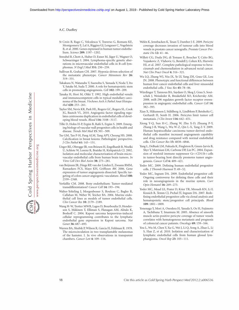
St Croix B, Rago C, Velculescu V, Traverso G, Romans KE,Montgomery E, Lal A, Riggins GJ, Lengauer C, VogelsteinB, et al. 2000. Genes expressed in human tumor endothe-lium. Science 289: 1197–1202.
Streubel B, Chott A, Huber D, Exner M, Jager U, Wagner O,Schwarzinger I. 2004. Lymphoma-specific genetic aber-rations in microvascular endothelial cells in B-cell lym-phomas. N Engl J Med 351: 250–259.
Sullivan R, Graham CH. 2007. Hypoxia-driven selection ofthe metastatic phenotype. Cancer Metastasis Rev 26:319–331.
Takakura N, Watanabe T, Suenobu S, Yamada Y, Noda T, ItoY, Satake M, Suda T. 2000. A role for hematopoietic stemcells in promoting angiogenesis. Cell 102: 199–209.
Tanaka H, Hori M, Ohki T. 1992. High endothelial venuleand immunocompetent cells in typical medullary carci-noma of the breast. Virchows Arch A Pathol Anat Histopa-thol 420: 253–261.
Taylor SM, Nevis KR, Park HL, Rogers GC, Rogers SL, CookJG, Bautch VL. 2010. Angiogenic factor signaling regu-lates centrosome duplication in endothelial cells of devel-oping blood vessels. Blood 116: 3108–3117.
Tilki D, Hohn H-P, Ergun B, Rafii S, Ergun S. 2009. Emerg-ing biology of vascular wall progenitor cells in health anddisease. Trends Mol Med 15: 501–509.
Tse GM, Tan P-H, Pang ALM, Tang APY, Cheung HS. 2008.Calcification in breast lesions: Pathologists’ perspective.J Clin Pathol 61: 145–151.
Unger RE, Oltrogge JB, von Briesen H, Engelhardt B, WoelkiU, Schlote W, Lorenz R, Bratzke H, Kirkpatrick CJ. 2002.Isolation and molecular characterization of brain micro-vascular endothelial cells from human brain tumors. InVitro Cell Dev Biol Anim 38: 273–281.
van Beijnum JR, Dings RP, van der Linden E, Zwaans BMM,Ramaekers FCS, Mayo KH, Griffioen AW. 2006. Geneexpression of tumor angiogenesis dissected: Specific tar-geting of colon cancer angiogenic vasculature. Blood 108:2339–2348.
Verfaillie CM. 2008. Bony endothelium: Tumor-mediatedtransdifferentiation? Cancer Cell 14: 193–194.
Walter-Yohrling J, Morgenbesser S, Rouleau C, Bagley R,Callahan M, Weber W, Teicher BA. 2004. Murine endo-thelial cell lines as models of tumor endothelial cells.Clin Cancer Res 10: 2179–2189.
Wang H-W, Trotter MWB, Lagos D, Bourboulia D, Hender-son S, Makinen T, Elliman S, Flanagan AM, Alitalo K,Boshoff C. 2004. Kaposi sarcoma herpesvirus-inducedcellular reprogramming contributes to the lymphaticendothelial gene expression in Kaposi sarcoma. NatGenet 36: 687–693.
Warren BA, Shubik P, Wilson R, Garcia H, Feldman R. 1978.The microcirculation in two transplantable melanomasof the hamster. I. In vivo observations in transparentchambers. Cancer Lett 4: 109–116.
Welen K, Jennbacken K, Tesan T, Damber J-E. 2009. Pericytecoverage decreases invasion of tumour cells into bloodvessels in prostate cancer xenografts. Prostate Cancer Pro-static Dis 12: 41–46.
Willett CG, Duda DG, di Tomaso E, Boucher Y, Czito BG,Vujaskovic Z, Vlahovic G, Bendell J, Cohen KS, HurwitzHI, et al. 2007. Complete pathological response to beva-cizumab and chemoradiation in advanced rectal cancer.Nat Clin Pract Oncol 4: 316–321.
Wu LQ, Zhang WJ, Niu JX, Ye LY, Yang ZH, Grau GE, LouJN. 2008. Phenotypic and functional differences betweenhuman liver cancer endothelial cells and liver sinusoidalendothelial cells. J Vasc Res 45: 78–86.
Wurdinger T, Tannous BA, Saydam O, Skog J, Grau S, Sout-schek J, Weissleder R, Breakefield XO, Krichevsky AM.2008. miR-296 regulates growth factor receptor overex-pression in angiogenic endothelial cells. Cancer Cell 14:382–393.
Xian X, Hakansson J, Stahlberg A, Lindblom P, Betsholtz C,Gerhardt H, Semb H. 2006. Pericytes limit tumor cellmetastasis. J Clin Invest 116: 642–651.
Xiong Y-Q, Sun H-C, Zhang W, Zhu X-D, Zhuang P-Y,Zhang J-B, Wang L, Wu W-Z, Qin L-X, Tang Z-Y. 2009.Human hepatocellular carcinoma tumor-derived endo-thelial cells manifest increased angiogenesis capabilityand drug resistance compared with normal endothelialcells. Clin Cancer Res 15: 4838–4846.
Yang L, DeBusk LM, Fukuda K, Fingleton B, Green-Jarvis B,Shyr Y, Matrisian LM, Carbone DP, Lin PC. 2004. Expan-sion of myeloid immune suppressor GrþCD11bþcellsin tumor-bearing host directly promotes tumor angio-genesis. Cancer Cell 6: 409–421.
Yoder MC. 2009. Defining human endothelial progenitorcells. J Thromb Haemost 7: 49–52.
Yoder MC, Ingram DA. 2009. Endothelial progenitor cell:Ongoing controversy for defining these cells and theirrole in neoangiogenesis in the murine system. CurrOpin Hematol 16: 269–273.
Yoder MC, Mead LE, Prater D, Krier TR, Mroueh KN, Li F,Krasich R, Temm CJ, Prchal JT, Ingram DA. 2007. Rede-fining endothelial progenitor cells via clonal analysis andhematopoietic stem/progenitor cell principals. Blood109: 1801–1809.
Yonenaga Y, Mori A, Onodera H, Yasuda S, Oe H, FujimotoA, Tachibana T, Imamura M. 2005. Absence of smoothmuscle actin-positive pericyte coverage of tumor vesselscorrelates with hematogenous metastasis and prognosisof colorectal cancer patients. Oncology 69: 159–166.
You L, Wu M, Chen Y, Xu G, Wei J, Li Q, Song A, Zhao L, LiS, Han Z, et al. 2010. Isolation and characterization oflymphatic endothelial cells from human glossal lym-phangioma. Oncol Rep 23: 105–111.
A.C. Dudley
18 Cite this article as Cold Spring Harb Perspect Med 2012;2:a006536
ww
w.p
ersp
ecti
vesi
nm
edic
ine.
org
on August 1, 2019 - Published by Cold Spring Harbor Laboratory Press http://perspectivesinmedicine.cshlp.org/Downloaded from
October 18, 20112012; doi: 10.1101/cshperspect.a006536 originally published onlineCold Spring Harb Perspect Med
Andrew C. Dudley Tumor Endothelial Cells
Subject Collection Angiogenesis
miRNAs as Modulators of Angiogenesis
William C. SessaShira Landskroner-Eiger, Isabelle Moneke and Malformation Syndromes
Arteriovenous Malformations and Other Vascular
Y. LiKevin J. Whitehead, Matthew C.P. Smith and Dean
VEGF and Notch in Tip and Stalk Cell SelectionRaquel Blanco and Holger Gerhardt Development
Molecular Parallels between Neural and Vascular
Anne Eichmann and Jean-Léon Thomas
Regulating AngiogenesisThe Role of the Tumor Microenvironment in
Randolph S. WatnickResponses, Resistance, and the Path ForwardThe VEGF Pathway in Cancer and Disease:
Mark W. Kieran, Raghu Kalluri and Yoon-Jae Cho
DisordersAngiogenic Factors in Preeclampsia and Related
Ana Sofia Cerdeira and S. Ananth Karumanchi
Common Polymorphisms in AngiogenesisMichael S. Rogers and Robert J. D'Amato
Anti-VEGF Therapies in the ClinicKellen L. Meadows and Herbert I. Hurwitz Signaling in Physiology and Pathology
Endothelial Cell-to-Cell Junctions: Adhesion and
Maria Grazia Lampugnani
Tie Signaling Pathway−Angiopoietin The Complex Role of Angiopoietin-2 in the
Gavin Thurston and Christopher DalyCells to OrganismVEGF-Directed Blood Vessel Patterning: From
Victoria L. Bautch
Disease-Restricted ActivityPlGF: A Multitasking Cytokine with
Mieke Dewerchin and Peter CarmelietModels for Therapeutic TrialsVascular Anomalies: From Genetics toward
VikkulaMelanie Uebelhoer, Laurence M. Boon and Miikka
Human Endothelial Progenitor CellsMervin C. Yoder Growth Factor Receptors
Signal Transduction by Vascular Endothelial
Sina Koch and Lena Claesson-Welsh
http://perspectivesinmedicine.cshlp.org/cgi/collection/ For additional articles in this collection, see
Copyright © 2012 Cold Spring Harbor Laboratory Press; all rights reserved
on August 1, 2019 - Published by Cold Spring Harbor Laboratory Press http://perspectivesinmedicine.cshlp.org/Downloaded from


![A vaccine targeting angiomotin induces an antibody response which alters tumor … · 2020. 1. 18. · by normal, but overexpressed by endothelial cells of tumor vessels [8] also](https://img.pdfslide.net/doc/110x75/60ff2f5ea75c100b011c893b/a-vaccine-targeting-angiomotin-induces-an-antibody-response-which-alters-tumor-2020.jpg)