Embed Size (px)
Citation preview

TFi
TCFPa
b
c
d
e
f
g
h
i
j
k
l
a
ARRA
KNBEE
opA
h0n
Lung Cancer 98 (2016) 1–8
Contents lists available at ScienceDirect
Lung Cancer
jou rn al hom epage: www.elsev ier .com/ locate / lungcan
umor marker analyses from the phase III, placebo-controlled,ASTACT-2 study of intercalated erlotinib with gemcitabine/platinumn the first-line treatment of advanced non-small-cell lung cancer�
ony Moka,∗, Guia Ladrerab, Vichien Srimuninnimitc, Virote Sriuranpongd,hong-Jen Yue, Sumitra Thongprasert f, Jennifer Sandoval-Tang, Jin Soo Leeh,atima Fuerte i, David S. Shamesj, Barbara Klughammerk, Matt Trumank,ablo Perez-Morenok, Yi-Long Wul,∗
State Key Laboratory of Southern China, Department of Clinical Oncology, Chinese University of Hong Kong, Shatin, New Territories, Hong Kong, ChinaLung Center of the Philippines, Quezon Ave., Diliman, Quezon City, Metro Manila, PhilippinesSiriraj Hospital, 2 Wanglang Road, Bangkok, Bangkoknoi 10700, ThailandKing Chulalongkorn Memorial Hospital, 1873 Rama 4 Road, Bangkok, Pathumwan 10330, ThailandNational Taiwan University Hospital, No. 7, Zhongshan S Rd., Zhongzheng District, Taipei, TaiwanFaculty of Medicine, Chiangmai University, Chiang Mai, ThailandPhilippine General Hospital, Taft Avenue Ermita, Brgy 670 Zone 72, Manila, 1000 Metro Manila, PhilippinesNational Cancer Center, Goyang, Republic of KoreaRizal Medical Center, Pasig Blvd, Pasig, Metro Manila, PhilippinesOncology Biomarker Development, Genentech Inc., South San Francisco, CA 94080, United StatesF. Hoffmann-La Roche Ltd., Basel, SwitzerlandGuangdong Lung Cancer Institute, Guangdong General Hospital, Guangdong Academy of Medical Sciences, Guangzhou, China
r t i c l e i n f o
rticle history:eceived 30 November 2015eceived in revised form 20 April 2016ccepted 30 April 2016
eywords:SCLCiomarkersGFRrlotinib
a b s t r a c t
Objectives: The FASTACT-2 study of intercalated erlotinib with chemotherapy in Asian patients found thatEGFR mutations were the main driver behind the significant progression-free survival (PFS) benefit notedin the overall population. Further exploratory biomarker analyses were conducted to provide additionalinsight.Materials and methods: This multicenter, randomized, placebo-controlled, double-blind, phase III studyinvestigated intercalated first-line erlotinib or placebo with gemcitabine/platinum, followed by mainte-nance erlotinib or placebo, for patients with stage IIIB/IV non-small cell lung cancer (NSCLC). Provisionof samples for biomarker analysis was encouraged but not mandatory. The following biomarkers wereanalyzed (in order of priority): EGFR mutation by cobas® test, KRAS mutation by cobas® KRAS test, HER2by immunohistochemistry (IHC), HER3 by IHC, ERCC1 by IHC, EGFR gene copy number by fluorescencein-situ hybridization (FISH) and EGFR by IHC. All subgroups were assessed for PFS (primary endpoint),
overall survival (OS), non-progression rate and objective response rate. Results: Overall, 256 patients provided samples for analysis. Considerable overlap was noted amongbiomarkers, except for EGFR and KRAS mutations, which are mutually exclusive. Other than EGFRmutations (p < 0.0001), no other biomarkers were significantly predictive of outcomes in a treatment-by-biomarker interaction test, although ERCC1 IHC-positive status was predictive of improved OS for theerlotinib arm versus placebo in EGFR wild-type patients (median 18.4 vs 9.5 months; hazard ratio [HR]HR = 0.32, 95% confidence intervals [CI]: 0.14–0.69, p = 0.0024).Abbreviations: EGFR, epidermal growth factor receptor; TKIs, tyrosine-kinase inhibitors; NSCLC, non-small-cell lung cancer; PFS, progression-free survival; FISH, flu-rescence in-situ hybridization; IHC, immunohistochemistry; ERCC1, excision repair cross-complementation group 1; ECOG, Eastern Cooperative Oncology Group; PS,erformance status; RECIST, Response Evaluation Criteria in Solid Tumors; OS, overall survival; NPR, non-progression rate; ORR, objective response rate; FACT-L, Functionalssessment of Cancer Therapy—Lung (quality of life questionnaire).� These data were previously presented at the European Society for Medical Oncology Annual Congress 2012, 28 September-2 October, Vienna, Austria.∗ Corresponding authors.
E-mail addresses: [email protected] (T. Mok), [email protected] (Y.-L. Wu).
ttp://dx.doi.org/10.1016/j.lungcan.2016.04.023169-5002/© 2016 The Authors. Published by Elsevier Ireland Ltd. This is an open access article under the CC BY-NC-ND license (http://creativecommons.org/licenses/by-c-nd/4.0/).
2 T. Mok et al. / Lung Cancer 98 (2016) 1–8
Conclusion: Activating EGFR mutations were predictive for improved treatment outcomes with a first-lineintercalated regimen of chemotherapy and erlotinib in NSCLC. ERCC1 status may have some predictivevalue in EGFR wild-type disease, but requires further investigation.
s. Pu
1
ttw(dfwtoE2Ce
bopFacAeet(dbEc6eba(hirtcSmr
2
2
dwgasI
© 2016 The Author
. Introduction
Activating mutations in the epidermal growth factor recep-or (EGFR) are now validated as predictive biomarkers for EGFRyrosine-kinase inhibitors (TKIs) as first-line treatment of patientsith locally advanced or metastatic non-small-cell lung cancer
NSCLC) [1–3] and, ideally, all patients should be tested at initialiagnosis. However, EGFR mutation analysis is still not availableor all patients and, in many cases, treatment decisions are madehile EGFR mutation status is unknown [4,5]. Prevalence of EGFR
esting varies geographically and over time. In a retrospective studyf 1503 patients in Korea, the proportion of patients undergoingGFR testing evolved from 23.3% (between January 2007 and July008) to 63.5% (between October 2009 and July 2010) [6], while inanada in 2010/2011, it was estimated that only 38% of potentiallyligible patients had EGFR testing initiated [7].
Patients with unknown EGFR mutation status should generallye treated with systemic chemotherapy, but a potential alternativeption is sequential combination of chemotherapy and an EGFR TKI,articularly in countries with high rates of EGFR mutations [8,9].ASTACT-2 (First-line Asian Sequential Tarceva and Chemother-py Trial) was a large, confirmatory, phase III trial of sequentialhemotherapy and erlotinib in a non-selected population ofsian patients with advanced NSCLC. FASTACT-2 met its primaryndpoint: patients treated with chemotherapy intercalated withrlotinib had significantly longer progression-free survival (PFS)han patients treated with chemotherapy intercalated with placebomedian PFS 7.6 vs 6.0 months; hazard ratio [HR] = 0.57, 95% confi-ence interval [CI]: 0.47–0.69; p < 0.0001) [9]. Previously reportediomarker analyses of FASTACT-2 confirmed that only patients withGFR mutation-positive NSCLC benefited significantly from inter-alated chemotherapy and erlotinib (median PFS 16.8 months vs.9 months; HR = 0.25, 95% CI: 0.16–0.39; p < 0.0001). Additionalxploratory analysis of important biomarkers was conducted onaseline tumor samples. These were markers that had been associ-ted with clinical outcomes for EGFR TKIs in NSCLC or other cancersKRAS mutations, EGFR gene copy number by fluorescence in-situybridization [FISH] and EGFR expression by immunohistochem-
stry [IHC]) [10–12] and markers for which there was scientificationale to anticipate prognostic or predictive effects (HER2 pro-ein expression by IHC, HER3 expression by IHC, and excision repairross-complementation group 1 [ERCC1] expression by IHC) (seeuppl. Table S1 Table S1 for further details). The current report sum-arizes the prevalence and overlap of these biomarkers, and their
elationships with efficacy outcomes in the FASTACT-2 study.
. Methods
.1. Study design and population
FASTACT-2 was a multicenter, randomized, placebo-controlled,ouble-blind, phase III study of intercalated erlotinib or placeboith gemcitabine plus either carboplatin or cisplatin (at investi-
ators’ discretion), followed by maintenance erlotinib or placebo,s first-line treatment in patients with stage IIIB/IV NSCLC. Thetudy was undertaken in 28 Asian centers across China, Hong Kong,ndonesia, South Korea, the Philippines, Taiwan, and Thailand.
blished by Elsevier Ireland Ltd. This is an open access article under the CCBY-NC-ND license (http://creativecommons.org/licenses/by-nc-nd/4.0/).
Patients aged 18 years and older, with stage IIIB/IV NSCLC, an East-ern Cooperative Oncology Group (ECOG) performance status (PS)of 0 or 1 and measurable disease according to the Response Eval-uation Criteria in Solid Tumors (RECIST version 1.0) were eligible.Patients were randomly assigned to treatment in a 1:1 ratio andwere stratified by disease stage, tumor histology, smoking status,and chemotherapy regimen. The full study methodology has beendescribed previously [9]. Patients were randomized to receive sixcycles of gemcitabine plus platinum with either sequential erlotinibor placebo on days 15–28 of each cycle. Patients who did notprogress during the six cycles of sequential treatment continued toreceive erlotinib or placebo until disease progression, unacceptabletoxicity or death. At disease progression, treatment was unblinded;patients in the placebo group had the option to be crossed over toopen-label erlotinib; patients in the erlotinib group could receivefurther treatment at the discretion of the investigator.
FASTACT-2 was approved by the institutional review board orethics committee of each participating center and was performedin accordance with the principles of the Declaration of Helsinki andGuidelines for Good Clinical Practice. All patients provided writteninformed consent prior to any study-related procedure. The trialwas registered on ClinicalTrials.gov (identifier NCT00883779).
2.2. Biomarker analysis
Patients provided separate consent for biomarker analysis.Tumor samples from first diagnosis or from biopsy at least 14 daysprior to first dose of study drug could be provided for biomarkeranalysis (10–20 slides of a formalin-fixed, paraffin-embeddedsample for histological procedures and 10 slides for cytologicalprocedures).
Biomarkers analyzed were (in order of priority): EGFR mutation,KRAS mutation, HER2 by IHC, HER3 by IHC, ERCC1 by IHC, EGFRgene copy number by FISH and EGFR by IHC. The methodologiesand criteria used are shown in Suppl. Table S2.
2.3. Statistical analyses
The primary endpoint of the study was PFS. Secondary end-points included overall survival (OS); PFS and OS in subgroups;non-progression rate (NPR); objective response rate (ORR); dura-tion of response; and quality of life (FACT-L). Disease control rateat 16 weeks was assessed in a post-hoc analysis. The methods andresults for these outcomes have been reported previously [9]. In thisreport, we analyzed PFS and OS by EGFR and KRAS mutation status,EGFR protein expression status and EGFR gene copy status (pre-specified secondary analyses). The evaluation of tumor biomarkersand correlation with treatment and outcomes was an exploratoryobjective. All patients who provided samples suitable for analysiswere included in the analysis of biomarker data. The study was notpowered for biomarker analysis; a sample size of 450 patients wasestimated based on detecting an HR of 0.75 for PFS at 80% powerwith a 2-sided log-rank test and an � level of 5% (documented in
Wu et al., 2013).A multivariate model of all patients was generated using astepwise selection procedure for PFS and OS, using the follow-ing covariates: treatment, age, ECOG PS, sex, disease stage, disease

T. Mok et al. / Lung Cancer 98 (2016) 1–8 3
SORT
hscw0aha
KaOaBe8
3
se(2mst9tsbaE(tb
been described previously [9]. The biomarker evaluable populationhad similar baseline characteristics to the overall population. Clin-ical characteristics were balanced between the treatment groups
Fig. 1. CON
istology, smoking status, chemotherapy regimen, EGFR mutationtatus, EGFR IHC status, HER2 status and HER3 status. Covariatesould enter the model if they were significant at the 0.1 level andere dropped from the model if their significance fell below the
.1 level. Treatment by biomarker interactions were assessed bydding appropriate terms to the final derived model. Patients mustave had a result for all selected covariates to be included in thesenalyses.
PFS and OS by biomarker status were assessed by use of theaplan–Meier methods, with treatment effect expressed as a HRnd two-sided 95% CI. There was no adjustment for multiple testing.RR and NPR were analyzed by logistic regression and expresseds percentage differences between treatment groups with 95% CI.iomarker subgroup analyses were presented graphically using for-st plots. Statistical analyses were carried out using SAS (version.2). The analysis was undertaken at a cut-off date of 22 June 2012.
. Results
A total of 256 patients provided tissue specimens that wereuitable for exploratory evaluation (129 in the chemotherapy plusrlotinib group and 127 in the chemotherapy plus placebo group)Table 1). Patient disposition in the study is shown in Fig. 1. Of the41 patients evaluated for EGFR mutation, eight had single resistantutations in exon 20 (one patient with T790M; one with S768I and
ix with insertion mutations) and were therefore excluded fromhe EGFR mutation data analyses. Of the 233 remaining patients,7 (41.6%) were confirmed as having EGFR mutation-positive sta-us and 136 (58.4%) had EGFR wild-type status. Other biomarkerubgroups are detailed in Table 1. All biomarkers were balancedetween the treatment groups. Considerable overlap was notedmong the various biomarker subgroups, with the exception of
GFR and KRAS mutations, which are typically mutually exclusiveFig. 2). In addition, some patients had more than one EGFR muta-ion (Suppl. Table S3) Statistically significant associations betweeniomarker subgroups were observed for EGFR mutations and EGFRdiagram.
FISH (p = 0.002); EGFR IHC and ERCC1 IHC (p = 0.016); EGFR IHC andHER2 IHC (p = 0.025); and HER2 IHC and HER3 IHC (p = 0.006).
3.1. Clinical characteristics
Baseline patient characteristics for the overall population have
Fig. 2. Overlap of biomarkers in patients with tumor marker data in FASTACT-2.

4
T. M
ok et
al. /
Lung Cancer
98 (2016)
1–8
Table 1Baseline patient characteristics for the overall population and the biomarker-evaluable populations.
All patients All biomarker-evaluablepatients
EGFR Mut+ EGFR WT EGFR unknown KRAS Mut+ KRAS WT EGFR IHC+ EGFR IHC− EGFR FISH+ EGFR FISH− HER2 IHC + HER2 IHC−
(n = 451) (n = 256) (n = 97) (n = 136) (n = 210) (n = 21) (n = 202) (n = 76) (n = 37) (n = 34) (n = 48) (n = 82) (n = 93)
GCEn = 226
GCPn = 225
GCEn = 129
GCPn = 127
GCEn = 49
GCPn = 48
GCEn = 69
GCPn = 67
GCEn = 106
GCPn = 104
GCEn = 10
GCPn = 11
GCEn = 101
GCPn = 101
GCEn = 40
GCPn = 36
GCEn = 12
GCPn = 25
GCEn = 14
GCPn = 20
GCEn = 25
GCPn = 23
GCEn = 41
GCPn = 41
GCEn = 45
GCPn = 48
Sex, %Male 58 62 53 61 43 48 59 76 66 63 80 73 51 60 53 58 58 60 50 65 44 52 56 61 56 56Female 42 38 47 39 57 52 41 24 34 37 20 27 49 40 48 42 42 40 50 35 56 48 44 39 44 44Disease stage, %IIIB 9 11 8 9 2 4 16 12 8 13 10 0 12 10 3 3 17 20 0 10 4 9 7 10 13 8IV 91 89 92 91 98 96 84 88 92 87 90 100 88 90 98 97 83 80 100 90 96 91 93 90 87 92ECOG PS, %0 26 26 26 21 27 26 30 25 24 27 20 36 31 25 28 33 33 20 36 30 32 30 34 25 18 191 74 74 74 79 73 74 70 75 76 73 80 64 69 75 73 67 67 80 64 70 68 70 66 75 82 81Smoking status, %Current smoker 29 29 26 29 16 15 32 39 33 31 60 55 22 24 23 17 17 28 21 20 20 9 27 34 24 23Former smoker 22 23 19 21 12 17 25 30 25 23 20 18 20 26 23 31 42 20 29 35 16 30 24 22 18 21Never smoker 50 48 55 50 71 69 43 31 42 46 20 27 58 50 55 53 42 52 50 45 64 61 49 44 58 56Histology, %Adenocarcinoma 77 75 80 75 92 92 70 67 75 70 90 91 76 78 80 75 83 88 86 90 76 74 90 93 76 69Non-adenocarcinoma
23 25 20 25 8 8 30 33 25 30 10 9 24 22 20 25 17 12 14 10 24 26 10 7 24 31
HER3 IHC+ (n = 71) HER3 IHC− (n = 70) ERCC1+ (n = 70) ERCC1− (n = 71)
GC-E n = 39 GC-P n = 32 GC-E n = 29 GC-P n = 41 GC-E n = 35 GC-P n = 35 GC-E n = 34 GC-P n = 37
Sex, %Male 49 63 55 51 49 57 53 57Female 51 38 45 49 51 43 47 43Disease stage, %IIIB 8 13 3 5 6 9 12 8IV 92 88 97 95 94 91 88 92ECOG PS, %0 31 28 24 25 29 18 26 351 69 72 76 75 71 82 74 65Smoking status, %Current smoker 18 34 28 20 11 26 29 22Former smoker 23 22 21 22 20 23 24 22Never smoker 59 44 52 59 69 51 47 57Histology, %Adenocarcinoma 90 84 66 76 77 66 82 95Non-adenocarcinoma 10 16 34 24 23 34 18 5
% values rounded to the nearest whole number.ECOG PS: Eastern Cooperative Oncology Group performance status; Mut+: mutation positive; WT: wild type.
g Canc
(s
3
vaa
wbw(as
sampAvtopa(
tubpgct
aooB
3E
bEoa(Ctpae1t4
a
4
a
T. Mok et al. / Lun
Table 1). The majority of clinical characteristics for each biomarkerubtype were consistent with the overall biomarker population.
.2. Clinical endpoints by biomarker subgroups
Due to the well-known, dominant biology associated with acti-ating mutations in EGFR and sensitivity to erlotinib and gefitinib,n analysis of the individual biomarkers in subgroups both withnd without activating mutations was performed.
With the exception of EGFR mutations, no other biomarkersere predictive of outcomes with a positive treatment-by-
iomarker interaction test (Suppl. Fig. S1A). The prolonged PFSith the intercalated regimen was statistically significant for most
9/15) of the biomarker groups; however, as this analysis did notccount for multiple testing and was not hierarchical, the p valueshould be interpreted with caution.
Patients in the following biomarker subgroups had statisticallyignificantly prolonged OS when receiving intercalated chemother-py plus erlotinib compared with chemotherapy plus placebo: EGFRutation positive, EGFR IHC positive, ERCC1 IHC positive, HER2 IHC
ositive, HER3 IHC positive, EGFR FISH positive (Suppl. Fig. S1B).gain, this analysis does not account for multiple testing and palues should be interpreted with caution. It should be noted thathese are all the biomarker ‘positive’ subgroups with the exceptionf KRAS mutation-positive NSCLC (as this group consisted of only 21atients, achieving statistical significance would be difficult in thisnalysis). Further data is provided on the HER2 and HER3 subgroupssee Supplementary information).
The pattern of effects of biomarker status on NPR was similaro that seen with PFS, with the exception that patients with EGFRnknown and EGFR wild-type status NSCLC did not appear to gainenefit from chemotherapy plus erlotinib versus chemotherapylus placebo (Suppl. Table S4). For ORR, all biomarker sub-roups showed a benefit from chemotherapy plus erlotinib versushemotherapy plus placebo, and the majority (13/15) were statis-ically significant.
It should be noted that a significant benefit for chemother-py plus erlotinib compared with chemotherapy plus placebo wasbserved in PFS, OS and ORR (but not NPR) for the overall subgroupf patients who were evaluated for biomarkers (Suppl. Fig. S1A and).
.3. Predictive power of biomarker status in patients with knownGFR mutation-positive or EGFR wild-type NSCLC
Considering EGFR mutation to be the most potent predictiveiomarker, we analyzed each biomarker subgroup according toGFR mutation status. None of the biomarkers were predictivef PFS or OS in patients with sensitizing EGFR mutations (Fig. 3And B). EGFR FISH positivity was predictive of better OS outcomesFig. 3D; HR for EGFR FISH-positive vs FISH-negative = 0.34, 95%I: 0.14–0.80; p = 0.0142) but not PFS benefit (Fig. 3C); however,his was only for patients with EGFR wild-type NSCLC. A significantrolongation of OS was observed for patients with EGFR wild-typend ERCC1 IHC-positive status who received chemotherapy plusrlotinib compared with chemotherapy plus placebo (median OS8.4 vs 9.5 months; HR = 0.32, 95% CI: 0.14–0.69; p = 0.0024), buthis was not mirrored in the PFS results: median PFS was 7.5 versus.6 months (HR = 0.55, 95% CI: 0.27–1.12, p = 0.0941) (Fig. 4).
Exploratory multivariate analyses were also carried out; thesere presented as Supplementary information.
. Discussion
FASTACT-2 was the first randomized phase III study to shown improvement in both PFS and OS with a first-line intercalated
er 98 (2016) 1–8 5
regiment of chemotherapy and erlotinib, although the benefit wasconfined largely to a subgroup of patients whose tumor tissuetested positive for an activating EGFR mutation (exon 19 dele-tions and L858R) or had unknown EGFR mutation status. Thecurrent report reinforces the fact that the predictive power ofEGFR mutation dominates over other molecular biomarkers forEGFR TKI-treated NSCLC. KRAS mutation is known to be generallymutually exclusive of EGFR mutation, thus the benefit seen in theKRAS wild-type subgroup was best explained by the presence ofEGFR mutations, as confirmed when the EGFR wild-type and KRASwild-type subgroup was analyzed (Fig. 3C and D). EGFR IHC sta-tus was not independently predictive of treatment outcomes inthis study, as both EGFR IHC-positive and −negative subgroupshad significant survival benefits with the intercalated regimen.Similar findings were observed in the IPASS study (first-line gefi-tinib vs carboplatin/paclitaxel), which reported an interaction pvalue of 0.214 with EGFR expression by IHC [13], in the SAT-URN study of maintenance erlotinib therapy [14] and in the BR.21study of second-/third-line therapy [12]. Consistent with previousstudies, EGFR FISH was not independently predictive of outcome.Survival benefit was greater in the EGFR FISH-positive subgroup(HR = 0.27, p = 0.0112), compared with the EGFR FISH-negative sub-group (HR = 0.65, p = 0.1554), but again this was likely due to ahigher incidence of EGFR mutations in the former subgroup. In theIPASS study, 78% of patients with EGFR FISH-positive status testedpositive for the EGFR mutation, while only 33% of patients who wereconfirmed to have EGFR FISH-negative status had the mutation.In FASTACT-2, similar findings were observed with 21/34 (62%)and 12/48 (25%) patients with EGFR FISH-positive and -negativestatus, respectively, having EGFR mutations. Patients with EGFRmutation-positive disease who had EGFR IHC-negative status orEGFR FISH-negative status failed to attain statistical significant ben-efit, which may be explained by the small sample size (n = 12 in bothgroups).
The HER/ErbB family of receptor tyrosine kinases are majordrivers of cellular growth and proliferation in both normal andcancer cells. Ligand dependent activation of ErbB signaling is medi-ated through ligand dependent homo- and heterodimerizationbetween members of the receptor family [15]. More specifically,EGFR phosphorylation has been shown to occur through homod-imerization between HER1–HER1 and heterodimerization betweenHER1–HER2, as well as HER1–HER3 [16–18]. It was of interest,therefore, to determine whether any additional clinical activityof erlotinib might be accounted for by overexpression and con-comitant activation of EGFR through other ErbB family members.Approximately 40% of patients assessed for HER2 or HER3 sta-tus also had EGFR activating mutations. No specific associationwas seen between EGFR mutations and HER2 or HER3 expression.Despite the better survival outcomes with the intercalated combi-nation in patients with positive HER2 or HER3 expression (Suppl.Fig. S2), neither of these biomarkers were considered to be inde-pendent predictors as their interaction tests were negative.
ERCC1 may be an important factor for the benefit of the interca-lated treatment regimen. The ERCC1 protein plays a role in restoringDNA from platinum damage, by its involvement in the nucleotideexcision repair pathway and the interstrand cross-link repair path-way [19]. ERCC1 may be a potential predictor of treatment outcomewith cytotoxic chemotherapy [19–21], although results have beenmixed and there have been some controversies regarding the qual-ity of ERCC1 reagents [22,23]. Generally patients with low ERCC1expression were reported to have better tumor response and longersurvival with platinum-based regimens [24]. The observation in
FASTACT-2 was contrary to current thinking, finding that patientswith ERCC1 IHC-positive status, among the EGFR wild-type popu-lation, benefited more from the intercalated regimen than patientswith ERCC1 IHC-negative status. A pre-clinical study on the poten-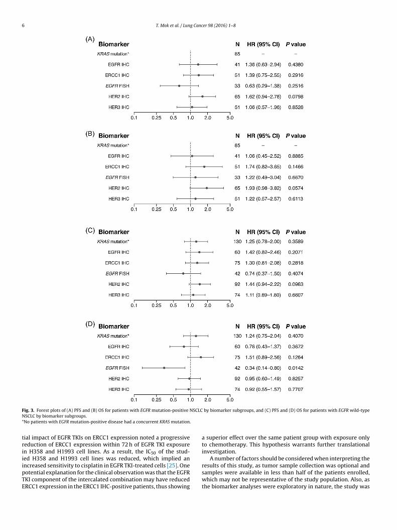
6 T. Mok et al. / Lung Cancer 98 (2016) 1–8
F NSCLCN* .
triiipTE
ig. 3. Forest plots of (A) PFS and (B) OS for patients with EGFR mutation-positive
SCLC by biomarker subgroups.No patients with EGFR mutation-positive disease had a concurrent KRAS mutation
ial impact of EGFR TKIs on ERCC1 expression noted a progressiveeduction of ERCC1 expression within 72 h of EGFR TKI exposuren H358 and H1993 cell lines. As a result, the IC50 of the stud-ed H358 and H1993 cell lines was reduced, which implied anncreased sensitivity to cisplatin in EGFR TKI-treated cells [25]. One
otential explanation for the clinical observation was that the EGFRKI component of the intercalated combination may have reducedRCC1 expression in the ERCC1 IHC-positive patients, thus showingby biomarker subgroups, and (C) PFS and (D) OS for patients with EGFR wild-type
a superior effect over the same patient group with exposure onlyto chemotherapy. This hypothesis warrants further translationalinvestigation.
A number of factors should be considered when interpreting theresults of this study, as tumor sample collection was optional and
samples were available in less than half of the patients enrolled,which may not be representative of the study population. Also, asthe biomarker analyses were exploratory in nature, the study was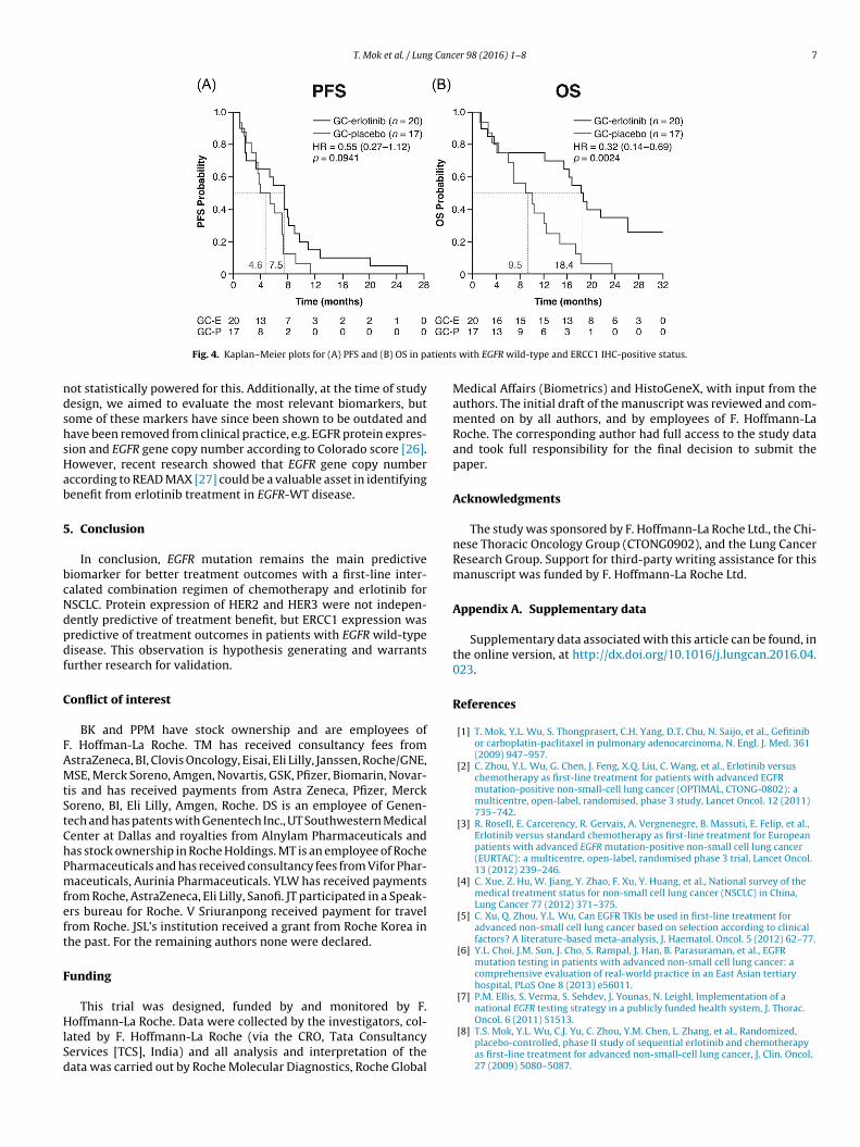
T. Mok et al. / Lung Cancer 98 (2016) 1–8 7
tients
ndshsHab
5
bcNdpdf
C
FAMtStChPmfeft
F
HlSd
Fig. 4. Kaplan–Meier plots for (A) PFS and (B) OS in pa
ot statistically powered for this. Additionally, at the time of studyesign, we aimed to evaluate the most relevant biomarkers, butome of these markers have since been shown to be outdated andave been removed from clinical practice, e.g. EGFR protein expres-ion and EGFR gene copy number according to Colorado score [26].owever, recent research showed that EGFR gene copy numberccording to READ MAX [27] could be a valuable asset in identifyingenefit from erlotinib treatment in EGFR-WT disease.
. Conclusion
In conclusion, EGFR mutation remains the main predictiveiomarker for better treatment outcomes with a first-line inter-alated combination regimen of chemotherapy and erlotinib forSCLC. Protein expression of HER2 and HER3 were not indepen-ently predictive of treatment benefit, but ERCC1 expression wasredictive of treatment outcomes in patients with EGFR wild-typeisease. This observation is hypothesis generating and warrantsurther research for validation.
onflict of interest
BK and PPM have stock ownership and are employees of. Hoffman-La Roche. TM has received consultancy fees fromstraZeneca, BI, Clovis Oncology, Eisai, Eli Lilly, Janssen, Roche/GNE,SE, Merck Soreno, Amgen, Novartis, GSK, Pfizer, Biomarin, Novar-
is and has received payments from Astra Zeneca, Pfizer, Merckoreno, BI, Eli Lilly, Amgen, Roche. DS is an employee of Genen-ech and has patents with Genentech Inc., UT Southwestern Medicalenter at Dallas and royalties from Alnylam Pharmaceuticals andas stock ownership in Roche Holdings. MT is an employee of Rocheharmaceuticals and has received consultancy fees from Vifor Phar-aceuticals, Aurinia Pharmaceuticals. YLW has received payments
rom Roche, AstraZeneca, Eli Lilly, Sanofi. JT participated in a Speak-rs bureau for Roche. V Sriuranpong received payment for travelrom Roche. JSL’s institution received a grant from Roche Korea inhe past. For the remaining authors none were declared.
unding
This trial was designed, funded by and monitored by F.
offmann-La Roche. Data were collected by the investigators, col-ated by F. Hoffmann-La Roche (via the CRO, Tata Consultancyervices [TCS], India) and all analysis and interpretation of theata was carried out by Roche Molecular Diagnostics, Roche Global
with EGFR wild-type and ERCC1 IHC-positive status.
Medical Affairs (Biometrics) and HistoGeneX, with input from theauthors. The initial draft of the manuscript was reviewed and com-mented on by all authors, and by employees of F. Hoffmann-LaRoche. The corresponding author had full access to the study dataand took full responsibility for the final decision to submit thepaper.
Acknowledgments
The study was sponsored by F. Hoffmann-La Roche Ltd., the Chi-nese Thoracic Oncology Group (CTONG0902), and the Lung CancerResearch Group. Support for third-party writing assistance for thismanuscript was funded by F. Hoffmann-La Roche Ltd.
Appendix A. Supplementary data
Supplementary data associated with this article can be found, inthe online version, at http://dx.doi.org/10.1016/j.lungcan.2016.04.023.
References
[1] T. Mok, Y.L. Wu, S. Thongprasert, C.H. Yang, D.T. Chu, N. Saijo, et al., Gefitinibor carboplatin-paclitaxel in pulmonary adenocarcinoma, N. Engl. J. Med. 361(2009) 947–957.
[2] C. Zhou, Y.L. Wu, G. Chen, J. Feng, X.Q. Liu, C. Wang, et al., Erlotinib versuschemotherapy as first-line treatment for patients with advanced EGFRmutation-positive non-small-cell lung cancer (OPTIMAL, CTONG-0802): amulticentre, open-label, randomised, phase 3 study, Lancet Oncol. 12 (2011)735–742.
[3] R. Rosell, E. Carcerency, R. Gervais, A. Vergnenegre, B. Massuti, E. Felip, et al.,Erlotinib versus standard chemotherapy as first-line treatment for Europeanpatients with advanced EGFR mutation-positive non-small cell lung cancer(EURTAC): a multicentre, open-label, randomised phase 3 trial, Lancet Oncol.13 (2012) 239–246.
[4] C. Xue, Z. Hu, W. Jiang, Y. Zhao, F. Xu, Y. Huang, et al., National survey of themedical treatment status for non-small cell lung cancer (NSCLC) in China,Lung Cancer 77 (2012) 371–375.
[5] C. Xu, Q. Zhou, Y.L. Wu, Can EGFR TKIs be used in first-line treatment foradvanced non-small cell lung cancer based on selection according to clinicalfactors? A literature-based meta-analysis, J. Haematol. Oncol. 5 (2012) 62–77.
[6] Y.L. Choi, J.M. Sun, J. Cho, S. Rampal, J. Han, B. Parasuraman, et al., EGFRmutation testing in patients with advanced non-small cell lung cancer: acomprehensive evaluation of real-world practice in an East Asian tertiaryhospital, PLoS One 8 (2013) e56011.
[7] P.M. Ellis, S. Verma, S. Sehdev, J. Younas, N. Leighl, Implementation of anational EGFR testing strategy in a publicly funded health system, J. Thorac.
Oncol. 6 (2011) S1513.[8] T.S. Mok, Y.L. Wu, C.J. Yu, C. Zhou, Y.M. Chen, L. Zhang, et al., Randomized,placebo-controlled, phase II study of sequential erlotinib and chemotherapyas first-line treatment for advanced non-small-cell lung cancer, J. Clin. Oncol.27 (2009) 5080–5087.

8 g Canc
[
[
[
[
[
[
[
[
[
[
[
[
[
[
[
[
[
Oncol. 24 (2006) 5034–5042.[27] J. Moecks, D. Soulieres, B. Klughammer, ReadMax—a novel reading and
T. Mok et al. / Lun
[9] J.L. Wu, J.S. Lee, S. Thongprasert, C.J. Yu, L. Zhang, G. Ladrera, et al., Intercalatedcombination of chemotherapy and erlotinib for patients with advanced stagenon-small-cell lung cancer (FASTACT-2): a randomised, double-blind trial,Lancet Oncol. 14 (2013) 777–786.
10] D.A. Eberhard, B.E. Johnson, L.C. Amler, A.D. Goddard, S.L. Heldens, R.S. Herbst,et al., Mutations in the epidermal growth factor receptor and in KRAS arepredictive and prognostic indicators in patients with non-small-cell lungcancer treated with chemotherapy alone and in combination with erlotinib, J.Clin. Oncol. 23 (2005) 5900–5909.
11] W. Brugger, N. Triller, M. Blasinska-Morawiec, S. Curescu, R. Sakalauskas, G.M.Manikhas, et al., Prospective molecular marker analyses of EGFR and KRASfrom a randomized, placebo-controlled study of erlotinib maintenancetherapy in advanced non-small-cell lung cancer, J. Clin. Oncol. 29 (2011)4113–4120.
12] C.-Q. Zhu, G. da Cunha Santos, K. Ding, A. Sakurada, J. Cutz, C, N. Liu, et al., Roleof KRAS and EGFR as biomarkers of response to erlotinib in National CancerInstitute of Canada Clinical Trials Group Study BR.21, J. Clin. Oncol. 26 (2008)428–475.
13] M. Fukuoka, Y.-L. Wu, S. Thongprasert, P. Sunpaweravong, S.S. Leong, V.Sriuranpong, et al., Biomarker analyses and final overall survival results froma phase III, randomized, open-label, first-line study of gefitinib versuscarboplatin/paclitaxel in clinically selected patients with advancednon-small-cell lung cancer in Asia (IPASS), J. Clin. Oncol. 29 (2011) 2866–2874.
14] J. Mazières, W. Brugger, F. Cappuzzo, P. Middel, A. Frosch, I. Bara, et al.,Evaluation of EGFR protein expression by immunohistochemistry usingH-score and the magnification rule: re-analysis of the SATURN study, LungCancer 82 (2013) 231–237.
15] H. Shankaran, Y. Zhang, Y. Tan, H. Resat, Model-based analysis of HERactivation in cells co-expressing EGFR, HER2 and HER3, PLoS Comput. Biol. 9(8) (2013) e1003201.
16] G. Schaefer, L. Haber, L.M. Crocker, S. Shia, L. Shao, D. Dowbenko, et al., A
two-in-one antibody against HER3 and EGFR has superior inhibitory activitycompared with monospecific antibodies, Cancer Cell 20 (2011) 472–486.17] T.R. Wilson, D.Y. Lee, L. Berry, D.S. Shames, J. Settleman,Neuregulin-1-mediated autocrine signaling underlies sensitivity to HER2kinase inhibitors in a subset of human cancers, Cancer Cell 20 (2011) 158–172.
er 98 (2016) 1–8
18] D.S. Shames, J. Carbon, K. Walter, A.M. Jubb, C. Kozlowski, T. Januario, et al.,High heregulin expression is associated with activated HER3 and may definean actionable biomarker in patients with squamous cell carcinomas of thehead and neck, PLoS One 8 (2013) e56765.
19] E. Felip, P. Martinez, Can sensitivity to cytotoxic chemotherapy be predictedby biomarkers? Ann. Oncol. 23 (2012) 189–192.
20] K. Azuma, Y. Komohara, T. Sasada, Y. Terazaki, J. Ikeda, T. Hoshino, et al.,Excision repair cross-complementation group 1 predicts progression-free andoverall survival in non-small cell lung cancer patients treated withplatinum-based chemotherapy, Cancer Sci. 98 (2007) 1336–1343.
21] P. Ceppi, M. Volante, S. Novello, I. Rapa, K.D. Danenberg, P.V. Danenberg, et al.,ERCC1 and RRM1 gene expressions but not EGFR are predictive of shortersurvival in advanced non-small-cell lung cancer treated with cisplatin andgemcitabine, Ann. Oncol. 17 (2006) 1818–1825.
22] K.A. Olaussen, A. Dunant, P. Fouret, E. Brambilla, F. André, V. Haddad, et al.,DNA repair by ERCC1 in non-small-cell lung cancer and cisplatin-basedadjuvant chemotherapy, N. Engl. J. Med. 355 (2006) 983–991.
23] L. Friboulet, K.A. Olaussen, J.P. Pignon, F.A. Shepherd, M.S. Tsao, S. Graziano,et al., ERCC1 isoform expression and DNA repair in non-small-cell lungcancer, N. Engl. J. Med. 368 (2013) 1101–1110.
24] D. Yan, P. Wei, G. An, W. Chen, Prognostic potential of ERCC1 proteinexpression and clinicopathologic factors in stage III/N2 non-small cell lungcancer, J. Cardiothorac. Surg. 8 (2013) 149.
25] L.H.T. Cheong, C.W.C. Hui, F. Xu, T.S.K. Mok, C.H. Wong, Down-regulation ofERCC1 by EGFR-TKI improves platinum-based chemotherapy efficacy fornon-small cell lung cancer (NSCLC), J. Thorac. Oncol. 9 (Suppl. 9) (2014) S7–52.
26] F.R. Hirsch, M. Verella-Garcia, P.A. Bunn Jr., W.A. Franklin, R. Dziadziuszko,Molecular predictors of outcome with gefitinib in a phase IIIplacebo-controlled study in advanced non-small-cell lung cancer, J. Clin.
scoring approach for EGFR gene copy number to predict therapeutic benefit oferlotinib treatment in EGFR wild-type non-small-cell lung cancer, J. Pathol.Clin. Res. 1 (2015) 134–143.