Embed Size (px)
Citation preview
Two Cases of Streptococcal Infections of Cultured Tilapia in Asia
KEI YUASA1, TAKASHI KAMAISHI1, KISHIO HATAI2, MELIYA BAHNNAN3 and PRASATPORN BORISUTPETH4
1National Research Institute of Aquaculture, Fisheries Research Agency, Japan2Laboratory of Fish Disease, Nippon Veterinary and Animal University, Japan
3Jambi Freshwater Aquaculture Center, Indonesia4Faculty of Veterinary Medicine, Khon-Kaen University, Thailand
ABSTRACT
Tilapia (Oreochromis niloticus) is one of the most popular cultured species in both Thailand and Indonesia. Initial outbreak of mass mortality in cultured tilapia in Thailand was observed in floating net cages of the Mekong River in Mukudahan City, northeast Thailand in May 2001. The mortality in the cages lasted approximately two weeks and reached around 40 to 60%. Affected fish showed abdominal distention due to ascites, a watery-like substance in the intestinal cavity and abscesses on the peduncle. In tilapia ponds, lower total percentage overall mortalities were observed daily, and continuously in the ponds with running water from the irrigation channel in Lubuk Linggau City, South Sumatra in 2002 and 2003. These fish had opaque discoloured eyes with bilateral exophthalmia. The bacteria isolated from the brains and other organs of the affected tilapia from Thailand and Indonesia were identified as Gram-positive, non-motile cocci. These bacterial isolates from infected tilapia in both Thailand and Indonesia were identified as Streptococcus agalactiae and S. iniae, respectively.
Yuasa, K., Kamaishi, T., Hatai, K., Bahnnan, M. and Borisuthpeth, P. 2008. Two cases of streptococcal infections of cultured tilapia in Asia, pp. 259-268. In Bondad-Reantaso, M.G., Mohan, C.V., Crumlish, M. and Subasinghe, R.P. (eds.). Diseases in Asian Aquaculture VI. Fish Health Section, Asian Fisheries Society, Manila, Philippines.
Corresponding author: Kei Yuasa, [email protected]
INTRODUCTION
Tilapia Oreochromis niloticus is one of the most popular fish species produced in freshwater aquaculture in many Asian countries. This fish species is popular due to its rapid growth and high adaptability to the environment. A serious obstacle to the sustainability and development of tilapia culture, however, is the frequent occurrences of mortality due to bacterial infection. The most commonly reported bacterial infection resulting in high mortalities was associated with the β-hemolytic streptococci, Streptococcus iniae (Sako, 1993; Ramesh et al., 1994; Sugita, 1996). Streptococcus shiloi was found responsible for a bacterial meningoencephalitis in tilapia in Israel (Eldar et al., 1994), which has become a synonym for S. iniae (Eldar et al., 1995). The Lansfield B type cocci identified as Streptococcus agalactiae was more known to cause mastitis of cattle, but recently this species has been found in wild mullet Liza klunzingeri, causing mass mortality in Kuwait in 2001 (Evans et al., 2002). A streptococcus that was recovered and isolated from infected tilapia was originally identified as Streptococcus difficile (Eldar et al., 1994) but this has been finally characterized as S. agalactiae (Evans et al., 2002).
Identification of S. iniae and S. agalactiae can be completed by biochemical tests using API 20 strep or API 32 strep test (BioMerieux, USA) or a serological test using the Slidex strepto kit for grouping of β-hemolytic streptococci groups (BioMerieux, USA) (Yuasa et al., 1999; Evans et al., 2002). Recently an oligonucleotide probe array has been recognized as a rapid and accurate diagnosis for bacterial infection of fish (Matsuyama et al., 2006). In the present study, S. agalactiae and S. iniae were both recovered from diseased tilapia farmed in two different aquaculture systems in Thailand and Indonesia, respectively.
MATERIALS AND METHODS
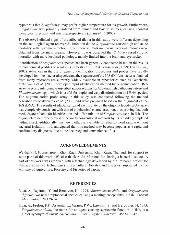
Mortality and clinical signs of fish in the sampling sitesIn May 2001, mass mortality of cultured tilapia occurred in the net cages floating on Mekong River in Mukdahan City, Morth-east Thailand (Figure 1; Plate 1, Photo A). In a colony of floating cages including 10 units (farms), 3 units located in the middle of the floating cages were affected by the mortality. Cumulative mortalities of the units reached 40-60% during two weeks, and total losses of fish in each unit reached around 2 tons. Affected fish, around 300-500 g in body weight, showed expanded abdomen, deposit of water-like in the intestine and swelling at the peduncle with grayish pus inside (Plate 1, Photos B, C, and D). Bacterial detection and identification in Thailand were attempted in one of the units.
On the other hand, in Indonesia, mass mortalities of cultured tilapia were observed at several farms where fish were reared in concrete ponds with running water system in Lubuk Linggau, South Sumatra, Indonesia in 2002 and 2003 (Figure 1; Plate 1, Photo E). Inlet water for all farms was supplied from an irrigation channel and water temperature in the channel was at around 23oC through the year. Mortality was lower than that in the Thailand case, maximum of 0.1-0.2% per day, but continuously observed throughout
the year. Affected fish characteristically showed the bilateral exophthalmia with opaque coloration (Plate 1, Photo F). Bacterial detection from affected fish in Indonesia was attempted at 3 farms.
Detection and isolation of the bacteriaRecovery of the bacteria was performed by insertion of flamed needle into the spleen, kidney, liver and grayish pus in peduncle of 5 moribund fish in Thailand and into additional brains and eye socket of 5 moribund per site in Indonesia, and streaked directly on to brain heart infusion (BHI) agar and incubated at 25oC for 2 days. The colony growth was observed after 2 days and sub-cultured onto fresh BHI agar if the culture onto fresh BHI agar if the cultures were not pure.
A small section of each of the organs in the fish in Indonesia was aseptically removed from the animals with clinical signs of disease and a tissue smear prepared on a clean microscope glass slide. This was fixed with absolute methanol for 3 min and stained with 5% Giemsa solution for 30 min at room temperature, and viewed by light microscopy at x 100 magnification under immersion oil.
Bacterial identification testsConventional identification of bacteria Single colonies of pure isolated bacteria was applied on the slide glass with a drop of distilled water, left to air dry and then heat-fixed by passing through a flame. These slides were then gram-stained using Gram stain kit (Nissui, Japan). Production of oxidase was observed by using the cytocrome-oxidase paper test (Nissui, Japan). Catalase production was confirmed by bubble production in 3% hydrogen peroxide (Wako, Japan). The oxidative-fermentative (O/F) test, production of H2S and haemolysis tests were conducted using O/F medium (Nissui, Japan), SIM medium (Nissui, Japan) and 5% sheep blood agar (Nissui, Japan), respectively. Motility of bacteria was confirmed by turbidity in SIM medium. Growth of bacteria at 45 oC was determined by incubation in BHI broth (Nissui, Japan) for 24 hr. Additional biochemical profiles of the pure isolates were identified using API 20 Strep. kit (BioMerieux, USA) examined at 25oC or 35oC. Isolates used in the tests were B44005 for tilapia from Thailand and JF0375 and JF0381 from tilapia of Indonesia. Additionally, 2 known bacterial isolates identified as NJM0101 from yellowtail Seriola quinqueradiata of Japan and 8-1 Br from mullet Liza klunzingeri of Kuwait were used as references of Lactococcus garvieae and Streptococcus agalactiae, respectively (Table 1).
Oligonucleotide probe array Oligonucleotide probes targeting one or two specific regions for each species in the 16S rDNA were used for spotting and immobilizing on a nylon membrane (Table 2). Prepared probes included 5 kinds of species causing streptococci in fish. These probes were designed from the unique regions to each species based on the alignment of sequences described in a GenBank database. A specific probe for S. agalactiae and Enterococcus sp. was prepared. Two probes were prepared for L. garvieae, S. iniae or S. dysgalactiae, which were used for spotting and immobilizing on a nylon membrane. Extracted DNA of each isolate (Table
1) was amplified using universal primer set AGAGTTTGATCMTGGCTCAG (20F) and GGTTACCTTGTTACGACTT (1500R), by PCR in order to amplify the conservative region of each isolate. The PCR reaction was conducted by temperature cycling on an iCycler (Bio-Rad, USA) including an initial denaturation (940C for 4 mins), 30 cycles of core PCR production (30s at 940C, 30s at 720C), and a final elongation (7 mins at 720C ). Hybridization of Digoxigenin (DIG)-labeled PCR product with each probe was conducted on the nylon membrane that was prepared according to the description by Matsuyama et al. (2006). Polaroid film was exposed with luminescent signals produced by alkaline phosphatase-conjugated anti-DIG antibody to observe white spots due to the luminescent exposure. Lactococcus garvieae, S. iniae and S. dysgalactiae were identified when two spots for each species were reacted since the result of single spot was not enough to identify these species and S. agalactiae and Enterococcus sp. were identified when one spot was reacted.
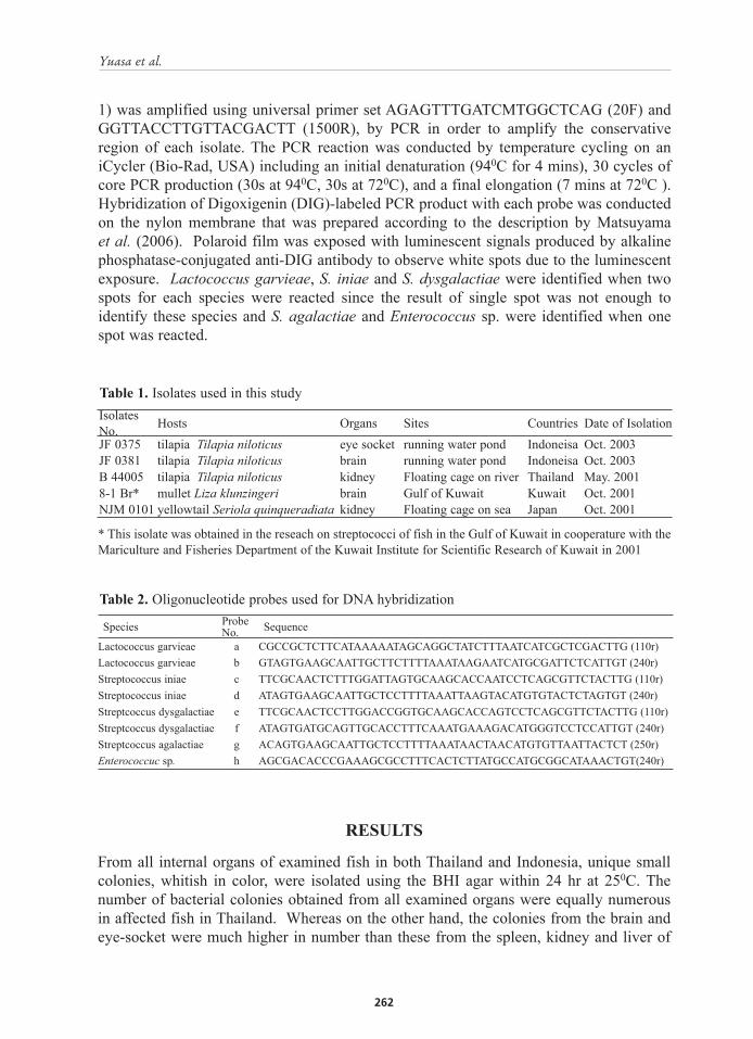
Isolates No. Hosts Organs Sites Countries Date of IsolationJF 0375 tilapia Tilapia niloticus eye socket running water pond Indoneisa Oct. 2003JF 0381 tilapia Tilapia niloticus brain running water pond Indoneisa Oct. 2003B 44005 tilapia Tilapia niloticus kidney Floating cage on river Thailand May. 20018-1 Br* mullet Liza klunzingeri brain Gulf of Kuwait Kuwait Oct. 2001NJM 0101 yellowtail Seriola quinqueradiata kidney Floating cage on sea Japan Oct. 2001
Table 1. Isolates used in this study
* This isolate was obtained in the reseach on streptococci of fish in the Gulf of Kuwait in cooperature with the Mariculture and Fisheries Department of the Kuwait Institute for Scientific Research of Kuwait in 2001
Species Probe No. Sequence
Lactococcus garvieae a CGCCGCTCTTCATAAAAATAGCAGGCTATCTTTAATCATCGCTCGACTTG (110r)Lactococcus garvieae b GTAGTGAAGCAATTGCTTCTTTTAAATAAGAATCATGCGATTCTCATTGT (240r)Streptococcus iniae c TTCGCAACTCTTTGGATTAGTGCAAGCACCAATCCTCAGCGTTCTACTTG (110r)Streptococcus iniae d ATAGTGAAGCAATTGCTCCTTTTAAATTAAGTACATGTGTACTCTAGTGT (240r)Streptcoccus dysgalactiae e TTCGCAACTCCTTGGACCGGTGCAAGCACCAGTCCTCAGCGTTCTACTTG (110r)Streptcoccus dysgalactiae f ATAGTGATGCAGTTGCACCTTTCAAATGAAAGACATGGGTCCTCCATTGT (240r)Streptcoccus agalactiae g ACAGTGAAGCAATTGCTCCTTTTAAATAACTAACATGTGTTAATTACTCT (250r)Enterococcuc sp. h AGCGACACCCGAAAGCGCCTTTCACTCTTATGCCATGCGGCATAAACTGT(240r)
Table 2. Oligonucleotide probes used for DNA hybridization
RESULTS
From all internal organs of examined fish in both Thailand and Indonesia, unique small colonies, whitish in color, were isolated using the BHI agar within 24 hr at 250C. The number of bacterial colonies obtained from all examined organs were equally numerous in affected fish in Thailand. Whereas on the other hand, the colonies from the brain and eye-socket were much higher in number than these from the spleen, kidney and liver of
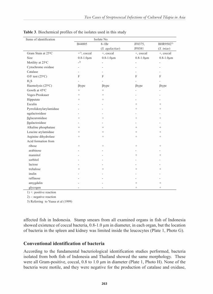
Items of identification Isolate No. B44005 8-1Br JF0375, BHR95023)
(S. agalactiae) JF0381 (S. iniae)Gram Stain at 25oC +1), coccal +, coccal +, coccal +, coccalSize 0.8-1.0µm 0.8-1.0µm 0.8-1.0µm 0.8-1.0µmMotility at 25oC -2) - - -Cytochrome oxidase - - - -Catalase - - - -O/F test (25oC) F F F FH2S - - - -Haemolysis (25oC) βtype βtype βtype βtypeGowth at 45oC + + - -Voges-Proskauer + + - -Hipputate + + - -Esculin - - + +Pyrrolidcnylarylamidase - - + +αgalactosidase - - - -βglucuronidase + + + +βgalactosidase - - - -Alkaline phosphatase + + + +Leucine arylamidase + + + +Arginine dihydrolase + + + +Acid formation from ribose + + + + arabinose - - - - mannitol - - + + sorbitol - - - - lactose - - - - trehalose + + + + inulin - - - - raffinose - - - - amygdalin - - + + glycogen - - + +1) +: positive reaction2) -: negative reaction3) Referring to Yuasa et al (1999)
Table 3. Biochemical profiles of the isolates used in this study
affected fish in Indonesia. Stamp smears from all examined organs in fish of Indonesia showed existence of coccal bacteria, 0.8-1.0 µm in diameter, in each organ, but the location of bacteria in the spleen and kidney was limited inside the leucocytes (Plate 1, Photo G).
Conventional identification of bacteriaAccording to the fundamental bacteriological identification studies performed, bacteria isolated from both fish of Indonesia and Thailand showed the same morphology. These were all Gram-positive, coccal, 0.8 to 1.0 μm in diameter (Plate 1, Photo H). None of the bacteria were motile, and they were negative for the production of catalase and oxidase,
utilized glucose fermentative and showed β haemolysis on blood agar. The main difference between the Thai and Indonesian isolates was growth in the Thai isolate at 450C and none from the Indonesian isolate at this temperature.
Biochemical identification using API 20 strep at 250C and 350C were ended within 24 hr and the results between both temperatures were the same. The result of API 20 strep for isolates JF0375 and JF0381 from Indonesia were consistent with the result described for S. iniae (Yuasa et al., 1999) recovered from infected rabbitfish in Bahrain. The API 20 strep results for isolate B4405 from Thailand was consistent with the result for the isolate 8-1 Br from wild mullet, which was identified as S. agalactiae at the Laboratory of Fish Pathology of Nippon Veterinary and Animal Science University.
Oligonucleotide probe arrayIn the oligonucleotide probe array, the PCR products from conservative regions of B4405 or 8-1 Br and NJM0101 were hybridized with only oligonucleotide spots for S. agalactiae and L. garvieae, respectively. The PCR products from conservative regions of JF0375 and JF0381 were hybridized with two oligonucleotide spots for S. iniae as well as an oligonucleotide spot for S. dysgalactiae, but not hybridized with another spot for S. dysgalactiae (Figure 2). In this case, JF0375 and JF0381 were identified as S. iniae.
DISCUSSION
In this study, S. agalactiae and S. iniae, were recovered and identified from affected fish at a site in Thailand and S. iniae recovered from the affected fish at several sites in Indonesia, respectively. Streptococcus spp. are recognized globally as causative agents of streptococcal infections in several kinds of seawater and freshwater fishes. However, all isolates from tilapia in Indonesia were identified as S. iniae; no S. agalactiae was isolated through the year. This may be due to the lower water temperature of Indonesian culture system favouring the recovery of S. agalactiae. Actually mass mortality of wild mullet Liza klunzingeri due to S. agalactiae in Kuwait occurred when temperature of seawater was higher than usual (Evans et al., 2002). Unfortunately water temperature at a sampling site in Thailand was not taken in this study; but our sampling was conducted in the hottest season, expectedly with higher temperature; more than 280C in the Mekong River at the sampling site. On the contrary, streptococci due to S. iniae has been reported in tilapia culture in Japan, a temperate country, and S. agalactiae has not been detected from any sick fish species in Japan yet.
Mortality of rabbitfish Siganus canaliculatus due to S. iniae in Bahrain were more frequently observed in May to June and September to October when water temperature is lower than that in July to August (personal data). These findings suggest that occurrences of streptococcal infection due to two species are water temperature- dependent. It may be considered that S. agalactiae may be a pathogen suited to more tropical regions compared with S. iniae which may prefer the cooler, temperate regions. In this study, it was found that the S. agalactiae grew at 450C in the medium, whereas S. iniae did not, thus supporting the
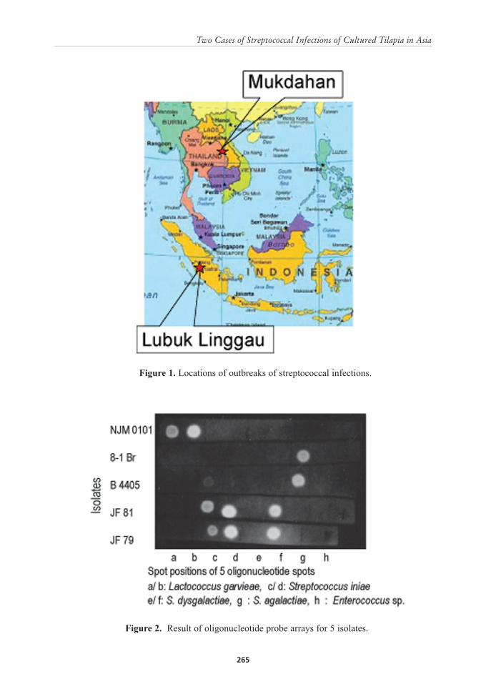
Figure 1. Locations of outbreaks of streptococcal infections.
Figure 2. Result of oligonucleotide probe arrays for 5 isolates.
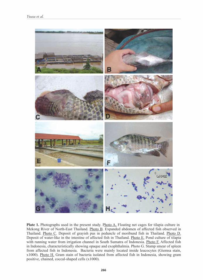
Plate 1. Photographs used in the present study. Photo A. Floating net cages for tilapia culture in Mekong River of North-East Thailand. Photo B. Expanded abdomen of affected fish observed in Thailand. Photo C. Deposit of grayish pus in peduncle of moribund fish in Thailand. Photo D. Deposit of water-like in the intestine of affected fish in Thailand. Photo E. Pond culture of tilapia with running water from irrigation channel in South Sumatra of Indonesia. Photo F. Affected fish in Indonesia, characteristically showing opaque and exophthalmia. Photo G. Stamp smear of spleen from affected fish in Indonesia. Bacteria were mainly located inside leucocytes (Giemsa stain, x1000). Photo H. Gram stain of bacteria isolated from affected fish in Indonesia, showing gram positive, chained, coccal-shaped cells (x1000).
hypothesis that S. agalactiae may prefer higher temperature for its growth. Furthermore, S. agalactiae was primarily isolated from human and bovine sources, causing neonatal meningitis infections and mastitis, respectively (Evans et al. 2002).
The observed clinical signs of the affected tilapia in this study were different depending on the aetiological agent recovered. Infection due to S. agalactiae caused high and acute mortality with systemic infection. From these animals numerous bacterial colonies were obtained from the main organs. However, it was observed that S. iniae caused chronic mortality with more localised pathology, mainly limited into the brain and eye socket.
Identification of Streptococcus species has been generally conducted based on the results of biochemical profiles or serology (Ramesh et al., 1994; Yuasa et al., 1999; Evans et al., 2002). Advances in the use of genetic identification procedures and probes have rapidly developed for other bacterial species and the sequences of the 16S rDNA in bacteria obtained from many microbes are currently widely available in repositories such as Genebank. Matsuyama et al. (2006) developed rapid identification method by oligonucleotide DNA array targeting intergenic transcribed spacer regions for bacterial fish pathogens Vibrio and Photobacterium spp., which is useful for rapid and easy discrimination of Vibrio species. The oligonucleotide probe array in this study was conducted following the method described by Matsuyama et al. (2006) and were prepared based on the alignment of the 16S rDNA. The results of identification of each isolate by the oligonucleotide probe array was completely consistent with that of biochemical characterization, thus proving that both methods are reliable for identification and differentiation of Streptococcus spp. in fish. The oligonucleotide probe array is superior to conventional methods by its rapidity (completed within 8 hrs). Additionally, this new method is available for ethanol-fixed sample without bacterial isolation. It is anticipated that this method may become popular as a rapid and confirmatory diagnosis, due to the accuracy and convenience of use.
ACKNOWLEDGEMENTS
We thank N. Kitancharoen, Khon-Kaen University, Khon-Kaen, Thailand, for support to some parts of this work. We also thank A. Al. Marzouk for sharing a bacterial isolate. A part of this work was archived with a technology developed by the ‘research project for utilizing advanced technologies in agriculture, forestry and fisheries’ supported by the Ministry of Agriculture, Forestry and Fisheries of Japan.
REFERENCES
Eldar, A., Bejerano, Y. and Bercovier, H. 1994. Streptococcus shiloi and Streptococcus difficile: two new streptococcal species causing a meningoencephalitis in fish. Current Microbiology 28:139-143.
Eldar, A., Frelier, P.F., Assenta, L., Varner, P.W., Lawhon, S. and Bercovier, H. 1995. Streptococcus shiloi, the name for an agent causing septicemic function in fish, is a junior synonym of Streptococcus iniae. Inter. J. System. Bacteriol. 45: 840-842.
Evans J. J., Klesius, P.H., Gilbert, P.M., Shoemaker, C.A., Sarawi, M.A.Al., Landsberg, J., Duremdez, R., Marzouk, A. Al. and Zenki, S. Al. 2002. Characterization of β-haemolytic group B Streptococcus agalactiae in cultured seabream, Sparus auratus L., and wild mullet, Liza klunzingeri (Day), in Kuwait. J. Fish Dis.s 25:505-513.
Matsuyama, T., Kamaishi, T. and Oseko, N. 2006. Rapid discrimination of fish pathogenic Vibrio and Photobacterium species by oligonucleotide DNA array. Fish Pathol. 41:105-112.
Parera, R., Sterling, P.J., Collins, M.D. and Lewis, D.H. 1994. Streptococcus iniae associated with mortality of Tilapia nilotica x T. aurea hybrids. J. Aquat. Anim. Health 6:335-340.
Sako, H. 1993. A comparative study on the properties and pathogenicities of β-hemolytic Streptococcus sp. isolated from marine and freshwater fishes. Suisanzoshoku 41:387-395.
Sugita, A. 1996. A case of streptococcosis in dusky spinefoot. Fish Pathol. 31:47-48.
Yuasa, K., Kitancharoen, N., Yasuoka, K. and Faisal, A. 1999. Streptococcus iniae, the causative agent of mass mortality in rabbitfish Siganus canaliculatus in Bahrain. J. Aquat. Anim. Health 11:87-93.