Embed Size (px)
Citation preview
1919THE EWHA MEDICAL JOURNALTHE EWHA MEDICAL JOURNAL
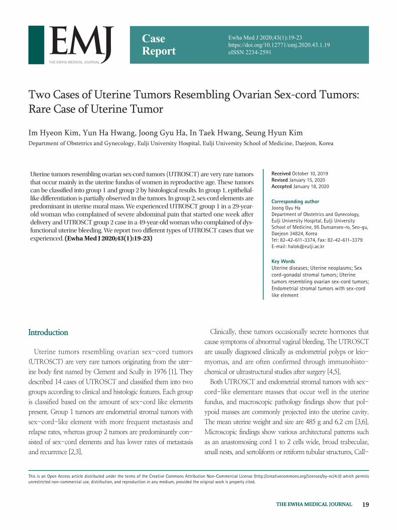
Two Cases of Uterine Tumors Resembling Ovarian Sex-cord Tumors: Rare Case of Uterine Tumor
Im Hyeon Kim, Yun Ha Hwang, Joong Gyu Ha, In Taek Hwang, Seung Hyun KimDepartment of Obstetrics and Gynecology, Eulji University Hospital, Eulji University School of Medicine, Daejeon, Korea
Case Report
Ewha Med J 2020;43(1):19-23https://doi.org/10.12771/emj.2020.43.1.19eISSN 2234-2591
Uterine tumors resembling ovarian sexcord tumors (UTROSCT) are very rare tumors that occur mainly in the uterine fundus of women in reproductive age. These tumors can be classified into group 1 and group 2 by histological results. In group 1, epitheliallike differentiation is partially observed in the tumors. In group 2, sexcord elements are predominant in uterine mural mass. We experienced UTROSCT group 1 in a 29yearold woman who complained of severe abdominal pain that started one week after delivery and UTROSCT group 2 case in a 49yearold woman who complained of dysfunctional uterine bleeding. We report two different types of UTROSCT cases that we experienced. (Ewha Med J 2020;43(1):19-23)
Received October 10, 2019Revised January 15, 2020Accepted January 18, 2020
Corresponding authorJoong Gyu HaDepartment of Obstetrics and Gynecology, Eulji University Hospital, Eulji University School of Medicine, 95 Dunsanseo-ro, Seo-gu, Daejeon 34824, KoreaTel: 82-42-611-3374, Fax: 82-42-611-3379E-mail: [email protected]
Key WordsUterine diseases; Uterine neoplasms; Sex cord-gonadal stromal tumors; Uterine tumors resembling ovarian sex-cord tumors; Endometrial stromal tumors with sex-cord like element
This is an Open Access article distributed under the terms of the Creative Commons Attribution Non-Commercial License (http://creativecommons.org/licenses/by-nc/4.0) which permits unrestricted non-commercial use, distribution, and reproduction in any medium, provided the original work is properly cited.
Introduction
Uterine tumors resembling ovarian sex-cord tumors
(UTROSCT) are very rare tumors originating from the uter-
ine body first named by Clement and Scully in 1976 [1]. They
described 14 cases of UTROSCT and classified them into two
groups according to clinical and histologic features. Each group
is classified based on the amount of sex-cord like elements
present. Group 1 tumors are endometrial stromal tumors with
sex-cord-like element with more frequent metastasis and
relapse rates, whereas group 2 tumors are predominantly con-
sisted of sex-cord elements and has lower rates of metastasis
and recurrence [2,3].
Clinically, these tumors occasionally secrete hormones that
cause symptoms of abnormal vaginal bleeding. The UTROSCT
are usually diagnosed clinically as endometrial polyps or leio-
myomas, and are often confirmed through immunohisto-
chemical or ultrastructural studies after surgery [4,5].
Both UTROSCT and endometrial stromal tumors with sex-
cord-like elementare masses that occur well in the uterine
fundus, and macroscopic pathology findings show that pol-
ypoid masses are commonly projected into the uterine cavity.
The mean uterine weight and size are 485 g and 6.2 cm [3,6].
Microscopic findings show various architectural patterns such
as an anastomosing cord 1 to 2 cells wide, broad trabeculae,
small nests, and sertoliform or retiform tubular structures, Call-
20 THE EWHA MEDICAL JOURNAL
Kim IH, et al
Exner-like bodies and diffuse sheets of uniform granulosa cell
tumor-like area. Neoplastic cells mainly have round to ovoid
nuclei, little nuclear pleomorphism, inconspicuous nucleoli,
scant, indistinct eosinophilic cytoplasm and are small in size
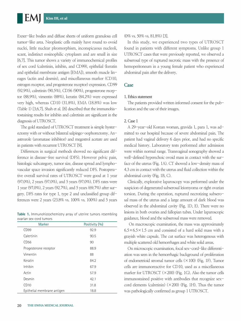
[6,7]. This tumor shows a variety of immunochemical profiles
of sex cord (calretinin, inhibin, and CD99), epithelial (keratin
and epithelial membrane antigen [EMA]), smooth muscle lin-
eages (actin and desmin), and miscellaneous marker (CD10,
estrogen receptor, and progesterone receptor) expression. CD99
(92.9%), calretinin (90.5%), CD56 (90%), progesterone recep-
tor (88.9%), vimentin (88%), keratin (84.2%) were expressed
very high, whereas CD10 (31.8%), EMA (18.8%) was low
(Table 1) [3,6,7]. Shah et al. [8] described that the immunohis-
tostaining results for inhibin and calretinin are significant in the
diagnosis of UTROSCT.
The gold standard of UTROSCT treatment is simple hyster-
ectomy with or without bilateral salpingo-oophorectomy. An-
astrozole (aromatase inhibitor) and megestrol acetate are used
in patients with recurrent UTROSCT [9].
Differences in surgical methods showed no significant dif-
ference in disease-free survival (DFS). However pelvic pain,
histologic subcategory, tumor size, disease spread and lympho-
vascular space invasion significantly reduced DFS. Postopera-
tive overall survival rates of UTROSCT were good at 1 year
(97.0%), 2 years (97.0%), and 5 years (97.0%). DFS rates were
1 year (97.0%), 2 years (92.7%), and 5 years (69.7%) after sur-
gery. DFS rates for type 1, type 2 and unclassified group dif-
ferences were 2 years (23.8% vs. 100% vs. 100%) and 5 years
(0% vs. 50% vs. 81.8%) [3].
In this study, we experienced two types of UTROSCT
found in patients with different symptoms. Unlike group 1
UTROSCT cases that were previously reported, we observed a
subserosal type of ruptured necrotic mass with the presence of
hemoperitoneum in a young female patient who experienced
abdominal pain after the delivery.
Case
1. Ethics statement
The patients provided written informed consent for the pub-
lication and the use of their images.
2. Case 1
A 29-year-old Korean woman, gravida 1, para 1, was ad-
mitted to our hospital because of severe abdominal pain. The
patient had vaginal delivery 6 days prior, and had no specific
medical history. Laboratory tests performed after admission
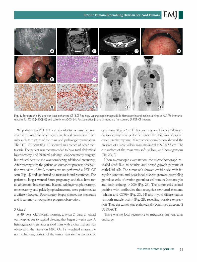
were within normal range. Transvaginal sonography showed a
well-defined hyperechoic ovoid mass in contact with the sur-
face of the uterus (Fig. 1A). CT showed a low-density mass of
4.3 cm in contact with the uterus and fluid collection within the
abdominal cavity (Fig. 1B, C).
Clinically, explorative laparoscopy was performed under the
suspicion of degenerated subserosal leiomyoma or right ovarian
torsion. During the operation, ruptured necrotizing subsero-
sal mass of the uterus and a large amount of dark blood was
observed in the abdominal cavity (Fig. 1D, E). There were no
lesions in both ovaries and fallopian tubes. Under laparoscopic
guidance, blood and the subserosal mass were removed.
On macroscopic examination, the mass was approximately
6.5×6.5×1.5 cm and consisted of a hard solid mass with a
grayish white capsule. The cut surface was heterogenous with
multiple scattered old hemorrhages and white solid areas.
On microscopic examination, focal sex-cord-like differenti-
ation was seen in the hemorrhagic background of proliferation
of endometrioid stromal tumor cells (×100) (Fig. 1F). Tumor
cells are immunoreactive for CD10, used as a miscellaneous
marker for UTROSCT (×200) (Fig. 1G). Also the tumor cells
immunostained positive with antibodies that recognize sex-
cord elements (calretinin) (×200) (Fig. 1H). Thus the tumor
was pathologically confirmed as group 1 UTROSCT.
Table 1. Immunohistochemistry array of uterine tumors resembling ovarian sex-cord tumors
Marker Positivity (%)CD99 92.9
Calertinin 90.5
CD56 90
Progesterone receptor 88.9
Vimentin 88
Keratin 84.2
Inhibin 67.9
Actin 57.9
Desmin 42.1
CD10 31.8
Epithelial membrane antigen 18.8
21THE EWHA MEDICAL JOURNAL
Uterine Tumors Resembling Ovarian Sex-cord Tumors
We performed a PET-CT scan in order to confirm the pres-
ence of metastasis to other organs in clinical correlation to re-
sults such as rupture of the mass and pathologic examination.
The PET-CT scan (Fig. 1I) showed an absence of other me-
tastasis. The patient was recommended to have total abdominal
hysterectomy and bilateral salpingo-oophorectomy surgery,
but refused because she was considering additional pregnancy.
After meeting with the patient, an outpatient progress observa-
tion was taken. After 3 months, we re-performed a PET-CT
scan (Fig. 1J) and confirmed no metastasis and recurrence. The
patient no longer wanted future pregnancy, and thus, have to-
tal abdominal hysterectomy, bilateral salpingo-oophorectomy,
omentectomy, and pelvic lymphadenectomy were performed at
a different hospital. Post-surgery biopsy showed no metastasis
and is currently on outpatient progress observation.
3. Case 2
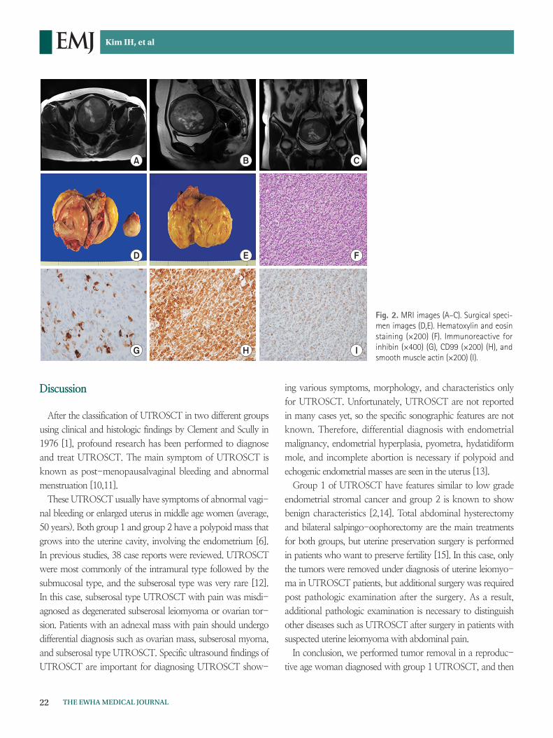
A 49-year-old Korean woman, gravida 2, para 2, visited
our hospital due to vaginal bleeding that began 3 weeks ago. A
heterogeneously enhancing solid mass with a clear margin was
observed in the uterus on MRI. On T2-weighted images, the
non-enhancing portion of the tumor was seen as necrotic or
cystic tissue (Fig. 2A-C). Hysterectomy and bilateral salpingo-
oophorectomy were performed under the diagnosis of degen-
erated uterine myoma. Macroscopic examination showed the
presence of a large yellow mass measured as 9.0×7.5 cm. The
cut surface of the mass was soft, yellow, and homogeneous
(Fig. 2D, E).
Upon microscopic examination, the microphotograph re-
vealed cord-like, trabecular, and nested growth patterns of
epithelioid cells. The tumor cells showed ovoid nuclei with ir-
regular contours and occasional nuclear grooves, resembling
granulosa cells of ovarian granulosa cell tumors (hematoxylin
and eosin staining, ×200) (Fig. 2F). The tumor cells stained
positive with antibodies that recognize sex-cord elements
(inhibin and CD99) (Fig. 2G, H) and myoid differentiation
(smooth muscle actin) (Fig. 2I), revealing positive expres-
sion. Thus the tumor was pathologically confirmed as group 2
UTROSCT.
There was no local recurrence or metastasis one year after
discharge.
A B C D
E F
M
G H I J
Fig. 1. Sonographic (A) and contrast-enhanced CT (B,C) findings. Laparoscopic images (D,E). Hematoxylin and eosin staining (×100) (F). Immuno-reactive for CD10 (×200) (G) and calretinin (×200) (H). Postoperative (I) and 3 months after surgery (J) PET-CT images.
22 THE EWHA MEDICAL JOURNAL
Kim IH, et al
Discussion
After the classification of UTROSCT in two different groups
using clinical and histologic findings by Clement and Scully in
1976 [1], profound research has been performed to diagnose
and treat UTROSCT. The main symptom of UTROSCT is
known as post-menopausalvaginal bleeding and abnormal
menstruation [10,11].
These UTROSCT usually have symptoms of abnormal vagi-
nal bleeding or enlarged uterus in middle age women (average,
50 years). Both group 1 and group 2 have a polypoid mass that
grows into the uterine cavity, involving the endometrium [6].
In previous studies, 38 case reports were reviewed. UTROSCT
were most commonly of the intramural type followed by the
submucosal type, and the subserosal type was very rare [12].
In this case, subserosal type UTROSCT with pain was misdi-
agnosed as degenerated subserosal leiomyoma or ovarian tor-
sion. Patients with an adnexal mass with pain should undergo
differential diagnosis such as ovarian mass, subserosal myoma,
and subserosal type UTROSCT. Specific ultrasound findings of
UTROSCT are important for diagnosing UTROSCT show-
ing various symptoms, morphology, and characteristics only
for UTROSCT. Unfortunately, UTROSCT are not reported
in many cases yet, so the specific sonographic features are not
known. Therefore, differential diagnosis with endometrial
malignancy, endometrial hyperplasia, pyometra, hydatidiform
mole, and incomplete abortion is necessary if polypoid and
echogenic endometrial masses are seen in the uterus [13].
Group 1 of UTROSCT have features similar to low grade
endometrial stromal cancer and group 2 is known to show
benign characteristics [2,14]. Total abdominal hysterectomy
and bilateral salpingo-oophorectomy are the main treatments
for both groups, but uterine preservation surgery is performed
in patients who want to preserve fertility [15]. In this case, only
the tumors were removed under diagnosis of uterine leiomyo-
ma in UTROSCT patients, but additional surgery was required
post pathologic examination after the surgery. As a result,
additional pathologic examination is necessary to distinguish
other diseases such as UTROSCT after surgery in patients with
suspected uterine leiomyoma with abdominal pain.
In conclusion, we performed tumor removal in a reproduc-
tive age woman diagnosed with group 1 UTROSCT, and then
A B C
D E F
G H I
Fig. 2. MRI images (A–C). Surgical speci-men images (D,E). Hematoxylin and eosin staining (×200) (F). Immunoreactive for inhibin (×400) (G), CD99 (×200) (H), and smooth muscle actin (×200) (I).
23THE EWHA MEDICAL JOURNAL
Uterine Tumors Resembling Ovarian Sex-cord Tumors
performed additional surgery. Also a woman with abnormal
vaginal bleeding diagnosed as group 1 UTROSCT underwent
curative surgical treatment. Like our case, the patient may be
diagnosed with UTROSCT after surgery and may need ad-
ditional treatment or evaluation. Therefore, it is essential to
confirm pathologic results in patients clinically diagnosed with
leiomyoma. If the patients have symptoms such as abnormal
vaginal bleeding and abdominal pain with uterine mass, the
possibility of UTROSCT should also be considered.
Furthermore, because of the rarity, preoperative radiologic
and laboratory diagnostic tools for UTROSCT are yet to be
established. Therefore, we conclude that further researches are
needed to improve the rate of diagnosis.
References
1. Clement PB, Scully RE. Uterine tumors resembling ovarian sexcord tumors: a linicopathologic analysis of fourteen cases. Am J Clin Pathol 1976;66:512525.
2. Stefanovic A, Jeremic K, Kadija S, Mitrovic M, Filimonovic D, JankovicRaznatovic S, et al. Uterine tumor resembling ovarian sex cord tumor. Case report and review of literature. Eur J Gynaecol Oncol 2013;34:275277.
3. Blake EA, Sheridan TB, Wang KL, Takiuchi T, Kodama M, Sawada K, et al. Clinical characteristics and outcomes of uterine tumors resembling ovarian sexcord tumors (UTROSCT): a systematic review of literature. Eur J Obstet Gynecol Reprod Biol 2014;181:163170.
4. Carta G, Crisman G, Margiotta G, Mastrocola N, Di Fonso A, Coletti G. Uterine tumors resembling ovarian sex cord tumors. A case report. Eur J Gynaecol Oncol 2010;31:456458.
5. Giordano G, Lombardi M, Brigati F, Mancini C, Silini EM. Clini
copathologic feature of 2 new cases of uterine tumors resembling ovarian sex cord tumors. Int J Gynecol Pathol 2010;29:459467.
6. Czernobilsky B. Uterine tumors resembling ovarian sex cord tumors: an update. Int J Gynecol Pathol 2008;27:229235.
7. Pradhan D, Mohanty SK. Uterine tumors resembling ovarian sex cord tumors. Arch Pathol Lab Med 2013;137:18321836.
8. Shah VI, Freites ON, Maxwell P, McCluggage WG. Inhibin is more specific than calretinin as an immunohistochemical marker for differentiating sarcomatoid granulosa cell tumour of the ovary from other spindle cell neoplasms. J Clin Pathol 2003;56:221224.
9. Blinman P, Tattersall MH. A case of uterine tumour resembling ovarian sex cord tumour responding to secondline, single agent anastrazole. Intern Med J 2009;39:617619.
10. O'Meara AC, Giger OT, Kurrer M, Schaer G. Case report: recurrence of a uterine tumor resembling ovarian sexcord tumor. Gynecol Oncol 2009;114:140142.
11. Suzuki C, Matsumoto T, Fukunaga M, Itoga T, Furugen Y, Kurosaki Y, et al. Uterine tumors resembling ovarian sexcord tumors producing parathyroid hormonerelated protein of the uterine cervix. Pathol Int 2002;52:164168.
12. Hauptmann S, Nadjari B, Kraus J, Turnwald W, Dietel M. Uterine tumor resembling ovarian sexcord tumor: a case report and review of the literature. Virchows Arch 2001;439:97101.
13. Franco A, Aquino NM, Malik SL, Navarro C. Sonographic presentation of uterine sex cordstromal tumor. J Clin Ultrasound 1999;27:199201.
14. Umeda S, Tateno M, Miyagi E, Sakurai K, Tanaka R, Tateishi Y, et al. Uterine tumors resembling ovarian sex cord tumors (UTROSCT) with metastasis: clinicopathological study of two cases. Int J Clin Exp Pathol 2014;7:10511059.
15. Hillard JB, Malpica A, Ramirez PT. Conservative management of a uterine tumor resembling an ovarian sex cordstromal tumor. Gynecol Oncol 2004;92:347352.


![UTERINE AND OVARIAN SARCOMAS: CLINICAL AND ... · Keywords: uterine sarcoma, ovarian sarcoma INTRODUCTION Sarcomas account for about 5% of uterine neo-plasms [1]. The most common](https://img.pdfslide.net/doc/110x75/5f8cf10c47e4f35e95087e30/uterine-and-ovarian-sarcomas-clinical-and-keywords-uterine-sarcoma-ovarian.jpg)