Embed Size (px)
Citation preview

Chehade et al. BMC Nephrology 2013, 14:65http://www.biomedcentral.com/1471-2369/14/65
CASE REPORT Open Access
Two new families with hereditary minimal changediseaseHassib Chehade1, Francois Cachat1, Eric Girardin1, Samuel Rotman2, Antonio Jorge Correia3,Florence Fellmann4 and Olivier Bonny5*
Abstract
Background: Steroid-sensitive idiopathic nephrotic syndrome (SSINS) is most often encountered in sporadic casesof minimal change disease (MCD). Only rare cases of familial forms of MCD have been reported and most of themonly in one generation. The scarcity of data has precluded unraveling the underlying genetic defect and candidategene approaches have been unsuccessful. Here we report two families with related SSINS cases and review therelated literature.
Case presentation: Two siblings and a cousin (first family), and a father and his son (second family), are reportedwith SSINS due to MCD. Patients have been followed up for more than 12 years and a renal biopsy was performedin three cases, demonstrating typical features of MCD. The course of the disease was remarkable because of severalrelapses treated with steroids. In three cases, mycophenolate mofetil or cyclosporine was added.
Conclusion: Familial SSINS due to MCD is extremely rare and no genetic defect has been identified so far.Reporting cases of hereditary MCD will allow further genetic studies which will ultimately help unravel themolecular basis of this disease.
Keywords: Nephrotic syndrome, Minimal change disease, Heredity, Genetics, Steroids
BackgroundIdiopathic nephrotic syndrome (INS) in children iscaused by various entities that differ in their histopatho-logical forms and their clinical course [1]. Minimalchange disease (MCD) and focal segmental glome-rulosclerosis (FSGS) are the most common causes ofINS representing 80% and 20% of the cases respectively[1]. The clinical outcome of INS is determined by theresponsiveness to treatment by steroids. Most steroid-sensitive INS (SSINS) are due to MCD, while steroid-resistant INS (SRINS) are mostly represented by FSGS.Although INS is well known as a sporadic disease, fami-
lial occurrences with autosomal dominant or recessivemode of inheritance have been described especially inFSGS forms [2]. Several genes have been associated withor shown to be causative for some specific forms of INS,mostly steroid-resistant, including NPHS1, NPHS2, PLCE1,WT1, ACTN4, TRPC6, CD2AP, APOL1 or INF2 [3]. How-
* Correspondence: [email protected] of Nephrology, Lausanne University Hospital, Lausanne, SwitzerlandFull list of author information is available at the end of the article
© 2013 Chehade et al.; licensee BioMed CentrCommons Attribution License (http://creativecreproduction in any medium, provided the or
ever, reports of familial MCD are scarce and no causalgene has been identified yet. Here, we describe five casesof steroid sensitive MCD in two non-consanguineous fa-milies and perform a review of the literature. The objectiveof our report is to encourage physicians to identify andcharacterize genetic causes of MCD. This may help tounderstand more precisely the pathophysiological mecha-nisms of INS and provide a first step toward the identifica-tion of the underlying genetic cause.
Cases presentationFamily 1We describe a Portuguese non-consanguineous family(see pedigree: Figure 1) in which two siblings (cases 1 and2) and one cousin (case 3) were diagnosed with MCDINS.
Case 1A 6 year old boy, with no previous medical history,presented with fatigue and facial edema. Physical exam-ination showed moderate periorbital edema. Blood pres-sure was within normal range for age (105/69 mmHg).
al Ltd. This is an Open Access article distributed under the terms of the Creativeommons.org/licenses/by/2.0), which permits unrestricted use, distribution, andiginal work is properly cited.

7
2 7
n
n n
1 2
3
Figure 1 Extensive pedigree of the first family. Three membersof this Portuguese non-consanguineous family were affected bysteroid-sensitive nephrotic syndrome, type minimal change disease:two affected siblings (cases 1 and 2) and the first cousin onceremoved (case 3). The index case (case 1) is indicated by an arrowhead. The numbers inside figures indicate the number of males(square), females (rounds) or non-specified sex (lozenges) ofthe family.
Chehade et al. BMC Nephrology 2013, 14:65 Page 2 of 7http://www.biomedcentral.com/1471-2369/14/65
Heart sounds and lung examination were unremarkable.Abdomen was soft, not distended, and no mass, shiftingdullness or hepatosplenomegaly were found. He had nor-mal male genitalia with mild scrotal edema. The dorsalsurfaces of hands and feet had mild pitting edema. Urineanalysis by dipstick showed 4+ proteinuria with no he-maturia. Urine spot showed a nephrotic range proteinuria(protein/creatinine ratio of 2000 g/mol). The blood che-mistry panel was remarkable for plasma protein level of35 g/l and serum albumin of 10 g/l. BUN and creatininelevels were normal, and no electrolyte disturbance wasnoted. The diagnosis of INS was posed and the patientwas treated with oral prednisone (60 mg/m2/day b.i.d.).Feet edema and proteinuria gradually resolved over thecourse of treatment. He was followed up as outpatientand did monitor daily albuminuria with urine dipsticks.Corticosteroids were tapered off progressively andstopped. Three months later, the patient relapsed after aminor respiratory infection with re-appearance of protei-nuria. Treatment with steroids was re-initiated for twomonths. Eighteen months later, the patient remains com-pensated without steroids and has normal blood pressureand normal renal function.
Case 2The brother of case 1, a child with no previous medicalhistory, presented at 3 years of age with mild facial edemawithout any other clinical sign. Blood pressure was normal.Laboratory tests showed proteinuria of nephrotic range(protein/creatinine ratio 750 g/mol). Blood chemistryshowed low levels of total protein (45 g/l) and serum albu-min (18 g/l), but BUN and creatinine concentrations were
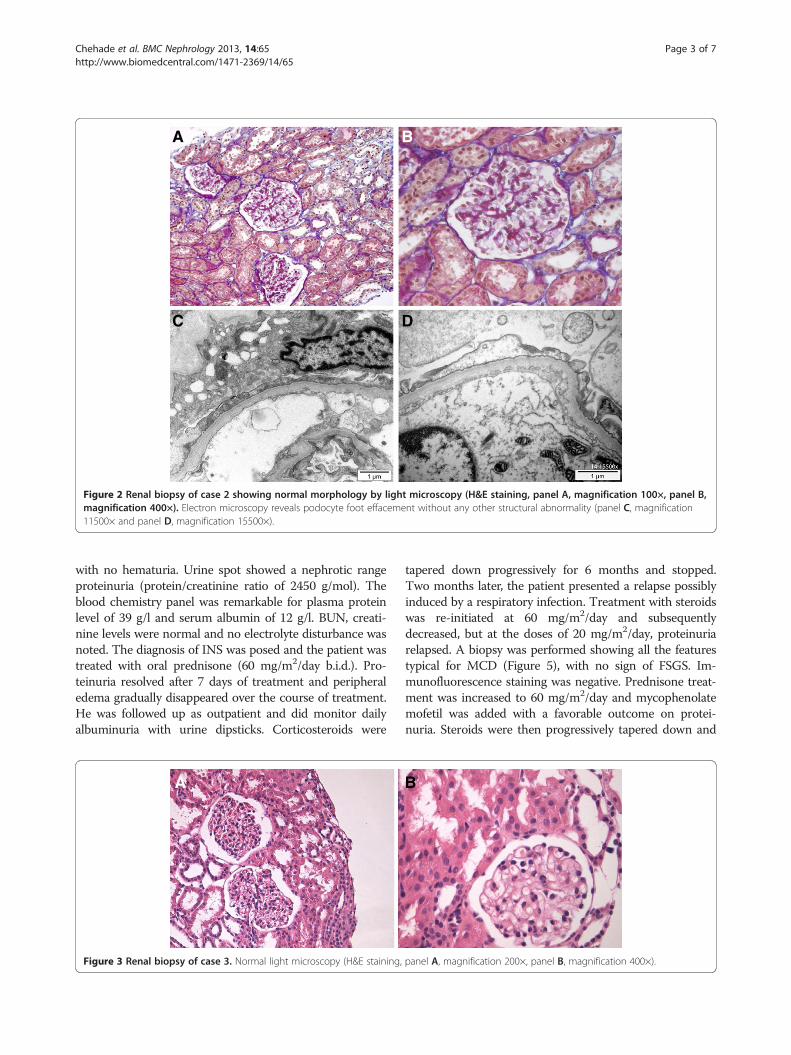
normal. INS was diagnosed and the infant was treated withoral prednisone 60 mg/m2/day b.i.d. He was followed upin the outpatient clinic and was monitoring proteinuriawith dipsticks every day. Edema and proteinuria graduallyresolved under treatment and steroids were tapered offwith an initial favorable course. However, 2 months afterthe interruption of the corticosteroids treatment, the pa-tient presented several relapses, all steroid-sensitive andevery time triggered by respiratory or gastro-intestinal viralinfections. A renal biopsy was performed and showed allthe typical features of MCD (Figure 2), but no sign ofFSGS. Immunofluorescence staining was negative. The pa-tient was treated with mycophenolate mofetil (MMF) inaddition to steroids with favorable outcome. Steroids werethen progressively tapered down and stopped.Asking for family history of the two siblings lead to
identification of a first cousin once removed with a historyof INS due to MCD (Figure 1). Of note, family history wasunremarkable for chronic kidney failure or renal graft.
Case 3This now 14 year old boy was initially diagnosed withnephrotic syndrome at the age of 2 and was successfullytreated with corticosteroids. He suffered from several re-lapses and was treated with oral cyclophosphamide. Arenal biopsy at age 3 shows normal morphology at lightmicroscopy, in particular no glomerulosclerosis andinterstitial fibrosis (Figure 3). No electronic microscopyhas been available. Another round of cyclophosphamidewas given at age 4 due to several relapses. At age 4.5,the child presented another relapse which was trea-ted with low dose corticosteroids and cyclosporine wasintroduced for a 12 month period. At age 8, anotherrelapse was treated with a full dose of corticosteroids(60 mg/m2/day) with favorable response and was main-tained afterwards at low dose on alternate days. A treat-ment by mycophenolate mofetil (600 mg/m2/day) wasintroduced. The course of the disease was since favor-able with rarer relapse episodes. At the last follow up(age 14), physical exam was normal with blood pressureof 111/65 mmHg and serum creatinine level was in thenormal range.
Family 2Here we describe a French non-consanguineous family(see pedigree: Figure 4) in which a father and his son(cases 4 and 5) were diagnosed with MCD INS.
Case 4A 4 year old boy, with no previous medical history,presented with periorbital edema. Blood pressure waswithin normal range for age (100/59 mmHg). He had mildedema of the dorsal surfaces of hands, feet and mild scrotaledema. Urine analysis by dipstick showed 4+ proteinuria
A B
DC
Figure 2 Renal biopsy of case 2 showing normal morphology by light microscopy (H&E staining, panel A, magnification 100×, panel B,magnification 400×). Electron microscopy reveals podocyte foot effacement without any other structural abnormality (panel C, magnification11500× and panel D, magnification 15500×).
Chehade et al. BMC Nephrology 2013, 14:65 Page 3 of 7http://www.biomedcentral.com/1471-2369/14/65
with no hematuria. Urine spot showed a nephrotic rangeproteinuria (protein/creatinine ratio of 2450 g/mol). Theblood chemistry panel was remarkable for plasma proteinlevel of 39 g/l and serum albumin of 12 g/l. BUN, creati-nine levels were normal and no electrolyte disturbance wasnoted. The diagnosis of INS was posed and the patient wastreated with oral prednisone (60 mg/m2/day b.i.d.). Pro-teinuria resolved after 7 days of treatment and peripheraledema gradually disappeared over the course of treatment.He was followed up as outpatient and did monitor dailyalbuminuria with urine dipsticks. Corticosteroids were
A
Figure 3 Renal biopsy of case 3. Normal light microscopy (H&E staining,
tapered down progressively for 6 months and stopped.Two months later, the patient presented a relapse possiblyinduced by a respiratory infection. Treatment with steroidswas re-initiated at 60 mg/m2/day and subsequentlydecreased, but at the doses of 20 mg/m2/day, proteinuriarelapsed. A biopsy was performed showing all the featurestypical for MCD (Figure 5), with no sign of FSGS. Im-munofluorescence staining was negative. Prednisone treat-ment was increased to 60 mg/m2/day and mycophenolatemofetil was added with a favorable outcome on protei-nuria. Steroids were then progressively tapered down and
B
panel A, magnification 200×, panel B, magnification 400×).

4
5
Figure 4 Pedigree of the second family. The index case isindicated by an arrow head.
A
C
Figure 5 Renal biopsy of case 4 shows normal light microscopy (H&E400×). Electron microscopy shows podocyte foot fusion (panel C, magnific
Chehade et al. BMC Nephrology 2013, 14:65 Page 4 of 7http://www.biomedcentral.com/1471-2369/14/65
stopped. Three months later, the patient remains compen-sated without steroids, has normal blood pressure and nor-mal renal function.
Case 5The 37 year old father of case 4 was hospitalized at theage of 10 for peripheral edema, nephrotic range protein-uria and hypoalbuminemia without renal failure or arte-rial hypertension. The diagnosis of nephrotic syndromewas made and treated with prednisone that was taperedoff progressively over 6 months. The evolution was thenfavorable without relapses. At present time, the patientdisplays normal renal function, normal blood pressure of120/78 mmHg and no proteinuria (protein/creatinineratio: 9 g/mol).
ConclusionIn the past years, many familial FSGS cases have beenreported and genetic studies have identified mutations inseveral genes coding for proteins of the slit diaphragmcomplex and the podocyte which leads to autosomalrecessive (NPHS1, NPHS2) or autosomal dominant(ACTN4, CD2AP, TRPC6 genes) steroid-resistant FSGS[1,4-6]. Identification of genes related to FSGS has con-tributed significantly to a better understanding of themolecular paths involved in SRINS and is an importantdeterminant for the course of the disease. For instance,relapses in renal transplant recipient carrying FSGS
B
D
staining, panel A, magnification 200×, panel B, magnificationation 6600× and panel D, magnification D, 11500×).

Table 1 Reported siblings with SSINS
References Year Familial cases with SSINS Histopathological confirmation of MCD
Roy S et al. [10] 1971 Identical twins Biopsy performed in 2 cases
Moncrieff MW et al. [11] 1973 18 cases in 9 families Biopsy performed in 12 cases
White RH et al. [12] 1973 12 cases from 24 centers in Europe Biopsy performed in 12 cases
Bader BI et al. [13] 1974 1 affected sibling pairs Biopsy performed in 1 case
McEnery PT et al. [14] 1989 2 cases in a family Data not available
Awadalla NB et al. [15] 1989 3 cases in a family Biopsy performed in 3 cases
Fuchshuber A et al. [7] 2001 32 cases in 15 families Biopsy performed in 12 cases
Ruf RG et al. [9] 2003 7 cases in 3 families Biopsy performed in 2 cases
Landau D et al. [8] 2007 6 cases in 2 related families No biopsy performed
Roberts IS et al. [16] 2008 2 cases in a family Biopsy performed in one case
Motoyama O et al. [17] 2009 2 cases in a family No biopsy performed
Chehade et al. BMC Nephrology 2013, 14:65 Page 5 of 7http://www.biomedcentral.com/1471-2369/14/65
genes mutations are rare as compared to FSGS kidneytransplant recipients without any gene mutation [3]. Butwhile important genetic clues have been identified for fa-milial steroid-resistant INS and FSGS, reports of genescausative for familial steroid sensitive INS and MCD arestill lacking. This might be due to the low prevalence ofthe disease and to the few numbers of cases described sofar. Here, we report two novel families with 5 cases ofMCD. We encourage clinicians to report their cases inorder to collect enough families to conduct geneticstudies.Literature review (using PubMed Advanced Search
Builder, date: 1960–1980 with the following key words:familial nephrotic syndrome, and the date: 1980–2011using the following key words: familial minimal changedisease, familial nephrotic syndrome) of familial cases ofSSINS revealed several reported cases within siblings(Table 1) and only sixteen families with SSINS affectingtwo generations (Table 2). Several interesting featurestaken from these reports may help in managing thesecases. In their report of fifteen families with childhood-
Table 2 Reported familial cases with SSINS in two generation
References Year Familial cases with SSINS in twogenerations
White RH et al. [12] 1973 A father and his daughter
Bader BI et al. [13] 1974 2 affected first cousins from aconsanguineous family
McEnery PT et al. [14] 1989 A father and his son
Two families with 2 affected first co
Awadalla NB et al. [15] 1989 3 cases in a family and a cousin
Landau D et al. [8] 2007 - 2 families with parent/child affecte
- 2 Bedouin consanguineous family14 affected members
- 5 non-related Bedouin families witaffected members
Motoyama O et al. [17] 2009 A father and a daughter
onset SSINS, Fuchshuber et al. [7] reported that the clin-ical course of the familial forms was equivalent to sporadicSSINS cases. A strong heritability of the age of onset ofthe disease was suggested. In this first large report of fa-milial SSINS, linkage with the candidate gene NPHS2 wasexcluded and the authors concluded the existence of adistinct gene locus for familial SSINS. Landau et al. [8]reported on several extensive Bedouin families affected bySSINS with similar clinical course - in terms of age of on-set, male predominance and spontaneous cure at puberty -compared to those in sporadic cases. By linkage analysis,the authors showed a complete absence of linkage withthe usual candidate genes loci implicated in nephrotic syn-drome or other glomerulopathies and they advised formore specific genome-wide screening with a densermarker set. In three families with SSINS, Ruf et al. [9] wereable to pinpoint a locus on chromosome 2p12-p13.2, andalso demonstrated clear evidence for genetic locus hetero-geneity upon examination of ten additional families withSSINS. The rare cases of familial SSINS reported in the lit-erature confirm that the disease course is similar to
s
Histopathological confirmation of MCD
Biopsy of the daughter only
Biopsy performed in both cases
usins Data not available
Biopsy performed in all 3 cases
d Biopsy performed in one case of the 14 affected Bedouinconsanguineous family members
with
h 10
No biopsy performed

Chehade et al. BMC Nephrology 2013, 14:65 Page 6 of 7http://www.biomedcentral.com/1471-2369/14/65
sporadic cases of SSINS, but clearly distinct from familialFSGS nephrotic syndrome.Cases of familial SSINS spread over two generations
have rarely been described (see list in Table 2). Outcome,in terms of renal function and blood pressure, is usuallyfavorable [7] compared to familial FSGS [18-20].As the majority of familial cases of SSINS reported in
the literature is limited to one generation of siblings(Table 1), the first genetic inheritance pattern suggestedwas autosomal recessive or a possible germinal mosai-cism. However, description of familial SSINS cases intwo generations (Table 2) with transmission from fatherto children broadens the disease inheritance possibilitiesto autosomal dominant transmission model with variablepenetrance. Altogether, analysis of the data issued fromthe literature does not allow definitive conclusions aboutthe inheritance pattern of familial MCD and is permis-sive for different possible transmission hypothesis, in-cluding autosomal recessive, autosomal dominant withvariable penetrance or genetic heterogeneity. In addition,a more complex inheritance pattern associated witholigogenic predisposition and possible environmental ef-fects is also possible. More reports of familial MCD areneeded in order to understand the disease transmissionpattern.In this report, case 1 presented with typical INS at age
six and the follow-up was marked by a single relapse oc-curring three months after the interruption of the steroids.Renal biopsy was not performed in that case due to rapidfavorable outcome. The second case presented with INS atthe age of 3 with an initial favorable disease course, latercomplicated by frequent relapses. A renal biopsy confirmedthe diagnosis of MCD. The child eventually showed a fa-vorable evolution after the introduction of MMF. Case 3, afirst cousin once removed, presented with a classical INSat age 6 and the renal biopsy showed typical MCD. Despitetreatment with corticosteroids, frequent relapses were ob-served and treated with cyclophosphamide, cyclosporine,and finally, MMF. These 3 cases had normal renal function(estimated GFR using the revised Schwartz formula were96, 97 and 99 ml/min 1.73 m2 respectively) and blood pres-sure. Pedigree of this family is compatible with an auto-somal dominant inheritance with variable penetrance, butother forms of heritability are possible. A de novo mutationin this family of two affected siblings and their cousinseems less probable though. In the second reported family,renal outcome was also favorable and the pedigree is com-patible with an autosomal dominant inheritance. Overall,all familial cases of MCD reported here had a clinical pres-entation in terms of age of onset, symptoms during the ini-tial phase, renal morphology and outcome close to otherfamilial cases described in the literature.We are aware of this report’s few limitations. First, it is
descriptive and does not propose precise genetic or mo-
lecular mechanisms that could explain familial MCD.However, this publication is meant to encourage furtherreports of similar rare cases that, once collected, mayallow wider genetic analysis. Second, recent reports havesuggested a role of CD80 induction in the occurrence ofsporadic MCD [21]. We did not dose soluble CD80 inthe urine of the patients presented here and thereforecould not conclude about the possible value of this bio-marker and this proposed pathophysiological mechanismfor familial MCD.In summary, here we describe five cases issued from
two families with steroid sensitive INS occurring in twogenerations. The clinical course of these cases was simi-lar to sporadic INS regarding the age of onset, clinicalpresentation and the presence of minor infection priorto the onset of recurrences, response to treatment anddisease outcome. This confirms the few previous ob-servations of familial MCD reported in the literature.The aim of this paper is to emphasize the importance ofidentifying these families in order to allow further gene-tic analysis, determine mode of inheritance and under-stand the mechanisms of INS appearance.
ConsentWritten informed consent was obtained from the patientfor publication of this Case report and any accompany-ing images. A copy of the written consent is available forreview by the Editor of this journal.
Competing interestNo part of this manuscript has been previously published. The authorsdeclare they have no conflict of interest related to this manuscript.
Authors’ contributionsHC, FC, EG, AJC treated and followed the patients. SR provided thehistopathological analysis. FF provided genetic counsels. OB and HCprepared the manuscript. All authors read and approved the finalmanuscript.
AcknowledgementsWe are thankful to the different members of the families studied here andthe members of the “Groupe des Maladies Rénales Génétiques” of theLausanne University Hospital. OB is the recipient of a Swiss NationalFoundation professorship grant PP00P3_133648.
Author details1Division of Pediatric Nephrology of West Switzerland, Lausanne UniversityHospital, Lausanne, Switzerland. 2Department of Pathology, LausanneUniversity Hospital, Lausanne, Switzerland. 3Pediatric Nephrology, Children’sHospital Coimbra, Coimbra, Portugal. 4Service of Medical Genetics, LausanneUniversity Hospital, Lausanne, Switzerland. 5Service of Nephrology, LausanneUniversity Hospital, Lausanne, Switzerland.
Received: 11 December 2012 Accepted: 15 March 2013Published: 22 March 2013
References1. Eddy AA, Symons JM: Nephrotic syndrome in childhood. Lancet 2003,
362(9384):629–639.2. Gbadegesin R, Lavin P, Foreman J, Winn M: Pathogenesis and therapy of
focal segmental glomerulosclerosis: an update. Pediatr Nephrol 2011,26(7):1001–1520.

Chehade et al. BMC Nephrology 2013, 14:65 Page 7 of 7http://www.biomedcentral.com/1471-2369/14/65
3. Shimizu A, Higo S, Fujita E, Mii A, Kaneko T: Focal segmentalglomerulosclerosis after renal transplantation. Clin Transplant 2011,25(Suppl 23):6–14.
4. Salomon R, Gubler MC, Niaudet P: Genetics of the nephrotic syndrome.Curr Opin Pediatr 2000, 12(2):129–134.
5. Kaplan JM, Kim SH, North KN, Rennke H, Correia LA, Tong HQ, Mathis BJ,Rodríguez-Pérez JC, Allen PG, Beggs AH, Pollak MR: Mutations in ACTN4,encoding alpha-actinin-4, cause familial focal segmentalglomerulosclerosis. Nat Genet 2000, 24(3):251–256.
6. Lahdenkari AT, Suvanto M, Kajantie E, Koskimies O, Kestilä M, Jalanko H:Clinical features and outcome of childhood minimal change nephroticsyndrome: is genetics involved? Pediatr Nephrol 2005, 20(8):1073–1080.
7. Fuchshuber A, Gribouval O, Ronner V, Kroiss S, Karle S, Brandis M,Hildebrandt F: Clinical and genetic evaluation of familial steroid-responsive nephrotic syndrome in childhood. J Am Soc Nephrol 2001,12(2):374–378.
8. Landau D, Oved T, Geiger D, Abizov L, Shalev H, Parvari R: Familial steroid-sensitive nephrotic syndrome in Southern Israel: clinical and geneticobservations. Pediatr Nephrol 2007, 22(5):661–669.
9. Ruf RG, Fuchshuber A, Karle SM, Lemainque A, Huck K, Wienker T, Otto E,Hildebrandt F: Identification of the first gene locus (SSNS1) for steroid-sensitive nephrotic syndrome on chromosome 2p. J Am Soc Nephrol2003, 14(7):1897–1900.
10. Roy S 3rd, Pitcock JA: Idiopathic nephrosis in identical twins. Am J DisChild 1971, 121(5):428–430.
11. Moncrieff MW, White RH, Glasgow EF, Winterborn MH, Cameron JS, Ogg CS:The familial nephrotic syndrome. II. A clinicopathological study. Clin Nephrol1973, 1(4):220–229.
12. White RH: The familial nephrotic syndrome. I. A European survey. Clin Nephrol1973, 1(4):215–219.
13. Bader PI, Grove J, Trygstad CW, Nance WE: Familial nephrotic syndrome.Am J Med 1974, 56(1):34–43.
14. McEnery PT, Welch TR: Major histocompatibility complex antigens insteroid-responsive nephrotic syndrome. Pediatr Nephrol 1989, 3(1):33–36.
15. Awadalla NB, Teebi AS, Elzouki AY, Shaltout A: Frequent relapser minimalchange nephrosis: an unrecognized X-linked disorder? Eur J Pediatr 1989,149(3):205–207.
16. Roberts IS, Gleadle JM: Familial nephropathy and multiple exostoses withexostosin-1 (EXT1) gene mutation. J Am Soc Nephrol 2008, 19(3):450–453.
17. Motoyama O, Sugawara H, Hatano M, Fujisawa T, Litaka K: Steroid-sensitivenephrotic syndrome in two families. Clin Exp Nephrol 2009, 13(2):170–173.
18. Branten AJ, van den Born J, Jansen JL, Assmann KJ, Wetzels JF: Familialnephropathy differing from minimal change nephropathy and focalglomerulosclerosis. Kidney Int 2001, 59(2):693–701.
19. Conlon PJ, Butterly D, Albers F, Rodby R, Gunnells JC, Howell DN: Clinicaland pathologic features of familial focal segmental glomerulosclerosis.Am J Kidney Dis 1995, 26(1):34–40.
20. Faubert PF, Porush JG: Familial focal segmental glomerulosclerosis: ninecases in four families and review of the literature. Am J Kidney Dis 1997,30(2):265–270.
21. Garin EH, Mu W, Arthur JM, Rivard CJ, Araya CE, Shimada M, Johnson RJ:Urinary CD80 is elevated in minimal change disease but not in focalsegmental glomerulosclerosis. Kidney Int 2010, 78(3):296–302.
doi:10.1186/1471-2369-14-65Cite this article as: Chehade et al.: Two new families with hereditaryminimal change disease. BMC Nephrology 2013 14:65.
Submit your next manuscript to BioMed Centraland take full advantage of:
• Convenient online submission
• Thorough peer review
• No space constraints or color figure charges
• Immediate publication on acceptance
• Inclusion in PubMed, CAS, Scopus and Google Scholar
• Research which is freely available for redistribution
Submit your manuscript at www.biomedcentral.com/submit