Embed Size (px)
Citation preview
Imaging AgentsDOI: 10.1002/anie.201104104
Ultrasmall Rigid Particles as Multimodal Probes for MedicalApplicationsFranÅois Lux, Anna Mignot, Pierre Mowat, C�dric Louis, Sandrine Dufort, Claire Bernhard,Franck Denat, Fr�d�ric Boschetti, Claire Brunet, Rodolphe Antoine, Philippe Dugourd,Sophie Laurent, Luce Vander Elst, Robert Muller, Lucie Sancey, V�ronique Josserand, Jean-Luc Coll, Vasile Stupar, Emmanuel Barbier, Chantal R�my, Alexis Broisat, Catherine Ghezzi,G�raldine Le Duc, St�phane Roux, Pascal Perriat,* and Olivier Tillement
Over the past two decades, nanoparticles have been devel-oped in the field of theranostic[1, 2] with the objective ofmeeting three requirements: 1) to exhibit long circulation inbody fluids with major accumulation in tumour tissues due to
their active or passive targeting properties (enhanced perme-ability and retention (EPR) effect);[3] 2) to be rapidlyeliminated through the renal route to ensure a sufficientdifference in concentration between healthy and diseasedzones; 3) to display therapeutic potential and contrast proper-ties.[4, 5] This latter requirement was reinforced by thesimultaneous development of devices combining imagingtechniques, such as 1) high-sensitive X-ray tomography,positron emission tomography, or single-photon emissioncomputed tomography and 2) magnetic resonance imaging(MRI) with high spatial resolution.[6, 7]
However, it is still a great challenge to ensure bothappropriate renal elimination (necessarily achieved usingparticles < 5.5 nm in size)[8] and multimodality (requiringmolecules that systematically enlarge the particle size to anon-adequate extent). For instance, quantum dots[9] or goldclusters[10] do not exhibit both features as they requirecoatings that are too prohibitive in size to ensure multi-modality. After reviewing the solutions proposed in scientificliterature,[11–13] the most promising strategies rely on theelaboration of silica- or polymeric-based structures thatincorporate different functional entities, such as dyes forfluorescence imaging, magnetic complexes for MRI, radio-active elements for scintigraphy or curie-therapy, heavyelements for interacting with X- or g-rays, neutron absorbersfor neutron-therapy or sensitizers for photodynamic therapy.Concerning multifunctional silica-based particles, even themost investigated technologies (i.e., Stçber or reverse emul-sion methods) failed to yield objects smaller than 10 nm insize.
Here we propose an original top-down method consistingin the fragmentation of sub-10 nm structures already possess-ing all the desired functions (see Supporting Information).Briefly, these starting structures consist of core (gadoliniumoxide)–shell (polysiloxane) particles developed by our groupwhich offer several features and functionalities, but are toolarge in size to escape hepatic clearance.[14] Gadolinium wasselected as contrast agent on account of its paramagneticproperties and because of its commercial use in approxi-mately 45% of all MRI analyses.[15] The starting structuresdisplayed an average core size of 3.5 nm and a shell thicknessof 0.5 nm. The fluorophore-encapsulated shell was renderedfunctionally active by modified 1,4,7,10-tetraazacyclodode-cane-1,4,7,10-tetraacetic acid (DOTA) ligands which are ableto chelate core gadolinium ions. In aqueous solutions, the
[*] Dr. A. Mignot, Prof. P. PerriatMat�riaux Ing�nierie et Science, INSA-Lyon, UMR 5510 CNRS,Universit� de Lyon, 69621 Villeurbanne Cedex (France)andCampus LyonTech La Doua, INSA-Lyon, B�timent Blaise Pascal7 avenue Jean Capelle, 69621 Villeurbanne cedex (France)E-mail: [email protected]
Dr. F. Lux, Dr. A. Mignot, Dr. P. Mowat, Prof. O. TillementLaboratoire de Physico-Chimie des Mat�riaux Luminescents, UMR5620 CNRS – Universit� Claude Bernard Lyon 1, Universit� de Lyon,69622 Villeurbanne Cedex (France)
Dr. A. Mignot, Dr. C. Louis, Dr. S. DufortNano-H SAS, 38070 Saint-Quentin Fallavier (France)
Dr. C. Bernhard, Prof. F. DenatInstitut de Chimie Mol�culaire de l’Universit� de Bourgogne, UMRCNRS 5260, Universit� de Bourgogne, 21078 Dijon Cedex (France)
Dr. F. BoschettiCheMatech, 21000 Dijon (France)
Dr. C. Brunet, Prof. R. Antoine, Prof. P. DugourdLaboratoire de Spectrom�trie Ionique et Mol�culaire, UMR CNRS5579, Universit� Claude Bernard Lyon 1, 69622 Villeurbanne Cedex(France)
Dr. S. Laurent, Prof. L. V. Elst, Prof. R. MullerUniv Mons, NMR & Mol Imaging Lab, Dept Gen Organ & BiomedChem, B-7000 Mons (Belgium)
Dr. L. Sancey, Dr. V. Josserand, Prof. J.-L. CollINSERM, CRI, U823, Inst Albert Bonniot, 38042 Grenoble 9 (France)
Dr. V. Stupar, Dr. E. Barbier, Dr. C. R�myUniversit� Grenoble 1, Grenoble Institut des Neurosciences, UMRS836, Grenoble (France)
Dr. V. Stupar, Dr. E. Barbier, Dr. C. R�myInserm, U826, Grenoble (France)
Dr. A. Broisat, Prof. C. GhezziLaboratoire radiopharmaceutique bioclinique, INSERM U877, Fac-ult� de m�decine de Grenoble, 38700 La Tronche (France)
Dr. G. Le DucEuropean Synchrotron Radiation Facility, ID 17 Biomedical Beamline,BP220, 38043 Grenoble (France)
Prof. S. RouxInstitut UTINAM, UMR 6213 CNRS – Universit� de Franche-Comt�,25030 BesanÅon Cedex (France)
Supporting information for this article is available on the WWWunder http://dx.doi.org/10.1002/anie.201104104.
12299Angew. Chem. Int. Ed. 2011, 50, 12299 –12303 � 2011 Wiley-VCH Verlag GmbH & Co. KGaA, Weinheim
ligands strongly accelerate the core dissolution leading to ahollow polysiloxane sphere. This latter collapses and frag-ments into small and rigid platforms (SRPs) of polysiloxane.These SRPs possess all the properties of the initial structure,bearing on their surface DOTA molecules that are partlychelated to dissolved gadolinium cations. DOTA was givenpreference over acyclic chelates, due to its higher complex-ation constant,[16] its lower kinetics that limit transmetallationwith endogen cations such as Ca2+ and reduced toxicity forpatients with severe renal dysfunction.[17,18] Our study aims toconfirm the relevance of this strategy 1) by reporting thechemical and size characterization of the SRPs obtained asdescribed above and 2) by demonstrating that they can bedetected in vivo by four different techniques, namely fluores-cence imaging, MRI, scintigraphy, and X-ray computedtomography.
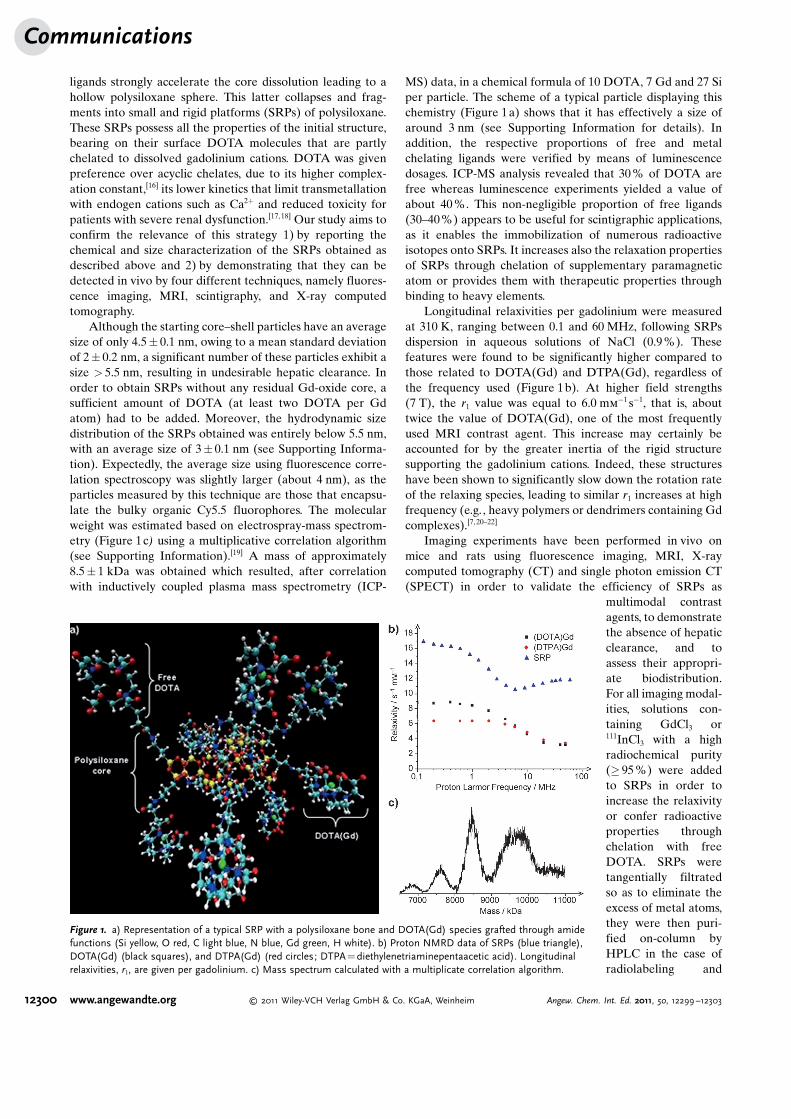
Although the starting core–shell particles have an averagesize of only 4.5� 0.1 nm, owing to a mean standard deviationof 2� 0.2 nm, a significant number of these particles exhibit asize > 5.5 nm, resulting in undesirable hepatic clearance. Inorder to obtain SRPs without any residual Gd-oxide core, asufficient amount of DOTA (at least two DOTA per Gdatom) had to be added. Moreover, the hydrodynamic sizedistribution of the SRPs obtained was entirely below 5.5 nm,with an average size of 3� 0.1 nm (see Supporting Informa-tion). Expectedly, the average size using fluorescence corre-lation spectroscopy was slightly larger (about 4 nm), as theparticles measured by this technique are those that encapsu-late the bulky organic Cy5.5 fluorophores. The molecularweight was estimated based on electrospray-mass spectrom-etry (Figure 1c) using a multiplicative correlation algorithm(see Supporting Information).[19] A mass of approximately8.5� 1 kDa was obtained which resulted, after correlationwith inductively coupled plasma mass spectrometry (ICP-
MS) data, in a chemical formula of 10 DOTA, 7 Gd and 27 Siper particle. The scheme of a typical particle displaying thischemistry (Figure 1a) shows that it has effectively a size ofaround 3 nm (see Supporting Information for details). Inaddition, the respective proportions of free and metalchelating ligands were verified by means of luminescencedosages. ICP-MS analysis revealed that 30 % of DOTA arefree whereas luminescence experiments yielded a value ofabout 40 %. This non-negligible proportion of free ligands(30–40%) appears to be useful for scintigraphic applications,as it enables the immobilization of numerous radioactiveisotopes onto SRPs. It increases also the relaxation propertiesof SRPs through chelation of supplementary paramagneticatom or provides them with therapeutic properties throughbinding to heavy elements.
Longitudinal relaxivities per gadolinium were measuredat 310 K, ranging between 0.1 and 60 MHz, following SRPsdispersion in aqueous solutions of NaCl (0.9 %). Thesefeatures were found to be significantly higher compared tothose related to DOTA(Gd) and DTPA(Gd), regardless ofthe frequency used (Figure 1b). At higher field strengths(7 T), the r1 value was equal to 6.0 mm
�1 s�1, that is, abouttwice the value of DOTA(Gd), one of the most frequentlyused MRI contrast agent. This increase may certainly beaccounted for by the greater inertia of the rigid structuresupporting the gadolinium cations. Indeed, these structureshave been shown to significantly slow down the rotation rateof the relaxing species, leading to similar r1 increases at highfrequency (e.g., heavy polymers or dendrimers containing Gdcomplexes).[7, 20–22]
Imaging experiments have been performed in vivo onmice and rats using fluorescence imaging, MRI, X-raycomputed tomography (CT) and single photon emission CT(SPECT) in order to validate the efficiency of SRPs as
multimodal contrastagents, to demonstratethe absence of hepaticclearance, and toassess their appropri-ate biodistribution.For all imaging modal-ities, solutions con-taining GdCl3 or111InCl3 with a highradiochemical purity(� 95 %) were addedto SRPs in order toincrease the relaxivityor confer radioactiveproperties throughchelation with freeDOTA. SRPs weretangentially filtratedso as to eliminate theexcess of metal atoms,they were then puri-fied on-column byHPLC in the case ofradiolabeling and
Figure 1. a) Representation of a typical SRP with a polysiloxane bone and DOTA(Gd) species grafted through amidefunctions (Si yellow, O red, C light blue, N blue, Gd green, H white). b) Proton NMRD data of SRPs (blue triangle),DOTA(Gd) (black squares), and DTPA(Gd) (red circles; DTPA= diethylenetriaminepentaacetic acid). Longitudinalrelaxivities, r1, are given per gadolinium. c) Mass spectrum calculated with a multiplicate correlation algorithm.
Communications
12300 www.angewandte.org � 2011 Wiley-VCH Verlag GmbH & Co. KGaA, Weinheim Angew. Chem. Int. Ed. 2011, 50, 12299 –12303

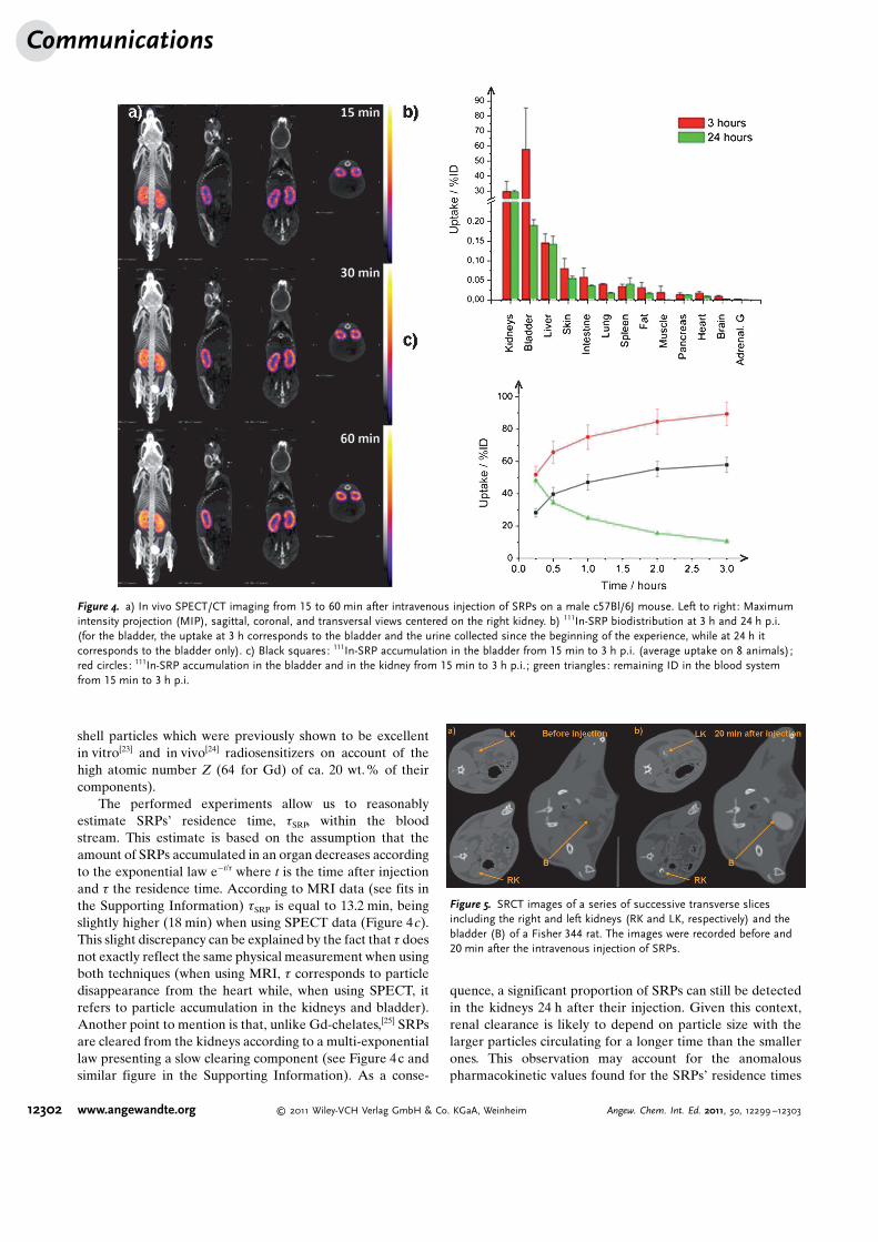
diluted in an aqueous solution composed of 145 mm NaCl and10 mm HEPES at pH 7.4. SRPs used for fluorescence studieswere doped by Cy5.5 during the sol–gel process (seeSupporting Information for details). To better evaluate theircontrast properties for fluorescence, injections (injectedvolume: 200 mL) were performed for three different particleconcentrations given in mmol of gadolinium per liter ofsolution: 10 mm, 20 mm, and 40 mm. Fluorescence imagingwas monitored over 24 h using three female swiss nude micefor various positions. In these experiments, SRPs were shownto only accumulate in kidneys and bladder (Figure 2). For allconcentrations, no undesirable accumulation in lung and liverwas detected, meaning that the particles are not recognized bythe mononuclear phagocytic system and thus could undergo aprolonged vascular retention. Twenty-four hours after theinjection, the mice were sacrificed, with fluorescence imagingof their organs confirming previous in vivo observations(Figure 2d). When performing MRI examinations at 7 T on8-weeks-old male c57B1/6J mice, the same conclusions weredrawn confirming the rapid clearance of SRPs by the renalroute (Figure 3). Kidney visualization was evident 5 min afterinjection (injected volume: 80 mL at 40 mm in Gd) and that ofbladder 25 min thereafter. On angiography images, brainblood vessels were highlighted, revealing rapid clearancefrom the blood stream. In comparison with DOTAREM(80 mL at 40 mm in Gd) administered under the sameconditions or with the absence of a contrast agent, anundoubtedly better contrast was achieved. This may beaccounted for by 1) the higher relaxivity of SRPs (twice thatof DOTAREM) and 2) the greater residence time of particlescompared to molecular compounds (see below for estimationof residence times). SPECT/CT imaging was performed on 8-weeks-old male c57B1/6J mice following injection throughthe tail vein (100 mL; 2.5 mm of gadolinium; 19.3� 2.2 MBq;colloidal stability over several days) (Figure 4). The countswere corrected for background and decay and expressed as apercentage of the injected dose (%ID). When mice weresacrificed 3 h following the injection, SRPs were, again,
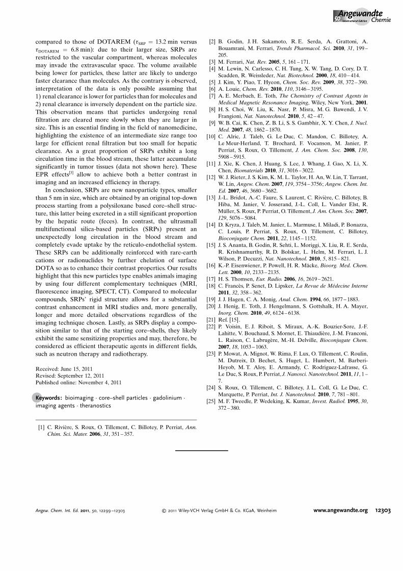
detected in the kidneys and bladder (29.8� 6.8%ID and57.9� 27.4%ID, respectively). For the other analyzed tissues,the uptake was lower than 0.2%ID (or 1%ID per gram), withno hepatic clearance being observed. In contrast, renalelimination occurred relatively fast since 12 min after injec-tion, 25% of the injected dose was detected in the bladder,with 50 % of the injected dose being detected at 74 min. Renalelimination was also highlighted by synchrotron radiation CT(SRCT) experiments performed at the biomedical beamlineof the European Synchroton Radiation Facility (ESRF). Infact, 20 min after SRP injection in Fisher 344 rats (1.4 mLinjected in saphena vein), the white coloration of bothbladder and kidneys confirmed that the particles were presentin these organs (Figure 5). Although the three other tech-niques were known to be more sensitive, we felt it importantto verify that SRPs could be imaged by the same radiationused for therapy (indeed, SRPs are parts of the starting core–
Figure 2. a–c) Fluorescence reflectance imaging of female swiss nude mice a) before, b) 90 min, and c) 3 h after SRP injection (concentration of10 mm in Gd) (K: kidneys, S: stomach, U: urine). d) Fluorescence reflectance imaging of some organs after dissection of the nude mouse.Stomach fluorescence is due to mice alimentation.
Figure 3. a) T1-weighted images of a slice including one kidney (K) andthe bladder (B) of a mouse before and after intravenous injection ofSRPs. b–d) T1-weighted images of the brain of a male c57Bl/6J mouseb) without contrast agent, c) with SRPs and d) with commercialDOTAREM at the same concentration of 40 mm.
12301Angew. Chem. Int. Ed. 2011, 50, 12299 –12303 � 2011 Wiley-VCH Verlag GmbH & Co. KGaA, Weinheim www.angewandte.org
shell particles which were previously shown to be excellentin vitro[23] and in vivo[24] radiosensitizers on account of thehigh atomic number Z (64 for Gd) of ca. 20 wt.% of theircomponents).
The performed experiments allow us to reasonablyestimate SRPs� residence time, tSRP, within the bloodstream. This estimate is based on the assumption that theamount of SRPs accumulated in an organ decreases accordingto the exponential law e�t/t where t is the time after injectionand t the residence time. According to MRI data (see fits inthe Supporting Information) tSRP is equal to 13.2 min, beingslightly higher (18 min) when using SPECT data (Figure 4c).This slight discrepancy can be explained by the fact that t doesnot exactly reflect the same physical measurement when usingboth techniques (when using MRI, t corresponds to particledisappearance from the heart while, when using SPECT, itrefers to particle accumulation in the kidneys and bladder).Another point to mention is that, unlike Gd-chelates,[25] SRPsare cleared from the kidneys according to a multi-exponentiallaw presenting a slow clearing component (see Figure 4c andsimilar figure in the Supporting Information). As a conse-
quence, a significant proportion of SRPs can still be detectedin the kidneys 24 h after their injection. Given this context,renal clearance is likely to depend on particle size with thelarger particles circulating for a longer time than the smallerones. This observation may account for the anomalouspharmacokinetic values found for the SRPs� residence times
Figure 4. a) In vivo SPECT/CT imaging from 15 to 60 min after intravenous injection of SRPs on a male c57Bl/6J mouse. Left to right: Maximumintensity projection (MIP), sagittal, coronal, and transversal views centered on the right kidney. b) 111In-SRP biodistribution at 3 h and 24 h p.i.(for the bladder, the uptake at 3 h corresponds to the bladder and the urine collected since the beginning of the experience, while at 24 h itcorresponds to the bladder only). c) Black squares: 111In-SRP accumulation in the bladder from 15 min to 3 h p.i. (average uptake on 8 animals);red circles: 111In-SRP accumulation in the bladder and in the kidney from 15 min to 3 h p.i. ; green triangles: remaining ID in the blood systemfrom 15 min to 3 h p.i.
Figure 5. SRCT images of a series of successive transverse slicesincluding the right and left kidneys (RK and LK, respectively) and thebladder (B) of a Fisher 344 rat. The images were recorded before and20 min after the intravenous injection of SRPs.
Communications
12302 www.angewandte.org � 2011 Wiley-VCH Verlag GmbH & Co. KGaA, Weinheim Angew. Chem. Int. Ed. 2011, 50, 12299 –12303
compared to those of DOTAREM (tSRP = 13.2 min versustDOTAREM = 6.8 min): due to their larger size, SRPs arerestricted to the vascular compartment, whereas moleculesmay invade the extravascular space. The volume availablebeing lower for particles, these latter are likely to undergofaster clearance than molecules. As the contrary is observed,interpretation of the data is only possible assuming that1) renal clearance is lower for particles than for molecules and2) renal clearance is inversely dependent on the particle size.This observation means that particles undergoing renalfiltration are cleared more slowly when they are larger insize. This is an essential finding in the field of nanomedicine,highlighting the existence of an intermediate size range toolarge for efficient renal filtration but too small for hepaticclearance. As a great proportion of SRPs exhibit a longcirculation time in the blood stream, these latter accumulatesignificantly in tumor tissues (data not shown here). TheseEPR effects[3] allow to achieve both a better contrast inimaging and an increased efficiency in therapy.
In conclusion, SRPs are new nanoparticle types, smallerthan 5 nm in size, which are obtained by an original top-downprocess starting from a polysiloxane based core–shell struc-ture, this latter being excreted in a still significant proportionby the hepatic route (feces). In contrast, the ultrasmallmultifunctional silica-based particles (SRPs) present anunexpectedly long circulation in the blood stream andcompletely evade uptake by the reticulo-endothelial system.These SRPs can be additionally reinforced with rare-earthcations or radionuclides by further chelation of surfaceDOTA so as to enhance their contrast properties. Our resultshighlight that this new particles type enables animals imagingby using four different complementary techniques (MRI,fluorescence imaging, SPECT, CT). Compared to molecularcompounds, SRPs� rigid structure allows for a substantialcontrast enhancement in MRI studies and, more generally,longer and more detailed observations regardless of theimaging technique chosen. Lastly, as SRPs display a compo-sition similar to that of the starting core–shells, they likelyexhibit the same sensitizing properties and may, therefore, beconsidered as efficient therapeutic agents in different fields,such as neutron therapy and radiotherapy.
Received: June 15, 2011Revised: September 12, 2011Published online: November 4, 2011
.Keywords: bioimaging · core–shell particles · gadolinium ·imaging agents · theranostics
[1] C. Rivi�re, S. Roux, O. Tillement, C. Billotey, P. Perriat, Ann.Chim. Sci. Mater. 2006, 31, 351 – 357.
[2] B. Godin, J. H. Sakamoto, R. E. Serda, A. Grattoni, A.Bouamrani, M. Ferrari, Trends Pharmacol. Sci. 2010, 31, 199 –205.
[3] M. Ferrari, Nat. Rev. 2005, 5, 161 – 171.[4] M. Lewin, N. Carlesso, C. H. Tung, X. W. Tang, D. Cory, D. T.
Scadden, R. Weissleder, Nat. Biotechnol. 2000, 18, 410 – 414.[5] J. Kim, Y. Piao, T. Hyeon, Chem. Soc. Rev. 2009, 38, 372 – 390.[6] A. Louie, Chem. Rev. 2010, 110, 3146 – 3195.[7] A. E. Merbach, E. Toth, The Chemistry of Contrast Agents in
Medical Magnetic Resonance Imaging, Wiley, New York, 2001.[8] H. S. Choi, W. Liu, K. Nasr, P. Misra, M. G. Bawendi, J. V.
Frangioni, Nat. Nanotechnol. 2010, 5, 42 – 47.[9] W. B. Cai, K. Chen, Z. B. Li, S. S. Gambhir, X. Y. Chen, J. Nucl.
Med. 2007, 48, 1862 – 1870.[10] C. Alric, J. Taleb, G. Le Duc, C. Mandon, C. Billotey, A.
Le Meur-Herland, T. Brochard, F. Vocanson, M. Janier, P.Perriat, S. Roux, O. Tillement, J. Am. Chem. Soc. 2008, 130,5908 – 5915.
[11] J. Xie, K. Chen, J. Huang, S. Lee, J. Whang, J. Gao, X. Li, X.Chen, Biomaterials 2010, 31, 3016 – 3022.
[12] W. J. Rieter, J. S. Kim, K. M. L. Taylor, H. An, W. Lin, T. Tarrant,W. Lin, Angew. Chem. 2007, 119, 3754 – 3756; Angew. Chem. Int.Ed. 2007, 46, 3680 – 3682.
[13] J.-L. Bridot, A.-C. Faure, S. Laurent, C. Rivi�re, C. Billotey, B.Hiba, M. Janier, V. Josserand, J.-L. Coll, L. Vander Elst, R.M�ller, S. Roux, P. Perriat, O. Tillement, J. Am. Chem. Soc. 2007,129, 5076 – 5084.
[14] D. Kryza, J. Taleb, M. Janier, L. Marmuse, I. Miladi, P. Bonazza,C. Louis, P. Perriat, S. Roux, O. Tillement, C. Billotey,Bioconjugate Chem. 2011, 22, 1145 – 1152.
[15] J. S. Ananta, B. Godin, R. Sehti, L. Moriggi, X. Liu, R. E. Serda,R. Krishnamurthy, R. D. Bolskar, L. Helm, M. Ferrari, L. J.Wilson, P. Decuzzi, Nat. Nanotechnol. 2010, 5, 815 – 821.
[16] K.-P. Eisenwiener, P. Powell, H. R. M�cke, Bioorg. Med. Chem.Lett. 2000, 10, 2133 – 2135.
[17] H. S. Thomsen, Eur. Radio. 2006, 16, 2619 – 2621.[18] C. Franc�s, P. Senet, D. Lipsker, La Revue de M�decine Interne
2011, 32, 358 – 362.[19] J. J. Hagen, C. A. Monig, Anal. Chem. 1994, 66, 1877 – 1883.[20] J. Henig, E. Toth, J. Hengelmann, S. Gottshalk, H. A. Mayer,
Inorg. Chem. 2010, 49, 6124 – 6138.[21] Ref. [15].[22] P. Voisin, E. J. Riboit, S. Miraux, A.-K. Bouzier-Sore, J.-F.
Lahitte, V. Bouchaud, S. Mornet, E. Thiaudi�re, J.-M. Franconi,L. Raison, C. Labrug�re, M.-H. Delville, Bioconjugate Chem.2007, 18, 1053 – 1063.
[23] P. Mowat, A. Mignot, W. Rima, F. Lux, O. Tillement, C. Roulin,M. Dutreix, D. Bechet, S. Huget, L. Humbert, M. Barberi-Heyob, M. T. Aloy, E. Armandy, C. Rodriguez-Lafrasse, G.Le Duc, S. Roux, P. Perriat, J. Nanosci. Nanotechnol. 2011, 11, 1 –7.
[24] S. Roux, O. Tillement, C. Billotey, J. L. Coll, G. Le Duc, C.Marquette, P. Perriat, Int. J. Nanotechnol. 2010, 7, 781 – 801.
[25] M. F. Tweedle, P. Wedeking, K. Kumar, Invest. Radiol. 1995, 30,372 – 380.
12303Angew. Chem. Int. Ed. 2011, 50, 12299 –12303 � 2011 Wiley-VCH Verlag GmbH & Co. KGaA, Weinheim www.angewandte.org


![Review Ultrasmall gold nanoparticles in cancer diagnosis ... · GNs (2, 4, and 6 nm) coated withzwitterionic ligands via gold-sulfur bonding [9], while Garcia et al. obtained ultrasmall](https://img.pdfslide.net/doc/110x75/5f5d9a8589543877274c94b3/review-ultrasmall-gold-nanoparticles-in-cancer-diagnosis-gns-2-4-and-6-nm.jpg)