Embed Size (px)
Citation preview

GE49CH24-Walhout ARI 31 October 2015 12:47
Understanding MetabolicRegulation at a Systems Level:Metabolite Sensing,Mathematical Predictions,and Model OrganismsEmma Watson, L. Safak Yilmaz,and Albertha J.M. Walhout∗
Program in Systems Biology, Program in Molecular Medicine, University of MassachusettsMedical School, Worcester, Massachusetts 01605; email: [email protected],[email protected], [email protected]
Annu. Rev. Genet. 2015. 49:553–75
The Annual Review of Genetics is online atgenet.annualreviews.org
This article’s doi:10.1146/annurev-genet-112414-055257
Copyright c© 2015 by Annual Reviews.All rights reserved
∗Corresponding author
Keywords
metabolic network, feedback loop, homeostasis, gene regulation, fluxbalance analysis, Caenorhabditis elegans
Abstract
Metabolic networks are extensively regulated to facilitate tissue-specificmetabolic programs and robustly maintain homeostasis in response to dietarychanges. Homeostatic metabolic regulation is achieved through metabolitesensing coupled to feedback regulation of metabolic enzyme activity or ex-pression. With a wealth of transcriptomic, proteomic, and metabolomic dataavailable for different cell types across various conditions, we are challengedwith understanding global metabolic network regulation and the resultingmetabolic outputs. Stoichiometric metabolic network modeling integratedwith “omics” data has addressed this challenge by generating nonintuitive,testable hypotheses about metabolic flux rewiring. Model organism stud-ies have also yielded novel insight into metabolic networks. This reviewcovers three topics: the feedback loops inherent in metabolic regulatorynetworks, metabolic network modeling, and interspecies studies utilizingCaenorhabditis elegans and various bacterial diets that have revealed novelmetabolic paradigms.
553
Click here to view this article'sonline features:
• Download figures as PPT slides• Navigate linked references• Download citations• Explore related articles• Search keywords
ANNUAL REVIEWS Further
Ann
u. R
ev. G
enet
. 201
5.49
:553
-575
. Dow
nloa
ded
from
ww
w.a
nnua
lrev
iew
s.or
g A
cces
s pr
ovid
ed b
y U
nive
rsity
of
Mas
sach
uset
ts M
edic
al S
choo
l - W
orce
ster
on
12/0
7/15
. For
per
sona
l use
onl
y.

GE49CH24-Walhout ARI 31 October 2015 12:47
INTRODUCTION
A metabolic network is a collection of biochemical reactions that maintains chemical homeostasis.This network forms the foundation for the cellular economy, as it controls amino acid levels forprotein and neurotransmitter biosynthesis, nucleic acids for DNA and RNA synthesis and repair,methyl and acetyl donors for building the epigenome and synthesizing lipids, and energy for everyanabolic, signaling, and general maintenance process.
Most of what is known about the human metabolic network comes from a century of enzymol-ogy research, in which enzyme activities were detected, functionally purified, and eventually genet-ically characterized. The current age of genomics has enabled the study of metazoan metabolismat a genome-wide, or systems, level. Genome sequencing has revealed a predicted parts list of thehuman metabolic network. Transcriptomic and proteomic studies have revealed that metabolicnetworks exhibit great diversity between tissues, during proliferation versus senescence, and inhealth versus disease. This differential use of metabolic subnetworks or pathways is referred to asmetabolic network rewiring. Today we are challenged with understanding which metabolic path-ways are employed in which tissues and under which conditions, and with dissecting the controlsystems that drive metabolic rewiring.
To explore metabolic network rewiring we must first consider the mechanisms and outputs ofmetabolic network regulation. Metabolic genes are extensively regulated at the levels of transcrip-tion (77), posttranscription (36, 120, 123), posttranslation (91, 109) including allostery throughdirect interactions with metabolites (3, 86, 155), and subcellular localization (104, 105). Further,the regulators are themselves regulated at multiple levels (11) and are connected to master en-docrine signaling pathways that coordinate metabolism across tissues (82).
Understanding how metabolic network regulation affects global outputs (phenotypes) is an-other major challenge. This can be addressed by stoichiometric metabolic network models thatare built by comprehensively annotating the collection of enzymes encoded by the genome andthe reactions they are likely to catalyze. The power of this approach is that such models can pro-vide nonintuitive metabolic and physiological hypotheses. An advantage of these models is that“omics” data can be integrated into the network model to investigate tissue- and condition-specificmetabolic programs, such as metabolite and enzyme dependencies of cancer cells (46).
A complementary approach to exploring metabolic network function is to utilize forward andreverse genetics to identify the genes responsible for metabolic phenotypes or metabolic rewiringevents in vivo. Metazoan model organisms such as Caenorhabditis elegans offer a platform forhigh-throughput genetic screening to uncover novel functions and biological roles of metabolicenzymes, as well as the regulatory networks involved in metabolic network rewiring. C. elegans isparticularly powerful because in addition to being genetically tractable itself, its bacterial diet canalso be subjected to systematic mutagenesis. This can be used to identify, in an unbiased fashion,the most important players in the orchestration of various metabolic states, such as those inducedby caloric restriction or different bacterially supplied nutrients.
This review summarizes what is known about the mechanisms of metabolic regulation froma metabolite-centered perspective, given that metabolites are both the currency of communi-cation between the metabolic network and its regulators, and the commodities that are ulti-mately regulated. We also discuss the methods by which mathematical models of metabolism arebuilt and integrated with gene expression data to generate hypotheses about global metabolicnetwork function. Finally, we explore genetics studies of C. elegans that have shed light onmetabolic network regulation and function, with emphasis on dissecting the metabolic responsesto diet and commensal relationships with bacteria such as those occurring in the mammalianintestine.
554 Watson · Yilmaz ·Walhout
Ann
u. R
ev. G
enet
. 201
5.49
:553
-575
. Dow
nloa
ded
from
ww
w.a
nnua
lrev
iew
s.or
g A
cces
s pr
ovid
ed b
y U
nive
rsity
of
Mas
sach
uset
ts M
edic
al S
choo
l - W
orce
ster
on
12/0
7/15
. For
per
sona
l use
onl
y.

GE49CH24-Walhout ARI 31 October 2015 12:47
MECHANISMS OF METAZOAN NUTRIENT SENSINGAND METABOLIC REGULATION
To maintain metabolic homeostasis, cells must monitor their energy states, the quantities of centralchemical building blocks, and essential micronutrients. This information enables the cell to takethe most appropriate action given its metabolic circumstances, whether that means a subtle shift inoxidative fuel choice or a dramatic rewiring of the metabolic network in response to starvation orhypoxia. How do cells sense the diverse repertoire of metabolites, and how do they use this infor-mation to make regulatory decisions? Organisms have evolved complex networks of proteins withmetabolite-sensing capabilities that relay information about the concentrations of key metabolitesto regulators or that are themselves direct regulators of metabolic genes. Metabolite sensing isachieved in large part through physical interactions between metabolites and regulators that canaffect the function of a regulator by altering its biophysical properties. Many nutrient-sensingregulators are transcription factors and members of signal transduction pathways that regulatemetabolic enzyme expression and function at various levels.
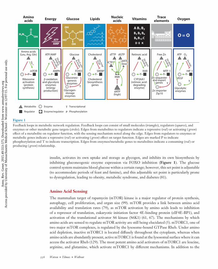
Metabolic regulation utilizes negative feedback loops (nFBLs), which are known to conferthe properties of robustness (80) and adaptability (93, 96) to biological networks. nFBLs inmetabolism center on metabolites, which need to be maintained within proper cellular concen-trations. Metabolic nFBLs include additional components such as enzymes to modify metabo-lite concentrations, regulators to regulate enzyme expression or activity, and sensors to monitormetabolite levels (81). For example, the cellular ATP control system operates as an nFBL to main-tain appropriate energy levels (Figure 1). In this system, AMP:ATP ratios are sensed by AMPK,which serves both sensor and regulator functions. AMPK is activated by increased AMP:ATP lev-els and phosphorylates glycolytic enzymes among other metabolic targets to enhance glycolyticflux, ultimately increasing ATP production. As ATP levels increase, AMPK is inhibited, whichcompletes the feedback loop. This section describes how specific metabolic control systems are en-coded by the genome, detailing the gene networks responsible for nutrient sensing and metabolicgene regulation as well as the mechanisms they employ.
Glucose Sensing
The insulin-signaling pathway is the major regulator of circulating glucose concentrations andglucose metabolism. It forms a multiorgan nFBL, which is initiated by glucose uptake in pancreaticβ-cells. These cells express GLUT2, the highest affinity glucose transporter, and thus are the firstcells to be affected by blood glucose spikes after a meal (41). Glucose is metabolized via glycol-ysis, the tricarboxylic acid (TCA) cycle, and ultimately the electron transport chain, resulting inincreased intracellular ATP:ADP ratios (63). This leads to depolarization of the cell through inhib-ited potassium export from ATP-sensitive potassium channels, which causes an influx of calciumand the release of insulin to the bloodstream through exocytosis of insulin-storage granules (63).Thus, pancreatic β-cells sense an increase in glucose by monitoring their own internal glucose ox-idation flux, reflected by ATP:ADP ratios. The release of insulin from the β-cells signifies to othertissues that glucose is abundant and should be imported, metabolized, and/or stored (as glycogen).
In the target tissue, the insulin-signaling pathway initiates with binding of insulin to theinsulin receptor, followed by phosphorylation of PI3K and AKT kinase, which are involved intransducing the signal (23). AKT activates GLUT4 translocation to the cell surface to promoteglucose import (76), inhibits a negative regulator of glycogen synthase (GSK3) to increaseglycogen production and thus glucose storage (34), and inhibits FOXO to halt transcriptionalactivation of gluconeogenesis enzymes (27, 115). Glucose, therefore, through its proxy signal
www.annualreviews.org • Metabolite Sensing, Mathematical Predictions, and Model Organisms 555
Ann
u. R
ev. G
enet
. 201
5.49
:553
-575
. Dow
nloa
ded
from
ww
w.a
nnua
lrev
iew
s.or
g A
cces
s pr
ovid
ed b
y U
nive
rsity
of
Mas
sach
uset
ts M
edic
al S
choo
l - W
orce
ster
on
12/0
7/15
. For
per
sona
l use
onl
y.
GE49CH24-Walhout ARI 31 October 2015 12:47
ATP
AMP
Aminoacids
Energy Glucose LipidsNucleic
acidsVitamins
Traceelements
Oxygen
Glucose
Glycogen-storage
enzymes
Gluconeo-genesis
enzymes
Cholesterol
Cholesterolbiosynthesis
genes
SREBPFOXO
Via In
sig/
SCA
PV
ia Insig
/
SCA
P
ATP/AMP
β-oxidationand glycolysis
enzymes(energy
production)
AMPK
Via d
irect
bin
din
g
and
LKB
1 V
ia direct
bin
din
g
and
LKB
1
Amino acids(Leu, Arg, Gln)
Ribosomeactivation(protein
synthesis)
mTOR
Via ?
Via β-cells
& in
sulin
Via β-cells
& in
sulin
O2
VEGF
Glycolyticenzymes
HIF-1
Via P
HD
ATPFree Zn
MTF-1
Via d
irect
bin
din
gV
ia direct
bin
din
g
Via d
irect
bin
din
gV
ia direct
bin
din
g
Zn importerZip10
Zn sequesterand export
genes
Zn sequesterand export
genes
Retinoic acid
CYP26A1(retinoic acid-
degradingenzyme)
RARTT
TT
TT
PPPP
PP
TT TT TT TT
TTTT
dATP(low)dATP(low)
dATP(high)
dCTPdCTP
dGTPdTTP
RNR
Metabolite
Regulator
Enzyme
Enzyme/regulator
Transcriptional
Phosphorylation
O
OH
R
H2N
OH
HO
O
OH
OH
OH
O = O
A B1 B2
B3 B5 B6
B9 B12 C
D E K
Figure 1Feedback loops in metabolic network regulation. Feedback loops can consist of small molecules (triangles), regulators (squares), andenzymes or other metabolic gene targets (circles). Edges from metabolites to regulators indicate a repressive (red ) or activating ( green)effect of a metabolite on regulator function, with the sensing mechanism noted along the edge. Edges from regulators to enzymes ormetabolic genes indicate a repressive (red ) or activating ( green) effect on target function. Edges are marked P to indicatephosphorylation and T to indicate transcription. Edges from enzymes/metabolic genes to metabolites indicate a consuming (red ) orproducing ( green) relationship.
insulin, activates its own uptake and storage as glycogen, and inhibits its own biosynthesis byinhibiting gluconeogenic enzyme expression via FOXO inhibition (Figure 1). The glucosecontrol system maintains blood glucose within a certain range; however, this set point is adjustable(to accommodate periods of feast and famine), and this adjustable set point is particularly proneto dysregulation, leading to obesity, metabolic syndrome, and diabetes (81).
Amino Acid Sensing
The mammalian target of rapamycin (mTOR) kinase is a major regulator of protein synthesis,autophagy, cell proliferation, and organ size (99). mTOR provides a link between amino acidavailability and translation rates (79), as mTOR activation by amino acids leads to inhibitionof a repressor of translation, eukaryotic initiation factor 4E-binding protein (eIF4E-BP1), andactivation of the translational activator S6 kinase (S6K1) (61, 67). The mechanisms by whichamino acids are sensed to regulate mTOR activity are still being elucidated (5). mTORC1, one oftwo major mTOR complexes, is regulated by the lysosome-bound GTPase Rheb. Under aminoacid depletion, inactive mTORC1 is located diffusely throughout the cytoplasm, whereas whenamino acids are abundantly present, active mTORC1 is found at the lysosomal surface where it canaccess the activator Rheb (129). The most potent amino acid activators of mTORC1 are leucine,arginine, and glutamine, which activate mTORC1 by different mechanisms. In addition to the
556 Watson · Yilmaz ·Walhout
Ann
u. R
ev. G
enet
. 201
5.49
:553
-575
. Dow
nloa
ded
from
ww
w.a
nnua
lrev
iew
s.or
g A
cces
s pr
ovid
ed b
y U
nive
rsity
of
Mas
sach
uset
ts M
edic
al S
choo
l - W
orce
ster
on
12/0
7/15
. For
per
sona
l use
onl
y.

GE49CH24-Walhout ARI 31 October 2015 12:47
lysosomal amino acid–sensing mechanism, leucine sensing may be achieved through interactionsbetween leucyl-tRNA synthase and the mTOR complex (56), whereas glutamine sensing involvesthe adenosine diphosphate ribosylation factor-1 (Arf1) (73), and arginine sensing requires thelysosomal SLC38A9 transporter (150).
Another mechanism of amino acid sensing is the monitoring of uncharged tRNAs, whichrepresent amino acid depletion. This is accomplished through the highly conserved Gcn2-eIF2α-Gcn4/ATF system. As amino acid levels decline during amino acid starvation, uncharged tRNAsaccumulate and bind Gcn2 through a domain with homology to histidyl tRNA synthetase. Thisdomain has higher affinity for uncharged tRNA than charged tRNA, and binding causes a confor-mational change that activates the kinase domain of Gcn2. Gcn2 phosphorylates eIF2α, resultingin a global decrease in translation rates, with the exception of certain mRNAs, such as those encod-ing the transcription factors Gcn4 and ATF. Transcriptional targets of Gcn4 include amino acidbiosynthetic enzymes and transporters (65). ATF drives the autophagy gene expression programin order to salvage amino acids from existing proteins (8).
Lipid Sensing
We focus on the mechanisms for sensing two types of lipid: cholesterol and free fatty acids. Choles-terol is an important component of mammalian cell membranes and also serves as a precursor in thesynthesis of bile acids and steroid hormones. Cholesterol sensing takes place in the endoplasmicreticulum membrane, where SREBP, a transcription factor, resides with its binding partners SCAPand Insig1 (39). When cholesterol levels in the membrane fall below a threshold, Insig1 dissociates,and the SCAP-SREBP complex translocates to the Golgi body, where SCAP-activated S1P andS2P proceed to cleave SREBP, freeing SREBP to migrate to the nucleus (125, 151). Once in thenucleus, SREBP activates the transcription of several genes in the de novo cholesterol biosynthe-sis pathway, including HMG-CoA reductase (68), the target of widely used cholesterol-loweringstatin drugs. Thus, via SREBP, cholesterol negatively regulates its own biosynthesis.
Fatty acids consist of saturated and unsaturated carbon chains with a carboxylic acid headgroup. Fatty acids serve both structural (as components of cellular membranes) and energy-storagepurposes (in their acyl-CoA form). The precise sensing mechanisms for different fatty acids arenot entirely clear, but it has been suggested that the peroxisome proliferator-activated receptor(PPAR) subgroup of nuclear hormone receptors sense the fatty acid milieu of the cell throughpromiscuous, weak binding of diverse fatty acid ligands (20, 147). The binding affinities observedin vitro [2–50 μM range for PPARα binding to free fatty acids (146)] may indicate that PPARs areactivated only when ligands reach high concentrations in vivo (128). PPARs regulate the expressionof enzymes involved in fatty acid oxidation (78), and increased intracellular levels of fatty acidsresult in increased PPAR-mediated expression of fatty acid oxidation genes (108). Another nuclearreceptor, HNF4-α, also binds lipids and regulates an overlapping set of lipid metabolism genes.Therefore, free fatty acids activate their own oxidation through PPARs and HNF4-α.
Nucleotide Sensing
Nucleotide pools are carefully managed, as nucleotide deficiencies lead to increased muta-genesis rates and genomic instability (12). During S phase, the demand for nucleotides spikesto support DNA replication, and enzymes involved in de novo nucleotide biosynthesis aretranscriptionally upregulated, including ribonucleotide reductase (RNR) (13), which convertsnucleoside diphosphates (NDPs) to deoxynucleoside diphosphates (dNDPs); these go on to formdeoxynucleoside triphosphates (dNTPs). RNR and other nucleotide metabolism genes are direct
www.annualreviews.org • Metabolite Sensing, Mathematical Predictions, and Model Organisms 557
Ann
u. R
ev. G
enet
. 201
5.49
:553
-575
. Dow
nloa
ded
from
ww
w.a
nnua
lrev
iew
s.or
g A
cces
s pr
ovid
ed b
y U
nive
rsity
of
Mas
sach
uset
ts M
edic
al S
choo
l - W
orce
ster
on
12/0
7/15
. For
per
sona
l use
onl
y.

GE49CH24-Walhout ARI 31 October 2015 12:47
targets of c-MYC, a master transcriptional regulator of the cell cycle (90). RNR is subject tofurther regulation through subcellular localization [for instance, it assembles at sites of DNAdamage (141)], proteolytic degradation (19), and an elegant system of allosteric feedback withvarious end product dNTPs to ensure proper nucleotide ratios (66). This autoregulation occursvia differential binding of ATP, dATP, dTTP, and dGTP to the specificity site, or S site. Theidentity of the S site–bound nucleotide dictates the substrate preference of RNR: ATP anddATP binding stimulates conversion of CDP to dCDP, dTTP (which is formed from dCTPthrough the de novo nucleotide synthesis pathway) activates conversion of GDP to dGDP, andfinally dGTP activates conversion of ADP to dADP. When dATP levels are high, dATP bindsa different site on RNR, the A site, to shut down all RNR activity and halt dNTP synthesis.Thus, RNR plays multiple vital roles in maintaining proper nucleotide pools; it is the enzymethat creates dNTPs, the sensor of dNTP ratios, and the regulator of those ratios.
Trace Element Sensing: Zinc
Trace metals, including cobalt, iron, and zinc, are micronutrients that function extensivelythroughout the metabolic network, where they bind enzymes to form metalloenzymes, whichutilize bioinorganic chemistry to catalyze metabolic reactions that are difficult to achieve with or-ganic chemistry alone. Trace metals are important resources that must be extracted from the dietand carefully incorporated into the cellular economy to avoid the toxicity associated with the over-abundance of highly reactive free ions. However, deficiencies in trace elements can be catastrophicfor an organism, as they can impair the function of hundreds if not thousands of different proteins.
For instance, zinc is predicted to bind 10% of the human proteome (4), serving both cat-alytic and structural roles. As a structural component, it generates protein-folding landscapes,which increases the complexity of protein-protein and protein-DNA interactions (101). In humanmetabolism, the catalytic activity of zinc is used by >100 zinc metalloenzymes (103). A classic ex-ample is carbonic anhydrase, which accommodates a zinc atom in its reaction center and reversiblyconverts carbon dioxide to bicarbonate (139).
An intricate network of zinc importers and exporters, intracellular zinc binding/transportingmetallothioneins, and zinc-sensing transcription factors such as MTF-1 (25) regulate the cellularreservoir of zinc ions. As with the other nutrient-sensing mechanisms, zinc sensing involves nFBLsto maintain zinc pools within an optimal cellular threshold. MTF-1, itself a zinc-finger protein,transports to the nucleus upon binding free zinc ions in the cytoplasm. In the nucleus, it binds tometal response elements (MREs) and thereby activates the expression of zinc-sequestering metal-lothioneins and zinc exporters. MTF-1 can also repress some targets, including the zinc importerZip10, which has an MRE downstream of its transcription start site, and binding of MTF-1 to thisMRE stalls Pol II progression (54). Whereas MTF-1 is conserved from insects to mammals, thereis no clear ortholog in the nematode C. elegans. C. elegans does exhibit transcriptional responsesto zinc and a C. elegans zinc-responsive DNA element was recently identified (122). However, thetranscription factor responsible for sensing zinc and binding this element is still unknown.
Vitamin Sensing
Vitamins are chemical cofactors required for the proper functioning of a species (the vitaminauxotroph) but provided by another species (the vitamin synthesizer). Vitamin auxotrophy is acornerstone of many symbiotic relationships (33, 62), including the relationship between humansand the vitamin-producing bacteria that make up our microbiota (84). Humans require a suite ofvitamins that function in diverse metabolic pathways, and flux through these pathways inherently
558 Watson · Yilmaz ·Walhout
Ann
u. R
ev. G
enet
. 201
5.49
:553
-575
. Dow
nloa
ded
from
ww
w.a
nnua
lrev
iew
s.or
g A
cces
s pr
ovid
ed b
y U
nive
rsity
of
Mas
sach
uset
ts M
edic
al S
choo
l - W
orce
ster
on
12/0
7/15
. For
per
sona
l use
onl
y.

GE49CH24-Walhout ARI 31 October 2015 12:47
depends on the quantity of vitamins derived from diet and provided by our microbiota. Vitamindeficiencies were once a major cause of disease and death in human populations, and continue tocause health problems in underdeveloped countries. In mammals, direct sensing of vitamin A andvitamin D occurs through binding to nuclear hormone receptors. Vitamin D, which is technicallya hormone rather than a vitamin given that it can be synthesized from cholesterol in the skinupon UV light exposure, regulates the uptake of calcium, iron, magnesium, and zinc (59). Thisregulation occurs through vitamin D receptor (VDR)-mediated transcriptional activation of thetransporters of these micronutrients (18). Vitamin A, a true vitamin, has a plethora of functions indiverse processes, including development, vision, and immunity (38). Vitamin A does not functionin anabolic or catabolic processes, as most other vitamins do, but rather serves as a light-sensingcofactor for rhodopsin and a signaling morphogen to regulate Hox genes during developmentvia retinoic acid receptor (RAR) binding (38). There are enzymes that modify dietary Vitamin A(retinol) to generate retinal (the form utilized by rhodopsin) and retinoic acid (the form that directlybinds and activates RAR), as well as enzymes that degrade retinoic acid. Vitamin A metabolism istightly regulated by positive and negative FBLs to maintain proper concentrations of the variousforms of Vitamin A during development, given that dysregulation can lead to teratogenesis (35,134). For instance, the retinoic acid–degrading enzyme CYP26A1 is directly activated by RARthrough binding of highly conserved retinoic acid response elements (RAREs) in its promoter (35).Thus, retinoic acid activates its own degradation, preventing deleteriously high levels of retinoicacid from building up. Retinoic acid also represses the expression of the enzymes involved in itssynthesis from retinol, although the mechanisms of this repression are unknown (35).
Less is known about the mechanisms employed by mammalian cells to sense other vitamins, ifthey do exist. However, gene expression studies have revealed regulatory responses to vitamins B1(thiamine) (47, 88, 142), B2 (riboflavin) (111), B3 (nicotinamide/niacin) (26, 29, 50), B6 [pyridoxal5′ phosphate (PLP)] (145, 158), B9 (folic acid) (7, 21, 87), C (ascorbic acid) (17, 74, 140), andE (tocopherol/tocotrienols) (83, 100, 110). Indirect vitamin-sensing mechanisms, in which fluxthrough the vitamin-dependent pathway is sensed as a proxy for vitamin abundance, may also exist.For instance, the nematode C. elegans exhibits a vitamin B12 response in which the expressionof several metabolic genes is tuned according to dietary vitamin B12 intake. However, geneticperturbation of either of the two vitamin B12–dependent pathways disrupts the vitamin B12responsiveness of several of these genes (154). This demonstrates that vitamin B12 itself is not themediator of transcriptional changes but rather suggests that some genes are regulated according toflux through vitamin B12–dependent pathways rather than the absolute quantities of the vitamin.Several C. elegans nuclear hormone receptors are implicated in this response (153), although theidentities of the metabolites that are sensed remain to be identified.
Energy Sensing
As the energy currency of the cell, ATP is a vital molecule required by many diverse cellularprocesses. A central regulator of energy homeostasis is AMPK, a heterotrimeric kinase that sensesAMP/ATP ratios and regulates ATP production accordingly. AMPK is activated by high AMPlevels and inhibited by high ATP levels (58). As energy is harvested from the hydrolysis of ATP toADP, increasing ADP levels push the adenylate kinase reaction (2ADP↔ ATP + AMP) towardAMP and ATP production to buffer the falling ATP levels. AMPK activity increases several-fold in vitro when AMP is present (24), and increases more than 100-fold when phosphorylatedby its upstream regulator, the LKB1-STRAD-MO25 complex (60). Although this complex doesnot sense AMP itself, it phosphorylates AMPK preferentially when AMP/ATP ratios are highbecause of conformational changes in AMPK induced by AMP or ADP binding, which enhances
www.annualreviews.org • Metabolite Sensing, Mathematical Predictions, and Model Organisms 559
Ann
u. R
ev. G
enet
. 201
5.49
:553
-575
. Dow
nloa
ded
from
ww
w.a
nnua
lrev
iew
s.or
g A
cces
s pr
ovid
ed b
y U
nive
rsity
of
Mas
sach
uset
ts M
edic
al S
choo
l - W
orce
ster
on
12/0
7/15
. For
per
sona
l use
onl
y.

GE49CH24-Walhout ARI 31 October 2015 12:47
phosphorylation site availability (156). There are many downstream targets of AMPK, includingthe central carbon metabolism enzymes 6-phosphofructo-2-kinase (glycolysis enzyme activatedby AMPK phosphorylation) and acetyl-CoA carboxylase (fatty acid synthesis enzyme inhibited byAMPK). The end result of AMPK activation is enhanced oxidative catabolism of glucose and fattyacids to fuel the electron transport chain to produce more ATP, and the inhibition of anabolicpathways such as gluconeogenesis and fatty acid synthesis (106).
Oxygen Sensing
Cellular oxygen levels must be closely monitored, as major metabolic network rewiring musttake place to survive periods of hypoxia. Primarily, a shift from oxidative phosphorylation(OXPHOS) to anaerobic glycolysis is needed for compensatory ATP production. The chief oxy-gen sensor, conserved in all metazoans, is the prolyl-hydroxylase and hypoxia-inducible factor 1(HIF-1) system. HIF-1 is a heterodimeric transcription factor, consisting of a basic helix-loop-helixDNA binding domain, a heterodimerization domain, and a transcriptional coactivator-bindingdomain. Under normoxic conditions, HIF-1 is hydroxylated by prolyl-hydroxylases (42), leadingto ubiquitination by a von Hippel–Lindau-guided E3 ubiquitin ligase (116) and proteasomaldegradation of HIF-1 (127). Under hypoxic conditions, the prolyl-hydroxylases fail to hydroxylateHIF-1 because this reaction requires molecular oxygen, and the HIF-1 protein is stabilized(130). Stable, active HIF-1 accumulates in the nucleus, where, in addition to upregulating theexpression of proangiogenic factors like vascular endothelial growth factor (VEGF) to increaseoxygen flow (89), it activates expression of glycolytic enzymes (133), resulting in increased fluxthrough glycolysis to compensate for the lack of mitochondrial ATP production.
The HIF-regulating prolyl-hydroxylases are Fe, O2, and α-ketoglutarate (α-KG, a TCA cycleintermediate) dependent, suggesting that the HIF-1 oxygen-sensing system also senses and re-sponds to changes in iron and α-KG levels through altered prolyl-hydroxylase substrate availability(95). A lack of α-KG may signify reduced flux through the TCA cycle, and therefore an impend-ing energy crisis, given that the TCA cycle produces reducing equivalents to fuel OXPHOS.Low TCA cycle flux and a lack of O2 would therefore lead to a decrease in ATP production viaOXPHOS and, in either situation, the HIF-1-mediated increase in glycolytic flux provides nec-essary compensation. Aberrant activation of HIF-1 under normoxic conditions is a common oc-currence in cancer cells and helps establish the aerobic glycolysis metabolic program conduciveto rapid proliferation (102, 121, 132).
Intercommunication Between Metabolic Regulatory Pathways
Regulatory cross talk between the various metabolite-specific sensing pathways is a useful wayfor the organism to coordinate metabolism with growth or to cope with nutrient stress. Forinstance, the insulin-signaling cascade activates mTOR to promote protein synthesis and cellgrowth. However, under nutrient stress, mTOR is inhibited by AMPK to slow translation ratesand halt cell growth and proliferation. Below are several examples of cross talk between pathways.
� The insulin-signaling pathway activates mTOR through AKT-mediated inhibition ofTSC1/TSC2, the major mTOR repressor (69).
� mTOR is inhibited by AMPK, both directly through phosphorylation of Raptor (55) andindirectly through activation of mTOR repressors TSC1 and TSC2 (70).
� AMPK activates FOXO (53), which upregulates genes involved in gluconeogenesis, lipidmetabolism, and autophagy.
560 Watson · Yilmaz ·Walhout
Ann
u. R
ev. G
enet
. 201
5.49
:553
-575
. Dow
nloa
ded
from
ww
w.a
nnua
lrev
iew
s.or
g A
cces
s pr
ovid
ed b
y U
nive
rsity
of
Mas
sach
uset
ts M
edic
al S
choo
l - W
orce
ster
on
12/0
7/15
. For
per
sona
l use
onl
y.

GE49CH24-Walhout ARI 31 October 2015 12:47
� mTOR can activate HIF-1a under normoxic conditions, leading to aerobic glycolysis (theWarburg effect). This occurs in proliferating macrophages (126) and cancer cells (57).
� mTOR can activate SREBP-1 (119) through Lipin-1 (118) to activate lipid synthesis for cellgrowth and proliferation.
� AMPK directly phosphorylates SREBP-1 to inhibit its cleavage and translocation into thenucleus, thus repressing SREBP-1-mediated transcriptional activation of lipid synthesis inmammalian liver (85).
BUILDING METABOLIC NETWORK MODELSAND INCORPORATING “OMICS” DATA
Metabolic regulation targets genes that encode metabolic enzymes or transporters. The output ofthis regulation is altered metabolic flux in regulated reactions and pathways, known as metabolicnetwork rewiring. A major challenge is to interpret local metabolic network rewiring in the contextof the entire metabolic network and to decipher how these rewiring events affect overall phenotypicoutputs. To address this challenge, it has become prudent to reconstruct and mathematically modelglobal metabolic networks for major model organisms and humans. In the following section, wefirst summarize the general principles of metabolic network reconstruction and modeling, andthen discuss the application of human metabolic models to the generation of tissue- and condition-specific metabolic networks and to the resolution of metabolic changes associated with disease.
Building Mathematical Models of Genome-Scale Metabolic Networks
Reconstruction of a metabolic model begins with annotation of gene-protein-reaction (GPR)associations from the literature and from bioinformatic analysis of gene sequences (131). Reactionsets for annotated enzymes are derived from several databases, including Brenda (131) and KEGG(75). Boolean language (AND, OR) is used to describe GPRs. For instance, if two isozymes cancatalyze the same reaction, they are associated with OR logic to that reaction, and enzyme complexsubunits that work in concert but are encoded by different genes are associated to their reactionwith AND logic (143). Although most metabolic network modeling efforts to date have targetedmicroorganisms (44), genome-scale reconstructions have also been built for several metazoans,including humans (40, 92, 144), mouse (138), Arabidopsis (37), and zebrafish (10). The most recentreconstruction of the human metabolic network is based on 1,789 enzyme-encoding genes, witha total of 7,440 reactions annotated, including those that transport metabolites between eightcompartments and involving 2,626 unique metabolites (144).
An ideal metabolic model can take the composition of nutrients from diet as input andgenerates as output a range of flux distributions that satisfy experimentally determined measure-ments of biomass composition and energy requirements of the organism. This is mathematicallyachieved by representing the network as a stoichiometry (S) matrix that consists of the coefficientsof metabolites (rows) in corresponding reactions (columns), such that these coefficients arenegative for the reactants and positive for the products of a given reaction (see the toy network inFigure 2a). This matrix is multiplied by a vector of fluxes (v), and the equation is set to equalzero, representing mass balance under steady state conditions (i.e., at every compound node, thetotal flux of consumption matches that of production). The solution of this equation alone givesthe null space of the S matrix. To narrow down the solution to a biologically meaningful spaceof feasible fluxes, a set of reaction constraints are applied based on available information of thebiochemistry. In particular, irreversible reactions are limited by a lower boundary of zero, i.e.,flux in the reverse direction is disallowed. In addition, an objective function is set to obtain a
www.annualreviews.org • Metabolite Sensing, Mathematical Predictions, and Model Organisms 561
Ann
u. R
ev. G
enet
. 201
5.49
:553
-575
. Dow
nloa
ded
from
ww
w.a
nnua
lrev
iew
s.or
g A
cces
s pr
ovid
ed b
y U
nive
rsity
of
Mas
sach
uset
ts M
edic
al S
choo
l - W
orce
ster
on
12/0
7/15
. For
per
sona
l use
onl
y.

GE49CH24-Walhout ARI 31 October 2015 12:47
particular flux distribution within this biological solution space to achieve a defined goal, suchas the maximization of growth (i.e., the flux of a biomass assembly reaction) or maximization ofenergy generation. This method is referred to as constraint-based flux balance analysis (FBA)(136). Importantly, after typical constraints are imposed, the same objective may still be achievedby multiple flux distributions, which implicates the existence of alternate pathways (Figure 2a).
a
b
A B
F
E
DC
Imp
ort
Imp
ort
Imp
ort
Imp
ort
Imp
ort
r2
r1t1
r3
r6r4
r5
Gene 4
Gene 5
Gene 3
Gene 6
Gene 2Gene 1
Gene9
Gene 8 Gene 7
AND
OR
A B
F
E
DC
r2
r1t1
r3
r6r4
r5
Gene 4
Gene 5
Gene 3
Gene 6
Gene 2Gene 1
Gene 9
Gene 8 Gene 7
AND
OR
A B
F
E
DC
r2
r1t1
r3
r6r4
r5
Gene 4
Gene 5
Gene 3
Gene 6
Gene 2Gene 1
Gene 9
Gene 8 Gene 7
AND
OR
Gene expression level
Low
Threshold
High
Tissue 1
Tissue 2
A B
F
E
DC
r2
r1t1
r3
r6r4
r5
A B
F
E
DC
r2
r1t1
r3
r6r4
r5
Tissue 1
Tissue 2
Gene-protein-reaction associations
Gene expression dataFilter reactions for
tissue-specific networks
A B
F
E
DC
Imp
ort
r2
r1t1
r3
r6r4
r5
0 0 0 0 0 1 0
1 –1 0 –1 0 0 0 0
0 0 0 1 –1 0 0 0
0 0 0 0 1 1 0 –1
0 0 1 0 0 –1 0 –2
0 1 –1 0 0 0 0 0
–1
Maximize objective f = vb
such that
S × v = 0
viLB ≤ vi ≤ vi
UB
A
B
F
E
C
D
r1 r2 r4 r5 r6 r3 t1 b
?
?
?
?
?
?
Fluxes (v)
Biomass
A B
F
E
DC
r3
r4Im
po
rt
r2
r1
t1
r6
r5
A B
F
E
DC
r3
r4
Imp
ort
r2
r1
t1
r6
r5
v1
v2
Solution space for vReactions (S)
Me
tab
oli
tes
Null space
Solution space
v1v2
b: 2E + D → Biomass
b
bBiomass
bBiomass
562 Watson · Yilmaz ·Walhout
Ann
u. R
ev. G
enet
. 201
5.49
:553
-575
. Dow
nloa
ded
from
ww
w.a
nnua
lrev
iew
s.or
g A
cces
s pr
ovid
ed b
y U
nive
rsity
of
Mas
sach
uset
ts M
edic
al S
choo
l - W
orce
ster
on
12/0
7/15
. For
per
sona
l use
onl
y.

GE49CH24-Walhout ARI 31 October 2015 12:47
Then, additional constraints based on experimental data, such as gene expression levels informingwhether a subset of reactions carry flux or not, can be utilized to narrow down the solution spacefurther by eliminating or favoring some of the alternative pathways (Figure 2b).
Using “Omics” Data to Extract Tissue- and Condition-SpecificMetabolic Network Models
One way to address the regulation of metabolic networks is to extract from the generic metabolicnetwork of an organism the metabolically rewired states that are tightly regulated, for instance,in different tissues or under specific conditions. Current methods of extraction constrain theglobal network models with tissue- or condition-specific experimental evidence, including geneexpression data (Figure 2b). For instance, mixed integer linear programming was used to penalizeFBA for introducing flux in reactions associated with lowly expressed genes and for lack of fluxin reactions associated with highly expressed genes, and this way, flux distributions in 10 humantissues were predicted based on transcriptomic and proteomic data sets (136). A more advancedand systematic optimization procedure called the model-building algorithm (MBA) was laterdeveloped to take into account not only expression data but also other tissue-specific information,such as phenotypic data and the presence of tissue-specific metabolites (71). This method uses setsof reactions grouped according to their probability of being functional in the targeted tissue basedon experimental information, and optimizes the metabolic network to maximize the number ofreactions in higher probability categories while minimizing those that are unlikely to be present.MBA was successfully used to derive a liver metabolic network that outperforms the generic humanmodel in predicting hepatic fluxes in different conditions and changes in biomarkers of hepaticdisorders related to metabolism (71).
Several other methods for deriving tissue-specific metabolic networks have been developedand validated since the development of MBA, including GIMME, which uses gene expressiondata combined with objective functions such as energy generation (9), mCADRE, which is similarto MBA (152), and INIT, which is also similar to MBA with a key exception that it allows fluximbalance to address metabolomics data (2). More about tissue-specific applications of metabolicnetwork modeling can be found in a recent review (124).
Constraint-Based Metabolic Models of Cancer
Cancer cells exhibit major metabolic network rewiring in central carbon pathways that allow un-controlled proliferation. Although the switch from OXPHOS to aerobic glycolysis in cancer cells
←−−−−−−−−−−−−−−−−−−−−−−−−−−−−−−−−−−−−−−−−−−−−−−−−−−−−−−−−−−−−−−−−−−−−−−−−−−−−−−−−−−−−−−−−−−Figure 2General principles of metabolic network modeling. (a) Toy network comprising metabolites (red letters, gray circles) converted byreactions (black letters, black rectangles). The metabolic network is designated by the stoichiometry (S) matrix with rows representingmetabolites and columns representing reactions. The values depict the stoichiometric ratios of metabolites that are consumed (negativevalues) and produced ( positive values) by reactions. Flux distribution (v) is determined by flux balance analysis (FBA), which satisfies massbalance at every node (compound) by the steady state equation (S × v = 0), while imposing the boundary conditions on v andmaximizing the objective function (rate of reaction 2E + D→ Biomass). Shown on the right is the solution space without anyconstraints ( yellow), biologically meaningful subspace with constraints (orange), and the landscape of alternative solutions that satisfy themaximum objective function (black curve). Two alternative solutions are shown. Arrow thickness and direction depict flux magnitudeand direction, respectively. (b) Gene-protein-reaction associations for the toy network from panel a, represented using Booleanlanguage. Gene expression data (colored in a scale from low to high) can be used to constrain fluxes within the global metabolic networkand to generate tissue- or condition-specific networks. Two different conditions are shown with corresponding flux distributions thatbest fit the expression data.
www.annualreviews.org • Metabolite Sensing, Mathematical Predictions, and Model Organisms 563
Ann
u. R
ev. G
enet
. 201
5.49
:553
-575
. Dow
nloa
ded
from
ww
w.a
nnua
lrev
iew
s.or
g A
cces
s pr
ovid
ed b
y U
nive
rsity
of
Mas
sach
uset
ts M
edic
al S
choo
l - W
orce
ster
on
12/0
7/15
. For
per
sona
l use
onl
y.

GE49CH24-Walhout ARI 31 October 2015 12:47
was first described by Otto Warburg more than 90 years ago, we are still determining its causesand consequences. It seems counterintuitive that rapidly proliferating cancer cells would prefer aless-efficient glucose catabolism with respect to ATP production (i.e., 1 mole of glucose yields 2moles ATP from glycolysis versus 32 moles ATP from OXPHOS) because proliferating cells stillrequire copious amounts of ATP. Recently, metabolic modeling of human cancer has been appliedto explore Warburg-like metabolic rewiring. Cancer metabolic models have accurately predicteda preference for flux through the glycolysis-branching pentose phosphate pathway to supply thecell with reducing equivalents (NADPH) for fatty acid synthesis and nucleotide biosynthesis pre-cursors. Other studies utilizing either small kinetic (148) or global stoichiometric (135) metabolicmodels of cancer have arrived at the same conclusion, which is that in the context of rapid prolifer-ation, glycolysis alone is actually more energetically efficient than OXPHOS if the costs of enzymesynthesis and molecular crowding are taken into account. By constraining a global stoichiometricmodel with gene expression data, cancer metabolic models have been built for individual tumorsto define metabolic phenotypes of premalignant versus malignant tumors (premalignant modelsactually supported higher growth rates) and estrogen receptor (ER)+ versus ER− breast cancers(72).
Metabolic modeling of an engineered fumarase-deficient cell line was used to generate hy-potheses that can explain how fumarase-deficient tumors manage to fuel the electron transportchain without a functional TCA cycle (48). This modeling discovered that these cells compensatefor lack of NADH production by synthesizing and degrading heme. Fumarase deficiency leads to abuildup of fumarate, which is believed to act as an oncometabolite by inhibiting prolyl-hydroxylasesfrom activating HIF-1 under normoxic conditions, leading to Warburg-like glucose metabolismand enhanced vascularization via HIF-1-mediated VEGF activation (48). Whether Warburg-likemetabolism is a driver or an enabler of tumorigenesis is a subject of debate. Certainly, manyproliferating cells exhibit aerobic glycolysis and do not become tumorigenic. Simultaneously,there is compelling evidence supporting the notion that metabolic disruptions such as fumarasedeficiency, succinate dehydrogenase deficiency, and neomorphic mutations in isocitrate dehydro-genase (114) can be oncogenic. Interestingly, these metabolic disturbances are each associatedwith specific types of cancer, suggesting context dependence for the transformative properties ofthe putative oncometabolites fumarate, succinate, and 2-hydroxyglutarate. Recently, cancer type–specific metabolic models were built and queried with commonly detected cancer type–specificenzyme mutations to explore the context dependence of known oncometabolites and predict noveloncometabolites, resulting in 15 newly predicted oncometabolites (112). Another study utilizedconstraint-based models of tumor cell metabolism to predict novel drug targets by determiningcancer-specific synthetic lethal relationships within the metabolic network (46).
NOVEL PARADIGMS IN METABOLIC NETWORK FUNCTIONAND REGULATION FROM CAENORHABDITIS ELEGANS
Although metabolic modeling compiles known metabolic data into a mathematical framework,it has limited ability to predict novel metabolic functions and pathways, and cannot criticallyassess the assignment of genes to reactions. This was illustrated in a recent study of metabolicmodels, which found that metabolic network reconstruction similarity between organisms did notcorrelate well with genome similarity or phylogenetic distance, and often metabolically diverseorganisms had highly similar metabolic network models (107). This is largely due to the fact thatmetabolic models are often built upon the framework of an existing model (107), and reactions andpathways outside of common central metabolic pathways are largely unexplored in most species,biochemically or genetically. Advancement in metabolic modeling will require complementary
564 Watson · Yilmaz ·Walhout
Ann
u. R
ev. G
enet
. 201
5.49
:553
-575
. Dow
nloa
ded
from
ww
w.a
nnua
lrev
iew
s.or
g A
cces
s pr
ovid
ed b
y U
nive
rsity
of
Mas
sach
uset
ts M
edic
al S
choo
l - W
orce
ster
on
12/0
7/15
. For
per
sona
l use
onl
y.

GE49CH24-Walhout ARI 31 October 2015 12:47
efforts in biochemistry and genetics to expand our knowledge about metabolism. Here, we focuson recent studies that have taken advantage of the genetic tractability of the nematode C. elegansto generate novel insights into metabolic gene function and regulation.
Caenorhabditis elegans Genetics Identify Novel Enzyme Activitiesand Biological Significance
C. elegans is a bacterivorous soil-dwelling nematode, consisting of 959 somatic cells, whose shortlife cycle (3 days) and genetic tractability make it amenable to high-throughput genetic screening.Much of the human metabolic network is conserved in C. elegans, which also has similar essentialnutrient requirements, devoted metabolic tissues (primarily the intestine, comprising 20 cells), andconserved metabolic regulatory pathways such as the insulin and TOR signaling pathways (14).In fact, the regulation of FOXO by insulin signaling and its relationship to aging were identifiedin C. elegans (115), as was the regulation of HIF-1 by prolyl-hydroxylases (42).
C. elegans can be used to genetically dissect metabolic phenotypes and explore metabolic en-zymes of unknown function. For instance, ACER-1, a conserved protein of unknown function,was identified as a novel acetyl-CoA hydrolase (converting acetyl-CoA into acetate and CoA) in asearch for binding partners of CRA-1, a conserved regulator of global histone acetylation, whichfunctions by an unknown mechanism (49). ACER-1 was found to bind and affect CRA-1-regulatedhistone acetylation, and metabolomics revealed elevated acetyl-CoA levels in an acer-1 mutant.Thus, a mechanism was proposed whereby CRA-1 regulates the levels of acetyl-CoA in the nucleusand thus histone acetylation by antagonizing ACER-1 acetyl-CoA hydrolase activity (49). Anotherprotein with novel metabolic activity identified in C. elegans is the GDP-D-glucose phosphorylaseC10F3.4, which converts GDP-D-glucose to GDP and D-glucose-1-phosphate (1). C. elegans mu-tants lacking C10F3.4 lost all GDP-D-glucose phosphorylase activity and built up the substrateGDP-D-glucose. The mouse ortholog of C10F3.4 was shown to have the same catalytic activityand to exhibit similar tissue expression patterns as C. elegans, with highest expression in neuronaland reproductive tissues (1). The exact function of GDP-D-glucose phosphorylation is unknownbut was postulated to be a metabolite repair reaction to deplete GDP-D-glucose produced non-specifically by the enzyme GDP-d-Man pyrophosphorylase, thus preventing the misincorporationof GDP-D-glucose into glycoproteins (1).
Other studies have identified new biological roles for known enzymes and have revealednovel relationships between metabolic pathways and cellular processes. For instance, mutation oftyrosine aminotransferase (tatn-1; breakdown of tyrosine to acetoacetate and fumarate) resultedin gene expression changes and enhanced the lifespan extension of insulin-signaling mutants (45).These effects were largely due to elevated tyrosine levels, which activated AMPK to repress FOXO(daf-16) (45). Another study identified a diet-specific phenotype for the proline catabolic enzymealh-6, which is the ortholog of human 1-pyrroline-5-carboxylate (P5C) dehydrogenase (P5CDH).alh-6 mutants exhibited reduced lifespan on the standard laboratory diet of Escherichia coli OP50but on a diet of E. coli HT115 alh-6 mutants and wild-type animals exhibited identical lifespans(117). The specific metabolic differences between these two E. coli strains as they relate to thelifespan of the alh-6 mutant are unknown, although metabolomic profiling has revealed myriaddifferences in metabolite levels. The buildup of the alh-6 substrate P5C was proposed to be thecause of premature aging in the mutant because knockdown of the upstream enzyme proline dehy-drogenase, which produces P5C from proline, rescued some of the effects of the alh-6 mutant onaging (117). Thus, a novel role for proline catabolism in metabolic flexibility, healthy aging, andadaptation to dietary changes was established, although the mechanisms underlying these rolesare unknown.
www.annualreviews.org • Metabolite Sensing, Mathematical Predictions, and Model Organisms 565
Ann
u. R
ev. G
enet
. 201
5.49
:553
-575
. Dow
nloa
ded
from
ww
w.a
nnua
lrev
iew
s.or
g A
cces
s pr
ovid
ed b
y U
nive
rsity
of
Mas
sach
uset
ts M
edic
al S
choo
l - W
orce
ster
on
12/0
7/15
. For
per
sona
l use
onl
y.

GE49CH24-Walhout ARI 31 October 2015 12:47
Caenorhabditis elegans Genetics Reveal Factors Involved in MetabolicResponse to Diet and the Microbiota
Diet and microbiota composition have profound impacts on host metabolic network regulation andfunction; however, these impacts are difficult to investigate because of the complexity and diversityof dietary inputs, as well as the genetic complexity and diversity within the human population andmicrobiota. A major advantage of using C. elegans is the ability to genetically dissect the regulatorymechanisms that enable metabolic network rewiring in response to changes in the bacterial diet andthe resulting physiological outputs. For instance, C. elegans has been used extensively to study theeffects of caloric restriction on metabolism and lifespan, and more recently to delineate regulatorynetworks controlling metabolic responses to dietary composition. The genetic tractability of bothC. elegans and its bacterial diets renders this natural predator-prey system an excellent model tostudy the principles of metabolic network regulation in response to diet, as well as to model theeffects of commensal bacteria on host metabolism and physiology (16, 97, 157). Recently, it wasdiscovered that the lifespan-extending effect of the antidiabetic drug metformin on C. elegans isdue to alteration of E. coli folate metabolism, which affects flux through the folate-dependentone-carbon cycle in C. elegans, as demonstrated through genetic inactivation of C. elegans one-carbon-cycle genes (15). Another study serendipitously isolated a spontaneous mutation in E. coliaroD, a shikimic acid pathway gene required for folate precursor biosynthesis, when screeningan RNAi feeding library for C. elegans lifespan extension (149), further demonstrating the linkbetween microbial folate production and C. elegans longevity.
C. elegans has been fed a variety of metabolically diverse bacterial species in the lab, and thephysiological and gene expression responses in C. elegans have been measured (28, 98). In one study,many of the genes that exhibit diet-induced expression changes were found to confer diet-specificphenotypes when mutated (28). Genetic inactivation of the regulators of metabolic gene expressioncan also confer diet-specific phenotypes. For instance, mutations in the nuclear hormone receptornhr-114 lead to complete sterility caused by a defective germline on a diet of E. coli OP50; however,nhr-114 mutants develop intact germlines and are fertile when fed E. coli HT115 (52). As with thealh-6 example, the differences between E. coli OP50 and HT115 that determine nhr-114 sterilityare unknown, although supplementation of E. coli OP50 with tryptophan could rescue the nhr-114sterility phenotype on this diet (52). A challenge for the future will be to determine the causalfactors among a sea of differences in nutrient compositions between different bacterial diets thatdrive effects in the animal.
Recently, interspecies genetics has been used to address this very challenge. Genetic screeningin both C. elegans and its bacterial diets, Comamonas aquatica and E. coli, revealed bacterially pro-duced vitamin B12 as the driver of several C. elegans gene expression and physiological changes(154). These screens employed a transgenic C. elegans reporter strain, in which green fluores-cent protein expression is driven by the promoter of the C. elegans gene acdh-1, which is stronglyrepressed by the Comamonas diet but activated by the E. coli diet (98). Mutations in Comamonasvitamin B12 biosynthetic enzymes were identified in strains that failed to repress acdh-1 promoteractivity. Exogenous supplementation of B12 was sufficient to repress acdh-1 expression on thelow-B12 diet of E. coli OP50. Networks of C. elegans metabolic and regulatory genes, includingnuclear hormone receptors, were required for the repression of the acdh-1 promoter by the vita-min B12–synthesizing Comamonas diet (153). Interestingly, the vitamin B12–mediated repressionof acdh-1 was dependent on flux through the vitamin B12–requiring propionate breakdown path-way, as mutants in this pathway resulted in constitutively activated acdh-1 expression (154). Thesegenes could also be activated by exogenously supplied propionate, and deletion of acdh-1 increasessensitivity to propionate-induced toxicity (154), suggesting that this gene may be involved in novel
566 Watson · Yilmaz ·Walhout
Ann
u. R
ev. G
enet
. 201
5.49
:553
-575
. Dow
nloa
ded
from
ww
w.a
nnua
lrev
iew
s.or
g A
cces
s pr
ovid
ed b
y U
nive
rsity
of
Mas
sach
uset
ts M
edic
al S
choo
l - W
orce
ster
on
12/0
7/15
. For
per
sona
l use
onl
y.

GE49CH24-Walhout ARI 31 October 2015 12:47
Mutant bacterial library dsRNA-expressing bacterial library
Observable phenotype
Control
guaA, purC, purE
cyaA, aroC, entB
lplA, entE, yeiC...
Bacterial genes affecting phenotype C. elegans genes affecting phenotype
Retest,
validate
Retest,
validate
Bacterial gene network C. elegans gene network
Interspecies network interactions
Dictate phenotype X
acdh-1, dhs-19,
dlat-1, F09F7.4,
F54D5.7, fum-1...
Figure 3Dissecting diet-related phenotypes in Caenorhabditis elegans with interspecies genetics. To determine gene networks involved in diet-related phenotypes in C. elegans, genetic screens can be performed in both the bacterial diet and in the animal. C. elegans can be fed alibrary of mutant bacteria to determine bacterial genes that, when mutated, lead to a particular phenotype. To identify C. elegans genesthat are involved in the diet-associated phenotype, animals can be fed a library of dsRNA-expressing bacteria targeting individual C.elegans genes. Hits from either screen should be retested and validated, and can be built into gene networks based on coexpression,cocomplex formation, and/or genetic interaction data, or metabolic pathway annotations. Hypotheses about interspecies gene networkinteractions and their consequences with respect to the metabolic phenotype can be generated and further tested.
alternative propionate breakdown or detoxification mechanisms on low-B12 diets. This approachutilizing interspecies systems biology serves as an example of how the effects of nutrient compo-sition within complex biotic diets on animal gene expression and physiology can be elucidated(Figure 3).
CONCLUSIONS AND FUTURE DIRECTIONS
Metabolic network modeling can benefit from improvements both in the biological knowledgethat is integrated into the model and in the computational methods of such integration (107). Al-though utilization of gene expression data to predict condition-specific fluxes has been advanced bynumerous methods (94, 112), techniques to integrate many other types of data sets of potential im-portance are in the early stages. In particular, methods that constrain FBA with metabolomics data
www.annualreviews.org • Metabolite Sensing, Mathematical Predictions, and Model Organisms 567
Ann
u. R
ev. G
enet
. 201
5.49
:553
-575
. Dow
nloa
ded
from
ww
w.a
nnua
lrev
iew
s.or
g A
cces
s pr
ovid
ed b
y U
nive
rsity
of
Mas
sach
uset
ts M
edic
al S
choo
l - W
orce
ster
on
12/0
7/15
. For
per
sona
l use
onl
y.

GE49CH24-Walhout ARI 31 October 2015 12:47
are emerging (148). Other challenges include the integration of kinetic parameters and metaboliteconcentrations. Kinetic parameters of central carbon pathways have been incorporated into theE. coli model (32). Certain human pathways, such as the one-carbon cycle (113), have also been stud-ied using kinetic models equipped with experimentally determined parameters. Thus, it is reason-able to expect the integration of kinetic data into genome-scale models of metazoan metabolism.
Perhaps the greatest future challenge for genome-scale metabolic network modeling with thegreatest potential benefit is the direct incorporation of regulatory networks (such as the regulatorynFBLs discussed in this review) into the model. An existing method named regulatory FBA convertsgene regulatory pathways to a Boolean formalism that dictates “on” or “off ” states of reactionswithin stoichiometric metabolic network models (31). This technique has been used to incorporateregulatory networks of E. coli (30) and yeast (64) into the respective genome-scale network models.Other Boolean formalism–based methods (6, 22, 32, 43, 51, 137), as well as a novel stochasticapproach (22), have also been developed, but applications have been limited to microorganismsto date. Direct integration of metazoan regulatory networks into genome-scale metabolic modelswill likely await better characterization of the paths from regulators to enzymatic reactions, whichis dependent on protein-metabolite, protein-protein, and protein-DNA interactions.
Recently, our lab has reconstructed a global metabolic network model for C. elegans (L.S.Yilmaz & A.J.M. Walhout, in preparation). C. elegans provides a unique opportunity to rapidlytest hypotheses generated by in silico FBA of the metabolic model, such as pathway usage ondifferent diets. Genetic screening in C. elegans can be used to improve GPR annotations and, byextrapolation, guide improvement of annotations within the human model.
DISCLOSURE STATEMENT
The authors are not aware of any affiliations, memberships, funding, or financial holdings thatmight be perceived as affecting the objectivity of this review.
LITERATURE CITED
1. Adler LN, Gomez TA, Clarke SG, Linster CL. 2011. A novel GDP-D-glucose phosphorylase involvedin quality control of the nucleoside diphosphate sugar pool in Caenorhabditis elegans and mammals.J. Biol. Chem. 286:21511–23
2. Agren R, Bordel S, Mardinoglu A, Pornputtapong N, Nookaew I, Nielsen J. 2012. Reconstruction ofgenome-scale active metabolic networks for 69 human cell types and 16 cancer types using INIT. PLoSComput. Biol. 8:e1002518
3. Ahmad MF, Dealwis CG. 2013. The structural basis for the allosteric regulation of ribonucleotidereductase. Prog. Mol. Biol. Transl. Sci. 117:389–410
4. Andreini C, Banci L, Bertini I, Rosato A. 2006. Counting the zinc-proteins encoded in the humangenome. J. Proteome Res. 5:196–201
5. Bar-Peled L, Sabatini DM. 2014. Regulation of mTORC1 by amino acids. Trends Cell Biol. 24:400–66. Barua D, Kim J, Reed JL. 2010. An automated phenotype-driven approach (GeneForce) for refining
metabolic and regulatory models. PLoS Comput. Biol. 6:e10009707. Barua S, Chadman KK, Kuizon S, Buenaventura D, Stapley NW, et al. 2014. Increasing maternal or
post-weaning folic acid alters gene expression and moderately changes behavior in the offspring. PLoSONE 9:e101674
8. B’Chir W, Maurin AC, Carraro V, Averous J, Jousse C, et al. 2013. The eIF2α/ATF4 pathway is essentialfor stress-induced autophagy gene expression. Nucleic Acids Res. 41:7683–99
9. Becker SA, Palsson BO. 2008. Context-specific metabolic networks are consistent with experiments.PLoS Comput. Biol. 4:e1000082
568 Watson · Yilmaz ·Walhout
Ann
u. R
ev. G
enet
. 201
5.49
:553
-575
. Dow
nloa
ded
from
ww
w.a
nnua
lrev
iew
s.or
g A
cces
s pr
ovid
ed b
y U
nive
rsity
of
Mas
sach
uset
ts M
edic
al S
choo
l - W
orce
ster
on
12/0
7/15
. For
per
sona
l use
onl
y.

GE49CH24-Walhout ARI 31 October 2015 12:47
10. Bekaert M. 2012. Reconstruction of Danio rerio metabolic model accounting for subcellular compart-mentalisation. PLoS ONE 7:e49903
11. Berrabah W, Aumercier P, Lefebvre P, Staels B. 2011. Control of nuclear receptor activities inmetabolism by post-translational modifications. FEBS Lett. 585:1640–50
12. Bester AC, Roniger M, Oren YS, Im MM, Sarni D, et al. 2011. Nucleotide deficiency promotes genomicinstability in early stages of cancer development. Cell 145:435–46
13. Bjorklund S, Skog S, Tribukait B, Thelander L. 1990. S-phase-specific expression of mammalian ri-bonucleotide reductase R1 and R2 subunit mRNAs. Biochemistry 29:5452–58
14. Braeckman BP, Houthoofd K, Vanfleteren JR. 2009. Intermediary metabolism. WormBook. doi:10.1895/wormbook.1.146.1
15. Cabreiro F, Au C, Leung KY, Vergara-Irigaray N, Cocheme HM, et al. 2013. Metformin retards agingin C. elegans by altering microbial folate and methionine metabolism. Cell 153:228–39
16. Cabreiro F, Gems D. 2013. Worms need microbes too: microbiota, health and aging in Caenorhabditiselegans. EMBO Mol. Med. 5:1300–10
17. Canali R, Natarelli L, Leoni G, Azzini E, Comitato R, et al. 2014. Vitamin C supplementation modulatesgene expression in peripheral blood mononuclear cells specifically upon an inflammatory stimulus: a pilotstudy in healthy subjects. Genes Nutr. 9:390
18. Carlberg C, Seuter S. 2009. A genomic perspective on vitamin D signaling. Anticancer Res. 29:3485–93
19. Chabes AL, Pfleger CM, Kirschner MW, Thelander L. 2003. Mouse ribonucleotide reductase R2 pro-tein: a new target for anaphase-promoting complex-Cdh1-mediated proteolysis. PNAS 100:3925–29
20. Chakravarthy MV, Lodhi IJ, Yin L, Malapaka RR, Xu HE, et al. 2009. Identification of a physiologicallyrelevant endogenous ligand for PPARα in liver. Cell 138:476–88
21. Champier J, Claustrat F, Nazaret N, Fevre Montange M, Claustrat B. 2012. Folate depletion changesgene expression of fatty acid metabolism, DNA synthesis, and circadian cycle in male mice. Nutr. Res.32:124–32
22. Chandrasekaran S, Price ND. 2010. Probabilistic integrative modeling of genome-scale metabolic andregulatory networks in Escherichia coli and Mycobacterium tuberculosis. PNAS 107:17845–50
23. Cheng Z, Tseng Y, White MF. 2010. Insulin signaling meets mitochondria in metabolism. TrendsEndocrinol. Metab. 21:589–98
24. Cheung PC, Salt IP, Davies SP, Hardie DG, Carling D. 2000. Characterization of AMP-activated proteinkinase gamma-subunit isoforms and their role in AMP binding. Biochem. J. 346(Pt. 3):659–69
25. Choi S, Bird AJ. 2014. Zinc’ing sensibly: controlling zinc homeostasis at the transcriptional level.Metallomics Integr. Biometal Sci. 6:1198–215
26. Choi S, Yoon H, Oh KS, Oh YT, Kim YI, et al. 2011. Widespread effects of nicotinic acid on geneexpression in insulin-sensitive tissues: implications for unwanted effects of nicotinic acid treatment.Metabolism 60:134–44
27. Cichy SB, Uddin S, Danilkovich A, Guo S, Klippel A, Unterman TG. 1998. Protein kinase B/Akt mediateseffects of insulin on hepatic insulin-like growth factor-binding protein-1 gene expression through aconserved insulin response sequence. J. Biol. Chem. 273:6482–87
28. Coolon JD, Jones KL, Todd TC, Carr BC, Herman MA. 2009. Caenorhabditis elegans genomic responseto soil bacteria predicts environment-specific genetic effects on life history traits. PLoS Genet. 5:e1000503
29. Couturier A, Keller J, Most E, Ringseis R, Eder K. 2014. Niacin in pharmacological doses altersmicroRNA expression in skeletal muscle of obese Zucker rats. PLoS ONE 9:e98313
30. Covert MW, Knight EM, Reed JL, Herrgard MJ, Palsson BO. 2004. Integrating high-throughput andcomputational data elucidates bacterial networks. Nature 429:92–96
31. Covert MW, Schilling CH, Palsson B. 2001. Regulation of gene expression in flux balance models ofmetabolism. J. Theor. Biol. 213:73–88
32. Covert MW, Xiao N, Chen TJ, Karr JR. 2008. Integrating metabolic, transcriptional regulatory andsignal transduction models in Escherichia coli. Bioinformatics 24:2044–50
33. Croft MT, Lawrence AD, Raux-Deery E, Warren MJ, Smith AG. 2005. Algae acquire vitamin B12through a symbiotic relationship with bacteria. Nature 438:90–93
www.annualreviews.org • Metabolite Sensing, Mathematical Predictions, and Model Organisms 569
Ann
u. R
ev. G
enet
. 201
5.49
:553
-575
. Dow
nloa
ded
from
ww
w.a
nnua
lrev
iew
s.or
g A
cces
s pr
ovid
ed b
y U
nive
rsity
of
Mas
sach
uset
ts M
edic
al S
choo
l - W
orce
ster
on
12/0
7/15
. For
per
sona
l use
onl
y.

GE49CH24-Walhout ARI 31 October 2015 12:47
34. Cross DA, Alessi DR, Cohen P, Andjelkovich M, Hemmings BA. 1995. Inhibition of glycogen synthasekinase-3 by insulin mediated by protein kinase B. Nature 378:785–89
35. D’Aniello E, Waxman JS. 2015. Input overload: contributions of retinoic acid signaling feedback mech-anisms to heart development and teratogenesis. Dev. Dyn. 244:513–23
36. David CJ, Chen M, Assanah M, Canoll P, Manley JL. 2010. HnRNP proteins controlled by c-Mycderegulate pyruvate kinase mRNA splicing in cancer. Nature 463:364–68
37. de Oliveira Dal’Molin CG, Quek LE, Palfreyman RW, Brumbley SM, Nielsen LK. 2010. AraGEM,a genome-scale reconstruction of the primary metabolic network in Arabidopsis. Plant Physiol. 152:579–89
38. di Masi A, Leboffe L, De Marinis E, Pagano F, Cicconi L, et al. 2015. Retinoic acid receptors: frommolecular mechanisms to cancer therapy. Mol. Asp. Med. 41:1–115
39. Dong XY, Tang SQ, Chen JD. 2012. Dual functions of Insig proteins in cholesterol homeostasis. LipidsHealth Dis. 11:173
40. Duarte NC, Becker SA, Jamshidi N, Thiele I, Mo ML, et al. 2007. Global reconstruction of the humanmetabolic network based on genomic and bibliomic data. PNAS 104:1777–82
41. Efrat S, Tal M, Lodish HF. 1994. The pancreatic beta-cell glucose sensor. Trends Biochem. Sci. 19:535–3842. Epstein AC, Gleadle JM, McNeill LA, Hewitson KS, O’Rourke J, et al. 2001. C. elegans EGL-9 and
mammalian homologs define a family of dioxygenases that regulate HIF by prolyl hydroxylation. Cell107:43–54
43. Faria JP, Overbeek R, Xia F, Rocha M, Rocha I, Henry CS. 2014. Genome-scale bacterial transcriptionalregulatory networks: reconstruction and integrated analysis with metabolic models. Brief. Bioinform.15:592–611
44. Feist AM, Herrgard MJ, Thiele I, Reed JL, Palsson BO. 2009. Reconstruction of biochemical networksin microorganisms. Nat. Rev. Microbiol. 7:129–43
45. Ferguson AA, Roy S, Kormanik KN, Kim Y, Dumas KJ, et al. 2013. TATN-1 mutations reveal anovel role for tyrosine as a metabolic signal that influences developmental decisions and longevity inCaenorhabditis elegans. PLoS Genet. 9:e1004020
46. Folger O, Jerby L, Frezza C, Gottlieb E, Ruppin E, Shlomi T. 2011. Predicting selective drug targetsin cancer through metabolic networks. Mol. Syst. Biol. 7:501
47. Fraser DA, Hessvik NP, Nikolic N, Aas V, Hanssen KF, et al. 2012. Benfotiamine increases glucoseoxidation and downregulates NADPH oxidase 4 expression in cultured human myotubes exposed toboth normal and high glucose concentrations. Genes Nutr. 7:459–69
48. Frezza C, Zheng L, Tennant DA, Papkovsky DB, Hedley BA, et al. 2011. Metabolic profiling of hypoxiccells revealed a catabolic signature required for cell survival. PLoS ONE 6:e24411
49. Gao J, Kim HM, Elia AE, Elledge SJ, Colaiacovo MP. 2015. NatB domain-containing CRA-1 antag-onizes hydrolase ACER-1 linking Acetyl-CoA metabolism to the initiation of recombination duringC. elegans meiosis. PLoS Genet. 11:e1005029
50. Giammona LM, Fuhrken PG, Papoutsakis ET, Miller WM. 2006. Nicotinamide (vitamin B3) in-creases the polyploidisation and proplatelet formation of cultured primary human megakaryocytes. Br. J.Haematol. 135:554–66
51. Goelzer A, Bekkal Brikci F, Martin-Verstraete I, Noirot P, Bessieres P, et al. 2008. Reconstruction andanalysis of the genetic and metabolic regulatory networks of the central metabolism of Bacillus subtilis.BMC Syst. Biol. 2:20
52. Gracida X, Eckmann CR. 2013. Fertility and germline stem cell maintenance under different dietsrequires nhr-114/HNF4 in C. elegans. Curr. Biol. 23:607–13
53. Greer EL, Oskoui PR, Banko MR, Maniar JM, Gygi MP, et al. 2007. The energy sensor AMP-activatedprotein kinase directly regulates the mammalian FOXO3 transcription factor. J. Biol. Chem. 282:30107–19
54. Gunther V, Lindert U, Schaffner W. 2012. The taste of heavy metals: gene regulation by MTF-1.Biochim. Biophys. Acta 1823:1416–25
55. Gwinn DM, Shackelford DB, Egan DF, Mihaylova MM, Mery A, et al. 2008. AMPK phosphorylationof raptor mediates a metabolic checkpoint. Mol. Cell 30:214–26
570 Watson · Yilmaz ·Walhout
Ann
u. R
ev. G
enet
. 201
5.49
:553
-575
. Dow
nloa
ded
from
ww
w.a
nnua
lrev
iew
s.or
g A
cces
s pr
ovid
ed b
y U
nive
rsity
of
Mas
sach
uset
ts M
edic
al S
choo
l - W
orce
ster
on
12/0
7/15
. For
per
sona
l use
onl
y.

GE49CH24-Walhout ARI 31 October 2015 12:47
56. Han JM, Jeong SJ, Park MC, Kim G, Kwon NH, et al. 2012. Leucyl-tRNA synthetase is an intracellularleucine sensor for the mTORC1-signaling pathway. Cell 149:410–24
57. Harada H, Itasaka S, Kizaka-Kondoh S, Shibuya K, Morinibu A, et al. 2009. The Akt/mTOR pathwayassures the synthesis of HIF-1αprotein in a glucose- and reoxygenation-dependent manner in irradiatedtumors. J. Biol. Chem. 284:5332–42
58. Hardie DG. 2014. AMPK: positive and negative regulation, and its role in whole-body energy homeo-stasis. Curr. Opin. Cell Biol. 33C:1–7
59. Haussler MR, Haussler CA, Bartik L, Whitfield GK, Hsieh JC, et al. 2008. Vitamin D receptor: molecularsignaling and actions of nutritional ligands in disease prevention. Nutr. Rev. 66:S98–112
60. Hawley SA, Boudeau J, Reid JL, Mustard KJ, Udd L, et al. 2003. Complexes between the LKB1 tu-mor suppressor, STRADα/β and MO25α/β are upstream kinases in the AMP-activated protein kinasecascade. J. Biol. 2:28
61. Hay N, Sonenberg N. 2004. Upstream and downstream of mTOR. Genes Dev. 18:1926–4562. Helliwell KE, Wheeler GL, Smith AG. 2013. Widespread decay of vitamin-related pathways: coinci-
dence or consequence? Trends Genet. 29:469–7863. Henquin JC. 2011. The dual control of insulin secretion by glucose involves triggering and amplifying
pathways in β-cells. Diabetes Res. Clin. Pract. 93(Suppl. 1):S27–3164. Herrgard MJ, Lee BS, Portnoy V, Palsson BO. 2006. Integrated analysis of regulatory and metabolic
networks reveals novel regulatory mechanisms in Saccharomyces cerevisiae. Genome Res. 16:627–3565. Hinnebusch AG. 2005. Translational regulation of GCN4 and the general amino acid control of yeast.
Annu. Rev. Microbiol. 59:407–5066. Hofer A, Crona M, Logan DT, Sjoberg BM. 2012. DNA building blocks: keeping control of manufacture.
Crit. Rev. Biochem. Mol. Biol. 47:50–6367. Holz MK, Ballif BA, Gygi SP, Blenis J. 2005. mTOR and S6K1 mediate assembly of the translation
preinitiation complex through dynamic protein interchange and ordered phosphorylation events. Cell123:569–80
68. Horton JD, Goldstein JL, Brown MS. 2002. SREBPs: activators of the complete program of cholesteroland fatty acid synthesis in the liver. J. Clin. Investig. 109:1125–31
69. Inoki K, Li Y, Zhu T, Wu J, Guan KL. 2002. TSC2 is phosphorylated and inhibited by Akt and suppressesmTOR signalling. Nat. Cell Biol. 4:648–57
70. Inoki K, Zhu T, Guan KL. 2003. TSC2 mediates cellular energy response to control cell growth andsurvival. Cell 115:577–90
71. Jerby L, Shlomi T, Ruppin E. 2010. Computational reconstruction of tissue-specific metabolic models:application to human liver metabolism. Mol. Syst. Biol. 6:401
72. Jerby L, Wolf L, Denkert C, Stein GY, Hilvo M, et al. 2012. Metabolic associations of reduced prolif-eration and oxidative stress in advanced breast cancer. Cancer Res. 72:5712–20
73. Jewell JL, Kim YC, Russell RC, Yu FX, Park HW, et al. 2015. Differential regulation of mTORC1 byleucine and glutamine. Science 347:194–98
74. Jun HJ, Kim S, Dawson K, Choi DW, Kim JS, et al. 2011. Effects of acute oral administration of vitaminC on the mouse liver transcriptome. J. Med. Food 14:181–94
75. Kanehisa M, Goto S, Sato Y, Furumichi M, Tanabe M. 2012. KEGG for integration and interpretationof large-scale molecular data sets. Nucleic Acids Res. 40:D109–14
76. Kanzaki M. 2006. Insulin receptor signals regulating GLUT4 translocation and actin dynamics. Endocr.J. 53:267–93
77. Karagianni P, Talianidis I. 2015. Transcription factor networks regulating hepatic fatty acid metabolism.Biochim. Biophys. Acta 1851:2–8
78. Kersten S. 2014. Integrated physiology and systems biology of PPARα. Mol. Metab. 3:354–7179. Kim SG, Buel GR, Blenis J. 2013. Nutrient regulation of the mTOR complex 1 signaling pathway. Mol.
Cells 35:463–7380. Kitano H. 2004. Biological robustness. Nat. Rev. Genet. 5:826–3781. Kotas ME, Medzhitov R. 2015. Homeostasis, inflammation, and disease susceptibility. Cell 160:816–2782. Kousteni S. 2012. FoxO1, the transcriptional chief of staff of energy metabolism. Bone 50:437–43
www.annualreviews.org • Metabolite Sensing, Mathematical Predictions, and Model Organisms 571
Ann
u. R
ev. G
enet
. 201
5.49
:553
-575
. Dow
nloa
ded
from
ww
w.a
nnua
lrev
iew
s.or
g A
cces
s pr
ovid
ed b
y U
nive
rsity
of
Mas
sach
uset
ts M
edic
al S
choo
l - W
orce
ster
on
12/0
7/15
. For
per
sona
l use
onl
y.
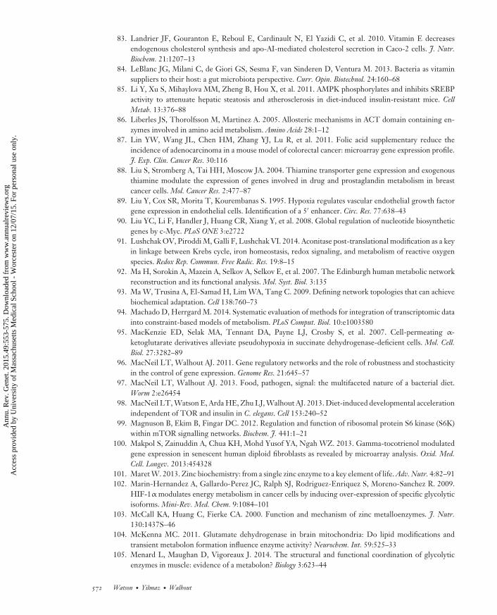
GE49CH24-Walhout ARI 31 October 2015 12:47
83. Landrier JF, Gouranton E, Reboul E, Cardinault N, El Yazidi C, et al. 2010. Vitamin E decreasesendogenous cholesterol synthesis and apo-AI-mediated cholesterol secretion in Caco-2 cells. J. Nutr.Biochem. 21:1207–13
84. LeBlanc JG, Milani C, de Giori GS, Sesma F, van Sinderen D, Ventura M. 2013. Bacteria as vitaminsuppliers to their host: a gut microbiota perspective. Curr. Opin. Biotechnol. 24:160–68
85. Li Y, Xu S, Mihaylova MM, Zheng B, Hou X, et al. 2011. AMPK phosphorylates and inhibits SREBPactivity to attenuate hepatic steatosis and atherosclerosis in diet-induced insulin-resistant mice. CellMetab. 13:376–88
86. Liberles JS, Thorolfsson M, Martinez A. 2005. Allosteric mechanisms in ACT domain containing en-zymes involved in amino acid metabolism. Amino Acids 28:1–12
87. Lin YW, Wang JL, Chen HM, Zhang YJ, Lu R, et al. 2011. Folic acid supplementary reduce theincidence of adenocarcinoma in a mouse model of colorectal cancer: microarray gene expression profile.J. Exp. Clin. Cancer Res. 30:116
88. Liu S, Stromberg A, Tai HH, Moscow JA. 2004. Thiamine transporter gene expression and exogenousthiamine modulate the expression of genes involved in drug and prostaglandin metabolism in breastcancer cells. Mol. Cancer Res. 2:477–87
89. Liu Y, Cox SR, Morita T, Kourembanas S. 1995. Hypoxia regulates vascular endothelial growth factorgene expression in endothelial cells. Identification of a 5′ enhancer. Circ. Res. 77:638–43
90. Liu YC, Li F, Handler J, Huang CR, Xiang Y, et al. 2008. Global regulation of nucleotide biosyntheticgenes by c-Myc. PLoS ONE 3:e2722
91. Lushchak OV, Piroddi M, Galli F, Lushchak VI. 2014. Aconitase post-translational modification as a keyin linkage between Krebs cycle, iron homeostasis, redox signaling, and metabolism of reactive oxygenspecies. Redox Rep. Commun. Free Radic. Res. 19:8–15
92. Ma H, Sorokin A, Mazein A, Selkov A, Selkov E, et al. 2007. The Edinburgh human metabolic networkreconstruction and its functional analysis. Mol. Syst. Biol. 3:135
93. Ma W, Trusina A, El-Samad H, Lim WA, Tang C. 2009. Defining network topologies that can achievebiochemical adaptation. Cell 138:760–73
94. Machado D, Herrgard M. 2014. Systematic evaluation of methods for integration of transcriptomic datainto constraint-based models of metabolism. PLoS Comput. Biol. 10:e1003580
95. MacKenzie ED, Selak MA, Tennant DA, Payne LJ, Crosby S, et al. 2007. Cell-permeating α-ketoglutarate derivatives alleviate pseudohypoxia in succinate dehydrogenase-deficient cells. Mol. Cell.Biol. 27:3282–89
96. MacNeil LT, Walhout AJ. 2011. Gene regulatory networks and the role of robustness and stochasticityin the control of gene expression. Genome Res. 21:645–57
97. MacNeil LT, Walhout AJ. 2013. Food, pathogen, signal: the multifaceted nature of a bacterial diet.Worm 2:e26454
98. MacNeil LT, Watson E, Arda HE, Zhu LJ, Walhout AJ. 2013. Diet-induced developmental accelerationindependent of TOR and insulin in C. elegans. Cell 153:240–52
99. Magnuson B, Ekim B, Fingar DC. 2012. Regulation and function of ribosomal protein S6 kinase (S6K)within mTOR signalling networks. Biochem. J. 441:1–21
100. Makpol S, Zainuddin A, Chua KH, Mohd Yusof YA, Ngah WZ. 2013. Gamma-tocotrienol modulatedgene expression in senescent human diploid fibroblasts as revealed by microarray analysis. Oxid. Med.Cell. Longev. 2013:454328
101. Maret W. 2013. Zinc biochemistry: from a single zinc enzyme to a key element of life. Adv. Nutr. 4:82–91102. Marin-Hernandez A, Gallardo-Perez JC, Ralph SJ, Rodriguez-Enriquez S, Moreno-Sanchez R. 2009.
HIF-1α modulates energy metabolism in cancer cells by inducing over-expression of specific glycolyticisoforms. Mini-Rev. Med. Chem. 9:1084–101
103. McCall KA, Huang C, Fierke CA. 2000. Function and mechanism of zinc metalloenzymes. J. Nutr.130:1437S–46
104. McKenna MC. 2011. Glutamate dehydrogenase in brain mitochondria: Do lipid modifications andtransient metabolon formation influence enzyme activity? Neurochem. Int. 59:525–33
105. Menard L, Maughan D, Vigoreaux J. 2014. The structural and functional coordination of glycolyticenzymes in muscle: evidence of a metabolon? Biology 3:623–44
572 Watson · Yilmaz ·Walhout
Ann
u. R
ev. G
enet
. 201
5.49
:553
-575
. Dow
nloa
ded
from
ww
w.a
nnua
lrev
iew
s.or
g A
cces
s pr
ovid
ed b
y U
nive
rsity
of
Mas
sach
uset
ts M
edic
al S
choo
l - W
orce
ster
on
12/0
7/15
. For
per
sona
l use
onl
y.

GE49CH24-Walhout ARI 31 October 2015 12:47
106. Mihaylova MM, Shaw RJ. 2011. The AMPK signalling pathway coordinates cell growth, autophagy andmetabolism. Nat. Cell Biol. 13:1016–23
107. Monk J, Nogales J, Palsson BO. 2014. Optimizing genome-scale network reconstructions. Nat. Biotechnol.32:447–52
108. Mottillo EP, Bloch AE, Leff T, Granneman JG. 2012. Lipolytic products activate peroxisomeproliferator-activated receptor (PPAR) α and δ in brown adipocytes to match fatty acid oxidation withsupply. J. Biol. Chem. 287:25038–48
109. Munday MR. 2002. Regulation of mammalian acetyl-CoA carboxylase. Biochem. Soc. Trans. 30:1059–64
110. Mustacich DJ, Gohil K, Bruno RS, Yan M, Leonard SW, et al. 2009. Alpha-tocopherol modulates genesinvolved in hepatic xenobiotic pathways in mice. J. Nutr. Biochem. 20:469–76
111. Nakano E, Mushtaq S, Heath PR, Lee ES, Bury JP, et al. 2011. Riboflavin depletion impairs cell prolif-eration in adult human duodenum: identification of potential effectors. Dig. Dis. Sci. 56:1007–19
112. Nam H, Campodonico M, Bordbar A, Hyduke DR, Kim S, et al. 2014. A systems approach to predictoncometabolites via context-specific genome-scale metabolic networks. PLoS Comput. Biol. 10:e1003837
113. Nijhout HF, Reed MC, Ulrich CM. 2008. Mathematical models of folate-mediated one-carbonmetabolism. Vitam. Horm. 79:45–82
114. Nowicki S, Gottlieb E. 2015. Oncometabolites: tailoring our genes. FEBS J. 282:2796–805115. Ogg S, Paradis S, Gottlieb S, Patterson GI, Lee L, et al. 1997. The Fork head transcription factor
DAF-16 transduces insulin-like metabolic and longevity signals in C. elegans. Nature 389:994–99116. Ohh M, Park CW, Ivan M, Hoffman MA, Kim TY, et al. 2000. Ubiquitination of hypoxia-inducible
factor requires direct binding to the β-domain of the von Hippel-Lindau protein. Nat. Cell Biol. 2:423–27117. Pang S, Curran SP. 2014. Adaptive capacity to bacterial diet modulates aging in C. elegans. Cell Metab.
19:221–31118. Peterson TR, Sengupta SS, Harris TE, Carmack AE, Kang SA, et al. 2011. mTOR complex 1 regulates
lipin 1 localization to control the SREBP pathway. Cell 146:408–20119. Porstmann T, Santos CR, Griffiths B, Cully M, Wu M, et al. 2008. SREBP activity is regulated by
mTORC1 and contributes to Akt-dependent cell growth. Cell Metab. 8:224–36120. Ravi S, Schilder RJ, Kimball SR. 2015. Role of precursor mRNA splicing in nutrient-induced alterations
in gene expression and metabolism. J. Nutr. 145:841–46121. Robey IF, Lien AD, Welsh SJ, Baggett BK, Gillies RJ. 2005. Hypoxia-inducible factor-1α and the
glycolytic phenotype in tumors. Neoplasia 7:324–30122. Roh HC, Dimitrov I, Deshmukh K, Zhao G, Warnhoff K, et al. 2015. A modular system of DNA
enhancer elements mediates tissue-specific activation of transcription by high dietary zinc in C. elegans.Nucleic Acids Res. 43:803–16
123. Rouault TA. 2002. Post-transcriptional regulation of human iron metabolism by iron regulatory proteins.Blood Cells Mol. Dis. 29:309–14
124. Ryu JY, Kim HU, Lee SY. 2015. Reconstruction of genome-scale human metabolic models using omicsdata. Integr. Biol. 7:859–68
125. Sakai J, Duncan EA, Rawson RB, Hua X, Brown MS, Goldstein JL. 1996. Sterol-regulated release ofSREBP-2 from cell membranes requires two sequential cleavages, one within a transmembrane segment.Cell 85:1037–46
126. Sakamoto T, Weng JS, Hara T, Yoshino S, Kozuka-Hata H, et al. 2014. Hypoxia-inducible factor 1regulation through cross talk between mTOR and MT1-MMP. Mol. Cell. Biol. 34:30–42
127. Salceda S, Caro J. 1997. Hypoxia-inducible factor 1α (HIF-1α) protein is rapidly degraded by theubiquitin-proteasome system under normoxic conditions. Its stabilization by hypoxia depends on redox-induced changes. J. Biol. Chem. 272:22642–47
128. Samarasinghe SP, Sutanto MM, Danos AM, Johnson DN, Brady MJ, Cohen RN. 2009. Altering PPARγ
ligand selectivity impairs adipogenesis by thiazolidinediones but not hormonal inducers. Obesity 17:965–72
129. Sancak Y, Peterson TR, Shaul YD, Lindquist RA, Thoreen CC, et al. 2008. The Rag GTPases bindraptor and mediate amino acid signaling to mTORC1. Science 320:1496–501
www.annualreviews.org • Metabolite Sensing, Mathematical Predictions, and Model Organisms 573
Ann
u. R
ev. G
enet
. 201
5.49
:553
-575
. Dow
nloa
ded
from
ww
w.a
nnua
lrev
iew
s.or
g A
cces
s pr
ovid
ed b
y U
nive
rsity
of
Mas
sach
uset
ts M
edic
al S
choo
l - W
orce
ster
on
12/0
7/15
. For
per
sona
l use
onl
y.
GE49CH24-Walhout ARI 31 October 2015 12:47
130. Schofield CJ, Ratcliffe PJ. 2004. Oxygen sensing by HIF hydroxylases. Nat. Rev. Mol. Cell Biol. 5:343–54131. Schomburg I, Chang A, Ebeling C, Gremse M, Heldt C, et al. 2004. BRENDA, the enzyme database:
updates and major new developments. Nucleic Acids Res. 32:D431–33132. Semenza GL. 2013. HIF-1 mediates metabolic responses to intratumoral hypoxia and oncogenic muta-
tions. J. Clin. Investig. 123:3664–71133. Semenza GL, Roth PH, Fang HM, Wang GL. 1994. Transcriptional regulation of genes encoding
glycolytic enzymes by hypoxia-inducible factor 1. J. Biol. Chem. 269:23757–63134. Shenefelt RE. 1972. Morphogenesis of malformations in hamsters caused by retinoic acid: relation to
dose and stage at treatment. Teratology 5:103–18135. Shlomi T, Benyamini T, Gottlieb E, Sharan R, Ruppin E. 2011. Genome-scale metabolic modeling elu-
cidates the role of proliferative adaptation in causing the Warburg effect. PLoS Comput. Biol. 7:e1002018136. Shlomi T, Cabili MN, Herrgard MJ, Palsson BO, Ruppin E. 2008. Network-based prediction of human
tissue-specific metabolism. Nat. Biotechnol. 26:1003–10137. Shlomi T, Eisenberg Y, Sharan R, Ruppin E. 2007. A genome-scale computational study of the interplay
between transcriptional regulation and metabolism. Mol. Syst. Biol. 3:101138. Sigurdsson MI, Jamshidi N, Steingrimsson E, Thiele I, Palsson BO. 2010. A detailed genome-wide
reconstruction of mouse metabolism based on human Recon 1. BMC Syst. Biol. 4:140139. Supuran CT. 2008. Carbonic anhydrases: an overview. Curr. Pharm. Des. 14:603–14140. Takahashi K, Kishimoto Y, Konishi T, Fujita Y, Ito M, et al. 2014. Ascorbic acid deficiency affects
genes for oxidation-reduction and lipid metabolism in livers from SMP30/GNL knockout mice. Biochim.Biophys. Acta 1840:2289–98
141. Tanaka H, Arakawa H, Yamaguchi T, Shiraishi K, Fukuda S, et al. 2000. A ribonucleotide reductasegene involved in a p53-dependent cell-cycle checkpoint for DNA damage. Nature 404:42–49
142. Tanaka T, Sohmiya K, Kono T, Terasaki F, Horie R, et al. 2007. Thiamine attenuates the hypertensionand metabolic abnormalities in CD36-defective SHR: uncoupling of glucose oxidation from cellularentry accompanied with enhanced protein O-GlcNAcylation in CD36 deficiency. Mol. Cell. Biochem.299:23–35
143. Thiele I, Palsson BO. 2010. A protocol for generating a high-quality genome-scale metabolic recon-struction. Nat. Protoc. 5:93–121
144. Thiele I, Swainston N, Fleming RM, Hoppe A, Sahoo S, et al. 2013. A community-driven global recon-struction of human metabolism. Nat. Biotechnol. 31:419–25
145. Toya K, Hirata A, Ohata T, Sanada Y, Kato N, Yanaka N. 2012. Regulation of colon gene expressionby vitamin B6 supplementation. Mol. Nutr. Food Res. 56:641–52
146. Tzameli I, Fang H, Ollero M, Shi H, Hamm JK, et al. 2004. Regulated production of a peroxisomeproliferator-activated receptor-γ ligand during an early phase of adipocyte differentiation in 3T3-L1adipocytes. J. Biol. Chem. 279:36093–102
147. Varga T, Czimmerer Z, Nagy L. 2011. PPARs are a unique set of fatty acid regulated transcriptionfactors controlling both lipid metabolism and inflammation. Biochim. Biophys. Acta 1812:1007–22
148. Vazquez A, Liu J, Zhou Y, Oltvai ZN. 2010. Catabolic efficiency of aerobic glycolysis: the Warburgeffect revisited. BMC Syst. Biol. 4:58
149. Virk B, Correia G, Dixon DP, Feyst I, Jia J, et al. 2012. Excessive folate synthesis limits lifespan in theC. elegans: E. coli aging model. BMC Biol. 10:67
150. Wang S, Tsun ZY, Wolfson RL, Shen K, Wyant GA, et al. 2015. Metabolism. Lysosomal amino acidtransporter SLC38A9 signals arginine sufficiency to mTORC1. Science 347:188–94
151. Wang X, Sato R, Brown MS, Hua X, Goldstein JL. 1994. SREBP-1, a membrane-bound transcriptionfactor released by sterol-regulated proteolysis. Cell 77:53–62
152. Wang Y, Eddy JA, Price ND. 2012. Reconstruction of genome-scale metabolic models for 126 humantissues using mCADRE. BMC Syst. Biol. 6:153
153. Watson E, MacNeil LT, Arda HE, Zhu LJ, Walhout AJ. 2013. Integration of metabolic and generegulatory networks modulates the C. elegans dietary response. Cell 153:253–66
154. Watson E, MacNeil LT, Ritter AD, Yilmaz LS, Rosebrock AP, et al. 2014. Interspecies systems biologyuncovers metabolites affecting C. elegans gene expression and life history traits. Cell 156:759–70
574 Watson · Yilmaz ·Walhout
Ann
u. R
ev. G
enet
. 201
5.49
:553
-575
. Dow
nloa
ded
from
ww
w.a
nnua
lrev
iew
s.or
g A
cces
s pr
ovid
ed b
y U
nive
rsity
of
Mas
sach
uset
ts M
edic
al S
choo
l - W
orce
ster
on
12/0
7/15
. For
per
sona
l use
onl
y.

GE49CH24-Walhout ARI 31 October 2015 12:47
155. Wegner A, Meiser J, Weindl D, Hiller K. 2014. How metabolites modulate metabolic flux. Curr. Opin.Biotechnol. 34:16–22
156. Xiao B, Sanders MJ, Underwood E, Heath R, Mayer FV, et al. 2011. Structure of mammalian AMPKand its regulation by ADP. Nature 472:230–33
157. Yilmaz LS, Walhout AJ. 2014. Worms, bacteria, and micronutrients: an elegant model of our diet. TrendsGenet. 30:496–503
158. Zhang P, Suidasari S, Hasegawa T, Yanaka N, Kato N. 2014. Vitamin B6 activates p53 and elevates p21gene expression in cancer cells and the mouse colon. Oncol. Rep. 31:2371–76
www.annualreviews.org • Metabolite Sensing, Mathematical Predictions, and Model Organisms 575
Ann
u. R
ev. G
enet
. 201
5.49
:553
-575
. Dow
nloa
ded
from
ww
w.a
nnua
lrev
iew
s.or
g A
cces
s pr
ovid
ed b
y U
nive
rsity
of
Mas
sach
uset
ts M
edic
al S
choo
l - W
orce
ster
on
12/0
7/15
. For
per
sona
l use
onl
y.
![Coordinate Regulation of Metabolite Glycosylation and · Coordinate Regulation of Metabolite Glycosylation and StressHormoneBiosynthesisbyTT8inArabidopsis1[OPEN] Amit Rai2,3, Shivshankar](https://img.pdfslide.net/doc/110x75/60342c778ae2d32d91662064/coordinate-regulation-of-metabolite-glycosylation-coordinate-regulation-of-metabolite.jpg)