Embed Size (px)
Citation preview
5/25/2012
1
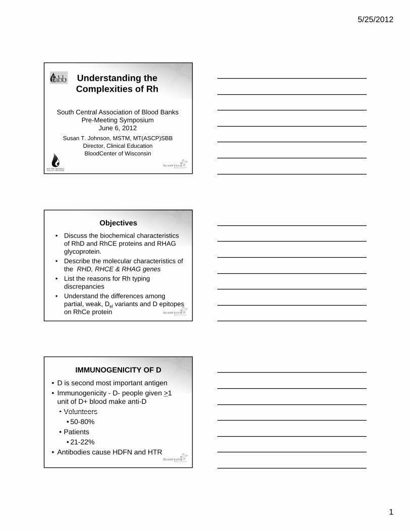
Understanding the Complexities of Rh
South Central Association of Blood BanksP M ti S i
Susan T. Johnson, MSTM, MT(ASCP)SBBDirector, Clinical EducationBloodCenter of Wisconsin
Pre-Meeting SymposiumJune 6, 2012
Objectives• Discuss the biochemical characteristics
of RhD and RhCE proteins and RHAG glycoprotein.
• Describe the molecular characteristics of the RHD, RHCE & RHAG genes
• List the reasons for Rh typing discrepancies
• Understand the differences among partial, weak, Del variants and D epitopes on RhCe protein
IMMUNOGENICITY OF D• D is second most important antigen• Immunogenicity - D- people given >1
unit of D+ blood make anti-D• VolunteersVolunteers
• 50-80% • Patients
• 21-22% • Antibodies cause HDFN and HTR
5/25/2012
2
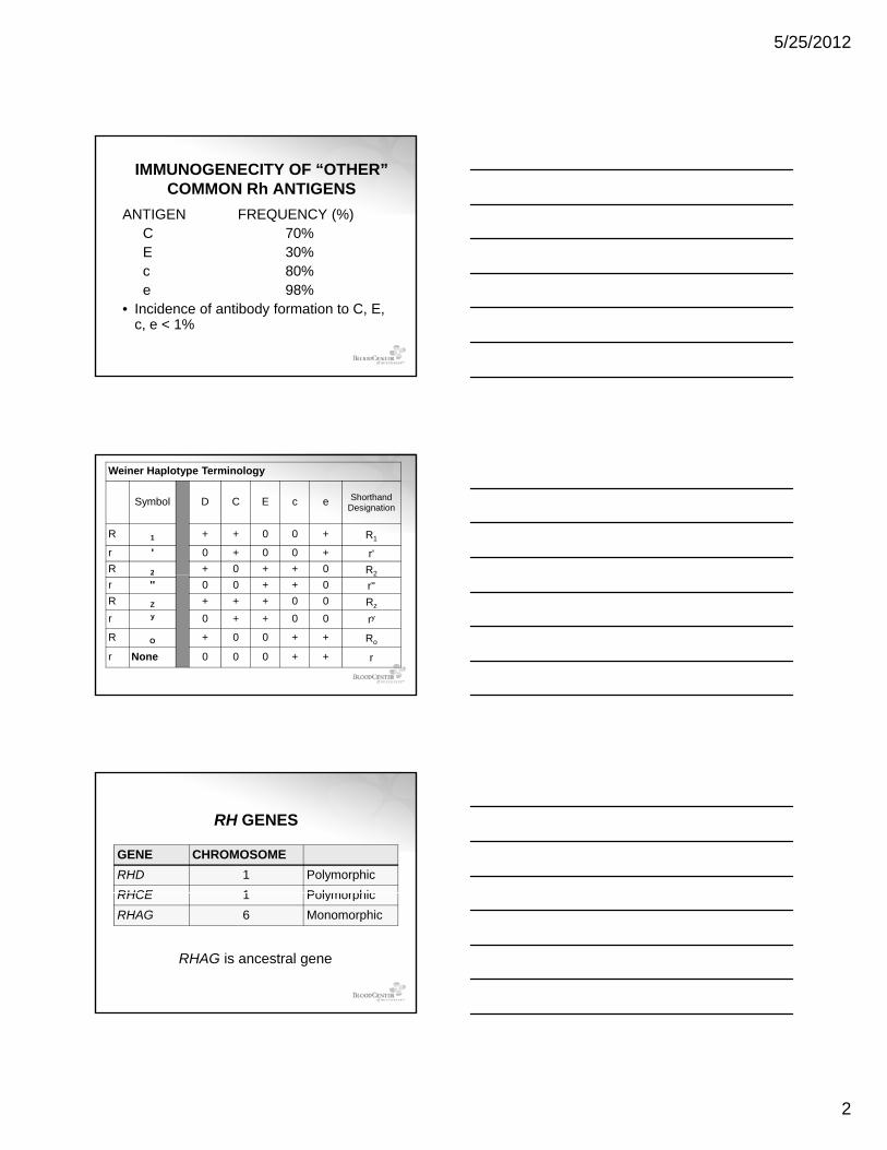
IMMUNOGENECITY OF “OTHER” COMMON Rh ANTIGENS
ANTIGEN FREQUENCY (%)C 70%E 30%E 30%c 80%e 98%
• Incidence of antibody formation to C, E, c, e < 1%
Weiner Haplotype Terminology
Symbol D C E c e ShorthandDesignation
R 1 + + 0 0 + R1
r ' 0 + 0 0 + r'R 2 + 0 + + 0 R22 2
r '' 0 0 + + 0 r''R Z + + + 0 0 Rz
r y 0 + + 0 0 ry
R O + 0 0 + + Ro
r None 0 0 0 + + r
RH GENES
GENE CHROMOSOMERHD 1 PolymorphicRHCE 1 PolymorphicRHCE 1 PolymorphicRHAG 6 Monomorphic
RHAG is ancestral gene
5/25/2012
3
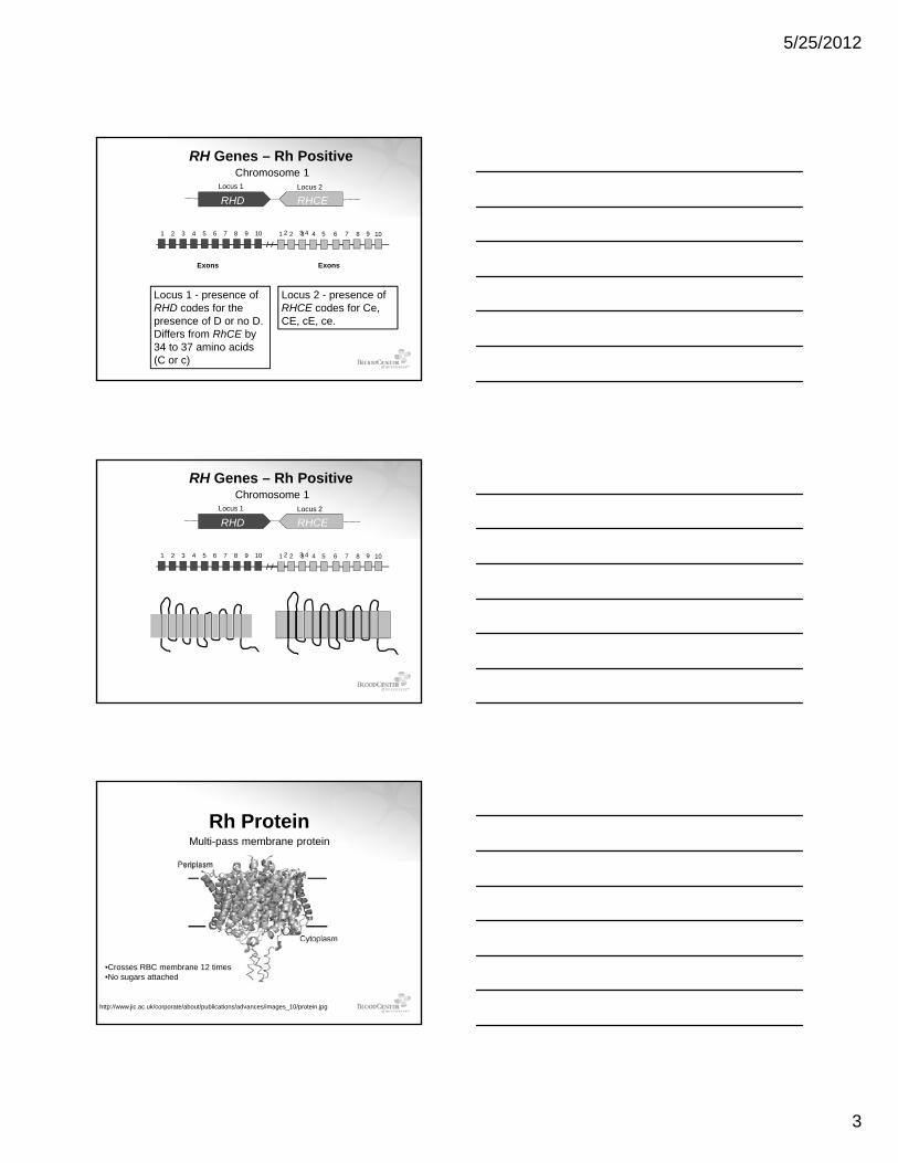
2 3 4 91 2 3 4 5 6 7 8 9 10 1 2 3 4 5 6 7 8 10
Locus 1 Locus 2
RH Genes – Rh Positive
RHD RHCE
Chromosome 1
Exons Exons
Locus 1 - presence of RHD codes for the presence of D or no D.Differs from RhCE by 34 to 37 amino acids (C or c)
Locus 2 - presence of RHCE codes for Ce, CE, cE, ce.
2 3 4 91 2 3 4 5 6 7 8 9 10 1 2 3 4 5 6 7 8 10
Locus 1 Locus 2
RH Genes – Rh Positive
RHD RHCE
Chromosome 1
Rh ProteinMulti-pass membrane protein
http://www.jic.ac.uk/corporate/about/publications/advances/images_10/protein.jpg
•Crosses RBC membrane 12 times•No sugars attached
5/25/2012
4
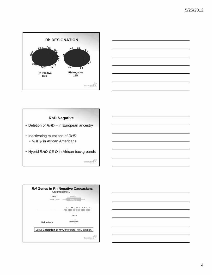
Rh DESIGNATION
DD D
D
D
D
c c
c
ee
eee
ec
cc
c c
ee
c e
Rh Positive85%
Rh Negative15%
DDD
cc
e
ee
ee
cc c
c e
RhD Negative
• Deletion of RHD – in European ancestry
• Inactivating mutations of RHDg• RHDψ in African Americans
• Hybrid RHD-CE-D in African backgrounds
1 3 4 5 6 7 8 91 2 3 4 5 6 7 8 10
Locus 1 Locus 2
RH Genes in Rh Negative Caucasians
RHCE
Chromosome 1
Exons
No D antigens ce antigens
Locus 1 deletion of RHD therefore, no D antigen.
5/25/2012
5
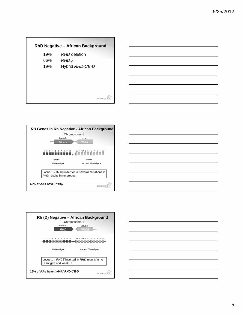
RhD Negative – African Background
19% RHD deletion66% RHDψ19% Hybrid RHD-CE-D
2 3 4 91 2 3 4 5 6 7 8 9 10 1 2 3 4 5 6 7 8 10
Locus 1 Locus 2
RH Genes in Rh Negative - African Background
RHDψ RHCE
Chromosome 1
Exons Exons
No D antigen C/c and E/e antigens
Locus 1 – 37 bp insertion & several mutations in RHD results in no product
66% of AAs have RHDψ
2 3 4 91 2 3 4 5 6 7 8 9 10 1 2 3 4 5 6 7 8 10
Locus 1 Locus 2
Rh (D) Negative – African Background
RHD RHCE
Chromosome 1
C/c and E/e antigensNo D antigen
Locus 1 – RHCE inserted in RHD results in no D antigen and weak C.
15% of AAs have hybrid RHD-CE-D
5/25/2012
6
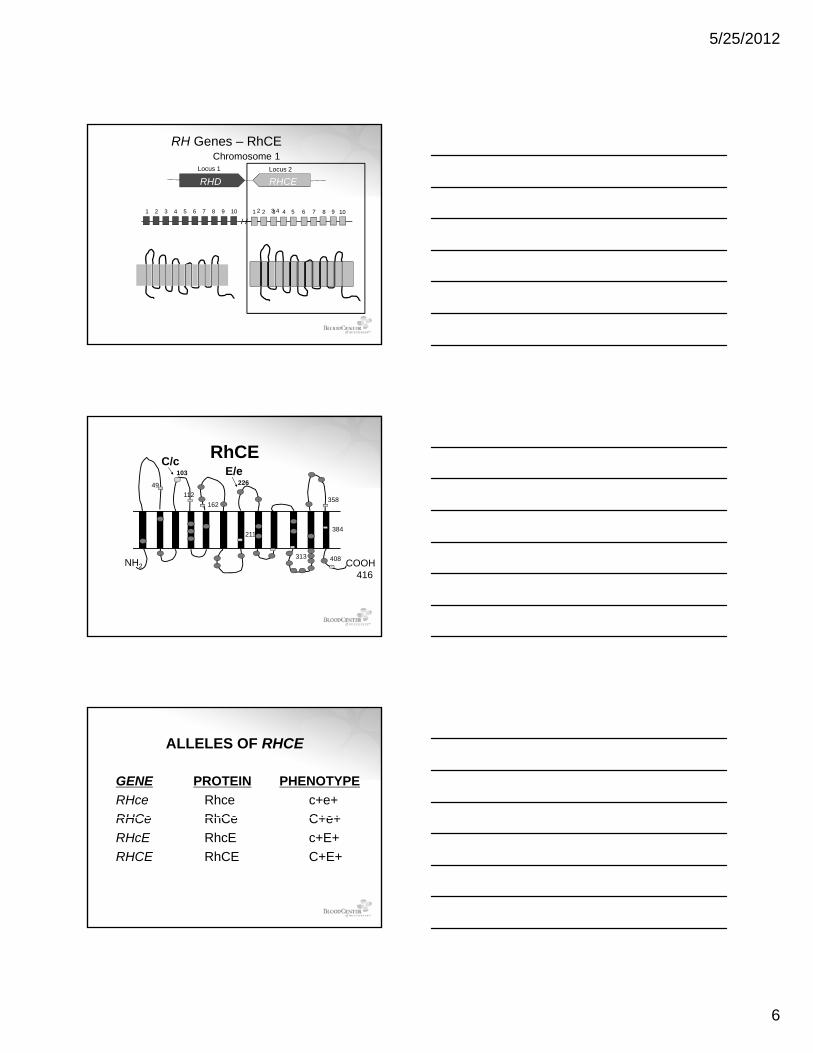
2 3 4 91 2 3 4 5 6 7 8 9 10 1 2 3 4 5 6 7 8 10
Locus 1 Locus 2
RH Genes – RhCE
RHD RHCE
Chromosome 1
RhCE49
112162
358
103226
C/cE/e
COOH416
NH2
211
313
384
408
ALLELES OF RHCE
GENERHceRHCe
PROTEINRhce RhCe
PHENOTYPEc+e+ C+e+RHCe
RHcERHCE
RhCeRhcERhCE
C+e+c+E+C+E+
5/25/2012
7
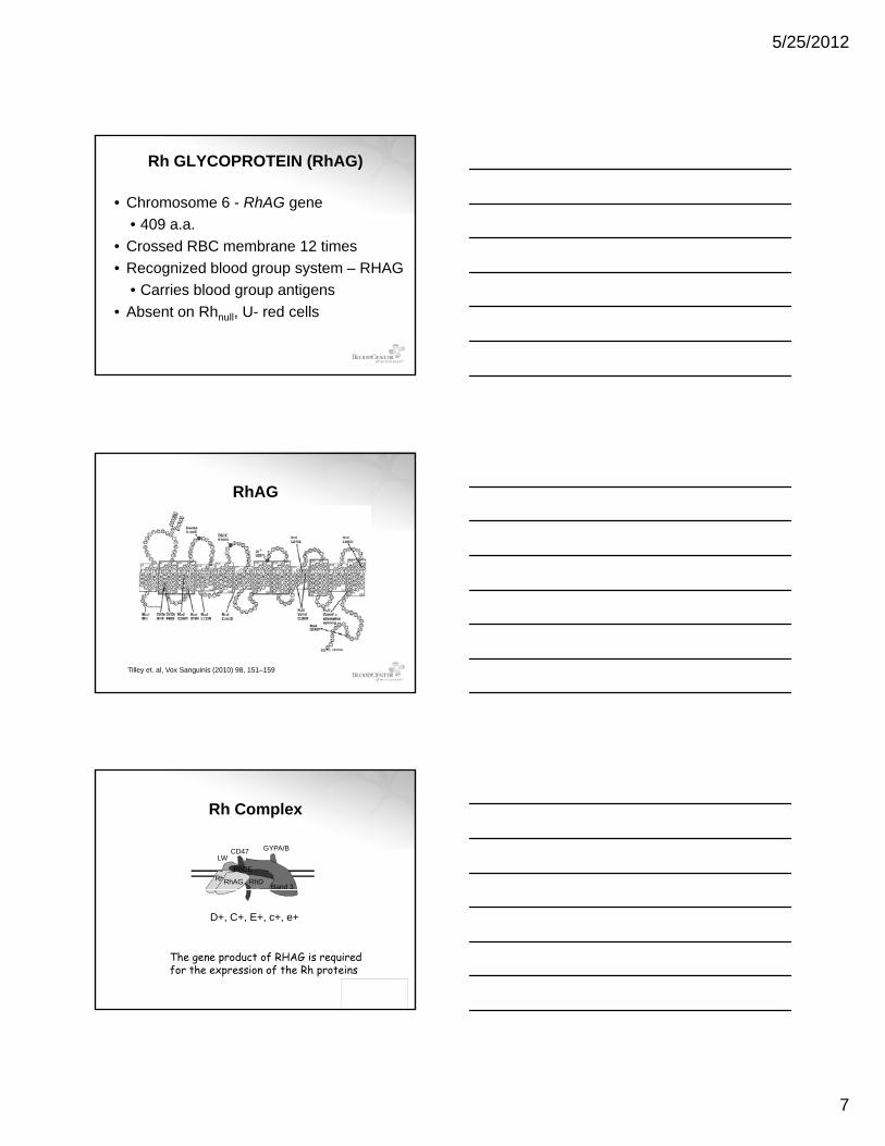
Rh GLYCOPROTEIN (RhAG)
• Chromosome 6 - RhAG gene• 409 a.a.
• Crossed RBC membrane 12 times• Recognized blood group system – RHAG
• Carries blood group antigens • Absent on Rhnull, U- red cells
RhAG
Tilley et. al, Vox Sanguinis (2010) 98, 151–159
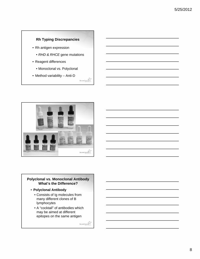
Rh Complex
RhD
RhCELW
CD47
Band 3
GYPA/B
RhAG
D+, C+, E+, c+, e+
The gene product of RHAG is required for the expression of the Rh proteins
5/25/2012
8
Rh Typing Discrepancies
• Rh antigen expression
• RHD & RHCE gene mutations
• Reagent differences
• Monoclonal vs. Polyclonal
• Method variability – Anti-D
Polyclonal vs. Monoclonal AntibodyWhat’s the Difference?
• Polyclonal Antibody• Consists of Ig molecules from
many different clones of B lymphocytes
• A “cocktail” of antibodies which may be aimed at different epitopes on the same antigen
5/25/2012
9
Polyclonal Antibodies
Normal immune response stimulates the production of many different immunoglobulins
Antigen
= immunoglobulin
immunoglobulins recognizing different epitopes on antigen
Monoclonal Antibody
Monoclonal antibody is specific for 1 epitope
Antigen
= immunoglobulin
Monoclonal versus PolyclonalAntigenAg
B cells
expansion
Antibodies
Monoclonal Polyclonal
5/25/2012
10
Monoclonal Reagent Types• Blend of monoclonal & polyclonal
antibodies• Blend of two or more monoclonal
antibodies, each secreted by a different ll licell line
• IgG or IgM, or combination of IgG + IgM• Why?
• D antigen has >30 different epitopes• Variant D antigens
CONTRIBUTORS OF VARIABILITY
VARIABLES
RHD Gene Weak D C in Trans to RHD
Partial D Del
D epitopes onRhCE Protein
ceCF R0Har or DHAR
Anti‐D Reagents Polyspecific Monoclonal Monoclonal Monoclonal Monoclonal
Variables Impacting RhD Typing
Slide and Modified Tube Human IgG
IgG IgM IgM
Human IgG
Blends
Testing Platform Test Tubes IS & IAT
Column Agglutination
Solid Phase Liquid Microtiter
Individual being Rh Typed
Transfusion Recipient
Obstetrical Patient
Cord Blood Donor Blood
Transfusion Technology Report Vol. #013 Immucor, Inc.
Why is Detecting RhD so challenging?so challenging?
5/25/2012
11
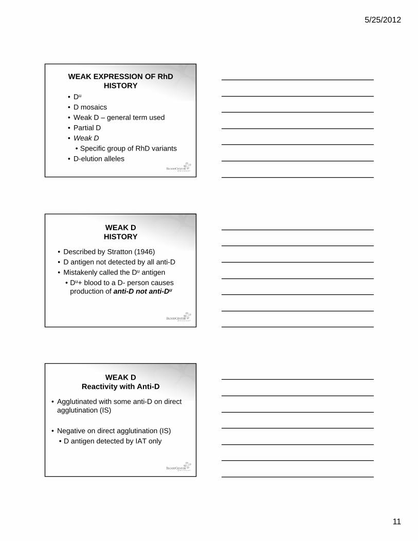
WEAK EXPRESSION OF RhDHISTORY
• Du
• D mosaics• Weak D – general term used g• Partial D• Weak D
• Specific group of RhD variants• D-elution alleles
WEAK D HISTORY
• Described by Stratton (1946)• D antigen not detected by all anti-D
Mi t k l ll d th Du ti• Mistakenly called the Du antigen• Du+ blood to a D- person causes
production of anti-D not anti-Du
WEAK D Reactivity with Anti-D
• Agglutinated with some anti-D on direct agglutination (IS)
• Negative on direct agglutination (IS)• D antigen detected by IAT only
5/25/2012
12
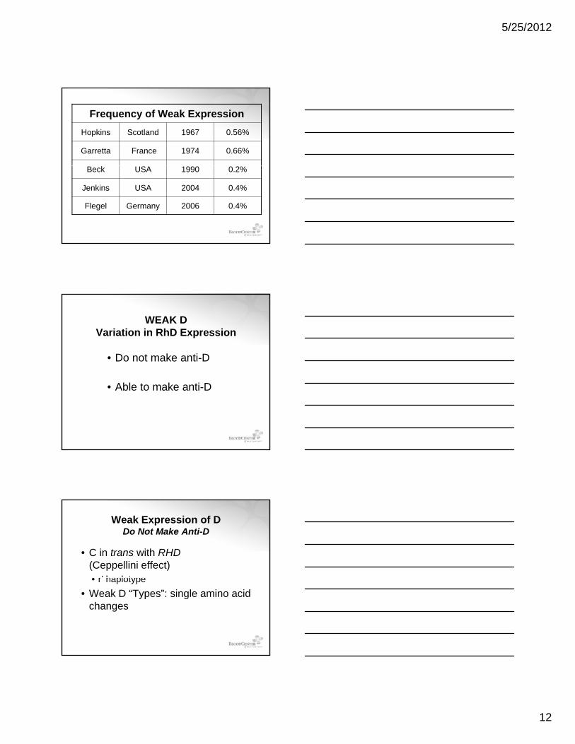
Frequency of Weak ExpressionHopkins Scotland 1967 0.56%
Garretta France 1974 0.66%
Beck USA 1990 0.2%
Jenkins USA 2004 0.4%
Flegel Germany 2006 0.4%
WEAK D Variation in RhD Expression
• Do not make anti-D
• Able to make anti-D
Weak Expression of DDo Not Make Anti-D
• C in trans with RHD(Ceppellini effect)• r’ haplotype• r haplotype
• Weak D “Types”: single amino acid changes
5/25/2012
13
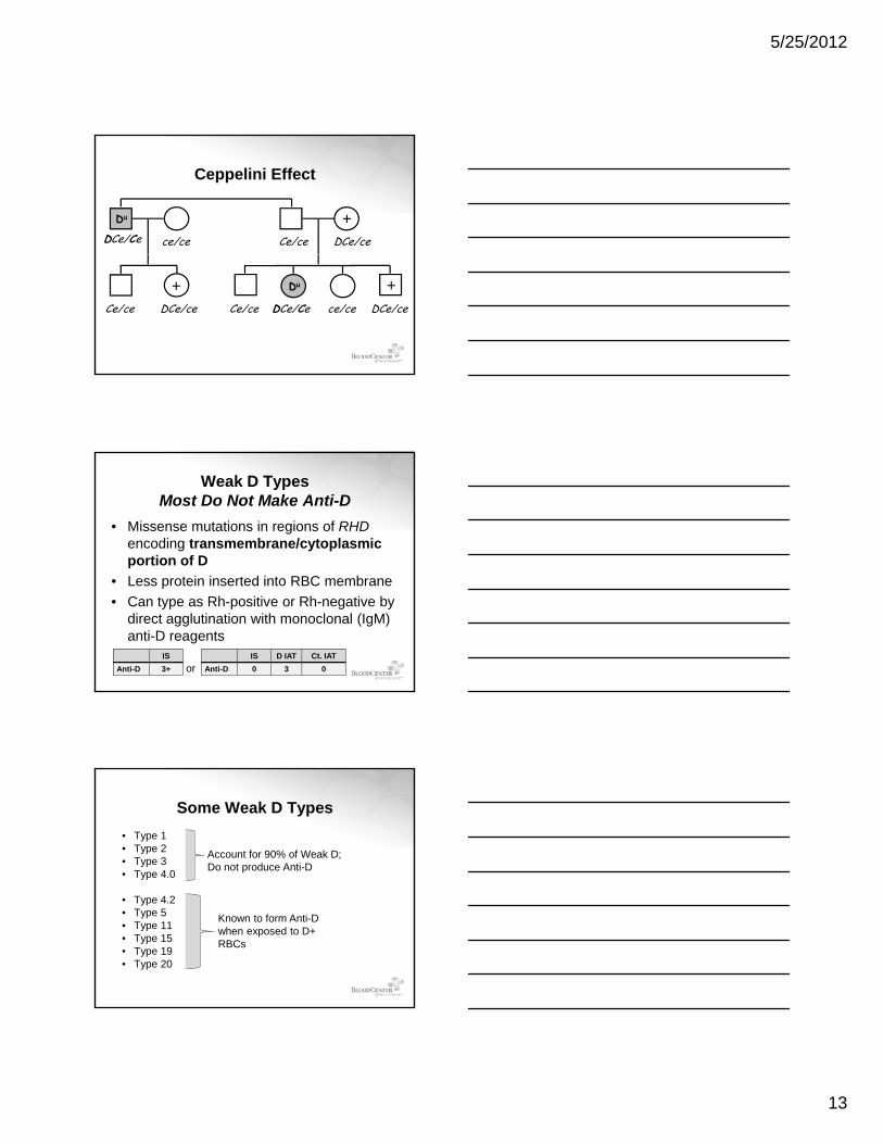
Ceppelini Effect
DCe/Ce ce/ce Ce/ce DCe/ce
Du +
Ce/ce DCe/ce DCe/CeCe/ce DCe/cece/ce
Du+ +
Weak D TypesMost Do Not Make Anti-D
• Missense mutations in regions of RHDencoding transmembrane/cytoplasmic portion of D
• Less protein inserted into RBC membrane• Can type as Rh-positive or Rh-negative by
direct agglutination with monoclonal (IgM) anti-D reagents
IS D IAT Ct. IATAnti-D 0 3 0
ISAnti-D 3+ or
Some Weak D Types • Type 1• Type 2• Type 3• Type 4.0
Account for 90% of Weak D; Do not produce Anti-D
• Type 4.2• Type 5• Type 11• Type 15• Type 19• Type 20
Known to form Anti-D when exposed to D+ RBCs
5/25/2012
14
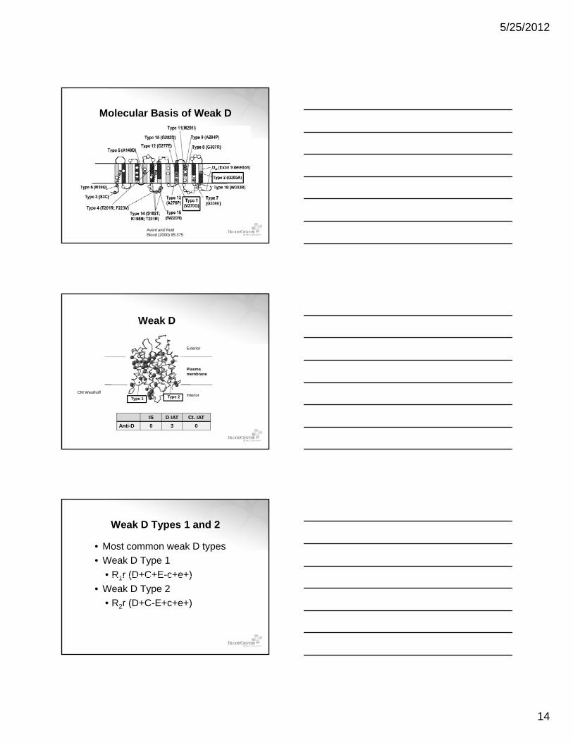
Molecular Basis of Weak D
Avent and ReidBlood (2000) 95:375
Plasma
Exterior
Weak D
membrane
InteriorType 1 Type 2
CM Westhoff
IS D IAT Ct. IATAnti-D 0 3 0
Weak D Types 1 and 2
• Most common weak D types• Weak D Type 1
• R1r (D+C+E-c+e+)R1r (D C E c e )• Weak D Type 2
• R2r (D+C-E+c+e+)
5/25/2012
15
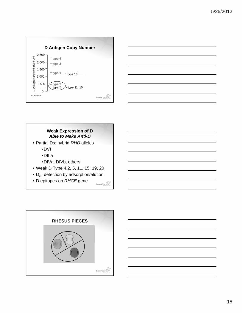
D Antigen Copy Number2,500
2,000
1 500ed B
lood
Cel
l
1,500
1,000
500
0
type 10
type 11, 15type 5D a
ntig
ens
per R
e
G Denomme
Weak Expression of DAble to Make Anti-D
• Partial Ds: hybrid RHD alleles• DVI• DIIIaDIIIa• DIVa, DIVb, others
• Weak D Type 4.2, 5, 11, 15, 19, 20• Del: detection by adsorption/elution• D epitopes on RHCE gene
RHESUS PIECES
5/25/2012
16
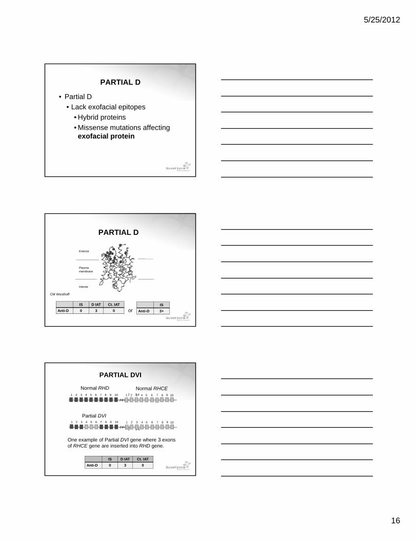
PARTIAL D
• Partial D• Lack exofacial epitopes
• Hybrid proteinsy p• Missense mutations affecting exofacial protein
Plasma
Exterior
PARTIAL D
membrane
Interior
CM Westhoff
IS D IAT Ct. IATAnti-D 0 3 0
ISAnti-D 3+or
2 3 4 91 2 3 4 5 6 7 8 9 10 1 2 3 4 5 6 7 8 10
Partial DVI
Normal RHD Normal RHCE
PARTIAL DVI
2 3 4
91 2 3 4 5 6 7 8 9 10 1 2 3 4 5 6 7 8 10
One example of Partial DVI gene where 3 exons of RHCE gene are inserted into RHD gene.
IS D IAT Ct. IATAnti-D 0 3 0
5/25/2012
17
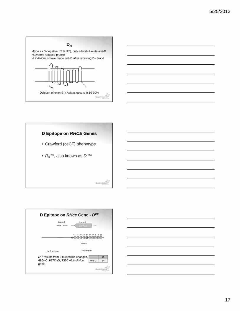
Del
•Type as D-negative (IS & IAT), only adsorb & elute anti-D•Severely reduced protein•2 individuals have made anti-D after receiving D+ blood
Deletion of exon 9 in Asians occurs in 10-30%
D Epitope on RHCE Genes
• Crawford (ceCF) phenotype
• R Har also known as DHAR• R0Har, also known as DHAR
1 3 4 5 6 7 8 91 2 3 4 5 6 7 8 10
Locus 1 Locus 2
RHCE
D Epitope on RHce Gene - DCF
Exons
No D antigens ce antigens
DCF results from 3 nucleotide changes, 48G>C, 697C>G, 733C>G in RHcegene.
ISAnti-D 3+
5/25/2012
18
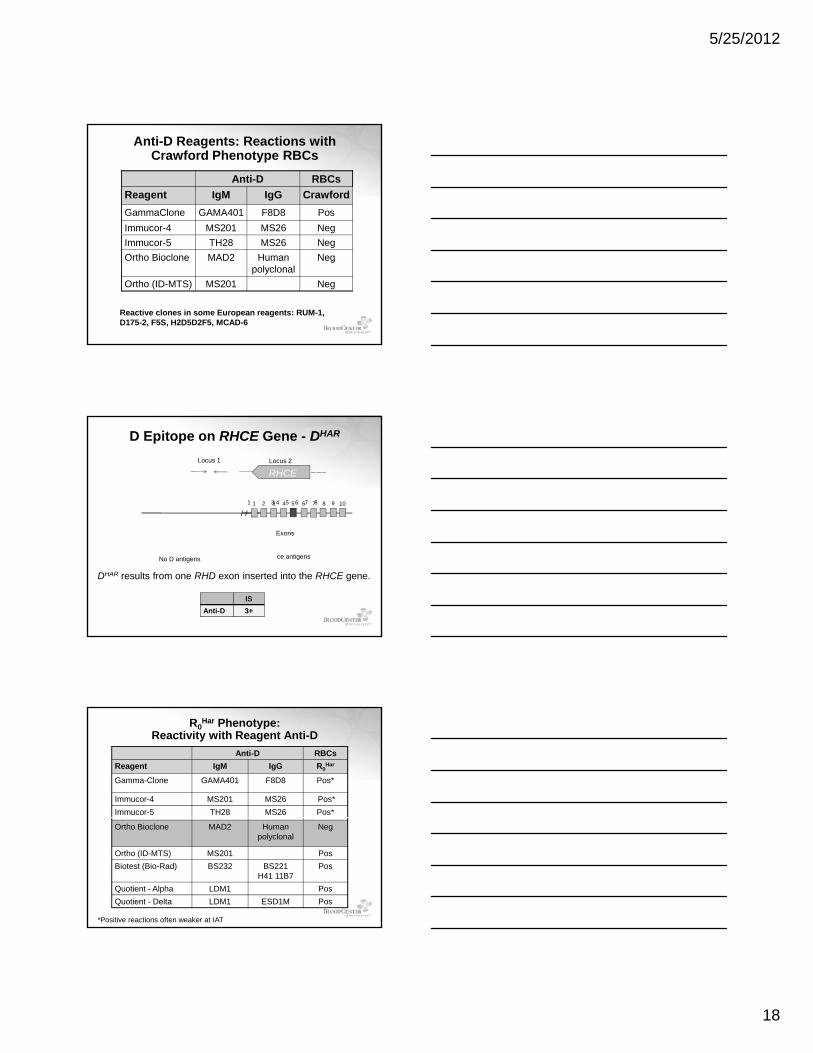
Anti-D Reagents: Reactions with Crawford Phenotype RBCs
Anti-D RBCsReagent IgM IgG CrawfordGammaClone GAMA401 F8D8 PosImmucor-4 MS201 MS26 NegImmucor-5 TH28 MS26 NegOrtho Bioclone MAD2 Human
polyclonalNeg
Ortho (ID-MTS) MS201 Neg
Reactive clones in some European reagents: RUM-1, D175-2, F5S, H2D5D2F5, MCAD-6
1 3 4 5 6 7 8 91 2 3 4 5 6 7 8 10
Locus 1 Locus 2
RHCE
D Epitope on RHCE Gene - DHAR
Exons
No D antigens ce antigens
DHAR results from one RHD exon inserted into the RHCE gene.
ISAnti-D 3+
R0Har Phenotype:
Reactivity with Reagent Anti-DAnti-D RBCs
Reagent IgM IgG R0Har
Gamma-Clone GAMA401 F8D8 Pos*
Immucor-4 MS201 MS26 Pos*Immucor-5 TH28 MS26 Pos*
Ortho Bioclone MAD2 Human polyclonal
Neg
Ortho (ID-MTS) MS201 PosBiotest (Bio-Rad) BS232 BS221
H41 11B7Pos
Quotient - Alpha LDM1 PosQuotient - Delta LDM1 ESD1M Pos
*Positive reactions often weaker at IAT
5/25/2012
19
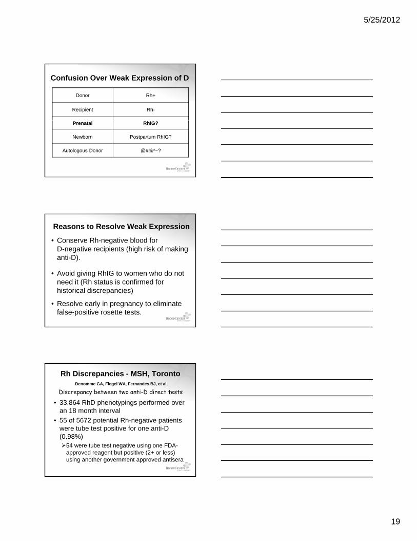
Confusion Over Weak Expression of D
Donor Rh+
Recipient Rh-
Prenatal RhIG?
Newborn Postpartum RhIG?
Autologous Donor @#!&*~?
Reasons to Resolve Weak Expression
• Conserve Rh-negative blood for D-negative recipients (high risk of making anti-D).
• Avoid giving RhIG to women who do not need it (Rh status is confirmed for historical discrepancies)
• Resolve early in pregnancy to eliminate false-positive rosette tests.
Rh Discrepancies - MSH, TorontoDenomme GA, Flegel WA, Fernandes BJ, et al.
• 33,864 RhD phenotypings performed over an 18 month interval
• 55 of 5672 potential Rh negative patients
Discrepancy between two anti-D direct tests
• 55 of 5672 potential Rh-negative patients were tube test positive for one anti-D (0.98%)
54 were tube test negative using one FDA-approved reagent but positive (2+ or less) using another government approved antisera
5/25/2012
20
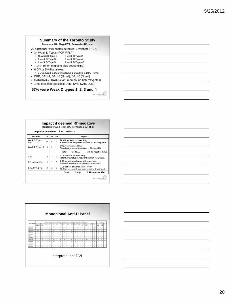
Summary of the Toronto StudyDenomme GA, Flegel WA, Fernandes BJ, et al.
20 functional RHD alleles detected; 1 wildtype (HDN)• 34 Weak D Types (PCR-RFLP):
• 16 weak D Type 1 8 weak D Type 2• 1 weak D Type 3 6 weak D Type 4• 1 weak D Type 5 2 weak D Type 42
• 7 DAR (exon mapping plus sequencing)• 6 DVa or DVa-like alleles:
• 3 DVa(Kou.) 1 DVaHK(E233K) 1 DVa-like 1 DTO (Novel)• DFR, DAU-4, DAU-5 (Novel), DAU-6 (Novel)• DAR/DAU-2, DAU-0/Cdes (compound heterozygotes)• 1 not identified (possible DIIIa, DVa, DAR, DOL)
57% were Weak D types 1, 2, 3 and 4
Impact if deemed Rh-negativeDenomme GA, Flegel WA, Fernandes BJ, et al.
RHD Allele OB TR NB Impact
Weak D Types 1-4 12 8 5 12 OB patients received Rhig
4 transfusion recipients received 12 Rh-neg RBCs
Weak D Type 42 1 1 - OB patient received RhigTransfusion recipient received 11 Rh-neg RBCs
T t l 21 RhIG 23 Rh ti RBC
Inappropriate use of blood products
Total: 21 RhIG 23 Rh-negative RBCs
DAR 3 1 3 3 OB patients received RhigPotential transfusion recipient was not transfused.
DVa and DVa-like 1 1 5 1 OB patient an delivered an Rh-neg infantPotential transfusion recipient not transfused
DAU, DFR, DTO 3 2 2 2 OB patient delivered an Rh+ infantNeither potential transfusion recipient transfused
Total: 7 Rhig 0 Rh-negative RBCs
Monoclonal Anti-D Panel
Interpretation: DVI
5/25/2012
21
Anti-D (std method)Negative
No
Inconclusive Rh NegativeRh Positive
Yes
No No
Anti-D (std method)>2+ agglutination score
No
Yes YesMatches Matches
Yes
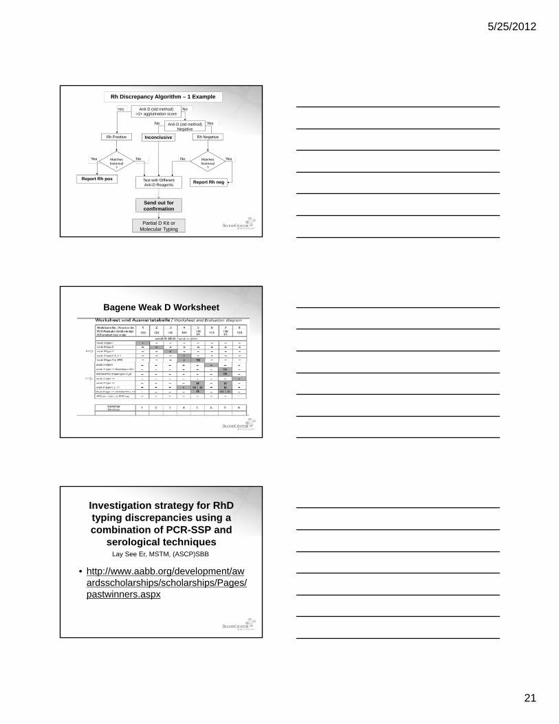
Rh Discrepancy Algorithm – 1 Example
Report Rh pos Test with Different Anti-D Reagents
Send out for confirmation
historical?
historical?
Partial D Kit or Molecular Typing
Report Rh neg
Bagene Weak D Worksheet
Investigation strategy for RhD typing discrepancies using a combination of PCR-SSP and
serological techniquesLay See Er, MSTM, (ASCP)SBB
• http://www.aabb.org/development/awardsscholarships/scholarships/Pages/pastwinners.aspx

5/25/2012
22
Bagene Weak D Kit Results
Weak D type 1
Lane 2: DNA ladderStart reading from lane 3Lane 1, 11,12: buffer load (no bands)
Bagene Weak D Kit Results
Weak D type 2Weak D type 2
Lane 2: DNA ladderStart reading from lane 3Lane 1, 11,12: buffer load (no bands)
Weak D type 2Weak D type 2
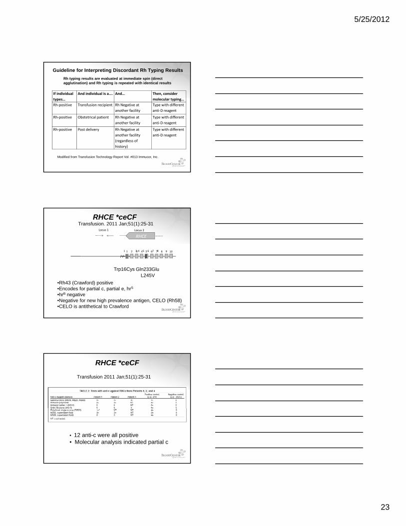
If individual types…
And individual is a…. And… Then, consider molecular typing…
Rh‐negative Transfusion recipient Donor record is Rh‐positive
Interpret Rh‐negative
Rh‐negative Obstetrical patient Donor record is Rh‐positive
Interpret Rh‐neg or Rh‐pos?
Rh ti P t d li D d i P f ti D IAT*
Guideline for Interpreting Discordant Rh Typing ResultsRh typing results are evaluated at immediate spin (direct agglutination) and Rh typing is repeated with identical results
Rh‐negative Post delivery Donor record is Rh‐positive
Perform anti‐D IAT*
Rh‐negative Transfusion recipient Facility history is Rh‐positive
Interpret Rh‐negative
Rh‐negative Obstetrical patient Facility history is Rh‐positive
Interpret Rh‐neg or Rh‐pos?
Rh‐negative Post delivery Facility history is Rh‐positive
Perform anti‐D IAT*
Modified from Transfusion Technology Report Vol. #013 Immucor, Inc.
5/25/2012
23
If individual types…
And individual is a…. And… Then, consider molecular typing…
Rh‐positive Transfusion recipient Rh Negative at another facility
Type with different anti‐D reagent
Rh positive Obstetrical patient Rh Negative at Type with different
Guideline for Interpreting Discordant Rh Typing ResultsRh typing results are evaluated at immediate spin (direct agglutination) and Rh typing is repeated with identical results
Rh‐positive Obstetrical patient Rh Negative at another facility
Type with different anti‐D reagent
Rh‐positive Post delivery Rh Negative at another facility (regardless of history)
Type with different anti‐D reagent
Modified from Transfusion Technology Report Vol. #013 Immucor, Inc.
1 3 4 5 6 7 8 91 2 3 4 5 6 7 8 10
Locus 1 Locus 2
RHCE
Transfusion. 2011 Jan;51(1):25-31RHCE *ceCF
L245VGln233GluTrp16Cys
•Rh43 (Crawford) positive•Encodes for partial c, partial e, hrS
•hrB negative•Negative for new high prevalence antigen, CELO (Rh58)•CELO is antithetical to Crawford
RHCE *ceCF
Transfusion 2011 Jan;51(1):25-31
• 12 anti-c were all positive • Molecular analysis indicated partial c
5/25/2012
24
Objectives• Discuss the biochemical characteristics
of RhD and RhCE proteins and RHAG glycoprotein.
• Describe the molecular characteristics of the RHD RHCE & RHAG genesthe RHD, RHCE & RHAG genes
• List the reasons for Rh typing discrepancies
• Understand the differences among partial, weak, Del variants and D epitopes on RhCe protein
References• Wagner FF, Gassner C, Mu¨ ller TH, et al. Molecular basis of weak
D phenotypes. Blood 1999; 93:385–393.• Denomme GA, Wagner FF, Fernandes BJ, et al. Partial D, weak D
types, and novel RHD alleles among 33 864 multiethnic patients: implications for anti-D alloimmunization and prevention. Transfusion 2005; 45:1554–1560.
• Flegel WA Denomme GA Yazer MH On the complexity of D• Flegel WA, Denomme GA, Yazer MH. On the complexity of D antigen typing: a handy decision tree in the age of molecular blood group diagnostics. J Obstet Gynaecol Can. 2007;29:746-52.
• Flegel WA. How I manage donors and patients with a weak D phenotype. Curr Opin Hematol 2006;13:476–483
• Flegel WA. Molecular genetics and clinical applications for RH. Transfusion and Apheresis Science 2011;44:81-91. 2.
• Sandler SG, Li W, Langeberg AL, Landy HJ. New Laboratory Procedures and Rh Blood Type Changes in a Pregnant Woman. Obstet Gynecol 2012;119:426–8.
Thank You


![The Complexities of Identity Formation[1]](https://img.pdfslide.net/doc/110x75/577d23a51a28ab4e1e9a612f/the-complexities-of-identity-formation1.jpg)