Embed Size (px)
Citation preview

1
Unit 3: Chemistry of Life (Cellular Homeostasis)
Targets Text 1) Describe the importance of the process of mitosis in the production of new body cells to make more cells for growth and repair
10.1, 10.2
2) Recognize that cells within organisms are specialized for different functions by which organelles they either possess or lack.
7.4
3) Describe the importance of a selectively permeable cell membrane in maintaining homeostasis by controlling what enters & leaves the cell.
7.3, 7.4
4) Use the processes of osmosis and diffusion to explain how, when cells are placed in different solutions (hypertonic, hypotonic, and isotonic), materials move both in and out of cells.
7.3
Key Terms Concentration Difference (gradient)
Concentration Diffusion Homeostasis Hypertonic Hypotonic
Impermeable Isotonic Osmosis Permeable Selectively Permeable Solute
Solution Solvent Facilitated Diffusion
Passive Transport
Active Transport Endocytosis
Exocytosis Phagocytosis Tissue Organ Organ System Receptor
Mitosis Cell Division Asexual Reproduction Chromatid Chromosomes Chromatin
Interphase Prophase Metaphase Anaphase Telophase Cytokinesis
Centrioles Centromere

2

3
Membrane Transport: Passive Transport Name: ______________________ Date: _________ Period: ______
Go to the following website: http://www.northland.cc.mn.us/biology/Biology1111/animations/transport1.html Pay attention, the “back” button can only be used at the end of the animation segment. (Firefox or Safari) If you want to pause, while your cursor is on the animation, hold the control button (ctrl) and click. This should freeze the animation, release & animation should continue. (Internet explorer) click on the “help” button on the internet explorer menu and hold, release this button when you are ready to continue. Click on passive transport Tour of the cell membrane 1. Passive transport is the movement of molecules across the cell that does not require ______ _____________________. 2. What are the three types of passive transport?
• • •
Click “next” Label the diagrams
Watch the rest of the animation, follow along carefully. Do not answer the questions below until the end of the segment. 3. Why is it important to note the lipid bilayer is fluid? 4. Which part of the lipid bilayer is polar (hydrophilic)? Non-polar (hydrophobic)? Label the diagram on the right. 5. Which type of molecules have difficulty passing through the membrane? 6. Which type of molecules pass easily through the membrane?

4
Click “Next” and go to “Simple Diffusion” 7. Diffusion is the movement of molecules from an area of __________________ to an area of ___________________. 8. The rate of diffusion is dependent on:
• steepness of the concentration gradient • • •
9. Diffusion ends when equilibrium is met.....What is meant by “equilibrium”? Click “Next” and go to “Facilitated Diffusion” 10. Facilitated diffusion is the passive transport of molecules down a concentration gradient with the aid of_____________________. 11. How does the protein “facilitate” the diffusion of the molecule? Click “Next” and go to “Osmosis” 12. How is osmosis almost exactly like diffusion? (what is the difference?) 13. Watch the remaining part of the animation & take your own notes, stop when you get to “Active Transport”....pay attention, you will hear more about hypertonic and hypotonic solutions in class!

5
Membrane Transport: Part II-Active Transport Name: ______________________ Date: _________ Period: ______
Using Firefox or Safari go to the following website: http://www.northland.cc.mn.us/biology/Biology1111/animations/transport1.html Pay attention, the “back” button can only be used at the end of the animation segment. If you want to pause, while your cursor is on the animation, hold the control buttom (ctrl) and click. This should freeze the animation, release & animation should continue. (Internet explorer) click on the “help” button on the internet explorer menu and hold, release this button when you are ready to continue. Click on active transport 1. Active transport is the _______________ of molecules against their _____________ ____________ with the expenditure of ___________( ___ ). 2. Name three types of active transport
• • •
Click next 3. In the diagram below add in the Hydrogen ions (H+) and label the protein pump. Before
After (draw the hydrogen ions)
4. What does “against the concentration gradient” mean? (think about how this is different from passive transport!) 5. What was needed for the ions to move “against the concentration gradient”? How did this needed component change? Click next to continue on to “cotransport” Cotransport

6
6. Define “cotransport”. 7. Explain cotransport using sucrose (sugar) and hydrogen as an example. Click next to continue on to “endocytosis” Endocytosis 8. Define “endocytosis” 9. Name three types of endocytosis:
• Phagocytosis ( ) • Pinocytosis ( ) • endocytosis
Click next to continue 10. What is a pseudopod? 11. 1) Draw a three step picture explaining Phagocytosis, 2) include labels on drawings: pseudopod, food & vesicle, define the inside & outside of the cell) & 3) write a caption describing what is happening in each step. Caption: Caption: Caption: Click next to continue on to “pinocytosis” 12. Watch the animation and then name two ways Phagocytosis if different from Pinocytosis. Click next to continue on to “receptor mediated” endocytosis 13. Draw a two step picture explaining receptor mediated endocytosis (include labels for coated pits and receptor proteins). Write a caption for both steps. Caption: Caption:
Demos: Magnificent Moving Molecules

7
Experiment #1:
Observations: Questions: 1. Why do the people who are closer smell it first? 2. Where was it most concentrated? 3. Where was it least concentrated? 4. The substance moved: (hint: look @ your answers to #2 and #3) 5. What is this process called?
Experiment #2:
Observations: Questions: 1. Where was it most concentrated? 2. Where was it least concentrated? 3. The substance moved: 4. What is this process called?
Experiment #3
Observations:

8
Before After
Questions: 1. What substance moved? 2. At the beginning, where was it most concentrated? 3. At the beginning, where was it least concentrated? 4. The substance moved: 5. What is this process called?
Experiment #4 Observations:
Before After
Questions: 1. What substance moved? 2. At the beginning, where was it most concentrated? 3. At the beginning, where was it least concentrated? 4. The substance moved: 5. What is this process called?
7.3 Cell Transport Name: _____________________________
Date: _______________ Period: ________

9
Go to Biology.com via PearsonSuccessNet, click explore, Unit 3 Cells, Ch 7, 7.3 Cell Transport, Activities, finally Art in Motion
TypeofActiveTransport
Usekeytoidentifywhatsubstancemovedduringanimation
DirectionofMovement
AdditionalNotes
Biology: The Internal Environment of Organisms Name: _______________________

10
Lab: Cells in Action-Part I Date: ____________Period: _____ How big is a cell? Most cells are so small you need a microscope to see them, but there are exceptions. A chicken egg is actually a single cell, although it is an unusually large one. It is protected by a hard shell that surrounds several soft membranes that you can see when you peel a hard-boiled egg. Placing a chicken egg in an acetic acid (vinegar) solution for three days causes the calcium in the hard shell to dissolve. As a result, what remains is a fragile chicken cell surrounded by a soft cell membrane. This membrane allows certain substances to pass in and out of the cell. Experiment 1-Problem: What will happen when the cell is placed into a 20% salt water environment. Write a prediction: Procedure:
1. Using a graduated cylinder, measure out 100 mL of 20% salt solution into your beaker.
2. Carefully get an egg and rinse it under tap water. Gently pat it dry with a paper towel.
3. Determine the mass of the egg on the electronic balance using the plastic weigh dish (make sure the reading is in grams not ounces). Record the mass of the egg (this is your initial mass).
4. Place the egg in the beaker containing the 100mL salt solution. Your egg should be
almost completely covered with solution, add more salt solution if necessary, but note how much you added (you will need to use the same amount in Experiment 2). Leave it in the solution for 15 minutes .
5. Empty the salt water down the drain, and remove the egg and gently pat it dry as you
did before. Clean and dry your beaker (you will use it again in Experiment 2).
6. Determine the mass of the egg, record it in your data table. (This is the mass of egg after salt solution)
Experiment 2-Problem: What will happen when the cell is placed into a distilled water environment? Write a prediction: Procedure:
7. Rinse the egg (use the same egg from Experiment 1) gently in tap water & gently pat it dry with a paper towel. Record initial mass of egg as you did in Experiment 1.
8. The egg will now be placed in a beaker of distilled water for 15 minutes (use the same
amount that you used to cover your egg in Experiment 1).
9. Remove the egg and follow the procedure as you did before to determine the mass of the egg. Record this information in your data table. Record all of your data on the class data projected overhead.
Experiment 3-Control: Your instructor will place an egg out for 15 minutes (not in solution), this will be your control group. Record the mass before and after the 15 minute period.

11
Intro to Lab: Cells in Action-Part 1 Name: ________________________
Date: ________ Period: ______
Work with your partner to complete the objectives below. Make sure you and your partner agree, one will be randomly graded from the pair.
Questions:
1. What will happen to an egg if it is placed in vinegar for 3 days? (be specific)
2. Why do the contents of the egg stay inside even though the shell has been removed?
3. Even though the egg (the cell) is not alive, what could be studied in this experiment?
4. What is the independent variable in this experiment? _________________________ 5. What is the dependent variable in this experiment? ________________________
6. Write a hypothesis (use proper format) for:
a. Experiment #1: _______________________________________________________
_____________________________________________________________________ b. Experiment #2: _______________________________________________________
_____________________________________________________________________
7. Indicate the proper axis for each variable (use answers from 4&5) in a graph of this experiment.
a. X-axis: b. Y-axis:
8. Identify at least 3 valid constants/controlled variables in this experimental design:
a. b. c.
Points: 10 9 8 7 6 5 4 3 2 1

12
9. Design a data table in the space below for recording ALL the results of the egg lab. (Include all proper components of a data table!) Correct answers to questions #4&5 will help you.
13
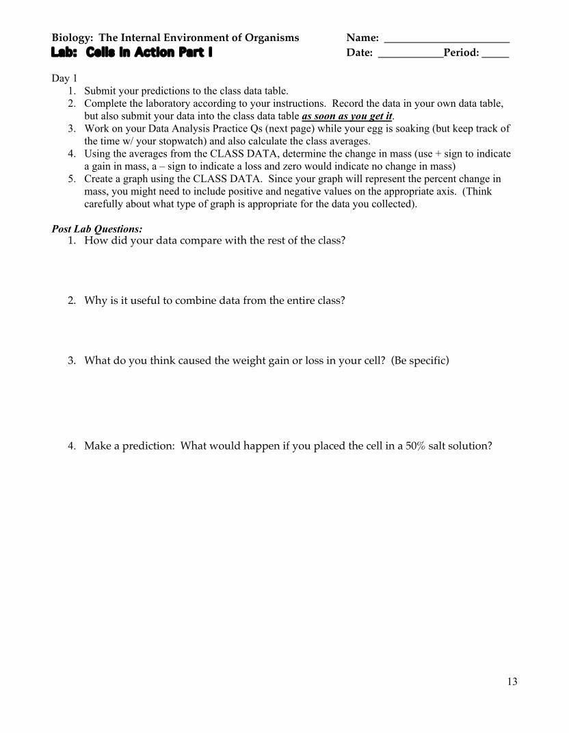
Biology: The Internal Environment of Organisms Name: _______________________ Lab: Cells in Action Part I Date: ____________Period: _____ Day 1
1. Submit your predictions to the class data table. 2. Complete the laboratory according to your instructions. Record the data in your own data table,
but also submit your data into the class data table as soon as you get it. 3. Work on your Data Analysis Practice Qs (next page) while your egg is soaking (but keep track of
the time w/ your stopwatch) and also calculate the class averages. 4. Using the averages from the CLASS DATA, determine the change in mass (use + sign to indicate
a gain in mass, a – sign to indicate a loss and zero would indicate no change in mass) 5. Create a graph using the CLASS DATA. Since your graph will represent the percent change in
mass, you might need to include positive and negative values on the appropriate axis. (Think carefully about what type of graph is appropriate for the data you collected).
Post Lab Questions:
1. How did your data compare with the rest of the class?
2. Why is it useful to combine data from the entire class?
3. What do you think caused the weight gain or loss in your cell? (Be specific)
4. Make a prediction: What would happen if you placed the cell in a 50% salt solution?

14
Name: ______________________ Date: _______ Period: _____
Biology Unit 3: Cells & Membrane Transport Data Analysis Practice In a biology class, students conducted and experiment to see how substances move from the small intestine into the bloodstream. The students filled an intestinal sac with glucose solution at a concentration of 275 mg glucose per 100 mL solution. The normal concentration of glucose in the bloodstream is 180 mg per 100 mL solution. The students measured glucose concentration in the sac at 15-minute intervals. They plotted the results on the graph below.
1. Over the 90 minutes of the experiment, the concentration of glucose in the intestine
A. increased. B. decreased. C. stayed the same. D. increased then decreased.
2. At 60 minutes the concentration of glucose in the intestine was
A. 225 mg% B. 100 mg% C. 60 mg% D. 45 mg%
3. Until what point in the experiment can the results be explained by diffusion alone?
A. 25 minutes B. 40 minutes C. 70 minutes D. 85 minutes
4. What can you conclude from this experiment about the movement of dissolved food and other substances from the small intestine into the bloodstream?
A. That dissolved substances move from the intestine into the bloodstream until the levels in the bloodstream are equal to that in the intestine.
B. That dissolved substances in the intestine do not move into the bloodstream. C. That dissolved substances in the intestine move into the bloodstream over time. D. That dissolved substances in the intestine absorb nutrients from the bloodstream.

15
X 100 = Percent Change
Class Predictions for Experiments 1 & 2 Gains Mass Loses Mass Mass stays the Same Salt Solution Distilled Water Class Data: Cells in Action-Part A
Types of Solutions/Liquid 20% Salt Solution Distilled Water No Solution (Control)
Partners Initial Mass of Egg (g)
Final Mass of Egg (g)
Initial Mass of Egg (g)
Final Mass of Egg (g)
Initial Mass of Egg (g)
Final Mass of Egg (g)
1 2 3 4 5 6 7 8 9 10 11 12
Average Calculate the percent increase or decrease in mass following each condition. How to calculate percent change: (Mass after 15 minutes) - (Mass initially)
(Mass Initially)
The % Change in an Egg’s Mass in Various Solutions Types of Solution Salt Solution Distilled Water No Solution
Class Average: % Change in Egg’s
Mass (g)

16
Biology: The Internal Environment of Organisms Name: _____________________ Post-Lab Discussion: Cells in Action: Part 1 Look at the Class Data. For most groups, what happened when: The egg was in concentrated salt solution: The egg was in distilled water: Conclusion: WHY does this happen? Review solutions: 2 parts of a solution ______________ and ________________ Some solutions are more concentrated than others. Some are less concentrated than others. Weʼll focus on the _________, which is ___________. Think of egg in beaker as 2 areas: ________ the egg, and ___________ the egg.

17
Summary: Egg in a Beaker of Distilled Water Egg in a Beaker of Salt Solution

18
Membranes & Membrane Transport Cell Membrane
Lipid Bilayer Channel Protein Selective Permeability
Passive Transport Movement of molecules: Three types:
Diffusion
Movement of molecules: Rate of Diffusion depends on:
Moving Down the Gradient
Membrane Transport: Facilitated Diffusion

19
Osmosis
Equilibrium Passive vs. Active Transport Endocytosis
Pinocytosis Phagocytosis
Exocytosis

20
Biology: The Internal Environment of Organisms Name: _____________________ Lab:CellsinAction:Part2 Date: ____________Period: _____ Observing Cell Activity PreLab Questions Think back to what you learned about solutions (Section 2.2 can refresh your memory) and use the background information in Section 7.3 to answer the questions below. 1. The substances in a solution are can be described by the terms solute and solvent. Dissolved substance: __________________ Dissolving agent: ____________________ 2. Which substance is present in the greater amount? ________________ 3. Describe the solute concentration outside the cell compared to inside the cell in each of the following solutions (using the terms high, low, equal)
Prepare a wet mount of onion skin
1. Remove 1 layer from your onion wedge.
2. Snap the layer backward, as demoed by your instructor.
3. Use the forceps to separate a piece of the transparent, tissue-thin layer from the outside of the original layer.
4. Lay a piece (smaller than coverslip) flat on a clean microscope slide.
5. Use a toothpick to smooth out any bubbles or wrinkles
6. Add a drop or two of water + 1 drop of iodine, then place cover slip over the onion skin.
7. Examine the onion skin under low power, focus.
8. Switch to medium power and focus. Final move to high power. Sketch the cells in the field to the right.
9. Don’t forget to write the TOTAL MAGNIFICATION below the field of view.
Outside the cell
Inside the Cell Isotonic
Hypertonic
Hypotonic
Onion Cells
Total Magnification ________

21
10. Test the effects of changing the external environment of
the cells you are viewing. To do this, place several drops of 20% salt solution against the edge of the cover slip opposite from a small piece of paper towel. Add more salt solution, if necessary, until you see changes in the cells. Draw your observations below.
Plasmolysis Go to the following website: http://www2.d125.org/science/secure/bio/unit3/plasmol.html You will see a four slide summary of today’s lab, sketch the first and last slide of the computerized version, use color , include LABELS!
Take your time making your drawings, you will be evaulated on your details! Use
the “next” button () in the upper right to advance through the 4 slides.
(This is especially helpful if your experiment did not go well)
DRAWWHATYOUSEEINSLIDES1&4ONTHENEXTPAGE
Onion Cells after addition of Salt Water
Total Magnification ________

22
Slide 1
Cover Slip
Slide 4
Cover Slip
Now answer the your post-lab questions (below), the online activity should match the events with your onion cell (previous 2 pages). If they didn’t
match, base your answers on the correct data from the online activity!
PostLab Questions 1. What changes (besides the orange (iodine) color getting lighter!) occurred when salt
water was added to the onion cells? (look @ your drawings above, the answer is there!)
2. What caused the change inside the cell to occurred? (think about the PreLab questions @ the start of this lab & use your text book to help you explain!)

23
Post-Lab : Cells in Action Part 2
Describe what happened to the onion cells in the salt solution. Describe what happens to red blood cells in Isotonic, Hypertonic & Hypotonic Solutions:
Hypotonic Solution Cell will __________ ( ) Less _________ outside the cell than inside Solute will ___________________ More ___________ ( ) outside the cell than inside Water undergoes __________________......moves _______ the cell causing it to _________
Another Example: Elodea • Salt solution: Hypertonic
Cell _______________ More ________ (____________) outside the cell than inside Salt will diffuse in More __________ (___________) inside the cell than outside Water undergoes osmosis (diffusion of water from an area of greater concentration to lesser)
What can be concluded based on the animation below?

24
Post Lab: Osmosis Practice Name: ________________________
Date: ______ Period: _____ For questions 1&2, a beaker is shown, with 2 solutions on either side of a selectively permeable membrane. Draw an arrow to show which way water will move across the membrane.
1. 2. 3. In the beaker in #1, which way would the solute move? _________________________
4. In the beaker in #2, which way would the solute move? _________________________ Types of Solutions 5. A semipermeable sac, with a solution containing 4% NaCl, 9%
glucose, and 10% albumin is suspended in a solution with the following composition: 10% NaCl, 10% glucose, and 40% albumin. Use the information to create a model in the beaker to the right. (write [ ] inside & out & use arrows to show movement)
6. Assume the sac is permeable to all substances except albumin.
Using the key choices, insert the letter and phrase indicating the correct event in the answer blanks.
a. _______________________________ Glucose
b. _______________________________ Water (solvent)
c. _______________________________ Albumin
d. _______________________________ NaCl
A. Moves into the sac B. Moves out of the sac C. Does not move

25
7. The figure above shows three microscopic fields (A-C) containing red blood cells. Arrows indicate the direction of net osmosis. Respond to the following questions, referring to the figure, by inserting your responses in the spaces provided.
a. Which microscope field contains a hypertonic solution? ____________________ The
cells in this field are said to be _____________________________________
b. Which microscopic field contains an isotonic bathing solution? ______________ What
does isotonic mean? ____________________________________________
c. Which microscopic field contains a hypotonic solution? ____________________ What is
happening to the cells in this field? ______________________________ Why?
____________________________________________________________
__________________________________________________________________

26
Unit 3 Crossword Review
ACROSS2.Theprocessoftakingmaterialintothecellbymeansofinfoldingsorpocketsofthecellmembrane;itrequiresenergy.3.Thisprocessdoesnotrequireenergy;movesmoleculesfromanareaofhighconcentrationtolowconcentration.6.Atypeofendocytosisinwhichthecelltakesinliquids.8.Thearrangementofmoleculesincellularmembranes.10.Apropertyofbiologicalmembranesthatallowssomesubstancestocrossandpreventsothersfromcrossing.11.Atypeofsolutionwheretheconcentrationofsolutesoutsideislessthantheconcentrationinside.12.Theprocessinwhichmoleculesmustpassacrossthemembranethroughcellmembranechannelsfromhightolowconcentration.
DOWN1.Themovementofasubstanceacrossabiologicalmembraneagainstitsconcentrationgradient(fromlowconcentrationtohighconcentration)withthehelpofenergyinput&specifictransportproteins.4.Thediffusionspecificallyofwatermolecules(notsolutemolecules).5.Thisiswhatwillhappentocellsinhypotonicsolutions.7.Thedissolvingagentinasolution.9.Someproteinsinthemembraneplaythisrolewhentheybindtomoleculeslikehormonesandthentriggerthecelltoactinsomecertainway.
Name: _____________________________ Date: _______________ Period: ________

27
Biology:TheInternalEnvironmentofOrganisms LAB:ACELLMODEL PreLab Questions After you have highlighted the lab (the next 3 pages) & completed the prompts (answer questions, write hypothesis, create data tables, etc.). Answer the questions below to check for understanding. Answer in COMPLETE sentences unless a fill-in-the blank is provided. CHECK YOUR SPELLING! 1. What will you do in this activity? 2. What will the dialysis tubing represent in this lab? (you need to think about this!) 3. What type of bio-molecule is Glucose? (this was discussed in Unit 2!) 4. What type of bio-molecule is Starch? (this was discussed in Unit 2!) 5. Given a solution of glucose & one of starch, which contains a solute large enough to be seen
by the naked eye? _______________________ 6. How will you test for the presence of starch? Describe a positive test. 7. What is the function of the string in this lab? (look at the diagram on the 2nd page of lab) 8. Where in your cell model will you put the glucose or starch solution? 9. What are micropores and why are they significant to this lab? 10. What does it mean if a Glucose Test Strip is an orange or brown color? 11. What are two safety precautions that you should take with Lugol’s iodine?
Name: _____________________________ Date: _______________ Period: ________

28
Biology: The Internal Environment of Organisms LAB: A CELL MODEL Introduction You have been exploring examples of interactions between internal and external environments, and the boundaries that separate them. These boundaries exist in every organism, from the smallest cell to large plants and animals (even YOU!). The cell is a compartment; the cell membrane serves as a boundary. In this activity, you will use dialysis tubing and different solutions to build a cell model that you can use to investigate how cell boundaries affect the internal cellular environment. Your investigation will help you develop an explanation for how boundaries and compartments help living systems maintain and regulate the conditions necessary for life. Objectives
• Construct a model of a cell using available materials. • Use the constructed model to investigate the availability of specific solutions to cross
cellular boundaries. Materials Read the following list of materials and notes to aid in your understanding of the cell model you will be constructing.
MaterialsMaterials NotesNotes
Dialysis Tubing (2 pieces)
A man-made cellophane-like material that has micropores (very tiny holes) in it. Molecules that are smaller than the holes can move through (permeate) the membrane. It is called “tubing” because it is cut into pieces that are 6
inches long and are open on both ends. Glucose Solution
(50 mL) A type of carbohydrate called a monosaccharide (simple sugar ) that
dissolves readily in water. (Glucose particles cannot be seen with the naked eye.)
Starch Solution (50 mL)
A type of complex carbohydrate called a polysaccharide that forms a suspension in water. (Starch particles are large enough to be seen with the
naked eye.
Glucose Test Strips (2 strips)
Used to test for the presence of glucose. Strips have a color sensor (which is blue) on the tip. If glucose is present, the strip will change color. A key can
be used to show how much glucose is present. Different colors indicate varying levels of glucose.
Lugolʼs Iodine Solution
(dropper bottle)
Used to test for the presence of starch. If starch is present, the surrounding solution will turn a blue-black color. 1 drop/1 mL starch.
Warning: Lugolʼs iodine solution is a poison if ingested. It is a strong irritant, and can stain clothing. Avoid skin/eye contact; do not ingest. If contact
occurs, flush affected area with water for 15 minutes; rinse mouth with water; call the teacher immediately.
String (4 pieces) Medium grade string.
Distilled Water Water with all of the impurities removed. Glassware or Plastic
Cups Beakers, Graduated Cylinders, etc.

29
Generalized Cell Model
Experiment #1: Cell Model #1 This model is designed to test if glucose will move through a semi-permeable membrane. Use the space below to indicate what you believe will happen when glucose is added to the cell model. Be very specific and include details such as the direction of the movement. ____________________________________________________________________________ ____________________________________________________________________________ ____________________________________________________________________________ Hypothesis Based upon your thoughts from above, write a hypothesis for this model. Procedure
1. Using a graduated cylinder, measure 100 mL of distilled water and pour it into your beaker. 2. Obtain one piece of dialysis tubing and, using string, tie one end of the tube shut. 3. Measure 15 mL of glucose solution into your graduated cylinder and pour this into the dialysis tubing using the funnel. Tie the other end shut with another piece of string. This is your model cell. Carefully rinse the entire surface of the cell model with tap water. Trim excess pieces of string. 4. Place the cell into the distilled water (it should cover your cell model) and wait 15 minutes. Record the time you started in your table. While you wait, construct your cell model for “Cell Model #2”. (steps 1&2 of next experiment) 5. After 15 minutes, test for the presence of glucose in the distilled water by dipping a glucose test strip into the beaker. Use the key for the glucose test strips to determine if glucose is present outside the cell. Record this data in your table.
Distilled Water
Dialysis Tubing
Indicator (Experiment 2 only)
String
Starch or Glucose Solution

30
6. Take a pair of scissors and open your cell model over a clean, dry beaker (the contents of the cell should fall inside the beaker). Use the key for the glucose test strips to determine if glucose is still present inside the cell. Record this data in your table. 7. Once all data is recorded, throw away your cell, and clean up your area. At this point, move on to the remaining part of “Cell Model #2.
Experiment #2: Cell Model #2 This model is designed to test if starch will move through a semi-permeable membrane. Use the space below to indicate what you believe will happen when starch is added to the cell model. Be very specific and include details such as the direction of the movement. ____________________________________________________________________________ ____________________________________________________________________________ ____________________________________________________________________________ ____________________________________________________________________________ Hypothesis Based upon your thoughts from above, write a hypothesis for this model. Procedure: Day 1
1. Obtain one piece of dialysis tubing and, using string, tie one end of the tube shut. 2. Measure 15 mL of starch solution into your graduated cylinder and pour this into the dialysis tubing. Tie the other end shut with another piece of string. This is your model cell. Carefully rinse the entire surface of the cell model with tap water! Trim excess pieces of string. 3. Using a graduated cylinder, measure 100 mL of distilled water and pour it into your beaker or plastic cup. 4. Using the dropper provided, place 10 drops of Lugolʼs (iodine) solution into the distilled water. (This will turn the water orange) 5. Place the cell into the distilled water (it should be covered by your distilled water & iodine) 6. You will be leaving this cell overnight to collect results. Label your beaker with your names and period # and place it in the appropriate place in the classroom. 7. Clean your area and return to your seats.

31
Procedure: Day 2
1. Examine your cell model. Record results in your data regarding the presences of iodine and starch both INSIDE and OUTSIDE the cell model. 2. Once your data is recorded, open your model over the container and empty liquid contents into sink. Clean up Lab Station.
Data and Results: Create a rough draft of data tables for Experiment 1 & 2. Donʼt forget to follow correct procedure (use a ruler, create a title, include any units).

32
Cell Model Analysis Questions
• Write in complete sentences & proofread your work (this includes spelling!) • Must be word processed • Include important vocabulary (include terms such as: solute, solvent, high concentration, low concentration, water, glucose, starch, iodine, diffusion, osmosis, etc.)
Questions: After completing the data analysis sections (below), write a paragraph to answer each question below. Answer the following in complete sentences. Cite all evidence from Experiment 1 and 2.
I. How did this lab represent diffusion? II. How did this lab represent osmosis?
III. How did this lab represent a semi-permeable membrane?
DATA ANALYSIS: A Cell Model For each cell model, draw detailed, labeled diagrams of the experimental setup before and after. Use arrows in front of brackets [ ], [ ] or = [ ] to illustrate concentrations of substances (donʼt forget put name of substance inside bracket). Add arrows to show any and all movement of chemicals/substances. (just like in practice activity!) Cell Model #1 [Distilled Water, Glucose Solution (Glucose + Water]
Before After
Name: _____________________________ Partner: ____________________________ Date: _______________ Period: ________

33
Cell Model #2 [Distilled Water, Starch Solution (Starch + Water), Lugolʼs Iodine Solution] For each cell model, draw detailed,labeled diagrams of the experimental setup before and after. Use arrows in front of brackets [ ], [ ] or = [ ] to illustrate concentrations of substances (donʼt forget put name of substance inside bracket). Add arrows to show any and all movement of chemicals/substances. (just like in practice activity!)
Before After
34
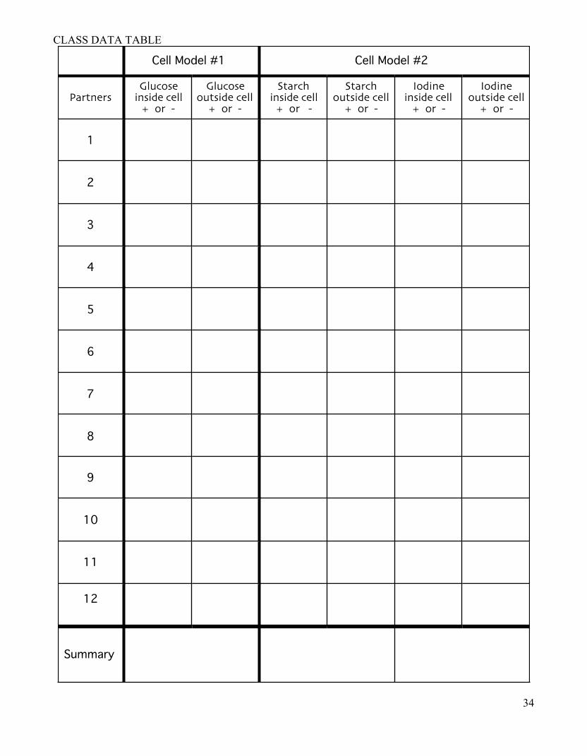
CLASS DATA TABLE

35
Post Lab Cell Model
• Micropores – –
• •
Cell Model #1 • Initial Setup
– External Environment
– Internal Environment
• Final Setup
– Conclusion

36
Cell Model #2 • Initial Setup
– External Environment
– Internal Environment
Starch + Iodine Overnight • Observations:
– External Environment
– Internal Environment
• Conclusion

37
7.4 Homeostasis & Cells
Go to Biology.com via PearsonSuccessNet, click explore, Unit 3 Cells, Ch 7, 7.4 Homeostasis & Cells, Activities, finally Data Analysis Data Analysis-Maximizing Mitochondria As you navigate through activity, answer the questions below.
1. What was the “French Paradox”?
2. What was one explanation proposed for this paradox?
3. What happened to fruit flies that were given concentrated doses of Resveratrol (RSV)?
4. Examine the RSV experiment with mice, which was the control group(s)?
5. Draw a conclusion from the graph “Resveratrol’s Impact on Weight Gain”.
6. Record the results for the normal diet + RSV & high fat diet + RSV below.
7. Interpret the graph “Resveratrol & Endurance”, how did RSV affect endurance? (be specific, don’t
just say increase or decrease).
8. Examine the graph which shows the relative area of muscle tissue occupied by mitochondria in both high-fat-diet mouse groups, which group has more slow –twitch, high-endurance muscle fibers?
Name: _____________________________ Date: _______________ Period: ________

38
9. Resveratrol’s Future: Why might athletes be interested in determining if resveratrol affects human in the same ways it affects mice?
10. Resveratrol and Longevity: Which two groups had the most similar longevity trend?
11. What does this graph (above) indicate about the addition of RSV to an unhealthy diet?

39
10.2 The Process of Cell Division
Go to Biology.com via PearsonSuccessNet, click explore, Unit 3 Cells, Ch 10, 10.2 The Process of Cell Division Activities, finally InterActive Art
1. The cell cycle is divided into three main stages, name them.
2. What happens during interphase?
3. During prophase, this separates __________________________, this begins to form _________________________, and this begins to break down ____________________________.
4. During metaphase what connects each chromosome to spindle fibers? _______________________.
5. During anaphase the sister chromatids separate into individual _____________________. The separated chromatids seem to move towards ________________________.
6. This structure forms during telophase, ________________________________.
Using your knowledge of the tutorial, sort the steps of the cell cycle. A. B. C. D. E. F.
(Beginning) ________ _________ _________ ________ ________ _________ (End)
Name: _____________________________ Date: _______________ Period: ________

40
THE BIOLOGY PROJECTCELL BIOLOGY Name: ____________________________________Date: _________Period: _______
http://www.biology.arizona.edu/cell_bio/activities/cell_cycle/cell_cycle.html Go to the web address listed above
Onl ine Onion Root Tips Determining time spent in different phases of the cell cycle Read the introduction to “Online Onion Root Tips” and answer the questions
below 1) Growth in an organism is carefully controlled by regulating the ___________________.
2) What type of microscope was used to examine the slice of root?__________________. 3) Scientists have identified how many different phases in the cell cycle? ________ Click on the Next button at the bottom of the page
Determining time spent in different phases of the cell cycle Read the description for each major phase, in the box draw a sketch of each and answer the
questions below. Interphase 1) What duplicates during this phase? ___________
2) What is not clearly visible during this phase? __________________________________________.
3) A dark spot may be visible, what is it? __________________________________________,
Prophase 4) What changes does the chromatin undergo so that we call them chromosomes? _________________________ ________________________________________________ 5) What structure dissolves during prophase? ________________________________________________
Metaphase 6) Spindle fibers align the chromosomes along the
middle of the cell nucleus, what is this line called? ________________________________________________
7) What is significant about this organization? ______________
________________________________________________
________________________________________________

41
Anaphase 8) What happens to the paired chromosomes during anaphase? _______________________________________
________________________________________________
________________________________________________
Telophase 9) What do you think the term “daughter nuclei” means? _________________________________________
10) What happens to the chromosomes? __________________
_______________________________________________
11) What is cytokinesis? _______________________________
Click Next at the bottom of the page
Determining time spent in different phases of the cell cycle Now that you are an expert on the five phases of the cell cycle, you will be presented with cells from the tip of an onion root. You will classify each cell based on its phase. Use those numbers to predict how much time a dividing cell spends in each phase. The table below will help you organize your information.
Interphase
Prophase
Metaphase
Anaphase
Telophase
Total
Number of cells
36
Percent of cells
100%
According to the results above predict which phase a cell is most likely to be in and which phase is least likely to be happening in a cell? _______________________ ________________________________________________________________ ________________________________________________________________

42
Biology:Unit3ReviewBaseball
• Today we will play baseball to review for the test. • Remember there is one out per inning and every pitch is a single.
Go to http://www6.district125.k12.il.us/science/secure/bio/bball/unit3.ppt Save the Powerpoint to your desktop, click “View Show” to play.
If innings end quickly, we will play a double or triple header (this means you havenʼt studied and could use the practice!)
Teams Inning 1 2 3 4 5 6 7 8 9
If you don’t finish this baseball game is downloadable online using the URL above…if you don’t have powerpoint you can view here
http://www6.district125.k12.il.us/science/secure/bio/bball/unit3.mov
Take Notes below and on back for any questions you (or your opponent) miss. This handout is due the day of the exam you must have a MINIMUM of 10 declarative facts recorded below for full credit. (attach an additional page if needed)
1.
2.
3.
4.
5.
6.
7.
8.
9.
10.
Name: Date: Period:

43
Unit 3: Additional Review Material Now go online & complete a practice test….take notes on any questions you missed http://www6.district125.k12.il.us/science/secure/bio/exampro/2b.htm
One more review! This one is organized by the four targets for this unit! 1. Describe the importance of the process of mitosis in the production of new body cells to make more cells
for growth and repair 2. Recognize that cells within organisms are specialized for different functions by which organelles they
either possess or lack. 3. Describe the importance of a selectively permeable cell membrane in maintaining homeostasis by
controlling what enters & leaves the cell. 4. Use the processes of osmosis and diffusion to explain how, when cells are placed in different solutions
(hypertonic, hypotonic, and isotonic), materials move both in and out of cells. http://www.quia.com/cb/618844.html
Name: Date: Period: