Embed Size (px)
Citation preview
Unit 4Animal Systems:Respiration, Circulation and ImmunityBiology 30Mr.Oosterom
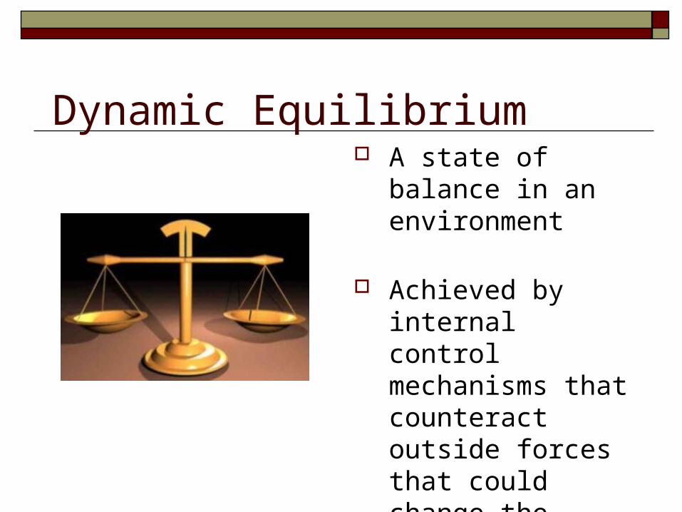
A state of balance in an environment
Achieved by internal control mechanisms that counteract outside forces that could change the inside environment (body)
Dynamic Equilibrium
Homeostasis
The steady state of conditions inside a living organism that allows it to function properly
Homeostasis is the dynamic equilibrium of the internal environment of the human body
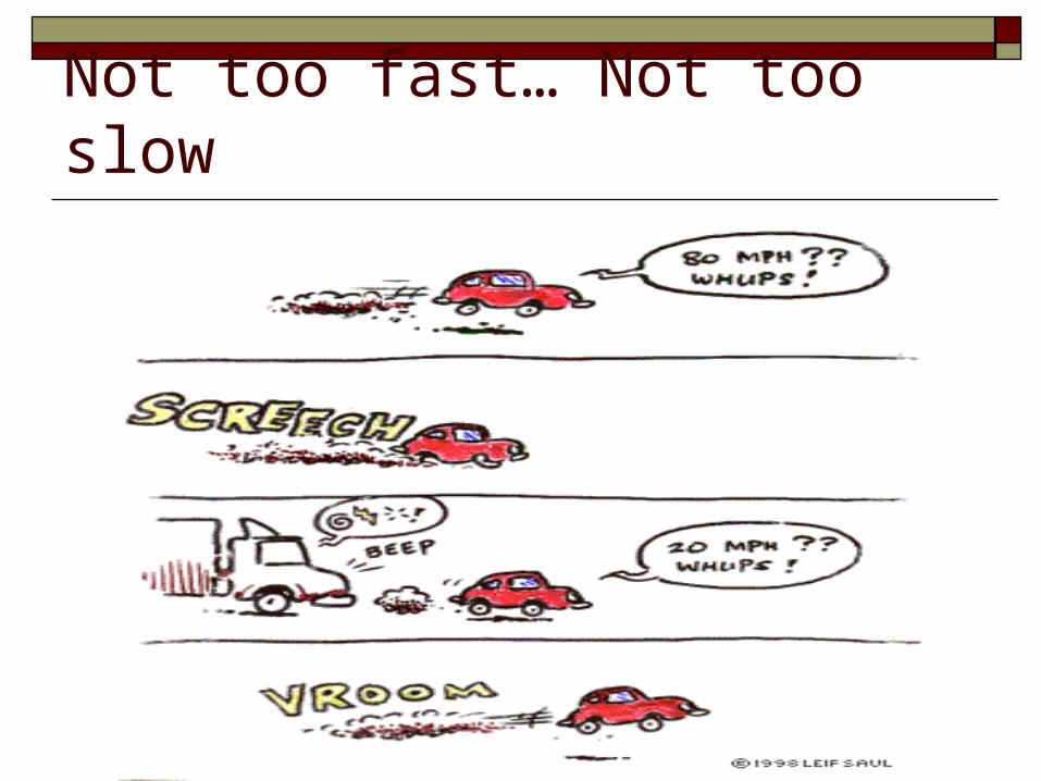
Not too fast… Not too slow
Examples of Homeostasis
Temperature Regulation Food and Water Balance Regulation of blood sugar levels Regulation of blood calcium
levels
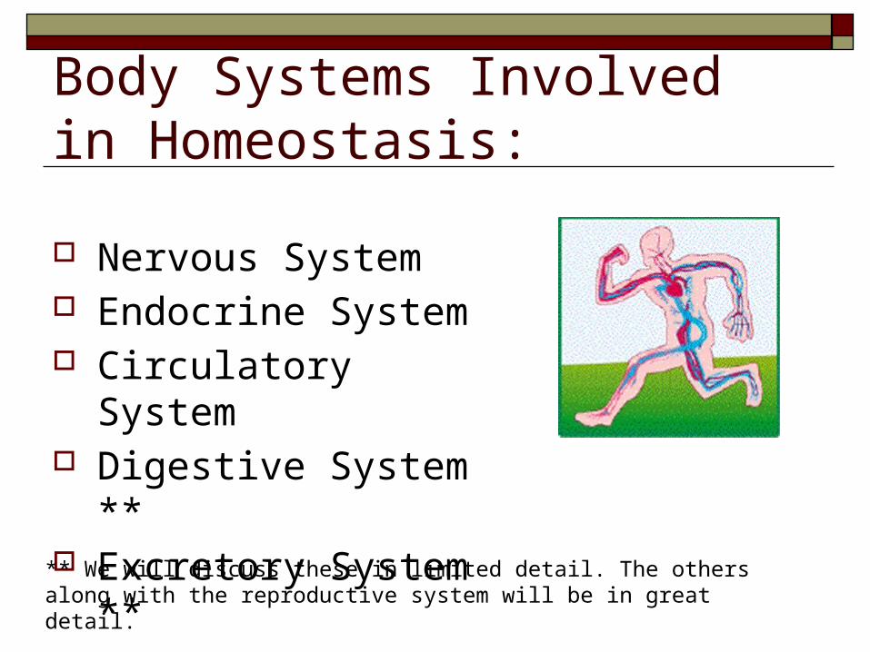
Body Systems Involved in Homeostasis:
Nervous System Endocrine System Circulatory System Digestive System ** Excretory System **
** We will discuss these in limited detail. The others along with the reproductive system will be in great detail.
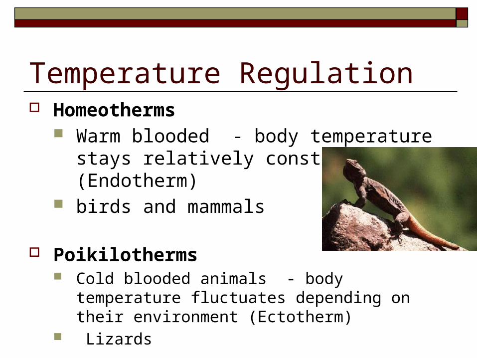
Temperature Regulation Homeotherms
Warm blooded - body temperature stays relatively constant (Endotherm)
birds and mammals
Poikilotherms Cold blooded animals - body temperature fluctuates
depending on their environment (Ectotherm) Lizards
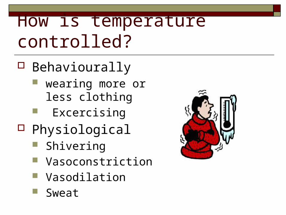
How is temperature controlled? Behaviourally
wearing more or less clothing
Excercising Physiological
Shivering Vasoconstriction Vasodilation Sweat
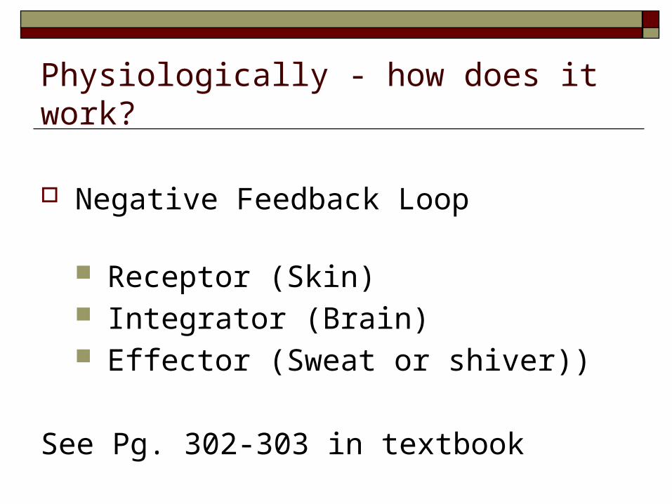
Physiologically - how does it work?
Negative Feedback Loop
Receptor (Skin) Integrator (Brain) Effector (Sweat or shiver))
See Pg. 302-303 in textbook
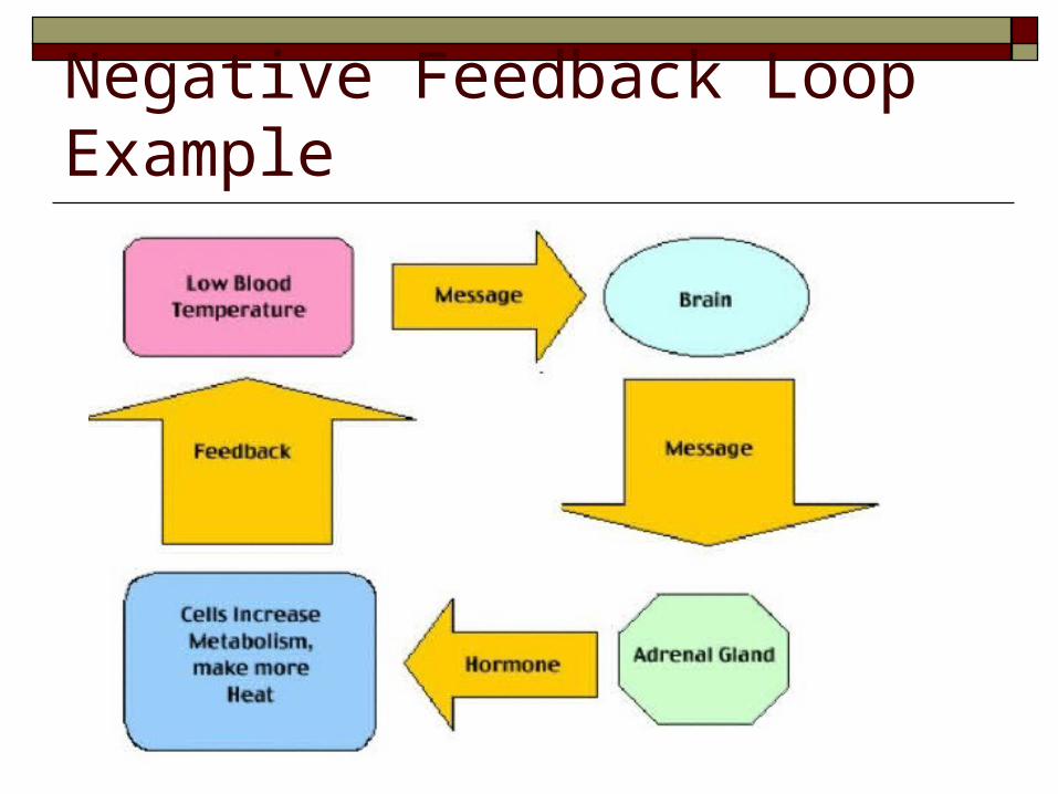
Negative Feedback Loop Example
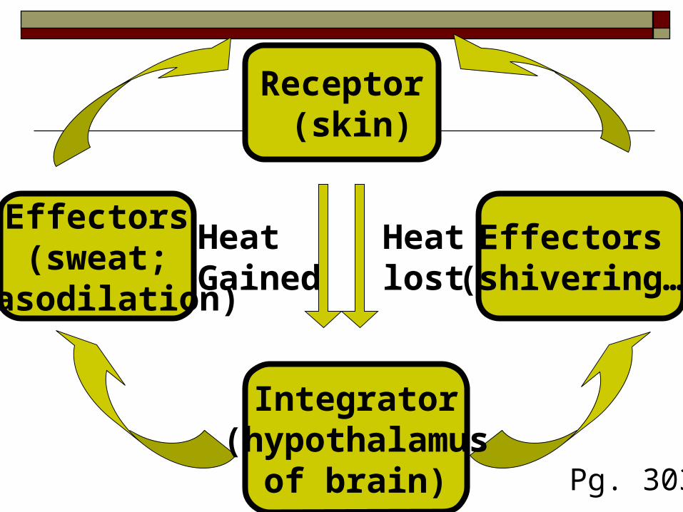
Integrator (hypothalamus
of brain)
Effectors(sweat;
vasodilation)
Receptor (skin)
Effectors (shivering…)
Heat lost
HeatGained
Pg. 303
Negative Feedback Loop
A process by which a receptor, an integrator and an effector detects, processes and produces a response to a change in a body constant (for example temperature) so that a reverse affect may take place, enabling the body to stay constant.
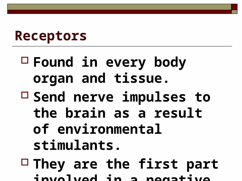
Receptors
Found in every body organ and tissue.
Send nerve impulses to the brain as a result of environmental stimulants.
They are the first part involved in a negative feedback loop.
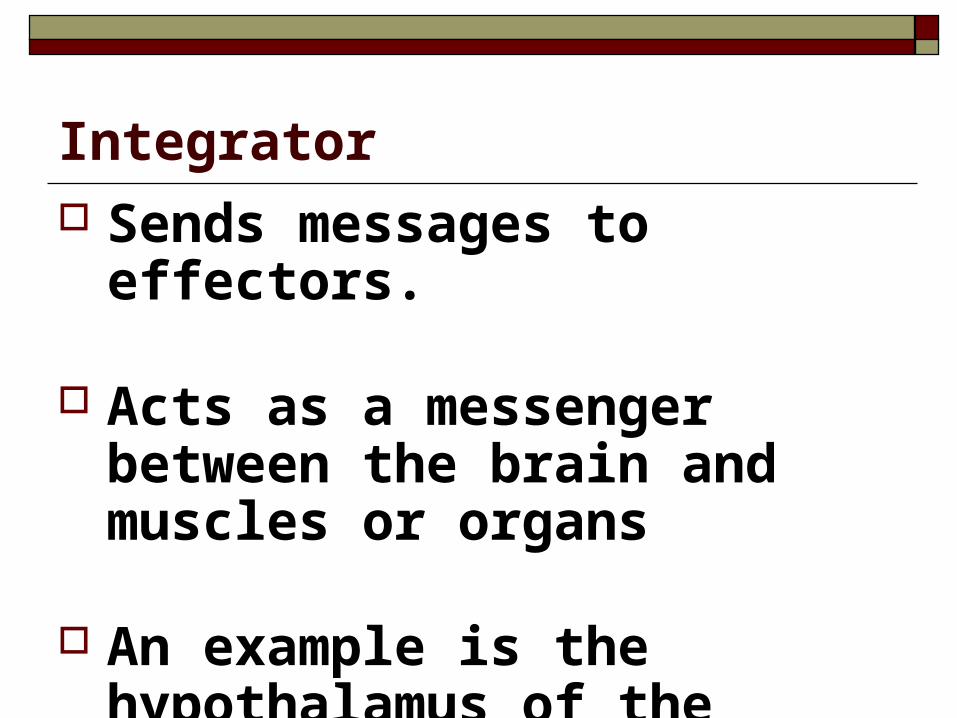
Integrator Sends messages to effectors.
Acts as a messenger between the
brain and muscles or organs
An example is the hypothalamus of the brain.
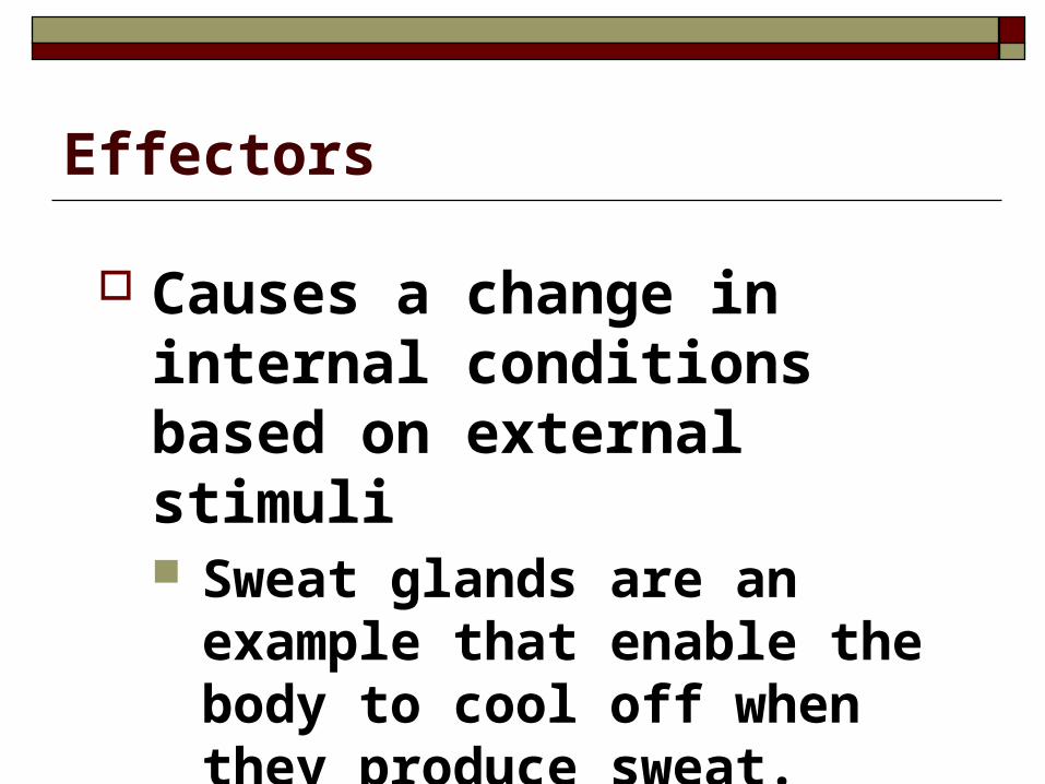
Effectors
Causes a change in internal conditions based on external stimuli Sweat glands are an example that
enable the body to cool off when they produce sweat.
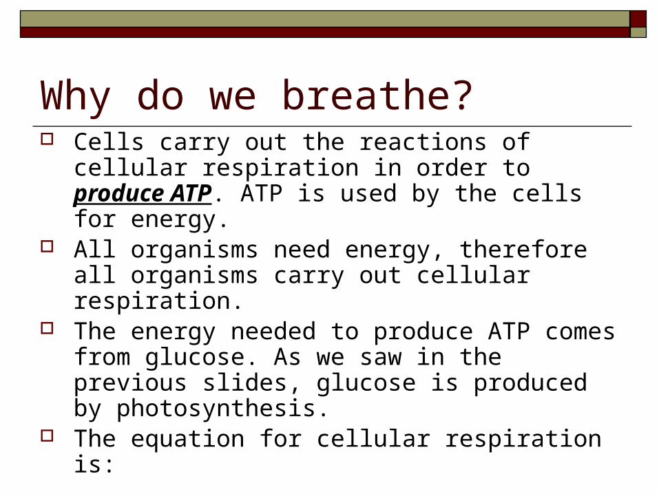
Why do we breathe? Cells carry out the reactions of cellular respiration in
order to produce ATP. ATP is used by the cells for energy.
All organisms need energy, therefore all organisms carry out cellular respiration.
The energy needed to produce ATP comes from glucose. As we saw in the previous slides, glucose is produced by photosynthesis.
The equation for cellular respiration is:
C6H12O6 + 6O2 6CO2 + 6H2O + 36 ATP
How do we get this oxygen, and get rid of the CO2?
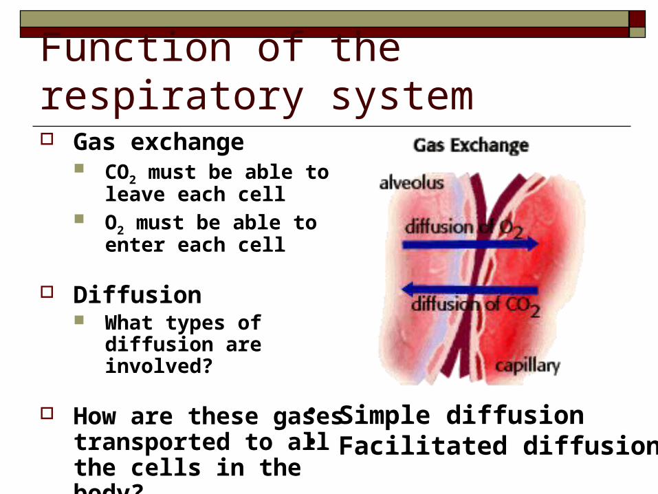
Function of the respiratory system Gas exchange
CO2 must be able to leave each cell
O2 must be able to enter each cell
Diffusion What types of diffusion are
involved?
How are these gases transported to all the cells in the body?
• Simple diffusion• Facilitated diffusion
The Requirements Though different organisms have different
respiratory systems, they function is the same There are TWO requirements for a respiratory
system1. Respiratory surface – There must be a large
surface area available for gas exchangeto take place efficiently
2. Moist environment
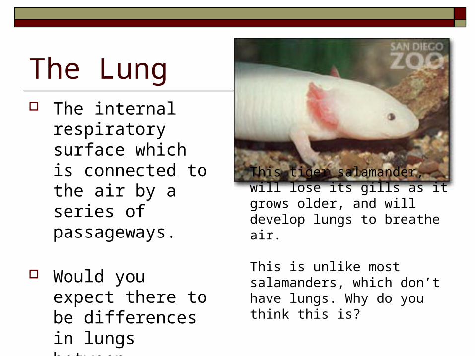
The Lung The internal
respiratory surface which is connected to the air by a series of passageways.
Would you expect there to be differences in lungs between species? Fig. 10.3
This tiger salamander, will lose its gills as it grows older, and will develop lungs to breathe air.
This is unlike most salamanders, which don’t have lungs. Why do you think this is?
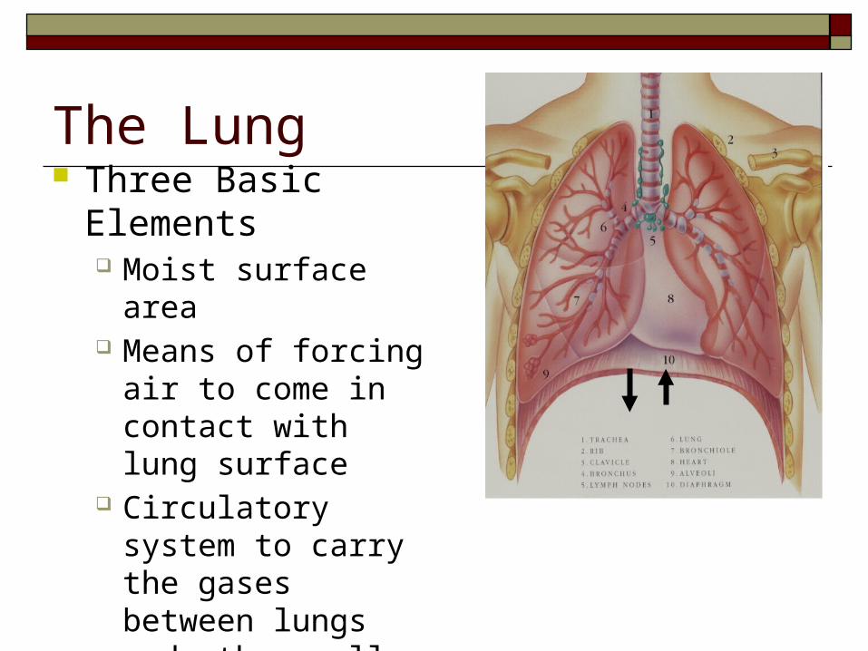
The Lung Three Basic Elements
Moist surface area Means of forcing air to
come in contact with lung surface
Circulatory system to carry the gases between lungs and other cells
Levels of Respiration At which locations does gas exchange occur?
External – Exchange of CO2 for O2 between the air and the blood
Internal – Exchange of CO2 for O2 between the blood and the cells
Cellular – Series of complex reactions that that take place in the mitochondria to make ATP

RespiratoryTract
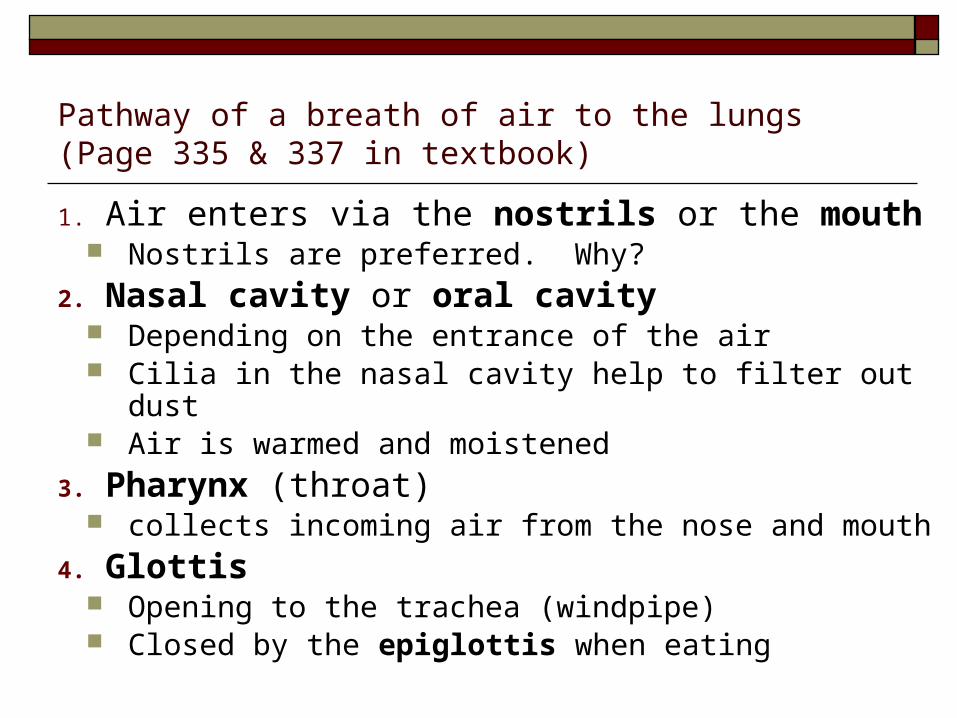
Pathway of a breath of air to the lungs(Page 335 & 337 in textbook)
1. Air enters via the nostrils or the mouth Nostrils are preferred. Why?
2. Nasal cavity or oral cavity Depending on the entrance of the air Cilia in the nasal cavity help to filter out dust Air is warmed and moistened
3. Pharynx (throat) collects incoming air from the nose and mouth
4. Glottis Opening to the trachea (windpipe) Closed by the epiglottis when eating
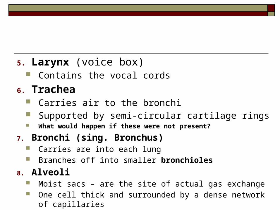
5. Larynx (voice box) Contains the vocal cords
6. Trachea Carries air to the bronchi Supported by semi-circular cartilage rings What would happen if these were not present?
7. Bronchi (sing. Bronchus) Carries are into each lung Branches off into smaller bronchioles
8. Alveoli Moist sacs – are the site of actual gas exchange One cell thick and surrounded by a dense network of capillaries
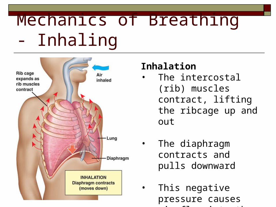
Mechanics of Breathing - Inhaling
Inhalation• The intercostal (rib)
muscles contract, lifting the ribcage up and out
• The diaphragm contracts and pulls downward
• This negative pressure causes air flow into the lungs enabling them to inflate
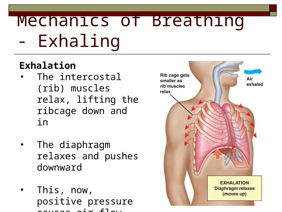
Mechanics of Breathing - ExhalingExhalation• The intercostal (rib)
muscles relax, lifting the ribcage down and in
• The diaphragm relaxes and pushes downward
• This, now, positive pressure causes air flow out of the lungs enabling them to deflate
Exchange of Gases
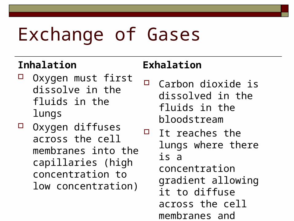
Oxygen must first dissolve in the fluids in the lungs
Oxygen diffuses across the cell membranes into the capillaries (high concentration to low concentration)
Carbon dioxide is dissolved in the fluids in the bloodstream
It reaches the lungs where there is a concentration gradient allowing it to diffuse across the cell membranes and into the fluids in the lungs
Inhalation Exhalation
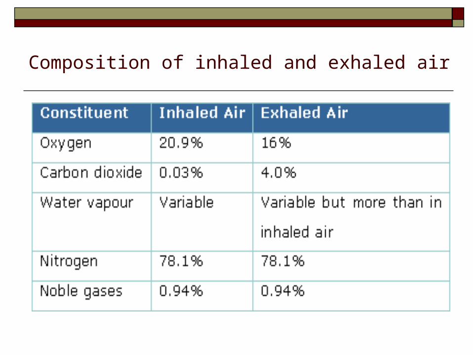
Composition of inhaled and exhaled air
Some Lung Capacity Humour
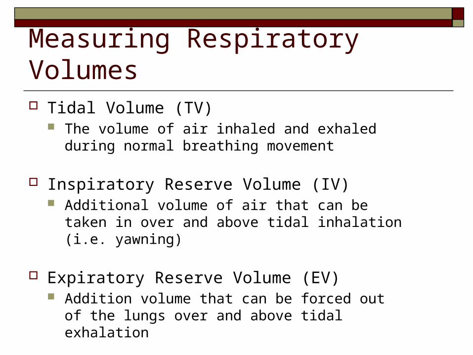
Measuring Respiratory Volumes Tidal Volume (TV)
The volume of air inhaled and exhaled during normal breathing movement
Inspiratory Reserve Volume (IV) Additional volume of air that can be taken in
over and above tidal inhalation (i.e. yawning)
Expiratory Reserve Volume (EV) Addition volume that can be forced out of the
lungs over and above tidal exhalation
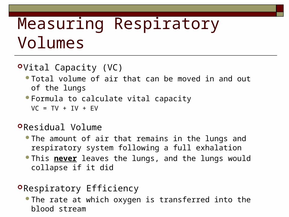
Measuring Respiratory Volumes Vital Capacity (VC)
Total volume of air that can be moved in and out of the lungs Formula to calculate vital capacity VC = TV + IV + EV
Residual Volume The amount of air that remains in the lungs and respiratory
system following a full exhalation This never leaves the lungs, and the lungs would collapse if it
did
Respiratory Efficiency The rate at which oxygen is transferred into the blood stream
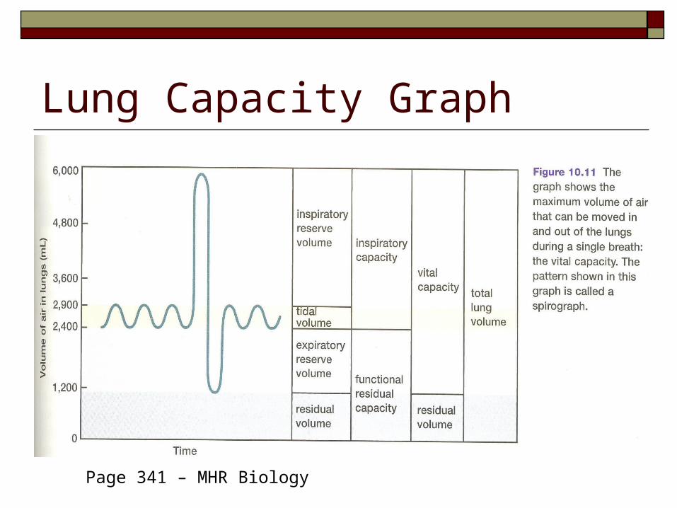
Lung Capacity Graph
Page 341 – MHR Biology
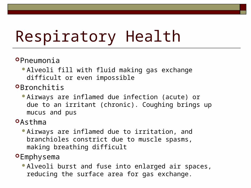
Respiratory Health Pneumonia
Alveoli fill with fluid making gas exchange difficult or even impossible
Bronchitis Airways are inflamed due infection (acute) or due to an
irritant (chronic). Coughing brings up mucus and pus Asthma
Airways are inflamed due to irritation, and branchioles constrict due to muscle spasms, making breathing difficult
Emphysema Alveoli burst and fuse into enlarged air spaces, reducing
the surface area for gas exchange.
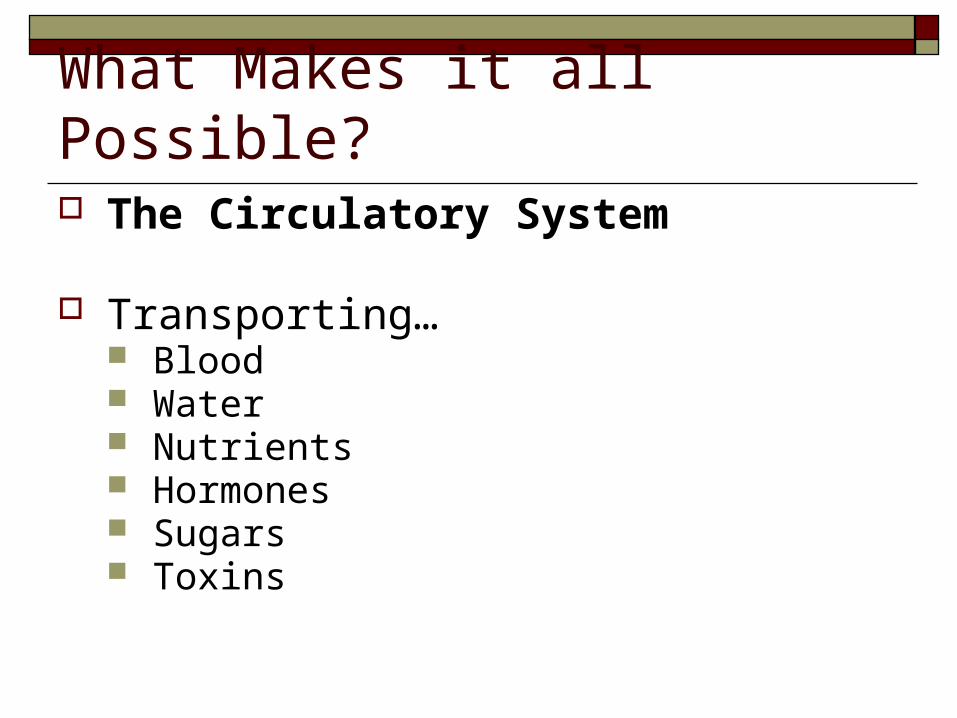
What Makes it all Possible? The Circulatory System
Transporting… Blood Water Nutrients Hormones Sugars Toxins
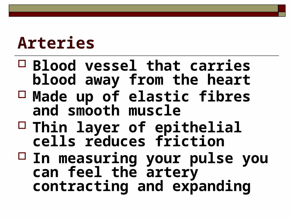
Arteries Blood vessel that carries blood away
from the heart Made up of elastic fibres and smooth
muscle Thin layer of epithelial cells reduces
friction In measuring your pulse you can feel
the artery contracting and expanding
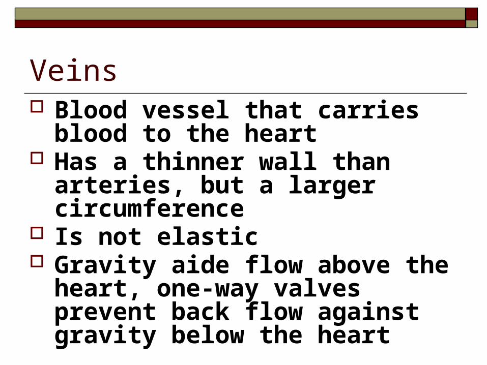
Veins Blood vessel that carries blood to the
heart Has a thinner wall than arteries, but a
larger circumference Is not elastic Gravity aide flow above the heart,
one-way valves prevent back flow against gravity below the heart
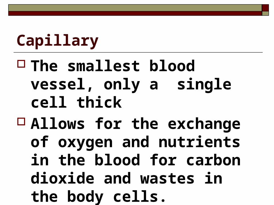
Capillary
The smallest blood vessel, only a single cell thick
Allows for the exchange of oxygen and nutrients in the blood for carbon dioxide and wastes in the body cells.

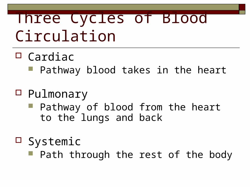
Three Cycles of Blood Circulation Cardiac
Pathway blood takes in the heart
Pulmonary Pathway of blood from the heart to the lungs and
back
Systemic Path through the rest of the body

Coronary/Cardiac Circulation
Circulation in and around the heart
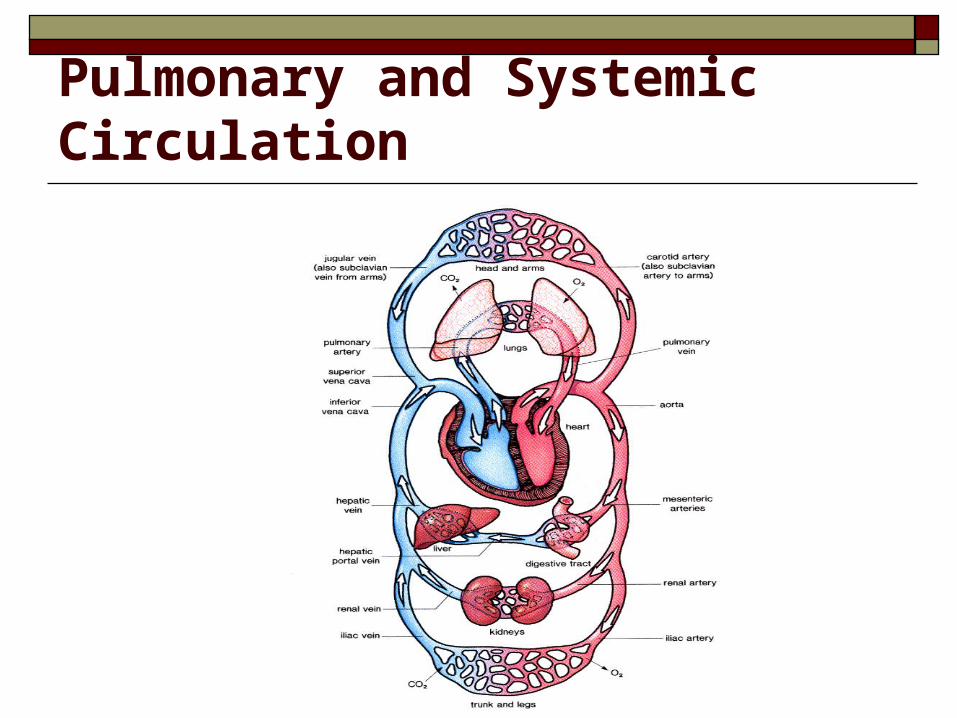
Pulmonary and Systemic Circulation
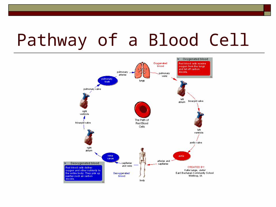
Pathway of a Blood Cell
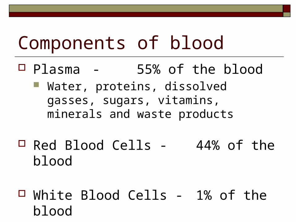
Components of blood Plasma - 55% of the blood
Water, proteins, dissolved gasses, sugars, vitamins, minerals and waste products
Red Blood Cells - 44% of the blood
White Blood Cells - 1% of the blood
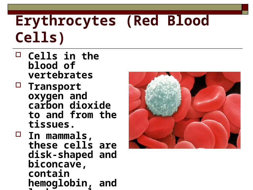
Erythrocytes (Red Blood Cells) Cells in the blood of
vertebrates Transport oxygen and
carbon dioxide to and from the tissues.
In mammals, these cells are disk-shaped and biconcave, contain hemoglobin, and lack a nucleus.
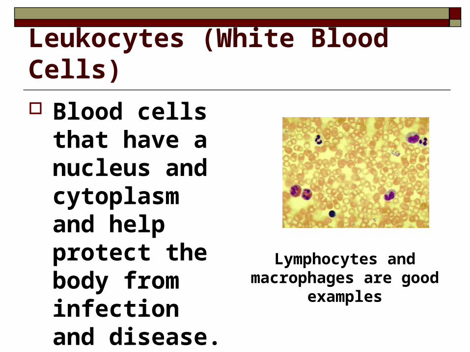
Leukocytes (White Blood Cells) Blood cells that
have a nucleus and cytoplasm and help protect the body from infection and disease.
Lymphocytes and macrophages are good
examples
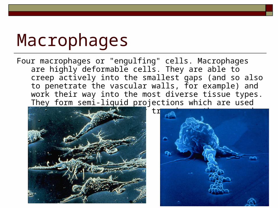
MacrophagesFour macrophages or "engulfing" cells. Macrophages are highly deformable
cells. They are able to creep actively into the smallest gaps (and so also to penetrate the vascular walls, for example) and work their way into the most diverse tissue types. They form semi-liquid projections which are used for motility and also for trapping pathogens and other foreign bodies.
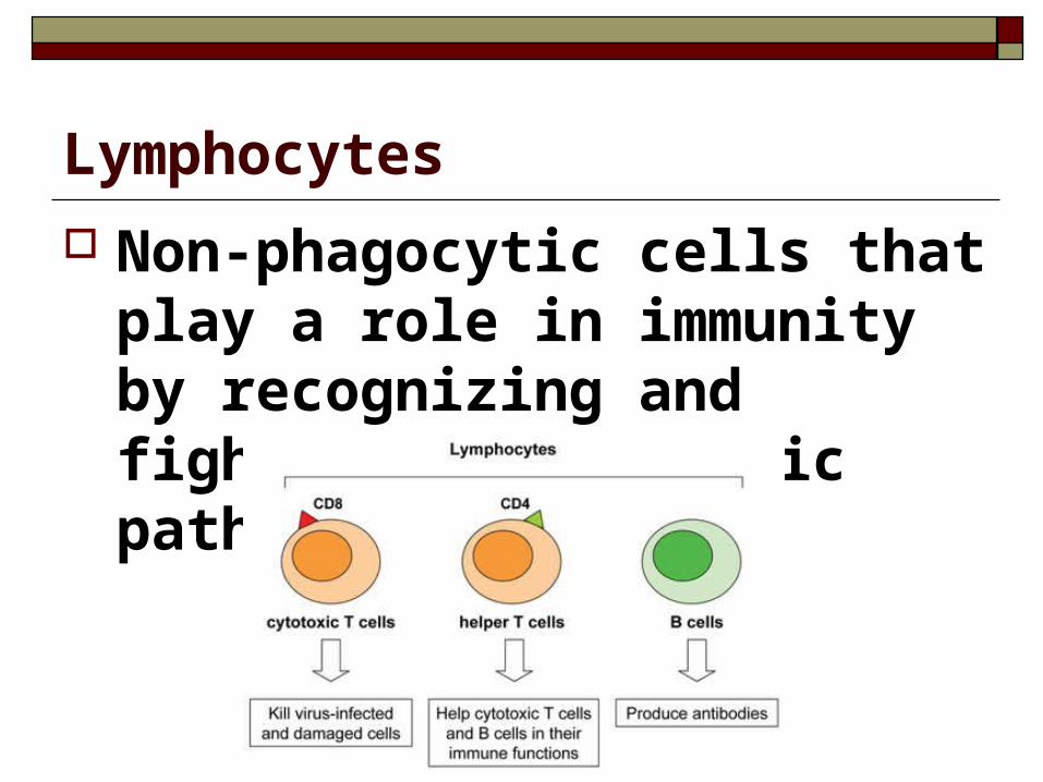
Lymphocytes
Non-phagocytic cells that play a role in immunity by recognizing and fighting off specific pathogens.
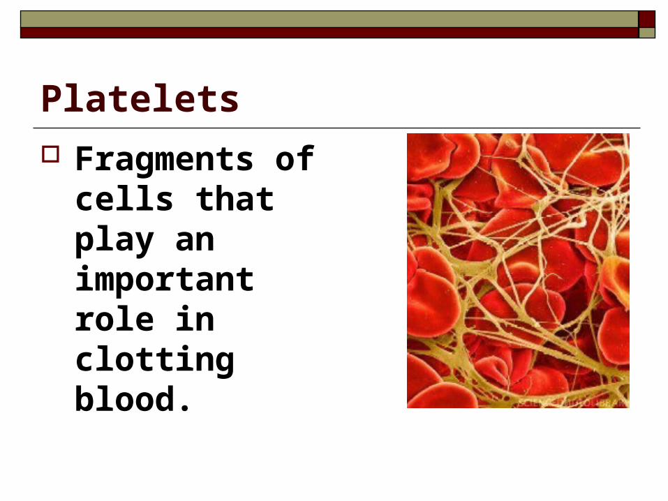
Platelets Fragments of
cells that play an important role in clotting blood.
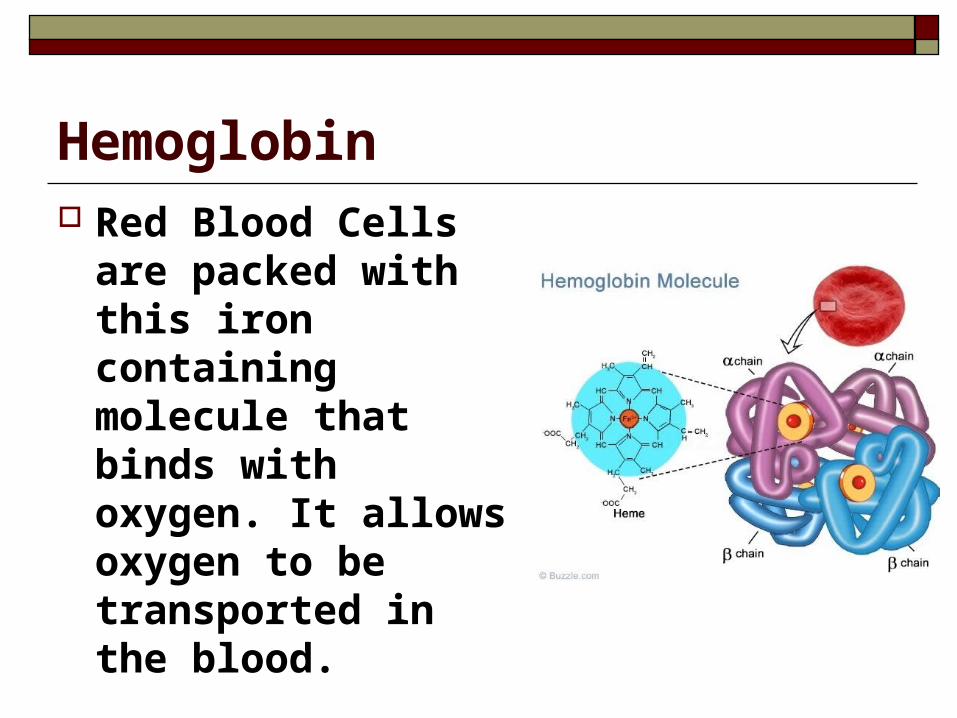
Hemoglobin Red Blood Cells
are packed with this iron containing molecule that binds with oxygen. It allows oxygen to be transported in the blood.
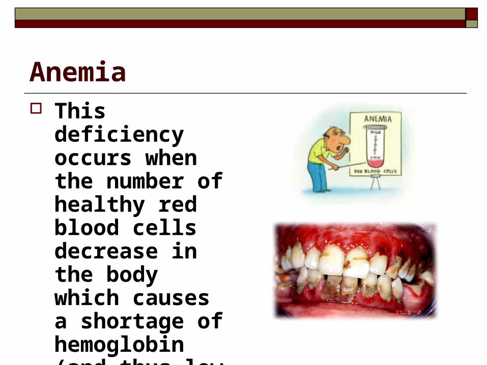
Anemia This deficiency
occurs when the number of healthy red blood cells decrease in the body which causes a shortage of hemoglobin (and thus low iron).
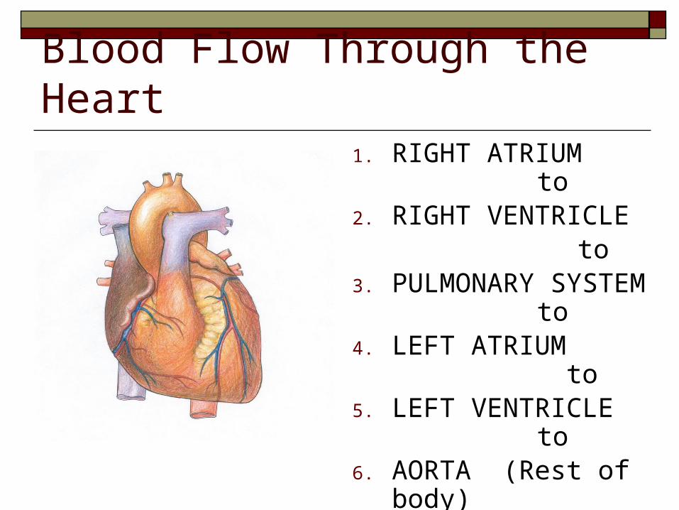
Blood Flow Through the Heart1. RIGHT ATRIUM
to2. RIGHT VENTRICLE
to3. PULMONARY SYSTEM
to4. LEFT ATRIUM
to5. LEFT VENTRICLE
to6. AORTA (Rest of body)
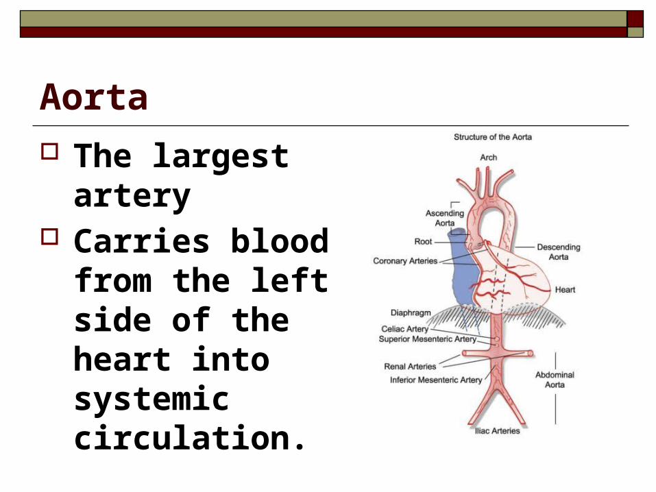
Aorta The largest artery Carries blood from
the left side of the heart into systemic circulation.
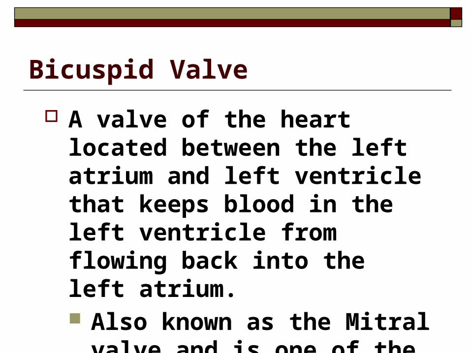
Bicuspid Valve
A valve of the heart located between the left atrium and left ventricle that keeps blood in the left ventricle from flowing back into the left atrium. Also known as the Mitral valve
and is one of the two atrioventricular valves.
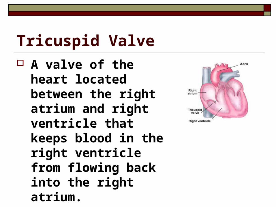
Tricuspid Valve A valve of the heart
located between the right atrium and right ventricle that keeps blood in the right ventricle from flowing back into the right atrium. It is one of the
atrioventricular valves
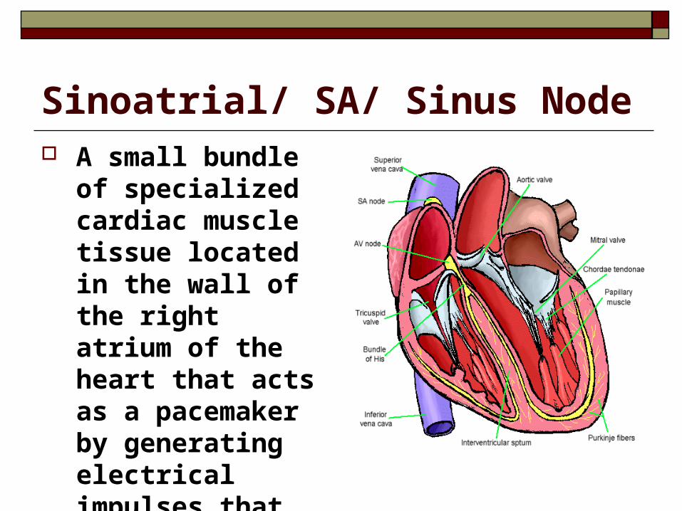
Sinoatrial/ SA/ Sinus Node A small bundle of
specialized cardiac muscle tissue located in the wall of the right atrium of the heart that acts as a pacemaker by generating electrical impulses that keep the heart beating.
Atrioventricular Valves
On both sides of the heart the atria and ventricles are separated from one another by this set of valves. (These are also called the bicuspid and tricuspid valves).
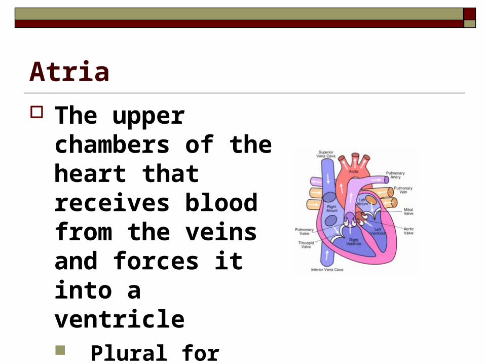
Atria The upper chambers
of the heart that receives blood from the veins and forces it into a ventricle Plural for atrium.
Left Ventricle The chamber on the left side of the
heart that receives arterial blood from the left atrium and contracts to force it into the aorta.
Septum The wall that separates the right and left ventricles.
Right Ventricle
The chamber on the right side of the heart that receives venous blood from the right atrium and forces it into the pulmonary artery.
Vena Cava
Either of two large veins that drain blood from the upper body (superior vena cava) and from the lower body (inferior vena cava) and empty into the right atrium of the heart.
Pulmonary Artery
A blood vessel that carries deoxygenated blood from the right ventricle of the heart to the lungs.
Pulmonary Vein
A blood vessel that carries oxygenated blood from the lungs to the left atrium of the heart.
Electrocardiogram
A device that measures the voltage of the electrical signals produced by the SA and AV nodes.
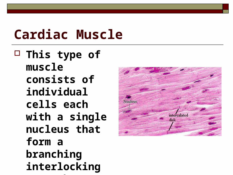
Cardiac Muscle This type of muscle
consists of individual cells each with a single nucleus that form a branching interlocking network.
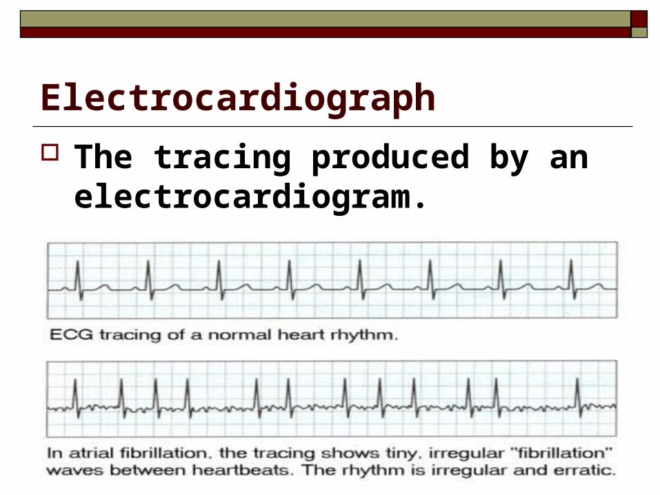
Electrocardiograph The tracing produced by an
electrocardiogram.
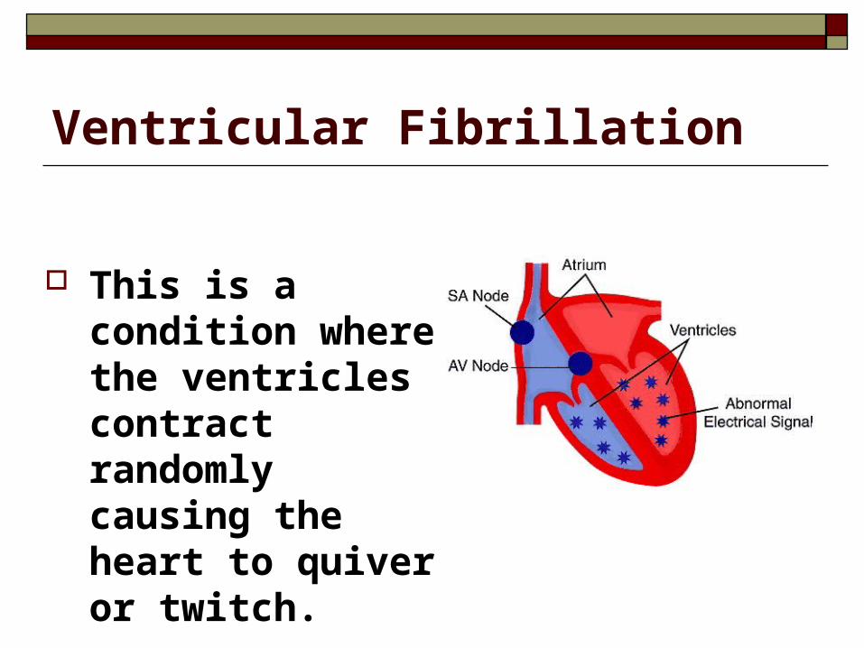
Ventricular Fibrillation
This is a condition where the ventricles contract randomly causing the heart to quiver or twitch.
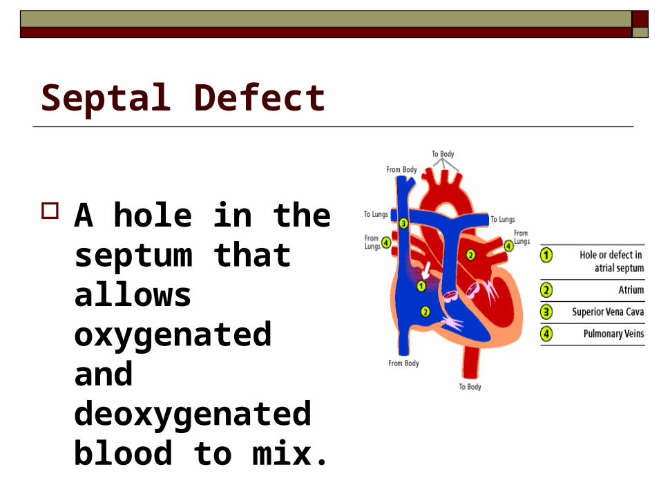
Septal Defect
A hole in the septum that allows oxygenated and deoxygenated blood to mix.
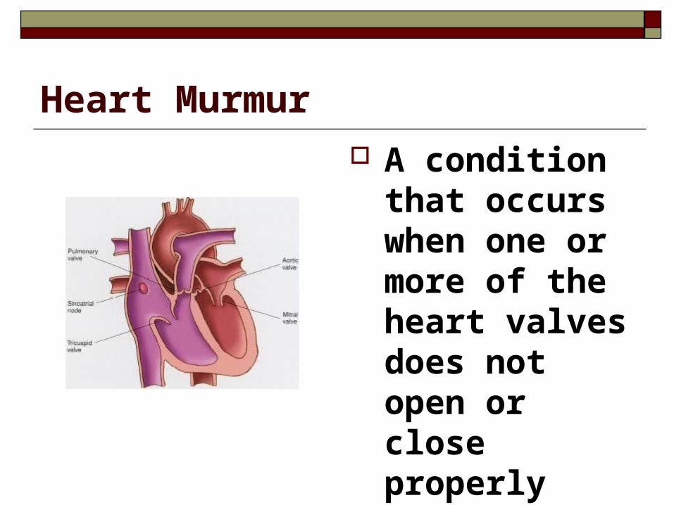
Heart Murmur A condition that
occurs when one or more of the heart valves does not open or close properly
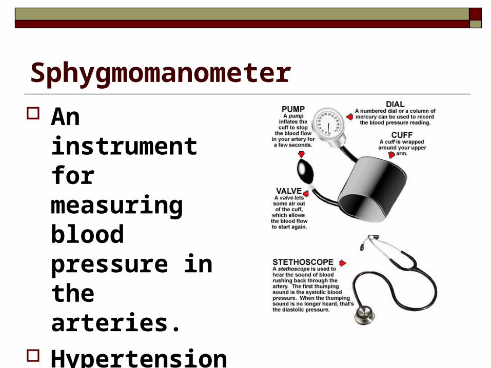
Sphygmomanometer An instrument
for measuring blood pressure in the arteries.
Hypertension Condition where
blood pressure is abnormally high
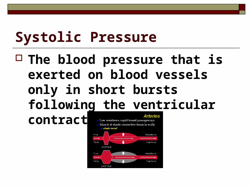
Systolic Pressure The blood pressure that is exerted on
blood vessels only in short bursts following the ventricular contractions.
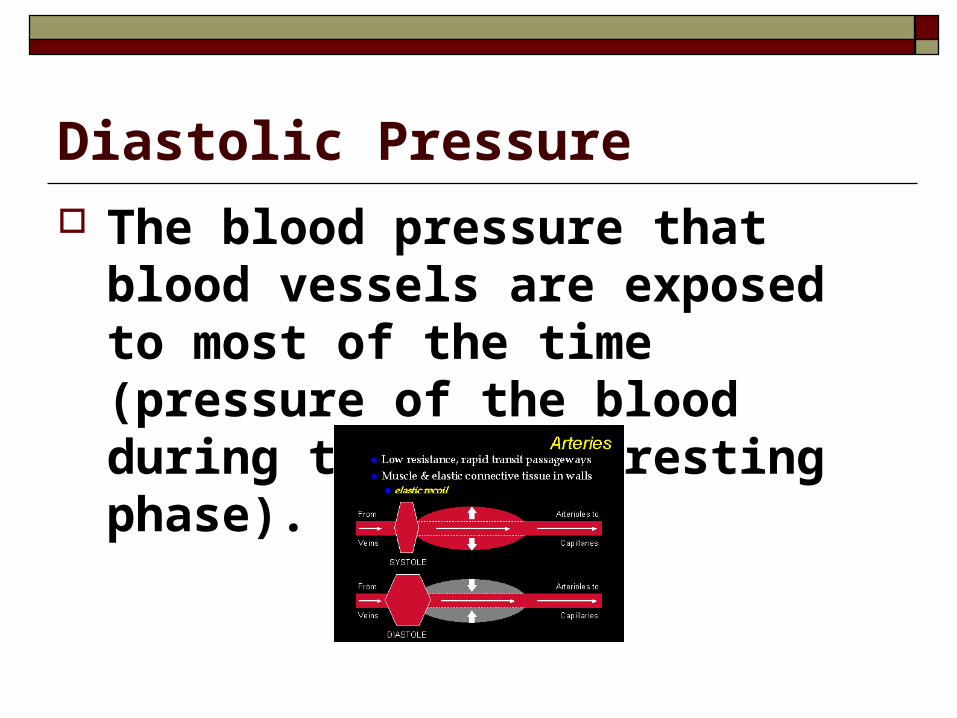
Diastolic Pressure The blood pressure that blood vessels
are exposed to most of the time (pressure of the blood during the hearts resting phase).
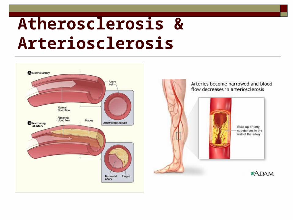
Atherosclerosis & Arteriosclerosis Atherosclerosis
A narrowing of the arteries caused by cholesterol or fatty tissue buildup called plaques, ON the inner lining of the artery wall.
Arteriosclerosis A condition where plaque material
becomes deposited UNDER the inner lining of the arteries
Atherosclerosis & Arteriosclerosis
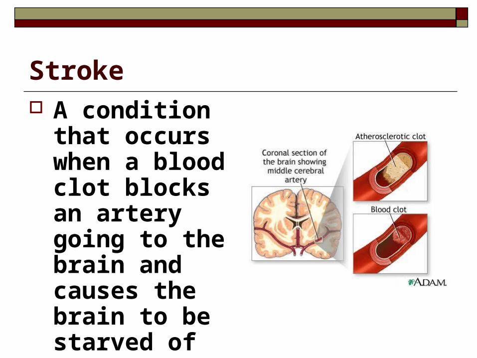
Stroke A condition that
occurs when a blood clot blocks an artery going to the brain and causes the brain to be starved of oxygen, killing the brain tissue
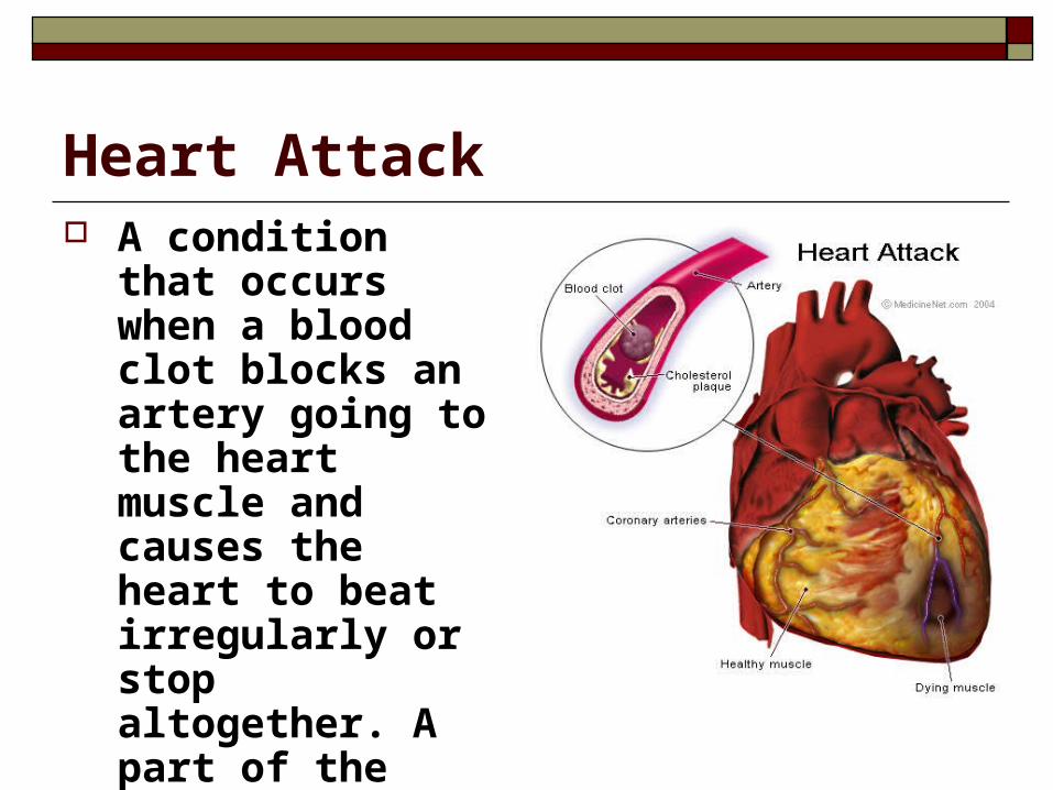
Heart Attack A condition that
occurs when a blood clot blocks an artery going to the heart muscle and causes the heart to beat irregularly or stop altogether. A part of the heart actually dies when this happens.

Clot Busting Drugs Medicines that help dissolve blood
clots in arteries, allowing blood to once again flow through them.
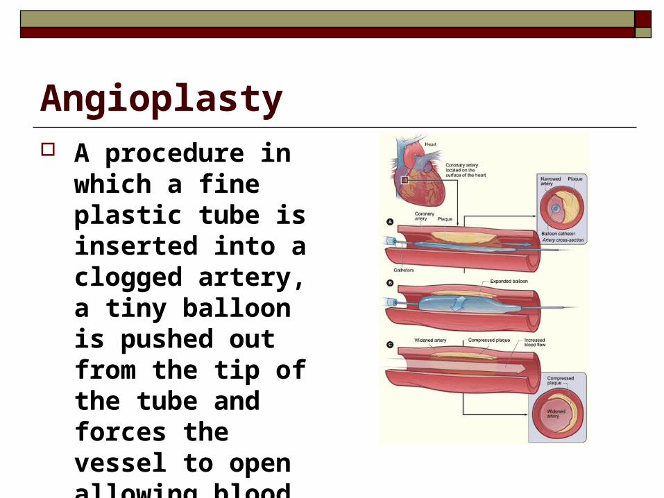
Angioplasty A procedure in which
a fine plastic tube is inserted into a clogged artery, a tiny balloon is pushed out from the tip of the tube and forces the vessel to open allowing blood to flow through.
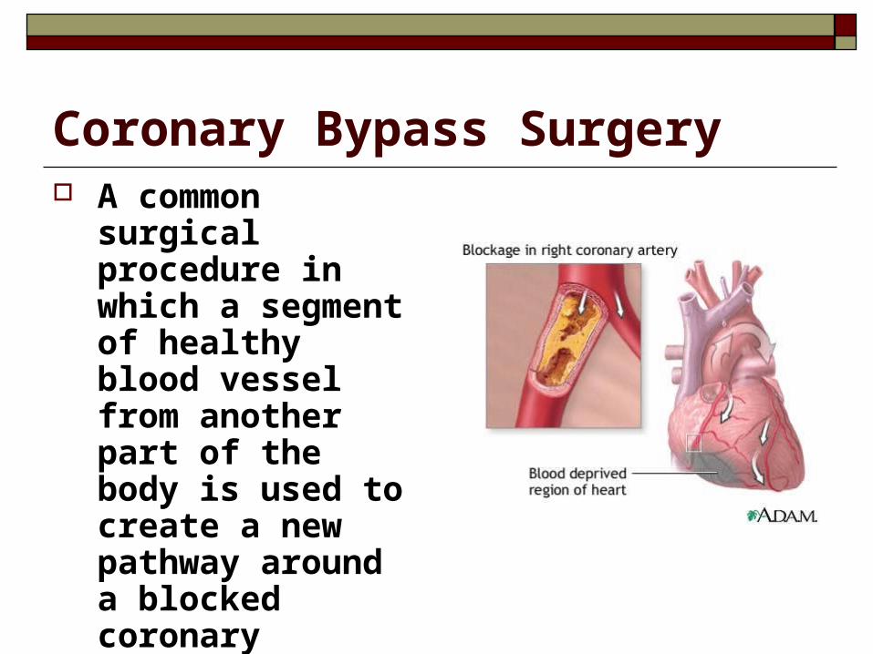
Coronary Bypass Surgery A common surgical
procedure in which a segment of healthy blood vessel from another part of the body is used to create a new pathway around a blocked coronary artery.
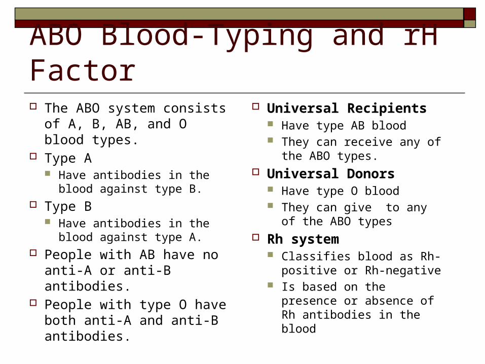
ABO Blood-Typing and rH Factor The ABO system consists of
A, B, AB, and O blood types. Type A
Have antibodies in the blood against type B.
Type B Have antibodies in the blood
against type A. People with AB have no anti-
A or anti-B antibodies. People with type O have both
anti-A and anti-B antibodies.
Universal Recipients Have type AB blood They can receive any of the
ABO types. Universal Donors
Have type O blood They can give to any of the
ABO types Rh system
Classifies blood as Rh-positive or Rh-negative
Is based on the presence or absence of Rh antibodies in the blood
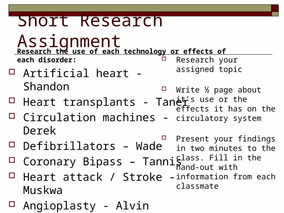
Short Research Assignment
Artificial heart - Shandon Heart transplants - Taner Circulation machines - Derek Defibrillators – Wade Coronary Bipass – Tannis Heart attack / Stroke - Muskwa Angioplasty - Alvin
Research your assigned topic
Write ½ page about it’s use or the effects it has on the circulatory system
Present your findings in two minutes to the class. Fill in the hand-out with information from each classmate
Research the use of each technology or effects of each disorder:
Immune System The system responsible for keeping your
body free from pathogens and preventing infection
Your body has two defense systems Non-specific defense Specific defence
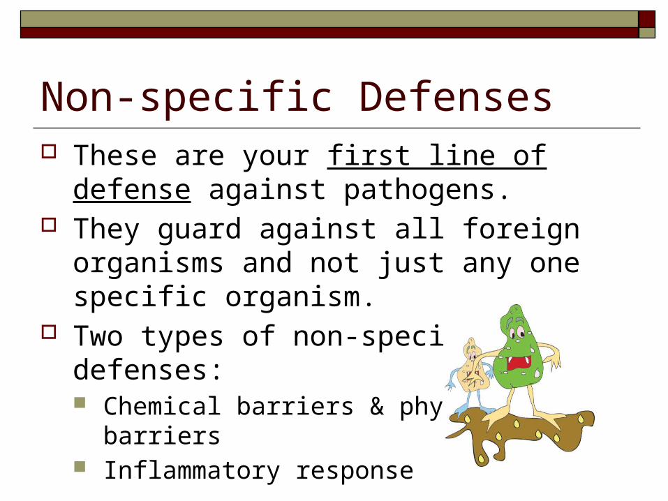
Non-specific Defenses These are your first line of defense against
pathogens. They guard against all foreign organisms and
not just any one specific organism. Two types of non-specific defenses:
Chemical barriers & physical barriers Inflammatory response
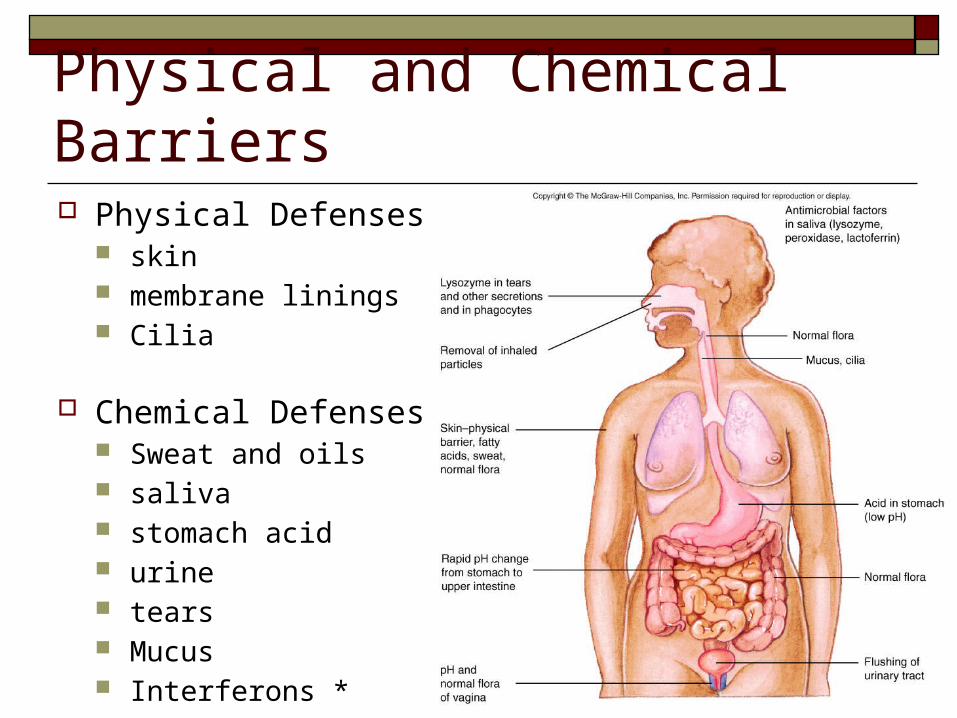
Physical and Chemical Barriers Physical Defenses
skin membrane linings Cilia
Chemical Defenses Sweat and oils saliva stomach acid urine tears Mucus Interferons *
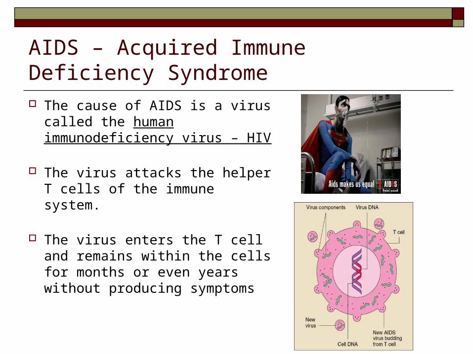
AIDS – Acquired Immune Deficiency Syndrome
The cause of AIDS is a virus called the human immunodeficiency virus – HIV
The virus attacks the helper T cells of the immune system.
The virus enters the T cell and remains within the cells for months or even years without producing symptoms
AIDS’ Disturbing Properties It is able to mutate giving it the ability to
produce different strains. HIV-1;1981, HIV-2; 1985 Dozens of subtypes worldwide for each strain
It causes change in the cell membrane of the T cell causing them to fuse together. This allows the virus to pass from cell to cell
without entering the bloodstream and becoming exposed to antibodies present in the blood
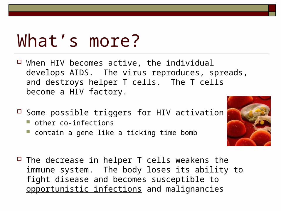
What’s more? When HIV becomes active, the individual develops AIDS.
The virus reproduces, spreads, and destroys helper T cells. The T cells become a HIV factory.
Some possible triggers for HIV activation are: other co-infections contain a gene like a ticking time bomb
The decrease in helper T cells weakens the immune system. The body loses its ability to fight disease and becomes susceptible to opportunistic infections and malignancies